ARTIGO ORIGINAL
AMP
STUDENT
RESUMO
Introdução: A biópsia do gânglio sentinela é atualmente o procedimento cirúrgico standard para o estadiamento ganglionar em
doentes com cancro da mama em estádio precoce, sendo realizado com recurso a diferentes técnicas, como a injeção de corantes vitais e/ou de radioisótopos e, mais recentemente, guiada por fluorescência utilizando o verde de indocianina. O objetivo deste estudo é avaliar a acuidade global do verde de indocianina na identificação do gânglio sentinela em doentes com cancro da mama e de acordo com fatores relacionados com a doente e com o tumor.
Material e Métodos: Estudo retrospetivo numa amostra aleatória de doentes com cancro da mama seguidas e tratadas no Centro
Hospitalar São João no período entre 2012 e 2016.
Resultados: Obtivemos uma taxa de deteção do gânglio sentinela no cancro da mama pelo verde de indocianina superior a 90% e
uma performance diagnóstica deste método similar às outras técnicas descritas na presença de metástases ganglionares.
Discussão: Não se verificaram diferenças estatisticamente significativas entre os três métodos relativamente às taxas de deteção no
subgrupo de mulheres mais velhas, normoponderais, nas que tinham realizado tratamento prévio à mama ou à axila e nas que foram submetidas a quimioterapia neo-adjuvante.
Conclusão: O verde de indocianina é um potencial método alternativo a outras técnicas de pesquisa do gânglio sentinela,
afigurando-se como opção futura para os hospitais que não possuem afigurando-serviço de Medicina Nuclear. Contudo, é esafigurando-sencial a realização de novos estudos no sentido de definir o perfil das doentes que maximiza a sua eficácia.
Palavras-chave: Biópsia do Gânglio Sentinela; Neoplasias da Mama; Verde de Indocianina
Fluorescence-Guided Sentinel Lymph Node Biopsy in
Breast Cancer: Detection Rate and Diagnostic Accuracy
Biópsia do Gânglio Sentinela Guiada por Fluorescência
no Cancro da Mama: Taxa de Deteção e
Performance
Diagnóstica
1. Estudante. Faculdade de Medicina. Universidade do Porto. Porto. Portugal.
2. Departamento de Cirurgia. Centro Hospitalar de São João/Faculdade de Medicina. Universidade do Porto. Porto. Portugal. 3. Centro de Epidemiologia Hospitalar. Centro Hospitalar de São João. Porto. Portugal.
4. Unidade de Epidemiologia. EPIUnit, Instituto de Saúde Pública. Universidade do Porto. Porto. Portugal.
5. Departamento de Ciências da Saúde Pública e Forenses, e Educação Médica. Faculdade de Medicina. Universidade do Porto. Porto. Portugal. Autor correspondente: Teresa Vaz. [email protected]
Recebido: 11 de fevereiro de 2018 - Aceite: 24 de setembro de 2018 | Copyright © Ordem dos Médicos 2018
Teresa VAZ1, Susy COSTA2, Bárbara PELETEIRO3,4,5
Acta Med Port 2018 Dec;31(12):706-713 ▪ https://doi.org/10.20344/amp.10395
ABSTRACT
Introduction: Sentinel lymph node biopsy is currently the standard surgical procedure for lymph node staging in patients with early
stage breast cancer. It is performed using different techniques, such as the injection of vital dyes and / or radioisotopes and, more recently, guided by fluorescence using Indocyanine green. The aim of this study is to assess the detection rate of sentinel lymph node using Indocyanine green in breast cancer patients according to factors related to the patient and the tumor.
Material and Methods: Retrospective study of a random sample of patients with breast cancer, treated and followed at Centro
Hospitalar São João, in Porto, between 2012 and 2016.
Results: Indocyanine green detection rate was over 90% and its diagnostic accuracy was similar to other methods described in the
presence of metastatic involvement of lymph nodes.
Discussion: There was no statistically significant difference between the three methods in the detection rates in subgroups of older
women, with normal weight and in those who underwent previous surgery in breast or axilla or neo-adjuvant chemotherapy.
Conclusion: Indocyanine green is a potential alternative method to other sentinel lymph node screening techniques, appearing as a
future option for breast cancer centers with no nuclear medicine department. However, it is essential to carry out further research in order to define the ideal patients’ profile that maximizes the method’s effectiveness.
Keywords: Breast Neoplasms; Indocyanine Green; Sentinel Lymph Node Biopsy
INTRODUCTION
Breast cancer is the second leading cancer worldwide, the most frequent in women and the fifth leading cause of cancer-related death worldwide, with an increasing inci-dence.1,2 It is the leading cause of cancer-related death in women and the second leading cause in developed coun-tries, second only to lung cancer.1
Axillary lymph node status is one the major prognos-tic factors in patients with early breast cancer and with the greatest impact in staging and in treatment decision.2-6
ARTIGO ORIGINAL
AMP
STUDENT
first few lymph node or nodes into which the tumour drains) biopsy was an important step towards a decline in axillary surgery and morbidity10-13 and is currently considered as the standard surgical procedure in patients with early breast cancer and no clinical or imaging evidence of axillary lymph node involvement.2,3,7,8,14,15
SLN detection has been obtained by the use of different techniques, among which the triple technique is currently the gold standard [combining the use of Tc-99m radioiso-tope (RI) - lymphoscintigraphy and intraoperative gamma probe detection – with vital blue dye (BD)], allowing for high-er accuracy and highhigh-er detection rates than each method used separately.14,16,17 However, there is still no consensus regarding which is the best technique as, despite the ad-vantages, different drawbacks are also involved, namely the use of radioactive elements and subsequent logistics involved with its manipulation and preoperative preparation, in the case of the RI.3,5,8,18,19 The chance of allergic reactions and tissue necrosis have been associated with the use of a BD.20 The need for alternative techniques has therefore emerged, namely the use of non-radioactive agents, such as indocyanine green (ICG),18 a low molecular weight, non-toxic and non-radioactive substance, currently used in dif-ferent medical areas.6,11,14,20,21 The ICG fluorescence-guided SLN biopsy has been recommended by different authors in recent studies as a safe and promising method for the detection of metastatic lymph nodes and with potential ben-efits when compared to the radioisotope.8,11
This study aimed at the assessment of ICG global ac-curacy for SLN detection in patients with breast cancer and according to patient and tumour-related factors.
MATERIAL AND METHODS Methodology
This was a retrospective study involving the analysis of medical records of patients with breast cancer attending the Breast Centre at Centro Hospitalar São João.
Sample and data collection
A group of 232 patients with breast cancer was involved in the study. Only patients having undergone surgery be-tween 2012 and 2016 in whom ICG was used in SLN de-tection were included in the study. Up to four patients were randomly selected each year and each month throughout the study period, by the use of a randomization app (www. random.org).
Data regarding patients, the tumour, the SLN detec-tion technique and the number of detected lymph nodes with each technique, as well as the presence of metastatic lymph nodes were collected. Data on previous local/region-al or systemic therapies were local/region-also obtained.
Surgical technique
The protocol that was validated for the use of Tc-99m in our institution was applied to the group of patients in whom the RI was one of the detection methods and was applied by using a preoperative peritumoral administration, carried
out approximately 12-18 hours before surgery.
A 1 mL dose of patent blue vital dye (S.A.L.F., 50 mg/2 mL, Italy) was injected in sub-areolar lymphatic plexus, fol-lowed by a gentle 3-5 min massaging technique.
The ICG fluorescence-guided SLN biopsy was carried out by using the Photodynamic Eye (PDE) system, Hama-matsu, Japan. ICG (VerDye 25 mg – 5 mg/mL, Diagnos-tic Green GmbH, Germany) was diluted in 5 mL of saline solution. Immediately before surgery and regardless of the tumour location, patients have received a peritumoral injec-tion (dose range, 0.5 – 2 mL), under general anaesthesia, followed by a 3-5 min massaging technique. Dose was based on patient’s body mass index (BMI), breast volume and ptosis as well as on tumour location. The lights of the operating room were turned off a few minutes later and the lymphatic drainage was visualized in real time up to the ax-illary region due to the fluorescence emitted by ICG and using the PDE infra-red probe.
All blue, radioactive (hot) or fluorescent lymph nodes, in addition to all lymph nodes clinically suspicious of meta-static involvement were considered as SLN. Their charac-teristics regarding the marking (blue vs. hot vs. green/fluo-rescent) were recorded.
Lymph nodes were subsequently processed by the standard method (inclusion of samples in paraffin wax and haematoxylin-eosin – HE staining) in the presence of ductal carcinoma in situ (DCIS) or after neoadjuvant chemothera-py (CT). The remaining cases were processed by using the OSNA molecular method (one-step nucleic acid amplifica-tion).22,23
Statistical analysis
Data were analysed with the STATA®, version 11.2
(StataCorp LP, College Station, Texas, EUA) software. The detection rate for each SLN detection method was deter-mined by the number of lymph nodes detected with each method, divided by the total number of lymph nodes de-tected in each patient. Estimated 95% confidence intervals (95% CI) were considered, assuming a binomial distribution and the binomial test was used for the comparison of the detection rate among the different methods, considering the diagnostic performance equality as null hypothesis. Mean and standard deviation of the number of lymph nodes de-tected were also calculated for each method and means were compared by use of Student’s t-test. These were ob-tained for the whole group of patients and by subgroups ac-cording to the patient’s characteristics, considering patient’s age (< 70 and ≥ 70 years of age), BMI (< 20, 20 - 25, 26 - 30 and > 30 kg/m2), previous breast or axillary treatment (no and yes) and the use of neoadjuvant CT (no and yes). The same procedures were carried out for the analysis of detec-tion of metastatic lymph nodes.
Ethical aspects
This study was submitted and approved by the Ethics
Committee of the Centro Hospitalar de São João /
ARTIGO ORIGINAL
AMP
STUDENT
retrospective nature of data collection, this study qualified as exempt of informed consent.
RESULTS
A median age of 58 years at diagnosis (range: 25-92)
and a median BMI of 25.71 kg/m2 (range: 18.29 – 43.28 kg/
m2) were found in our group of 232 patients. From our group of patients, 13 (5.6%) had undergone previous breast or ax-illary treatments and 14 (6.0%) had undergone neoadjuvant CT (Table 1).
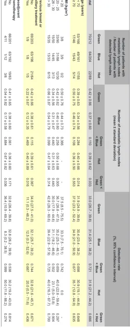
The global diagnostic performance of each of the three methods is shown in Table 2. Higher mean number (± standard deviation) of SLN was detected with the ICG when compared to the BD (1.66 ± 1.09 vs. 1.38 ± 0.90, p < 0.001) and lower when compared to the RI (1.66 ± 1.09 vs. 1.87 ± 1.01, p = 0.002).
The use of ICG (applied to the whole group of patients)
allowed for the detection of SLN in 212 patients, correspond-ing to a 91.4% detection rate (95% CI: 87.0% - 94.6%). BD was used in 228 patients, allowing for the detection of SLN in 204 of these (detection rate: 89.5%; 95%CI: 85.5% - 93.5%), while the RI was used in 71 patients and SLN were identified in 69 of these (detection rate: 97.2%; 95% CI: 90.2% - 99.6%). The ICG method was not lower than BD regarding the detection rate (p = 0.852), while a lower rate was found when compared to the RI method (p < 0.001).
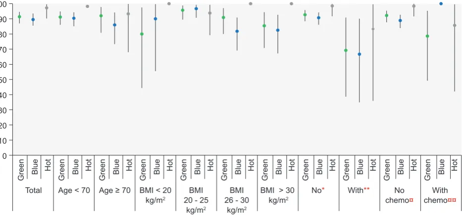
When comparing the detection rate of the three meth-ods according to patient’s characteristics (Table 1 and Fig. 1), we have found that ICG was not lower to BD in terms of SLN detection in any of the subgroups that were consid-ered, while a higher detection rate was found with the use of RI in patients under the age of 70 (p < 0.001), with no previous breast or axillary treatment (p < 0.001) and in pa-tients who did not undergo any neoadjuvant CT (p < 0.001). No statistically significant differences were found as regards the performance of the tests in the subgroup of older pa-tients (p = 0.433), normal-weight (BMI 20 - 25 kg/m2) (p = 0.841), patients with no previous breast or axillary treatment
(p = 0.159) and in patients who underwent neoadjuvant CT
(p = 0.323).
The diagnostic performance (detection of metastatic lymph nodes) of each method is shown in Table 3. The av-erage (± standard deviation) number of metastatic lymph nodes detected with the ICG method was similar to the BD (0.42 ± 0.66 vs. 0.37 ± 0.60, p = 0.110) and higher when compared to the RI (0.42 ± 0.66 vs. 0.39 ± 0.62, p = 0.033).
A 33% (95% CI: 26.7% - 39.8%), 31.4% (95% CI: 25.1% - 38.2%) and 31.9% (95% CI: 21.2% - 44.2%) detection rates were found with ICG, BD and RI methods, respective-ly. No statistically significant differences as regards the de-tection rate of metastatic lymph nodes were found between
Figure 1 – Lymph node detection rate and 95% confidence intervals, according to patient’s characteristics
* No previous breast or axillary treatment; ** With previous breast or axillary treatment; ¤With no neoadjuvant chemotherapy;¤¤With neoadjuvant chemotherapy;
100 90 80 70 60
40 30 20 10 0
Green Blue Green Blue Green Blue Green Blue Green Blue Green Blue Green Blue Green Blue Green Blue Green Blue Green Blue
Total Age < 70 Age ≥ 70 BMI < 20 kg/m2
BMI 20 - 25
kg/m2
BMI 26 - 30
kg/m2
BMI > 30 kg/m2
No* With** No
chemo¤ chemoWith ¤¤
Hot Hot Hot Hot Hot Hot Hot Hot Hot Hot Hot
50
Table 1 – Patient characteristics
n (%)
Total 232
Age (years) < 70
≥ 70 182 (78.4)50 (21.6)
BMI (kg/m2)
< 20 20 - 25 26 - 30 > 30
10 (5.0) 94 (47.0) 55 (27.5) 41 (20.5) Previous breast or axillary treatment
No
Yes 219 (94.4)13 (5.6)
Neoadjuvant chemotherapy No
ARTIGO ORIGINAL
AMP
STUDENT
the three methods, globally and according to patient’s char-acteristics (Fig. 2).
A total of 225 SLN were detected in patients who under-went the combined ICG/BD method [188 were fluorescent and blue (83.6%), 21 were only fluorescent (9.3%) and 16 were only blue (7.1%)]. A total of 73 metastatic lymph nodes were detected in this group [58 were fluorescent and blue (79.5%), nine were only fluorescent (12.3%) and six were only blue (8.2%)].
A total of 67 SLN were detected in patients who under-went the combined BD/RI method [61 were blue and hot (91.0%), two were only blue (3.0%) and four were only hot (6.0%)]. A total of 20 metastatic lymph nodes were detected in this group [14 were blue and hot (70.0%), two were only blue (10.0%) and three were only hot (15.0%)].
A total of 69 SLN were detected in patients who under-went the combined ICG/RI method [61 were fluorescent and hot (88.4%) and eight were hot (11.6%)]. A total of 22 metastatic lymph nodes were detected in this group [19 fluorescent and hot (86.4%) and three were hot (13.6%)].
A total of 67 SLN were detected in patients who under-went the three methods [56 were fluorescent, blue and hot (83.6%), two were green and hot (3.0%), five were blue and hot (7.5%), two were only blue (3.0%) and two only hot (3.0%)]. A total of 16 metastatic lymph nodes were detected in this group [12 were fluorescent, blue and hot (75.0%), one was green and hot (6.3%), one was blue and hot (6.3%), one was only blue (6.3%) and one only hot (6.3%)].
DISCUSSION
A > 90% detection rate with the use of the ICG fluores-cence-guided breast cancer SLN biopsy method and a sim-ilar diagnostic performance in the presence of metastatic lymph nodes when compared to the remaining techniques have been found in our study.
The axillary lymph node status remains as a relevant outcome factor in patients with early breast cancer and cru-cial for the selection of the more adequate adjuvant treat-ment, despite the increasing relevance of tumour biology in
treatment decision.8 Lymph node mapping and SLN biopsy
allow for lymph node staging with a high efficacy through a minimally invasive way, having been accepted as an alter-native to the axillary lymph node dissection and reducing the associated morbidity.2
The BD / technetium RI (Tc-99m) combined technique (lymphoscintigraphy and intraoperative detection) has been considered as the standard method, with 95 to 97% detec-tion rates, according to literature.2,24 However, expensive equipment and logistics related to the manipulation of ra-dioactive material and to preoperative preparation are re-quired with the use of RI, restricting its use to major centres (with a high number of patients) provided with a Nuclear Medicine department.3,5,11,18,19 The fact that no intraoperative visual information nor a structured and sequential dissec-tion is provided by this technique is worth mendissec-tioning, as lymph nodes are detected as hot spots, regardless of the
anatomical lymphatic flow.15-18,24 Table 2
– Number of lymph nodes and detection rate according to patient’
s characteristics
Number of patients with detected lymph nodes / Total number of patients
Number of metastatic lymph nodes
(mean ± standard deviation)
Detection rate
(%, 95% confidence interval)
Green
Blue
Hot
Green
Blue
Green < Blue
Hot
Green < Hot
Green
Blue
Green < Blue
Hot
Green < Hot
Total 212/232 204/228 69/71 1.66 ± 1.09
1.38 ± 0.90
< 0.001
1.87 ± 1.01
0.002
91.4 (87.0 - 94.6)
89.5 (85.5 - 93.5)
0.852
97.2
(90.2
- 99.6)
< 0.001
Age (years) < 70 ≥ 70 166/182 46/50 161/178 43/50 55/56 14/15 1.62 ± 1.05 1.82 ± 1.22
1.32 ± 0.79 1.58 ± 1.20 < 0.001 0.914 1.91 ± 0.98 1.73 ± 1.16 < 0.001 0.697 91.2 (86.1 - 94.9) 92.0 (80.8 - 97.8) 90.4 (85.1 - 94.3) 86.0 (73.3 - 94.2) 0.681 0.933 98.2 (90.4 - 99.9) 93.3 (68.0 - 99.8)
< 0.001 0.433
BMI (Kg/m
2)
< 20 20 - 25 26 - 30 > 30 8/10 90/94 50/55 35/41 9/10 88/91 45/55 33/40
2/2 30/32 13/13 15/15
1.90 ± 1.66 1.80 ± 1.06 1.49 ± 0.98 1.66 ± 1.24
1.50 ± 0.97 1.49 ± 0.85 1.22 ± 0.85 1.38 ± 1.10 0.767 0.997 0.978 0.921 3.00 ± 1.41 2.06 ± 1.13 1.62 ± 1.04 1.67 ± 0.62 0.033 0.009 0.166 0.476 80.0 (44.4 - 97.5) 95.7 (89.5 - 98.8) 90.9 (80.0 - 97.0) 85.4 (70.8 - 94.4) 90.0 (55.5 - 99.7) 96.7 (90.7 - 99.3) 81.8 (69.1 - 90.9) 82.5 (67.2- 92.7) 0.264 0.376 0.981 0.746
100.0 93.8 (79.2 - 99.2) 100.0 100.0
-0.841
-Previous breast or axillary treatment No Ye
s 203/219 9/13 196/216 8/12 64/65 5/6 1.68 ± 1.08 1.31 ± 1.25
1.41 ± 0.90 0.83 ± 0.72 < 0.001 0.903 1.91 ± 0.98 1.50 ± 1.38 0.001 0.295 92.7 (88.4 - 95.8) 69.2 (38.6 - 90.9) 90.7 (86.1 - 94.2) 66.7 (34.9 - 90.1) 0.874 0.677 98.5 (91.7 - 99.9) 83.3 (35.9 - 99.6)
< 0.001 0.159
Neoadjuvant chemotherapy No Ye
s
201/218 11/14 192/216 12/12 63/64 6/7 1.67 ± 1.08 1.50 ± 1.22
1.37 ± 0.91 1.50 ± 0.67 < 0.001 0.500 1.94 ± 1.02 1.28 ± 0.76 < 0.001 0.743 92.2 (87.8 - 95.4) 78.6 (49.2 - 95.3)
88.9 (83.9 - 92.7)
100.0 0.957 -98.4 (91.6 - 99.9) 85.7 (42.1 - 99.6)
< 0.001 0.323
Student’
ARTIGO ORIGINAL
AMP
STUDENT
As regards BD, despite its low cost and the absence of radiation, it may be associated with allergic reactions and the development of nodules and fat necrosis at the injec-tion site.12,20 In addition, it is not always easily visualised through the skin and the fat, as it does not penetrate into the dermis, preventing from the detection of SLN before the skin incision.4
The ICG is a non-radioactive dye with the ability to produce fluorescence and, when released, it binds to pro-teins and macromolecules that change the spectrum of emission close to infrared.14 This radiation, which is devel-oped a few seconds upon the injection is not visible to the human eye although the subcutaneous lymphatic vessels may be detected by using a high-sensitivity photodynamic chamber allowing to follow its flow towards the axillary re-gion and the SLN,12,15,25 with different advantages when compared to the standard methods.21 Its tissue penetration allows for percutaneous, intraoperative, real-time visuali-sation of the lymphatic vessels, giving an adequate orien-tation to the incision and location of the potential SLN.3,4,21 In addition, an anatomical order is possible for SLN detec-tion, allowing for a sequential dissection in an orderly way with the visualisation of the lymphatic tissue.15,16,18,24 Even though with a low complication rate and few adverse ef-fects, some limitations should be considered with the ICG fluorescence technique, including the worse identifica-tion of deeper SLN due to its low tissue penetraidentifica-tion (up to a 10-20 mm depth) making more difficult to follow the lymphatic flow within deeper tissues.13,20,25 There is also the possibility of an ICG leakage, producing the rupture of lymphatic vessels, making more difficult the distinction between the fluorescence from undamaged lymphatic vessels and from the surrounding tissue.13 No expensive material is required for the ICG technique,12 even though the need for a photodynamic chamber is certainly a
draw-back.20 Some surgeons and authors have also raised the
issue regarding the presence of an excessive number of fluorescent lymph nodes subsequently classified as sen-tinel nodes, increasing the average number of removed nodes and the procedure morbidity.13,15 Nevertheless, this was not found in our study, in which a lower number of SLN has been detected with the ICG method when com-pared to the RI.
Some factors could have had an influence on the SLN detection by the methods that were described, namely the patient’s age and BMI, some previous procedures as an ipsilateral breast surgery, the detection of an ipsilateral SLN and ipsilateral radiotherapy (RT) in addition to the use of neoadjuvant CT.26-29
Age-related decline in oestrogen level and anatomi-cal changes, with deposition of fat within the breast may affect lymphatic flow.27,28 In addition, the ability of lymph nodes to retain dyes and/or radiocolloids is reduced when these are replaced by fat.27,28 Nevertheless, in this study, when patients were stratified according to age, we have found that the LSN detection rate with the ICG method was not lower than the rate with the blue dye or the
Table 3
– Number of metastatic lymph nodes and detection rate according to patient’
s characteristics
Number of patients with
metastatic lymph nodes
/ Number of patients with
detected lymph nodes
Number of metastatic lymph nodes
(mean ± standard deviation)
Detection rate
(%, 95% confidence interval)
Green Blue Hot Green Blue Green < Blue Hot Green < Hot Green Blue Green < Blue Hot Green < Hot Total 70/212 64/204 22/69
0.42 ± 0.66
0.37 ± 0.60
0.1
10
0.39 ± 0.62
0.033
33.0 (26.7 - 39.8)
31.4 (25.1 - 38.2)
0.721
31.9 (21.2 - 44.2)
0.666 Age (years) < 70 ≥ 70 53/166 17/46 49/161 15/43 17/55 5/14
0.39 ± 0.63
0.50 ± 0.75
0.34 ± 0.56
0.46 ± 0.74
0.284
0.108
0.40 ± 0.66
0.36 ± 0.50
0.014
0.553
31.9 (24.9 - 39.6)
37.0 (23.2 - 52.4)
30.4 (23.4 - 38.2)
34.9 (21.0 - 50.9)
0.698
0.677
30.9 (19.1 - 44.8)
35.7 (12.8 - 64.9)
0.648 0.635 BMI (kg/m 2 ) < 20
20 - 25
26 - 30
> 30 3/8 33/90 15/50 15/35 3/9 31/88 14/45 13/33 0/2 12/30 3/13 6/15
0.50 ± 0.76
0.42 ± 0.62
0.34 ± 0.56
0.66 ± 0.87
0.44 ± 0.73
0.40 ± 0.60
0.31 ± 0.47
0.54 ± 0.79
0.366
0.258
0.440
0.099
-0.50 ± 0.68
0.31 ± 0.63
0.47 ± 0.64
-0.005
0.440
0.314
37.5 (8.5 - 75.5)
36.7 (26.8 - 47.5)
30.0 (17.9 - 44.6)
42.8 (26.3 - 60.6)
33.3 (7.5 - 70.1)
35.2 (25.3 - 46.1)
31.1 (18.2 - 46.6)
39.4 (22.9 - 57.9)
0.742
0.659
0.502
0.725
0.0
40.0 (22.6 - 59.4)
23.1 (5.0- 53.8)
40.0 (16.3 - 67.7)
-0.297 0.904 0.700 Previous breast or axillary treatment No Ye s 69/203 1/9 63/196 1/8 21/64 1/5
0.42 ± 0.66
0.22 ± 0.67
0.38 ± 0.61
0.12 ± 0.35
0.1
15
0.469
0.39 ± 0.61
0.40 ± 0.89
0.067
0.016
34.0 (27.5 - 41.0)
11.1 (0.3 - 48.2)
32.1 (25.7 - 39.2)
12.5 (0.3 - 52.6)
0.744
0.687
32.8 (21.6 - 45.7)
20.0 (0.5 - 71.6)
0.671 0.436 Neoadjuvant chemotherapy No Ye s 66/201 4/1 1 63/192 1/12 19/63 3/6
0.41 ± 0.65
0.54 ± 0.82
0.38 ± 0.60
0.17 ± 0.58
0.061
0.884
0.36 ± 0.60
0.67 ± 0.82
0.171
0.036
32.8 (26.4 - 39.8)
36.4 (10.9 - 69.2)
32.8 (26.2 - 39.9)
8.3 (0.2 - 38.5)
0.538
0.999
30.2 (19.2 - 43.0)
50.0 (1
1.8 - 88.2)
0.814
0.274
Student’
ARTIGO ORIGINAL
AMP
STUDENT
radioisotope in the group of patients aged 70 and older. Patient’s BMI may have an influence on LSN detection, as visualisation of the operating field could be impaired by a high fat content in subcutaneous and axillary tissue, pro-ducing mechanical pressure on the lymphatic vessels, pre-venting lymphatic flow of markers.28,29 Lymph nodes lose the ability to retain dyes and/or radioisotopes both in high-BMI patients and in older patients.29 When the detection rate of the three methods was analysed according to patient’s BMI and particularly in obese patients, in whom a lower detec-tion rate would be expected, no lower rate was found with the use of the ICG method when compared to blue dye and RI methods. However, no statistically significant differences were found between the three methods in normal-weight patients.
Previous breast treatments such as surgery and/or RT to the breast and the axillary region may lead to the de-velopment of fibrosis and subsequent changes in the lym-phatic flow, which may have an influence on SLN detection in patients with breast cancer recurrence.30-33 In addition, a correct SLN detection may become more difficult after neo-adjuvant CT due to changes in the structure of the lymphatic drainage system.34,35 In fact, important axillary anatomical changes may be induced by CT, namely a blockage of the lymphatic vessels (by cellular necrosis, tumour cell apopto-sis and fibroapopto-sis) and the development of collateral lymphatic drain pathways which ultimately may lead to a higher rate of false negative results.36-38 These limitations should there-fore be considered when SLN biopsy is carried out after these procedures. In this study, SLN detection rate with the ICG method was not lower when compared to blue dye and RI methods in patients who underwent previous breast or axillary treatments, while it was not lower when compared to the RI method in patients submitted to neoadjuvant CT. ICG’s flow through lymphatic vessels partially obliterated by
tumour cells, inflammation and previous surgery or RT-relat-ed fibrosis due to its low molecular weight and subsequent albumin-binding could be a possible explanation for these favourable results, which may correspond to an advantage regarding the RI method in this subgroup of patients.6
The detection of metastatic lymph nodes is the aim of SLN biopsy. Our results have shown that the three methods are equally valid for the detection of metastatic nodes, with no statistically significant differences between the different subgroups.
The analysis of a group of patients having undergone the three methods would allow for a valid comparison be-tween them; this was not possible, due to the retrospective nature of the study. In addition, some of the subgroups that were analysed have included a small number of patients, which may prevent from considering some comparisons as valid. Therefore, an increased number of patients, using a prospective study design would be crucial in further studies.
CONCLUSION
SLN biopsy is a very useful technique, preventing from radical surgery in breast cancer staging. However, varying results have been found in different institutions and each patient’s characteristics, in addition to tumour characteris-tics and detection method’s availability should be consid-ered by surgeons in order to prevent any failed detection with an influence on treatment and outcomes.
Our study has shown that ICG fluorescence-guided is a promising technique for SLN detection in breast cancer, due to its high accuracy and comparability with the other available methods, regardless of patient and tumour-relat-ed factors. Considering these aspects, we believe that this is apparently a reproducible and safe method that could avoid ionizing radiation and with the potential to reduce the costs of SLN detection, corresponding to a future option in Figure 2 – Metastatic lymph node detection rate and 95% confidence intervals, according to the patient’s characteristics
* No previous breast or axillary treatment; ** With previous breast or axillary treatment; ¤With no neoadjuvant chemotherapy;¤¤With neoadjuvant chemotherapy
100 90 80 70 60
40 30 20 10 0
Green Blue Green Blue Green Blue Green Blue Green Blue Green Blue Green Blue Green Blue Green Blue Green Blue Green Blue
Total Age < 70 Age ≥ 70 BMI < 20
kg/m2 20 - 25 BMI kg/m2
BMI 26 - 30
kg/m2
BMI > 30
kg/m2 No* With** chemoNo ¤ chemoWith ¤¤
Hot Hot Hot Hot Hot Hot Hot Hot Hot Hot Hot
ARTIGO ORIGINAL
AMP
STUDENT
hospitals with no Nuclear Medicine department, limited in terms of lymphoscintigraphy.
Further studies on which patient profile would maximize the efficacy of the ICG detection method are needed.
We hope that this study will contribute to further re-search in terms of the application and optimisation of ICG in fluorescence-guided SLN biopsy in breast cancer.
HUMAN AND ANIMAL PROTECTION
The authors declare that the followed procedures were according to regulations established by the Ethics and Clini-cal Research Committee and according to the Helsinki Dec-laration of the World Medical Association.
DATA CONFIDENTIALITY
The authors declare that they have followed the proto-cols of their work centre on the publication of patient data.
All data were anonymously collected and stored in a data-base.
CONFLICTS OF INTEREST
The authors declare that there were no conflicts of inter-est in writing this manuscript.
FINANCIAL SUPPORT
The authors declare that there was no financial support in writing this manuscript.
REFERENCES
1. Ferlay J, Soerjomataram I, Dikshit R, Eser S, Mathers C, Rebelo M, et al. Cancer incidence and mortality worldwide: sources, methods and major patterns in GLOBOCAN 2012. Int J Cancer. 2015;136:E359-86. 2. Gherghe M, Bordea C, Blidaru A. Sentinel lymph node biopsy (SLNB)
vs. axillary lymph node dissection (ALND) in the current surgical treatment of early stage breast cancer. J Med Life. 2015;8:176-80. 3. Toh U, Iwakuma N, Mishima M, Okabe M, Nakagawa S, Akagi Y.
Navigation surgery for intraoperative sentinel lymph node detection using Indocyanine green (ICG) fluorescence real-time imaging in breast cancer. Breast Cancer Res Treat. 2015;153:337-44.
4. Jung SY, Kim SK, Kim SW, Kwon Y, Lee ES, Kang HS, et al. Comparison of sentinel lymph node biopsy guided by the multimodal method of indocyanine green fluorescence, radioisotope, and blue dye versus the radioisotope method in breast cancer: a randomized controlled trial. Ann Surg Oncol. 2014;21:1254-9.
5. Guo W, Zhang L, Ji J, Gao W, Liu J, Tong M. Breast cancer sentinel lymph node mapping using near-infrared guided indocyanine green in comparison with blue dye. Tumour Biol. 2014;35:3073-8.
6. Stoffels I, Dissemond J, Poppel T, Schadendorf D, Klode J. Intraoperative fluorescence imaging for sentinel lymph node detection: prospective clinical trial to compare the usefulness of indocyanine green vs technetium Tc 99m for identification of sentinel lymph nodes. JAMA Surg. 2015;150:617-23.
7. Grischke EM, Rohm C, Hahn M, Helms G, Brucker S, Wallwiener D. ICG fluorescence technique for the detection of sentinel lymph nodes in breast cancer: results of a prospective open-label clinical trial. Geburtshilfe Frauenheilkd. 2015;75:935-40.
8. Samorani D, Fogacci T, Panzini I, Frisoni G, Accardi FG, Ricci M, et al. The use of indocyanine green to detect sentinel nodes in breast cancer: a prospective study. Eur J Surg Oncol. 2015;41:64-70. 9. Xiong L, Gazyakan E, Yang W, Engel H, Hunerbein M, Kneser U, et al.
Indocyanine green fluorescence-guided sentinel node biopsy: a meta-analysis on detection rate and diagnostic performance. Eur J Surg Oncol. 2014;40:843-9.
10. Zhang X, Li Y, Zhou Y, Mao F, Lin Y, Guan J, et al. Diagnostic performance of indocyanine green-guided sentinel lymph node biopsy in breast cancer: A meta-analysis. PLoS One. 2016;11:e0155597. 11. Sugie T, Ikeda T, Kawaguchi A, Shimizu A, Toi M. Sentinel lymph node
biopsy using indocyanine green fluorescence in early-stage breast cancer: a meta-analysis. Int J Clin Oncol. 2017;22:11-7.
12. Sugie T, Sawada T, Tagaya N, Kinoshita T, Yamagami K, Suwa H, et al. Comparison of the indocyanine green fluorescence and blue dye methods in detection of sentinel lymph nodes in early-stage breast cancer. Ann Surg Oncol. 2013;20:2213-8.
13. Cong BB, Sun X, Song XR, Liu YB, Zhao T, Cao XS, et al. Preparation study of indocyanine green-rituximab: A new receptor-targeted tracer for sentinel lymph node in breast cancer. Oncotarget. 2016;7:47526-35.
14. Coufal O, Fait V. Use of indocyanine green and the HyperEye system for detecting sentinel lymph nodes in breast cancer within a population of European patients: a pilot study. World J Surg Oncol. 2016;14:299. 15. Pitsinis V, Provenzano E, Kaklamanis L, Wishart GC, Benson JR.
Indocyanine green fluorescence mapping for sentinel lymph node
biopsy in early breast cancer. Surg Oncol. 2015;24:375-9.
16. Sugie T, Kinoshita T, Masuda N, Sawada T, Yamauchi A, Kuroi K, et al. Evaluation of the clinical utility of the ICG fluorescence method compared with the radioisotope method for sentinel lymph node biopsy in breast cancer. Ann Surg Oncol. 2016;23:44-50.
17. van der Vorst JR, Schaafsma BE, Verbeek FP, Hutteman M, Mieog JS, Lowik CW, et al. Randomized comparison of near-infrared fluorescence imaging using indocyanine green and 99(m) technetium with or without patent blue for the sentinel lymph node procedure in breast cancer patients. Ann Surg Oncol. 2012;19:4104-11.
18. Benson J. Indocyanine green fluorescence for sentinel lymph node detection in early breast cancer. Ann Surg Oncol. 2016;23:6-8. 19. Wishart GC, Loh SW, Jones L, Benson JR. A feasibility study (ICG-10)
of indocyanine green (ICG) fluorescence mapping for sentinel lymph node detection in early breast cancer. Eur J Surg Oncol. 2012;38:651-6.
20. Aydogan F, Arikan AE, Aytac E, Velidedeoglu M, Yilmaz MH, Sager MS, et al. Sentinel lymph node biopsy under fluorescent indocyanin green guidance: Initial experience. Ulus Cerrahi Derg. 2016;32:50-3. 21. He K, Chi C, Kou D, Huang W, Wu J, Wang Y, et al. Comparison
between the indocyanine green fluorescence and blue dye methods for sentinel lymph node biopsy using novel fluorescence image-guided resection equipment in different types of hospitals. Transl Res. 2016;178:74-80.
22. Raia-Barjat T, Trombert B, Khaddage A, Douchet C, Seffert P, Peoc’h M, et al. OSNA (one-step nucleic acid amplification) sentinel lymph node intraoperative molecular analysis in breast cancer: a cost-benefit analysis. Med Oncol. 2014;31:322.
23. Osako T, Iwase T, Kimura K, Yamashita K, Horii R, Yanagisawa A, et al. Intraoperative molecular assay for sentinel lymph node metastases in early stage breast cancer: a comparative analysis between one-step nucleic acid amplification whole node assay and routine frozen section histology. Cancer. 2011;117:4365-74.
24. Takeuchi M, Sugie T, Abdelazeem K, Kato H, Shinkura N, Takada M, et al. Lymphatic mapping with fluorescence navigation using indocyanine green and axillary surgery in patients with primary breast cancer. Breast J. 2012;18:535-41.
25. Ballardini B, Santoro L, Sangalli C, Gentilini O, Renne G, Lissidini G, et al. The indocyanine green method is equivalent to the (9)(9) mTc-labeled radiotracer method for identifying the sentinel node in breast cancer: a concordance and validation study. Eur J Surg Oncol. 2013;39:1332-6.
26. Boughey JC, Suman VJ, Mittendorf EA, Ahrendt GM, Wilke LG, Taback B, et al. Factors affecting sentinel lymph node identification rate after neoadjuvant chemotherapy for breast cancer patients enrolled in ACOSOG Z1071 (Alliance). Ann Surg. 2015;261:547-52.
27. Gschwantler-Kaulich D, Riegler-Keil M, Ruecklinger E, Singer CF, Seifert M, Kubista E. Factors influencing the identification rate of the sentinel node in breast cancer. Eur J Cancer Care. 2011;20:627-31. 28. Straalman K, Kristoffersen US, Galatius H, Lanng C. Factors
influencing sentinel lymph node identification failure in breast cancer surgery. Breast. 2008;17:167-71.
ARTIGO ORIGINAL
AMP
STUDENT
affecting failed localisation and false-negative rates of sentinel node biopsy in breast cancer-results of the ALMANAC validation phase. Breast Cancer Res Treat. 2006;99:203-8.
30. Vugts G, Maaskant-Braat AJ, Voogd AC, van Riet YE, Luiten EJ, Rutgers EJ, et al. Repeat sentinel node biopsy should be considered in patients with locally recurrent breast cancer. Breast Cancer Res Treat. 2015;153:549-56.
31. Vugts G, Maaskant-Braat AJ, Voogd AC, van Riet YE, Roumen RM, Luiten EJ, et al. Improving the success rate of repeat sentinel node biopsy in recurrent breast cancer. Ann Surg Oncol. 2015;22:S529-35. 32. Maaskant-Braat AJ, Voogd AC, Roumen RM, Nieuwenhuijzen GA.
Repeat sentinel node biopsy in patients with locally recurrent breast cancer: a systematic review and meta-analysis of the literature. Breast Cancer Res Treat. 2013;138:13-20.
33. Matsumoto A, Jinno H, Nakamura T, Saito J, Takahashi M, Hayashida T, et al. Technical feasibility of sentinel lymph node biopsy in patients with ipsilateral breast tumor recurrence and previous axillary surgery. Int J Surg. 2015;22:28-31.
34. Pilewskie M, Morrow M. Axillary nodal management following neoadjuvant chemotherapy: a review. JAMA Oncol. 2017;3:549-55. 35. Pinero-Madrona A, Escudero-Barea MJ, Fernandez-Robayna F,
Alberro-Aduriz JA, Garcia-Fernandez A, Vicente-Garcia F, et al. Selective sentinel lymph node biopsy after neoadjuvant chemotherapy in breast cancer: results of the GEICAM 2005-07 study. Cir Esp. 2015;93:23-9.
36. Aguiar PH, Pinheiro LG, Mota RM, Margotti NH, Rocha JI. Sentinel lymph node biopsy in patients with locally advanced breast cancer after neoadjuvant chemotherapy. Acta Cir Bras. 2012;27:912-6.
37. Pecha V, Kolarik D, Kozevnikova R, Hovorkova K, Hrabetova P, Halaska M, et al. Sentinel lymph node biopsy in breast cancer patients treated with neoadjuvant chemotherapy. Cancer. 2011;117:4606-16. 38. Kuehn T, Bauerfeind I, Fehm T, Fleige B, Hausschild M, Helms G, et