263
EVALUATION OF TWO DIFFERENT DENDRITIC CELL PREPARATIONS
WITH BCG REACTIVITY
Marek Fol1, Anna Nitecka-Blaźlak1, Piotr Szpakowski1, Murty V.V.S. Madiraju2, Wiesława Rudnicka1,
Magdalena Druszczyńska1, Joël Pestel3 and Magdalena Kowalewicz-Kulbat1,*
1Department of Immunology and Infectious Biology, Institute for Microbiology, Biotechnology and Immunology, University
of Lodz, 90-237 Lodz, Poland
2University of Texas Health Science Center at Tyler, Tyler TX 75708, USA
3 CNRS-UMR 8576, Unité de Glycobiologie Structurale et Fonctionnelle, IFR 147, Université Lille Nord de France, 59655 Villeneuve d’Ascq, France
*Corresponding author: [email protected]
Received: June 1, 2014; Revised: December 1, 2015; Accepted: December 3, 2015; Published online: February 24, 2016
Abstract: Dendritic cells (DCs) play a key-role in the immune response against intracellular bacterial pathogens, including mycobacteria. Monocyte-derived dendritic cells (MoDCs) are considered to behave as inflammatory cell populations. Differ-ent immunomagnetic methods (positive and negative) can be used to purify monocytes before their in vitro differentiation and their culture behavior can be expected to be different. In this study we evaluated the reactivity of two dendritic cell populations towards the Bacillus Calmette–Guérin (BCG) antigen. Monocytes were obtained from the blood of healthy donors, using positive and negative immunomagnetic separation methods. The expression of DC-SIGN, CD86, CD80, HLA-DR and CD40 on MoDCs was estimated by flow cytometry. The level of IL-12p70, IL-10 and TNF-α was measured by ELISA.Neither of the testedmethods affected the surface marker expression of DCs. No significant alteration in immuno-logical response, measured by cytokine production, was noted either. After BCG stimulation, the absence of IL-12, but the IL-23 production was observed in both cell preparations. Positive and negative magnetic separation methods are effective techniques to optimize the preparation of monocytes as the source of MoDCs for potential clinical application
Key words: dendritic cells; MACS separation; monocytes; BCG
INTRODUCTION
Dendritic cells play a significant role in the induc-tion and regulainduc-tion of a protective response against intracellular bacterial pathogens, including mycobac-teria [1-4]. The predominating DC subset involved in mycobacterial infections are monocyte-derived dendritic cells (MoDCs) [5,6]. It is suggested that the mechanism behind DC uptake of Mycobacterium tu-berculosis (Mtb) and Mycobacterium bovis BCG is me-diated by the intercellular adhesion molecule-3 grab-bing non-integrin (DC-SIGN) [7], which is present on the surface of human MoDCs, dermal DCs, lung interstitial DCs and in lymph nodes [8,9]. Upon infec-tion with Mtb or BCG in vitro, human DCs mature,
produce Th1-promoting cytokines, and activate IFN-γ-producing T cells [10-12]. In contrast, Hanekom et al. [3] and Larsen et al. [13] showed that human DCs infected with Mtb or BCG had decreased levels of MHC class II and costimulatory molecule CD80, produced TNF-α and IL-10 instead of IL-12, and had an impaired capacity to activate T cells.
genera-tion of dendritic cells for cellular immune therapies. Although monocyte recovery from PBMC (peripheral blood mononuclear cell) fractions is high (~80%), only 30% of the cells can be developed into mature highly immunostimulatory DCs [15,16]. Human DCs can be generated in vitro from peripheral blood CD14+
monocytes (hence they are termed monocyte-derived dendritic cells) or from CD34+ progenitors.
Recent studies showed that monocyte separation methods, flask adherence and magnetic activated cell sorting, provide different phenotypic and functional characteristics of the resultant DCs [17]. In this study, we evaluated, in vitro, the effect of two different im-munomagnetic methods of monocyte separation on the reactivity of MoDCs to the BCG antigen.
Experimental approaches were focused on the potential differences in the expression of costimula-tory molecules and surface receptors (CD86, CD80, CD40, HLA-DR and DC-SIGN) on MoDCs, which are known as crucial signals for T cell activation. Consid-ering the central role of cytokines produced by den-dritic cells in the negative or positive regulation of immune response, we also determined the production of IL-10, IL-12, IL-23 and TNF-α released by stimu-lated and unstimustimu-lated dendritic cells.
MATERIALS AND METHODS Blood donors
Blood was collected from 6 young healthy volunteers with a mean age of 30±3 years (range: 25-35 years), vaccinated with BCG according to state policy. All experiments were approved by the local Ethics Com-mittee. Agreement for participation in the study was signed by each donor before blood collection.
Isolation of monocytes
Peripheral blood (60-70 mL) was drawn in vacutainer tubes containing spray-coated heparin (Becton Dick-inson). After centrifugation (1000 rpm/min. for 15 min., RT) and plasma removal, blood was diluted in
RPMI 1640 medium (1:1, Sigma-Aldrich) and layered on Ficoll-Paque PLUS at a ratio of 4:3 (Amersham, Biosciences). After centrifugation at 400×g for 30 min, PBMCs were harvested, washed and resuspended in PBS (phosphate-buffered saline) supplemented with 0.5% BSA and 2 mM EDTA. PBMCs were counted in trypan blue dye, and immediately used for monocyte isolation either by positive or negative selection using a MACS system (Miltenyi Biotech, Germany), accord-ing to the protocol of manufacturer.
During positive separation, PBMCs were incu-bated with magnetic beads conjugated with mouse monoclonal anti-human CD14 antibody at 4°C for 30 min. After washing with MACS buffer, cells were centrifuged and applied onto as LS column placed in the magnetic field of a MACS separator. The magneti-cally labeled CD14+ cells were retained in the column
while the unlabeled CD14- cells passed through the
column. After removal from the magnetic field, mag-netically retained CD14+ cells (monocytes) were eluted
as a positively selected cell fraction.
In negative separation, monocytes were obtained from PBMCs through the depletion of B cells, T cells, natural killer cells, DCs, early erythroid cells, platelets and basophils by an indirect magnetic labeling using a cocktail of biotin conjugated antibodies against CD3, CD7, CD16, CD19, CD56, CD123 and glycophorin A as well as anti-biotin microbeads (monocyte isolation kit Miltenyi Biotech). After incubation, cells were ap-plied onto the LS column. The effluent of highly pure unlabeled monocytes was collected.
The purity of monocytes obtained through posi-tive and negaposi-tive separation was determined to be 96% to 99% on the basis of forward and side scatter gating in conjunction with CD14 staining using stand-ard flow cytometry (data now shown). The viability of the magnetically sorted cells was measured using trypan blue dye.
Monocyte-derived dendritic cell generation
supple-mented with 100 U/mL penicillin, 0.1 mg/mL strep-tomycin, L-glutamine (Polfa Tarchomin, Poland), and enriched with 10% (v/v) fetal calf serum (FCS, heat inactivated; Cambrex, Belgium). The cell density was adjusted to 1×10⁶/mL and monocytes were placed into
6-well tissue culture plates to differentiate into den-dritic cells by incubation for 6 days in RPMI-1640 (supplemented as above) in the presence of 25 ng/mL human granulocyte-macrophage colony-stimulating factor (GM-CSF) and 10 ng/mL human recombinant IL-4 (R&D Systems, USA). Then the cells were har-vested, pooled and counted before use.
Stimulation of DCs with antigens
Prepared immature DCs at a density of 1×10⁶ cells/
mL were placed into a 6-well plate and pulsed for 24 h at 37°C, 5% CO2 either with M. bovis BCG (ratio 1:1) or LPS (lipopolysaccharide, 1 µg/mL; Sigma) as DC maturation inducer. Unpulsed DCs (in medium alone) were used as the negative control.
DC preparation for flow cytometry
Antigen-pulsed or unpulsed DCs were collected from the 6-well plate using PBS/2mM EDTA. After wash-ing in PBS, the cells were incubated for 30 min at 4°C with the following monoclonal antibodies (mAbs): fluorescein isothiocyanate (FITC)-conjugated anti-CD86, anti-CD40, anti-HLA-DR, anti-DC-SIGN, Phycoerythrin (PE)-conjugated anti-CD80, or with isotype-matched control mAb. All monoclonal anti-bodies were purchased from Becton Dickinson. Af-ter two washings with PBS and centrifugation (1700 rpm/min, 10 min), DCs were analyzed using a flow cytofluorimeter FACS LSRII (Becton Dickinson). Data were analyzed using FlowJo software.
Cytokine measurement
Supernatants of pulsed and unpulsed DC cultures (1×10⁶/well) harvested after 24-h stimulation and
centrifugation (1600 rpm/min, 10 min.) were stored at -20°C until tested. The levels of IL-10, IL-12p70, IL-23 and TNF-α were quantified by ELISA Eli-pair
test (Diaclone). The test detection sensitivity was 5 pg/mL for IL-10, IL-12p70 and TNF-α and 20 pg/mL for IL-23.
Statistical analysis
Statistical analyses were performed with STATISTI-CA 8.0 PL program. Data are expressed as mean- or median±SEM. Differences between samples were ana-lyzed by Mann-Whitney U test (for impaired data). P values of ≤0.05 were considered significant.
RESULTS
Characteristics of cells after alternative separation methods
To assess the efficiency of positive and negative sepa-ration techniques, both cell prepasepa-rations obtained from the same healthy donors were analyzed by flow cytometry. First, the monocyte populations were stud-ied. The number of monocytes obtained with the posi-tive (7.2±2.0%) and negaposi-tive (6.5±2.3%) separation methods, with regard to the total number of PBMC, was similar (no statistically significant differences). In-terestingly, the efficacy of the two separation methods was comparable (Fig. 1) and varied from 71%-80.5%, indicating that both separation protocols deliver simi-lar source material. However, as indicated in dot-plot diagrams, a higher homogeneity of monocytes was obtained using the negative separation method com-pared to that obtained with the positive selection technique, suggesting that the negative separation technique might provide a monocyte population with higher purity (Fig. 1).
Fig. 1. Positive and negative selection techniques and their impact on the efficiency of monocyte isolation. Graphs show dot-blots of monocytes obtained by differential gradient centrifugation on Ficoll-Paque Plus, followed by positive or negative immunomag-netic separation of MACS System and analyzed by flow cytometry; a – unstained monocytes; b – stained with fluorescently (FITC) labeled isotype matched control mAbs; c – stained with fluores-cently (FITC) labeled specific anti-human CD14 mAbs.
Fig. 2. Populations of monocyte-derived dendritic cells (MoDCs) isolated using positive (a) and negative (b) monocyte immunomag-netic separation methods. Cells were analyzed by flow cytometry. The blue graph represents the isotype matched control, and the black graph cells stained with FITC labeled anti-human CD14 mAb. Populations were similarly numerous and localized at similar posi-tions in cytometry graphs regardless of used separation methods.
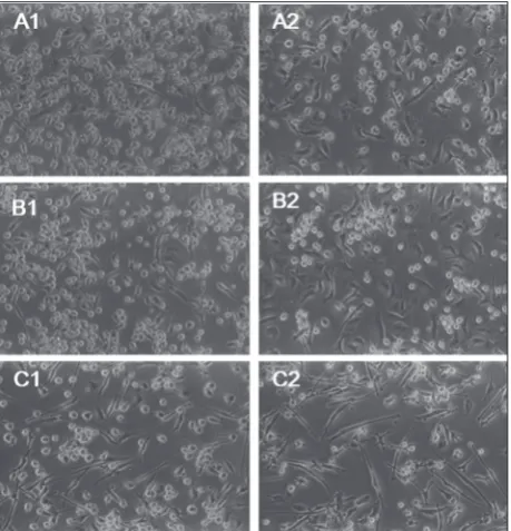
Fig. 3. Morphology of unstimulated (A), BCG (B) or LPS (C) 24 h-pulsed MoDCs obtained from human peripheral blood monocytes isolated by positive (1) and negative (2) separation methods. The cells were examined by inverted microscopy (×400).
In response to BCG and LPS stimulation, MoDCs de-veloped the morphology characteristic of mature cells (presence of dendrites) (Fig. 3). However, it is worth pointing out that, especially after LPS treatment, in the case of negative separation more elongated cells were observed in comparison to positive separation, where the cell morphology was more distinct disparate.
Evaluation of cell surface markers after Ag stimulation
To explore whether the type of monocyte isolation method affects the MoDC surface marker associ-ated with T cell polarization, their expression was investigated after stimulation with two different bac-terial products, LPS and BCG. As shown in Fig. 4, representative histograms indicate that similar sur-face marker modifications were observed in the DC populations prepared by both methods.
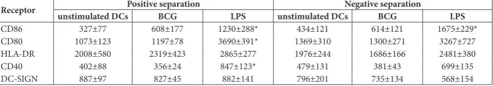
Further statistical analyses performed on all results revealed no significant differences in the expression of costimulatory molecules on unstimulated DCs or LPS-stimulated DCs, regardless of the MACS separation method used. Within the group of MoDCs obtained by the positive separation method we observed a sig-nificant increase in CD86 (p=0.027), CD80 (p=0.009) and CD40 (p=0.04) expression after LPS stimulation compared with unstimulated MoDCs (Table 1). In con-trast, in LPS-stimulated MoDCs isolated by the negative separation method, a significant enhancement was only observed for the CD86 marker (p=0.006), whereas a true tendency to increase the other surface parameters was noticed. In the presence of BCG antigen, in the
ma-jority of cases an increase in CD86 marker was detected. However, between BCG-treated or untreated dendritic cells derived from negative or positive monocyte selec-tion there were no statistically significant differences in the expression of all investigated DC surface mark-ers. Indeed, BCG bacilli did not induce any significant changes in the expression of DC-SIGN compared to unstimulated cells. Taken together, these results suggest that the positive or negative magnetic separation meth-ods generally did not affect the expression of surface markers involved in the immune synapse.
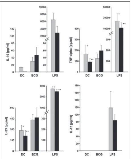
Evaluation of cytokine production after BCG stimulation
The release of TNF-α, IL-10, IL-12 and IL-23 by MoDCs in culture supernatants was evaluated to es-tablish the impact of MACS separation technique on cytokine production (Fig. 5). There were no signifi-cant differences in TNF-α production by BCG-pulsed MoDCs compared to unpulsed cells, but LPS induced significant TNF-α release, whatever the source of DC populations. Concerning IL-10 production, unpulsed MoDCs, derived from monocytes obtained by positive but not negative selection, released IL-10, however at a low level. In the presence of BCG, negatively-derived MoDCs secreted a higher amount of IL-10 (47±22 pg/mL) than positively generated MoDCs (30±11 pg/mL), but much less than the same DC populations stimulated with LPS (2891±1744 pg/mL vs. 6645±1755 pg/mL, respectively).
Interestingly, in the presence of BCG, both MoDC preparations failed to produce IL-12 p70. Indeed, only
Table 1. Median fluorescence intensity (MFI) for unstimulated DCs and BCG- or LPS-stimulated DCs obtained from monocytes by positive and negative separation techniques (number of subjects, n=6).
Receptor unstimulated DCsPositive separationBCG LPS unstimulated DCsNegative separationBCG LPS
CD86 327±77 608±177 1230±288* 434±121 614±121 1675±229*
CD80 1073±123 1197±78 3690±391* 1369±310 1300±271 3267±727
HLA-DR 2008±580 2319±423 2865±277 1976±244 1686±166 2481±380
CD40 402±88 356±24 847±123* 479±131 381±43 699±135
DC-SIGN 887±97 827±45 882±141 796±201 735±134 568±154
LPS-activated MoDCs, generated from monocytes iso-lated with positive and negative purification methods, secreted IL-12 at measureable and comparable levels. These results underlined the variable reactivity of DCs to BCG compared to LPS. As IL-12 consists of p35 and p40 subunits, and the p40 subunit is shared with IL-23, we determined the amount of secreted IL-23 in the su-pernatants of BCG-stimulated DC cultures. After BCG treatment, no statistically significant difference in IL-23 production, compared to unpulsed DCs generated from monocytes obtained by the positive and nega-tive methods, was observed. The mean value of IL-23 production by BCG-stimulated MoDCs was 431±78
pg/mL and 465±137 pg/mL for positive and negative separation, respectively. In contrast, LPS-stimulated MoDCs produced significantly more IL-23 (2750±231 pg/mL and 2510±201 pg/mL for positive and negative separation, respectively) compared to unpulsed DCs (280±76 pg/mL and 210±63 pg/mL, analogously).
DISCUSSION
It is known that in vitro cell reactivity might be differ-ent according to the method used to prepare cell pop-ulations. Concerning dendritic cells, it was reported that CD14+ cells can be isolated either by a positive
or negative magnetic sorting method, with better re-sults than by plastic adherence [18]. Moreover, it has been demonstrated that positively MACS-separated monocytes presented a better viability and purity of the cells in comparison to the adherence method [19]. Different manufacturers have proposed original and valuable separation systems. In the present study we used two immunomagnetic separation methods to evaluate the reactivity of DCs to BCG compared to LPS. Our experiments demonstrated that both tech-niques provided monocytes with high efficiency and these monocytes similarly differentiated into dendritic cells exhibiting similar characteristics.
The expression of costimulatory molecules can be used to evaluate the stage of differentiation and the degree of maturation of DCs during an in vitro cul-ture. We investigated the expression of some costimu-latory molecules following stimulation with two bacte-rial antigens at the surface of MoDCs obtained by two different immunomagnetic separation methods. CD80 and CD86 are present in the cellular membrane of monocytes/macrophages, dendritic cells, thymocytes and T and B cells, and participate in T cell activation by binding with costimulatory the molecules CD80-CD28 and CD86-CD80-CD28 [20]. Another costimulatory molecule, CD40 is a membrane glycoprotein that coparticipates in the induction of DC maturation by ligation with CD40L. CD40 together with CD80, CD86 and HLA-DR belong to the immune synapse and participate in signal transduction between antigen presenting cells and T lymphocytes [21,22].
The positive selection of monocytes and the nega-tive isolation technique, have been well described [23-26]. In our study, a comparison of the two methods applied on the same blood samples did not show any statistical differences in the expression of costimulato-ry molecules CD86, CD80, HLA-DR, CD40 and sur-face DC-SIGN receptor. This is in agreement with the study by Reuter et al. [23], in which both procedures of monocyte isolation had a similar effect concerning CD86 expression. Moreover, Elkord et al. [27] showed that the expression of CD80, CD86 and CD83 mol-ecules was the same, regardless of the method used to isolate CD14+ cells (positive magnetic separation
technique and plastic adherence method). The results of our study, in which we investigated the expression of CD86, CD80, HLA-DR and CD40 on the surface of unstimulated MoDCs, are in agreement with the study conducted by Gregori et al. [28], who reported the substantial expression of CD86 and CD80 on hu-man immature MoDCs. Thus, by taking into account the expression level of these costimulatory molecules, BCG-activated DCs might be considered as less ma-ture than LPS-stimulated DCs.
Concerning the DC-SIGN expression, it must be underlined that LPS-activated DCs have less DC-SIGN receptors than BCG-activated DCs. DC-DC-SIGN belongs to the lectin receptors type C and recognizes numerous bacterial- and viral- antigens. Among the components of mycobacteria recognized by DC-SIGN are lipoarabinomannan (LAM), arabinomannan (AM) and antigen 19 kDa [8]. Moreover, as we could see no differences in DC-SIGN expression by MoDCs isolated from monocytes by two different magnetic cell separation systems, we concluded that the type of isolation method does not have an impact on the ability of MoDCs for antigen uptake (M. bovis BCG). During the maturation process, MoDCs secrete cytokines, which can play an important role in T cell polarization. According to our data, both immu-nomagnetic methods of monocyte isolation gener-ated MoDCs capable of producing cytokines (TNF-α and IL-10) at detectable levels. It was noticed that positively, but not negatively, obtained unstimulated MoDCs produced IL-10. This production could be the
effect of cell activation resulting from the tendency toward cell clustering or antibody attachment to the surface of monocytes isolated by positive selection. However, BCG induced a higher production of IL-10 than unstimulated cells, indicating than despite a limited number of surface marker modifications, BCG tends to activate both DC populations to some extent, but less than LPS.
Finally, one important new finding from the cur-rent study is the observed lack of IL-12 production by BCG-stimulated MoDCs in contrast to LPS, which is considered as the best inducer of IL-12. The absence of IL-12p70 production by BCG-pulsed MoDCs suggests that additional stimuli are required to initiate IL-12 production. Indeed, the IL-12 synthesis pathway is a well-controlled mechanism. It is known that stimulated dendritic cells have a relatively short time in which to produce 12 [29]. Moreover, it was reported that IL-10 could reduce IL-12 production [30]. In contrast to IL-12, we observed the production of IL-23 by BCG-pulsed DCs. Interleukin 23 is a heterodimeric cytokine composed of the p40 and p19 subunits. It has been shown that M. bovis preferentially induced IL-23 to IL-12 by murine bone marrow-derived DCs [31].
To summarize, this study demonstrates that both positive and negative immunomagnetic selection can be used to isolate CD14+ cells with high efficiency. The
MoDCs generated in both conditions exhibit similar behavior in the presence of two different bacterial stimuli. Interestingly, the lack of IL-12 production by BCG-stimulated MoDCs was not related to the meth-od used to generate DCs. Further studies are required to establish the specific mechanisms that control the IL-12 synthesis within the BCG stimulation and to evaluate the influence on T cell response.
Acknowledgments: This work was supported by the Polish Min-istry of Science and Higher Education grants: N N401 015236, N N303 345035.
Conflict of interest disclosure: All authors declare that there are no competing financial interests. This includes employment, consultancies, stocks, honoraria, patents, grants and travel grants within three years of beginning the submitted work.
REFERENCES
1. Mortellaro A, Robinson L., Ricciardi-Castagnoli P. Spotlight on mycobacteria and dendritic cells: will novel targets to fight tuberculosis emerge? EMBO Mol Med. 2009;1(1):19-29. 2. Gościcka T, Walencka M, Fol M, Krupa A, Szeliga J.
Influ-ence of dendritic cells on the in vitro allogenic cytotoxic reaction of lymphoid cells derived from normal or Listeria
innocua-infected BALB/c mice. Arch Immunol Ther Exp.
2001;49(6):447-52.
3. Hanekom HW, Mendillo M, Manca C, Haslett PA, Siddiqui MR, Barry C 3rd, Kaplan G. Mycobacterium tuberculosis inhib-its maturation of human monocyte-derived dendritic cells in vitro. J Infect Dis. 2003;188(2):257-66.
4. Kono M, Nakamuram Y, Suda T, Uchijima M, Tsujimura K, Nagata T, Giermasz AS, Kalinski P, Nakamura H, Chida K. Enhancement of protective immunity against intracellular bacteria using type-1 polarized dendritic cell (DC) vaccine. Vaccine. 2012;30(16):2633-39.
5. Humphreys IR, Stewart GR, Turner DJ, Patel J, Karamanou D, Snelgrove RJ, Young DB. A role for dendritic cells in the dissemination of mycobacterial infection. Microbes Infect. 2006;8(5):1339-46.
6. Wolf AJ, Linas B, Trevejo-Nuñez GJ, Kincaid E, Tamura T, Takatsu K, Ernst JD. Mycobacterium tuberculosis infects den-dritic cells with high frequency and impairs their function in vivo. J Immunol. 2007;179(4):2509-19.
7. Hermann JL, Lagrange PH. Dendritic cells and Mycobac-terium tuberculosis: which is the Trojan horse? Pathol Biol (Paris). 2004;53(1):35-40.
8. Geijtenbeek TB, Van Vliet SJ, Koppel EA, Sanchez-Hernandez M, Vandenbroucke-Grauls CM, Appelmelk B, Van Kooyk Y. Mycobacteria target DC-SIGN to suppress dendritic cell function. J Exp Med. 2003;197(1):7-17.
9. Geijtenbeek TB, van Vliet SJ, Engering A, ‘t Hart BA, van Kooyk Y. Self- and nonself-recognition by C-type lectins on dendritic cells. Annu Rev Immunol. 2004;22:33-54.
10. Kim KD, Lee HG, Kim JK, Park SN, Choe IS, Choe YK, Kim SJ, Lee E, Lim JS. Enhanced antigen-presenting activ-ity and tumour necrosis factor-alpha-independent activation of dendritic cells following treatment with M. bovis bacillus Calmette-Guérin. Immunology. 1999;97(4):626-33.
11. Hickman SP, Chan J, Salgame P. Mycobacterium tuberculosis induces differential cytokine production from dendritic cells and macrophages with divergent effects on naïve T cell polari-zation. J Immunol. 2002;168(9):4636-42.
12. Rudnicka W. Molecular mechanisms of resistance to tuber-culosis. Post Mikrobiol. 2004;43(1):107-27.
13. Larsen J, Benn C, Fillie, Y, van der Kleij D, Aaby P, Yazdan-bakhsh M. BCG stimulated dendritic cells induce an intere-lukin–10 producing T-cell population with no T helper 1 or T helper 2 bias in vitro. Immunology. 2007;121(2):276-82. 14. Erdmann M, Schuler-Thurner B. Towards a Standardized
Protocol for the Generation of Monocyte-Derived Dendritic Cell Vaccine. Methods Mol Biol. 2010;595:149-163.
15. Strasser E, Eckstein R. Optimization of leukocyte collection and monocyte isolation for dendritic cell culture. Transfus Med Rev. 2010;24(2):130-9.
16. Steinman R, Idoyaga J. Features of the dendritic cell lineage. Immunol Rev. 2010;234(1):5-17.
17. Beikzadeh B, Delirezh N. Phenotypic and functional compar-ison of two distinct subsets of programmable cell of mono-cytic origin (PCMOs)-derived dendritic cells with conven-tional monocyte-derived dendritic cells. Cell Mol Immunol. 2015;DOI:10.1038/cmi.2014.135.
18. Meyer-Wentrup F, Burdach S. Efficacy of dendritic cell gen-eration for clinical use: recovery and purity of monocytes and mature dendritic cells after immunomagnetic sorting or adherence selection of CD14+ starting populations. J Hema-tother Stem Cell Res. 2003;12(3):289-99.
19. El-Sahrigy SA, Mohamed NA, Talkhan HA, Rahman AMA. Comparison between magnetic activated cell sorted mono-cytes and monocyte adherence techniques for in vitro genera-tion of immature dendritic cells: An Egyptian trial. Centr Eur J Immunol. 2015;40(1):18-24.
20. Rutkowski R, Moniuszko T. The role of costimulatory mol-ecules B7.1 (CD80) and B7.2 (CD86) in the pathomecha-nism of inflammatory process. Alergia Astma Immunol. 2001;6(2):87-94.
21. MacDonald AS, Straw AD, Dalton NM, Pearce EJ. Cutting edge: Th2 response induction by dendritic cells: a role for CD40. J Immunol. 2002;168(2):537-40.
22. Cella M, Engering A, Pinet V, Pieters J, Lanzavecchia A. Inflammatory stimuli induce accumulation of MHC class II complexes on dendritic cells. Nature. 1997;388(6644):782-7. 23. Reuter H, Spieker J, Gerlach S, Engels U, Pape W, Kolbe L,
Schmucker R, Wenck H, Diembeck W, Wittern KP, Reisinger K, Schepky AG. In vitro detection of contact allergens: devel-opment of an optimized protocol using human peripheral blood monocyte-derived dendritic cells. Toxicol In Vitro. 2011;25(1):315-23.
24. Pickl W, Majdic O, Kohl P, Stockl J, Riedl E, Schneinecker C, Bello-Fernandez C, Knapp W. Molecular and functional characteristics of dendritic cells generated from highly purified CD14+ peripheral blood monocytes. J Immunol. 1996;157(9):3850-9.
25. Hu Q, Frank I, Williams V, Santos J, Watts P, Griffin G, Moore JP, Pope M, Shattock RJ. Blockade of attachment and fusion receptors inhibits HIV-1 infection of human cervical tissue. J Exp Med. 2004;199(8):1065-75.
versus negative MACSTM protocol. Clin Hemorheol Micro-circ. 2011;48(1):57-63.
27. Elkord E, Williams PE, Kynaston, H, Rowbottom A. W. Human monocyte isolation methods influence cytokine pro-duction from in vitro generated dendritic cells. Immunology. 2005;114(2):204-12.
28. Gregori S, Tomasoni D, Pacciani V, Scirpoli M, Battaglia M, Magnani CF, Hauben E, Roncarolo MG. Differentia-tion of type 1 T regulatory cells (Tr1) by tolerogenic DC-10 requires the IL-10-dependent ILT4/HLA-G pathway. Blood. 2010;116(6):935-44.
29. Langenkamp A, Messi M, Lanzavecchia A, Sallusto F. Kinetics of dendritic cell activation: impact on priming of TH1, TH2 and nonpolarized T cells. Nat Immunol. 2000;1(4):311-6. 30. Redford PS , Murray PJ, O’Garra A. The role of IL-10 in
immune regulation during
31. M. tuberculosis infection. Mucosal Immunol. 2011;4:261-70. 32. Zhang X, Li S, Luo Y, Chen Y, Cheng S, Zhang G, Hu C, Chen