
REVIEW
Rupture signs on computed tomography, treatment, and outcome
of abdominal aortic aneurysms
Kim-Nhien Vu
&Youri Kaitoukov
&Florence Morin-Roy
&Claude Kauffmann
&Marie-France Giroux
&Éric Thérasse
&Gilles Soulez
&An Tang
Received: 27 December 2013 / Revised: 26 March 2014 / Accepted: 27 March 2014 / Published online: 1 May 2014 #The Author(s) 2014. This article is published with open access at Springerlink.com
Abstract
Objectives
Abdominal aortic aneurysm (AAA) rupture has a
high mortality rate. Although the diagnosis of a ruptured AAA
is usually straightforward, detection of impending rupture
signs can be more challenging. Early diagnosis of impending
AAA rupture can be lifesaving. Furthermore, differentiating
between impending and complete rupture has important
re-percussions on patient management and prognosis. The
pur-pose of this article is to classify and illustrate the entire
spectrum of AAA rupture signs and to review current
treat-ment options for ruptured AAAs.
Methods
Using medical illustrations supplemented with
com-puted tomography (CT), this essay showcases the various signs
of impending rupture and ruptured AAAs. Endovascular
aneu-rysm repair (EVAR) and open surgical repair are also discussed
as treatment options for ruptured AAAs.
Results
CT imaging findings of ruptured AAAs can be
categorised according to location: intramural, luminal,
and extraluminal. Intramural signs generally indicate
i m p e n d i n g A A A r u p t u r e , w h e r e a s l u m i n a l a n d
extraluminal signs imply complete rupture. EVAR has
emerged as an alternative and possibly less morbid
meth-od to treat ruptured AAAs.
Conclusions
AAA rupture occurs at the end of a continuum of
growth and wall weakening. This review describes the CT
imaging findings that may help identify impending rupture
prior to complete rupture.
Teaching Points
•
AAA rupture occurs at the end of a continuum of growth and
wall weakening.
•
Intramural imaging findings indicate impending AAA
rupture.
Based on an EPOS electronic poster This review article is an expanded version of an EPOS electronic poster [1]:
Vu KN, Kaitoukov Y, Morin-Roy F et al. (2014) Abdominal aortic aneurysms: rupture signs on computed tomography. Electronic exhibit at the ECR 2014. 20th annual meeting of the European Society of Radiol-ogy; Vienna, Austria. DOI:10.1594/ecr2014/C-0789
K.<N. Vu
:
Y. Kaitoukov:
F. Morin-Roy:
A. TangDepartment of Radiology, Centre hospitalier de l’Université de Montréal (CHUM), Hôpital Saint-Luc, 1058 Saint-Denis, Montréal, Québec, Canada H2X 3J4
K.<N. Vu
:
Y. Kaitoukov:
F. Morin-Roy:
C. Kauffmann:
M.<F. Giroux:
É. Thérasse:
G. Soulez:
A. TangDepartment of Radiology, Radio-Oncology and Nuclear Medicine, and Institute of Biomedical Engineering, Université de Montréal, 2900 Édouard-Montpetit, Montréal, Québec, Canada H3T 1J4
C. Kauffmann
:
M.<F. Giroux:
G. SoulezDepartment of Radiology, Centre hospitalier de l’Université de Montréal (CHUM), Hôpital Notre-Dame, 1560 Sherbrooke Est, Montréal, Québec, Canada H2L 4M1
C. Kauffmann
:
G. Soulez:
A. TangCentre de recherche du Centre hospitalier de l’Université de Montréal (CRCHUM), 900 Saint-Denis, Montréal, Québec, Canada H2X 0A9
É. Thérasse
Department of Radiology, Centre hospitalier de l’Université de Montréal (CHUM), Hôtel-Dieu de Montréal, 3840 Saint-Urbain, Montréal, Québec, Canada H2W 1T8
A. Tang (*)
Department of Radiology, Université de Montréal and Centre de recherche du Centre hospitalier de l’Université de Montréal (CRCH UM), Hôpital Saint-Luc, 1058 Saint-Denis, Montréal, Québec, Canada H2X 3J4
•
Luminal and extraluminal imaging findings imply complete
AAA rupture.
•
Some imaging findings are not specific to AAA ruptures and
can be seen in other pathologies.
•
EVAR has emerged as an alternative and possibly less
morbid method of treating ruptured AAAs.
Keywords
Abdominal aortic aneurysm (AAA) . Ruptured
aneurysm . Computed tomography . Endovascular aneurysm
repair (EVAR) . Vascular surgical procedures
Introduction
Abdominal aortic aneurysm (AAA) is defined as a focal
dilation of the aorta of more than 50 % of its expected
diameter [
2
]. Found in 1 % of the population over the age of
50 [
3
], AAAs are responsible for 1 % of all deaths amongst
Caucasian adults [
4
] and are the tenth leading cause of death in
men over the age of 55 [
5
]. Risk factors include age, male
gender, tobacco usage, family history, hypercholesterolaemia,
and hypertension [
6
]. Aneurysms may progress in size as a
result of gradual wall weakening [
7
], with rupture occurring at
the end of the growth spectrum.
A ruptured AAA often leads to disastrous consequences,
with 50 % of patients dying before reaching the hospital and
another 40 to 50 % mortality rate for those who undergo
surgery [
6
]. However, surgical treatment can be slightly
de-layed in haemodynamically stable patients who present with
an impending AAA rupture. These patients still require urgent
treatment, but can also benefit from preoperative imaging that
optimises surgical planning and reduces mortality rates to
those comparable to elective procedures (less than 5 %) [
6
,
8
]. It is therefore crucial to recognise imaging signs of
impending and complete AAA ruptures as proper
identifica-tion of these rupture signs will have important repercussions
on treatment promptness and prognosis.
The purpose of this review is to identify signs of
impending AAA rupture before rupture is completed.
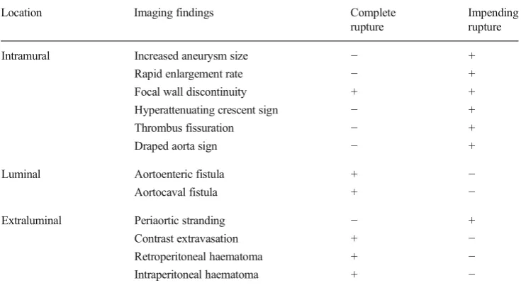
This article categorises imaging findings according to
l o c a t i o n : ( 1 ) i n t r a m u r a l , ( 2 ) l u m i n a l , a n d ( 3 )
extraluminal (Table
1
). Intramural signs generally
indi-cate impending AAA rupture, whereas luminal and
extraluminal signs imply complete rupture. Treatment
options for AAA ruptures will also be discussed.
It must be added that an aneurysm about to rupture
may not reveal any imaging findings. In addition, a
painful AAA, in itself, may indicate impending rupture,
even in the absence of CT imaging findings, and may
justify urgent repair [
6
].
At our institution, computed tomography (CT)
angi-ography using a multidetector scanner is the standard of
practice for evaluating AAA. Unenhanced images are
first acquired through the abdomen and pelvis. Then, a
bolus-tracked CT angiography is performed using 80
–
120 ml of intravenous contrast agent at an injection rate
of 5 ml/s. Parameters include 120 kV with
auto-milliamperage, 0.50
–
0.75 mm collimation and 1 mm
slice thickness, reconstructed every 0.9 mm. Delayed
venous phase scanning performed 70
–
90 s after initial
contrast injection can be helpful for confirmation of
rupture and imaging of active contrast extravasation.
Thin-slice acquisition allows for subsequent multiplanar
and curved planar reformation, which are useful for
planning emergency endovascular aneurysm repair
(EVAR). The role of EVAR in ruptured aneurysms has
been associated with lower morbidity even though this
has not been confirmed in a randomised trial [
9
,
10
].
Table 1 Classification of signs of impending and complete aortic aneurysm rupture according to location: intramural, luminal, and extraluminal
Location Imaging findings Complete
rupture
Impending rupture
Intramural Increased aneurysm size − +
Rapid enlargement rate − +
Focal wall discontinuity + +
Hyperattenuating crescent sign − +
Thrombus fissuration − +
Draped aorta sign − +
Luminal Aortoenteric fistula + −
Aortocaval fistula + −
Extraluminal Periaortic stranding − +
Contrast extravasation + −
Retroperitoneal haematoma + −
Intramural signs
Increased aneurysm size
Aneurysm size is the most important predictive factor for
AAA rupture [
11
,
12
]. Following Laplace
’
s law, wall tension
increases with vessel radius. Accordingly, rupture risk
in-creases with size, with a 3-15 % risk per year for those with
a 5
–
6 cm aneurysm, 10
–
20 % for 6
–
7 cm aneurysms, 20
–
40 % for 7
–
8 cm aneurysms, and 30
–
50 % for those with a
diameter greater than 8 cm [
13
]. Rupture risk was shown to be
substantially higher with larger AAAs, as reported in a series
of patients refusing or unfit for elective repair [
11
].
Rapid enlargement rate
The AAA growth rate is correlated to its diameter and to
the risk of rupture [
5
,
14
]. When smaller than 4 cm,
aneurysms grow at a slower rate of 1.0
–
1.5 mm per year,
whereas AAAs of more than 4 cm tend to have a greater
growth rate of 3 mm per year [
15
]. With every 5 mm
increase in diameter, there is a 0.5 mm per year increase
in growth rate and a doubling of rupture risk [
16
]. Active
smokers are also at higher risks of having rapidly
enlarg-ing AAAs [
17
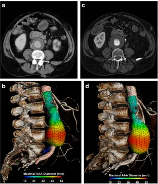
]. When the aneurysm growth rate exceeds
1 cm per year, the increased rupture risk justifies elective
AAA repair [
6
,
14
] (Fig.
1
).
Focal wall discontinuity
Many aneurysms are lined with circumferential wall
calcifications. In impending or complete AAA rupture,
a focal discontinuity of the intimal calcifications can be
seen indicating the rupture site, which is most
common-ly observed on the posterolateral wall [
18
]. Found in
8 % of ruptured aneurysms [
5
,
19
], this sign is most
useful when prior intact circumferential calcified walls
w e r e d o c u m e n t e d o n p r e v i o u s s c a n s ( F i g .
2
) .
Alternatively, an impending rupture site can also be
seen as a focal bulging of the aneurysm wall, also
described as an aortic
“
bleb.
”
This corresponds
histo-logically to inflammatory changes and a focal thinning
of elastic fibres [
20
].
Hyperattenuating crescent sign
Although intramural thrombus can be regarded as a protective
factor against rupture, in fact, intrinsic metabolic activity
within the thrombus weakens the underlying aortic wall
[
21
]. When intraluminal blood dissects into the thrombus
and comes into contact with the weakened aortic wall, rupture
risk is increased [
19
,
21
,
22
]. The dissecting blood can be seen
as a high-attenuating crescent underlying the intramural
thrombus or lying within the aortic wall [
7
]. The attenuation
of this crescent is greater than the aortic lumen on unenhanced
scans, and it remains greater than the psoas muscle on
en-hanced scans [
7
]. The sensitivity and specificity of the
hyperattenuating crescent sign to detect AAA rupture are
77 % and 93 %, respectively [
23
,
24
] (Fig.
3
).
Thrombus fissuration
F o l l o w i n g t h e s a m e c o n c e p t d e s c r i b e d f o r t h e
hyperattenuating crescent sign, blood dissection into
intramu-ral thrombus can be seen as thrombus fissuration [
7
]. This sign
is observed on enhanced CT as linear contrast infiltrations
from the patent aortic lumen through the intramural thrombus
(Fig.
4
). A study using mathematical models has suggested
that when thrombus fissurations extend from the aortic lumen
to the aneurysm wall, AAA wall tension may increase,
indi-cating impending aneurysm rupture [
25
].
Fig. 2 Focal wall discontinuity.aIllustration depicts a focal discontinu-ity (white arrow) of the calcified intima walls (C), indicating the AAA rupture site.bAxial unenhanced CT image of an asymptomatic 56-year-old man shows a 4.2 cm AAA with intact circumferential calcified walls. cAxial unenhanced CT image of the same patient who presented with severe lumbar pain 4 years later. The AAA now measures 5.7 cm, and a new 1 cm focal gap (white arrow) of the circumferential calcifications can be seen with periaortic fat infiltration near the rupture site (white arrowhead)
Draped aorta sign
A draped aorta sign can be seen in contained AAA ruptures,
when the rupture site is posterior and sealed by the adjacent
vertebral body [
8
]. The posterior wall of the aorta moulds to
the anterior surface of the vertebra. Normal fat planes between
the aneurysm and vertebra are lost. When the contained
rup-ture is chronic, smooth vertebral erosions can be observed in
up to 30 % of cases [
8
] (Fig.
5
).
Luminal signs
Aortoenteric fistula
Aortoenteric fistulas can be due to atherosclerotic disease,
without prior aortic surgery, or, much more commonly,
asso-ciated with a surgically repaired aneurysm. The latter fistulas
may develop long after the initial surgery [
26
]. Patients may
present with the classic triad of known AAA, abdominal pain,
and gastrointestinal bleeding. Although endoscopic
examina-tions are often negative, CT scans may show characteristic
findings. Imaging findings include intra- and extraluminal gas
within the aneurysmal sac and loss of fat planes between the
aneurysm and involved bowel segment. Contrast
extravasa-tion into the bowel lumen can sometimes be seen on enhanced
Fig. 4 Thrombus fissuration.aIllustration demonstrates blood (black arrow) dissecting from the aortic lumen (L) into a mural thrombus (T). The infiltrating blood is seen as linear fissurations (white arrows).bAxial andccoronal enhanced CT of a 64-year-old man shows linear infiltrations of contrast material (white arrows) within the hypodense mural thrombus. The patient underwent successful emergent AAA repair before complete rupture occurred
CT when the patient is actively bleeding. The third and fourth
portions of the duodenum are involved in over 50 % of
aortoenteric fistulas [
6
]. Aortoenteric fistulas have an up to
30
–
40 % mortality rate [
6
] (Fig.
6
).
Aortocaval fistula
An aortocaval fistula is a rare entity that affects less
than 1 % of AAAs and 2
–
4 % of ruptured aneurysms
[
27
,
28
]. They are associated with a 30 % mortality rate
[
6
]. Symptoms of congestive heart failure and venous
congestion can be observed clinically. On unenhanced
CT, normal fat planes between the AAA and inferior
vena cava (IVC) are obliterated, and the IVC may
appear dilated. On arterial phase CT angiography, the
AAA and IVC enhance simultaneously, whereas the
renal cortex and femoral arteries show later
enhance-ment [
28
] (Fig.
7
).
Extraluminal signs
Periaortic stranding
Periaortic fat stranding is frequently observed in impending
rupture and may be the earliest sign before complete AAA
rupture [
5
]. It represents oedema of the periaortic fat [
29
] and
can be seen before visualisation of a retroperitoneal
haematoma. Fat stranding is seen as an area of
high-attenuating fat surrounding a large aneurysm [
29
]. It can range
from a discrete and indistinct peripheral infiltration to a coarse
linear pattern [
29
] (Fig.
8
).
Contrast extravasation
Contrast extravasation is the most specific sign of a complete
AAA rupture. On arterial phase enhanced CT, contrast
mate-rial from the AAA lumen is seen escaping through the
bound-aries of the aneurysm wall into the retroperitoneal space.
Venous phase imaging can demonstrate an expanding pool
of contrast material that is increased in size compared to the
arterial phase images. This sign indicates unequivocal AAA
rupture (Fig.
9
).
Retroperitoneal haematoma
Given that AAAs are retroperitoneal and usually rupture
their posterior and posterolateral walls [
23
], the most
common sign of a ruptured AAA is the concomitant
presence of an AAA and an adjacent haematoma in
the retroperitoneal space [
2
,
19
]. Acute haemorrhage
can be seen as a high-attenuating fluid collection of
30 HU or more [
5
], although lower attenuation values
do not exclude haematoma, especially in patients with
anaemia. Bleeding may extend into multiple
retroperito-neal compartments, such as the perirenal, anterior and
posterior pararenal spaces, and along the psoas muscle
[
30
]. Large haematomas may displace the kidneys
later-ally and the small bowel anteriorly against the anterior
abdominal wall (Fig.
10
).
Intraperitoneal haematoma
Bleeding from a ruptured AAA may extend into the
intraperitoneal compartment. Haemoperitoneum can be
seen in the perihepatic space, within mesenteric folds,
along paracolic gutters, or within the pouch of Douglas.
Such intraperitoneal haematomas are more commonly
associated with rupture of the anterior or anterolateral
aortic aneurysm wall [
23
] (Fig.
11
).
Differential diagnosis
Some of the previously mentioned signs are not entirely
specific to impending or complete AAA rupture and can be
seen in other pathologies.
Intra- and extraluminal gas seen with aortoenteric fistulas
can also be found in infected (mycotic) aneurysms. Such
aneurysms often have a more saccular shape and irregular
walls that are devoid of calcifications [
5
]. Occasionally, signs
of primary infection sites such as vertebral osteomyelitis,
pyelonephritis, or retroperitoneal abscesses may provide clues
to the proper diagnosis. Clinically, these patients do not
pres-ent with gastrointestinal bleeding.
Periaortic stranding can also be seen in inflammatory
an-eurysms, associated with autoimmune diseases (lupus,
rheu-matoid arthritis, giant cell arteritis) and retroperitoneal fibrosis
[
31
]. In such cases, the aortic wall is thickened from long
standing inflammation. This thickening is outside the intimal
calcifications, may enhance with intravenous contrast
injec-tion, and frequently has more defined margins than periaortic
haematomas. Periaortic fibrosis can extend to adjacent organs,
such as the small bowel and ureters [
5
]. Malignancy, drug
toxicity, retroperitroneal injury, and infection are also
Fig. 7 Aortocaval fistula.a Axial arterial phase enhanced CT showssimultaneous enhancement of the AAA and IVC in an 83-year-old man with AAA rupture and retroperitoneal haematoma (R).bAxial enhanced CT of the same patient at a lower level demonstrates active contrast extravasation (white arrows) from the aortic aneurysm to the IVC with loss of normal fat planes between the structures. Retroperitoneal haematoma (R) can also be seen
associated with retroperitoneal fibrosis and need to be
includ-ed in the differential diagnosis of periaortic fibrosis [
32
].
Retroperitoneal haematoma can be the result of
haemor-rhage secondary to anticoagulation. Such haematomas are
often restricted to the posterior pararenal compartment,
typi-cally intramuscular within the psoas muscle, or at a distance
from the abdominal aorta. A
“
haematocrit effect
”
may also
be seen as a blood-fluid level [
33
]. Rarely, haematomas
secondary to other vascular injuries in the
retro-peritoneum, such as rupture of smaller lumbar, renal
capsular, or inferior pancreaticoduodenal arteries or
pseudoaneurysms, can mimic AAA rupture. Analysing
the images for other such vascular anomalies, relating
the arterial vascular anomalies to the anatomical site of
the haematoma, and correlating these to the clinical and
previous surgical history can help distinguish AAA
rup-ture from other arterial branch ruprup-tures.
Management
AAA rupture represents a major surgical emergency that
re-quires prompt intervention. However, an estimated 87.5 % of
patients admitted to the hospital with a ruptured AAA survived
longer than 2 h after their admission [
34
]. Furthermore, the
median time interval between admission and death from AAA
rupture was 10.5 h [
34
]. This suggests that most patients with
ruptured AAAs who reach the hospital alive are
haemodynam-ically stable enough to undergo preoperative imaging. CT
angi-ography permits confirmation of AAA rupture and evaluation
of AAA anatomy while intravenous fluid resuscitation is
initi-ated [
34
]. Pre-interventional imaging also allows assessing the
eligibility for an endovascular repair.
It has been reported that aneurysm repair can be slightly
delayed in haemodynamically stable patients who present with
symptomatic AAA without signs of complete rupture [
6
,
35
,
36
].
Urgent treatment is still warranted in these patients, who should
receive proper preoperative management and aneurysm repair
within 4 to 24 h of admission [
6
,
36
]. Such management
opti-mises preoperative conditions and reduces mortality rates to
those comparable to elective procedures (less than 5 %) [
6
,
35
,
36
]. Nevertheless, in haemodynamically unstable patients with
Fig. 9 Contrast extravasation.a-bAxial enhanced CT of a 75-year-oldwoman demonstrates active contrast extravasation (white arrows) from the aneurysm lumen (L) into the retroperitoneal space with massive retroperitoneal haematoma (R)
acute and complete AAA rupture, emergent and immediate
treatment is mandatory.
Open surgical repair is considered the standard treatment
for AAA. Surgical repair involves an extensive abdominal
Fig. 11 Intraperitoneal haematoma. a Illustration depicts AAA rupture of the left anterolateral wall (white arrow). Massive haem-orrhage extends into both the retroperitoneal (R, shown as trans-lucent red blood) and intraperitoneal (I, shown as more opaque blood) spaces. b Coronal unenhanced CT of a 51-year-old man demonstrates intraperitoneal haematoma involving the perihepatic space (PH), right (RG), and left (LG) paracolic gutters. Retroper-itoneal haematoma (white arrows) is also seen. Attenuation of 60 HU indicates acute haemorrhage. c Axial unenhanced CT of the same patient shows right (RG) and left (LG) paracolic gutters, as well as retroperitoneal haematoma (white arrows)
incision, clamping of the proximal abdominal aorta, and
su-turing of a graft that excludes the aneurysm [
6
] (Fig.
12
).
Despite important advances in technology and critical
care, very little or no significant reduction in mortality
rates in patients with open surgical repair of ruptured
AAA has been noted in over 2 decades, remaining
higher than 40 % [
37
–
40
].
EVAR was first introduced as an alternative to open
surgi-cal AAA repair in 1986 [
41
]. Under fluoroscopic guidance
using a transfemoral approach, a covered stent-graft bridges
the proximal neck of the AAA with one (mono-iliac
stent-graft) or both iliac arteries (bifurcated stent-stent-graft) to isolate its
walls from the arterial pressure (Fig.
13
) [
42
]. This excludes
the aneurysm from normal blood circulation, which leads to a
thrombosis of the aneurysmal sac. If a mono-iliac stent-graft is
used, a femoro-femoral bypass graft preserves opposite limb
perfusion [
42
].
In 1994, Yusuf et al. demonstrated that EVAR could also be
used in emergency situations, such as impending and even
complete AAA ruptures [
9
,
43
]. However, the logistical
chal-lenge of offering an endovascular service at any time limits the
widespread use of EVAR [
44
]. In addition, not all patients
presenting with a rupturing AAA are suitable for EVAR. Since
stent-graft sizing and selection need to be adapted to patient
anatomy, a relatively wide stent-graft inventory is required,
which can become cumbersome logistically and financially.
Pre-interventional imaging is required to evaluate aneurysm
anatomical suitability for an endovascular procedure. Some
favourable anatomical criteria include sufficient proximal
neck length, normal proximal and distal neck diameters,
lim-ited neck angulation, and a limlim-ited amount of mural thrombus
and calcification in the neck (Table
2
) [
6
,
45
–
49
]. This
imag-ing also needs to assess common femoral and iliac artery
anatomy as limited transfemoral access, seen in patients with
small iliac arteries or severe occlusive disease, may preclude
the possibility of an EVAR.
Refinement in technological equipment has allowed more
flexible inclusion criteria. For example, with the use of
intra-aortic balloon occlusion of the supra-renal aorta, EVAR has
become available to even haemodynamically unstable patients
[
49
]. The arrival of fenestrated stent-grafts allowed
deploy-ment of the stent-graft with coverage of the renal and superior
mesenteric arteries even in patients with short proximal AAA
necks, without inducing ischaemia in these aortic branch
territories, and thereby expanding anatomical suitability for
EVAR [
50
].
Fig. 13 Endovascular aneurysm repair.aCoronal enhanced CT image of an 85-year-old man presenting with an impending AAA rupture (same as Fig.5) demonstrates a large AAA (white arrow) with a long proximal neck (white arrowheads).b
Fluoroscopy image,caxial, andd 3D rendering of the same patient show a deployed aorto-biiliac stent-graft (white arrows)
Table 2 Anatomic features favourable for endovascular aneurysm repair [6,45–49]
Anatomic inclusion criteria
Proximal neck length more than 10 to 15 mm Proximal neck diameter less than 32 mm Iliac neck diameters less than 5 mm Neck angulation less than 60 to 90°
Unfortunately, because of differences in interventional
ex-perience levels amongst physicians, anatomical inclusion
criteria for EVAR still vary widely amongst medical centres,
with eligibility rates ranging from 34 to 100 % [
51
]. Most
studies, however, report eligibility rates over 50 % [
52
–
55
].
Outcome
A ruptured AAA often leads to disastrous consequences, with
an up to 90 % mortality rate [
56
,
57
]. Over half of the patients
do not reach the hospital alive [
37
,
58
]. Even for those who are
able to undergo open surgical repair, the mortality rate remains
higher than 40 % [
51
–
53
].
The arrival of EVAR for ruptured AAA has been associated
with lower mortality and morbidity, reduction in blood loss,
transfusion requirements, and hospital stay [
59
–
61
].
Short-term mortality rates vary from 11 to 45 % in the literature,
with most studies reporting a perioperative mortality rate
around 20 % [
62
–
64
]. Even in patients with haemodynamic
instability, the mortality rate with EVAR has been seen to be as
low as 18 % [
49
,
58
,
63
]. A study led by Bosch et al. found a
reduction in the 6-month mortality rate of EVAR over open
surgery by 26.5 % [
48
].
Such lower mortality and morbidity may be due to
the use of local, as opposed to general, anaesthesia, the
lower amount of blood loss, the minimally invasive
nature of EVAR, and fewer extra-aortic injuries
associ-ated with open surgery [
6
,
49
,
51
].
Despite improvements in perioperative mortality rates, no
studies have shown a long-term reduction in survival rates
with EVAR over open surgery [
65
–
67
]. Also, an irrefutable
reduction in mortality rates has not been confirmed in
randomised control studies [
9
,
10
,
44
].
Finally, EVAR is associated with endoleaks, which are the
most frequent long-term complications and may end up
caus-ing aneurysm rupture. CT imagcaus-ing at 1 and 12 months after
the initial intervention, followed by annual controls, is
there-fore recommended for early detection and prevention of
com-plications, such as endoleaks [
6
]. If an endoleak is observed
on follow-up imaging, it needs to be carefully analysed to
determine its origin. Types I (sealing zone endoleak) and III
(graft failure endoleak) need to be immediately addressed as
they should be considered the equivalent of an untreated AAA
[
42
]. Type II endoleaks (retrograde flow from a covered aortic
branch) are the most commonly encountered endoleaks [
42
].
If a type II endoleak is detected, a 6-month follow-up CT is
recommended [
6
], and re-intervention should be considered if
there is progression of the aneurysm diameter. Finally, if a
type V endoleak is suspected (endotension; progression of the
AAA diameter with no detectable endoleak on a dual-phase
CT), it should be closely followed with imaging and possibly
referred for surgical conversion.
Conclusion
AAA rupture occurs at the end of a continuum of growth and
wall weakening. Although ruptured AAAs are easily
diag-nosed, identifying impending rupture signs can be more
chal-lenging. This review describes the CT imaging findings that
may help identify impending rupture prior to complete
rup-ture, which has important consequences on treatment and
prognosis. Knowledge of alternative causes of retroperitoneal
stranding and haemorrhage may also facilitate the diagnosis.
EVAR has emerged as an alternative and possibly less morbid
method of treating ruptured AAAs over conventional open
surgical repair.
Sources of support that require acknowledgement Many of the figures, illustrations, and 3D rendering images were obtained through a project entitled“Morphologic Evaluation of Ruptured Abdominal Aortic Aneurysm by 3D Modeling”, which was funded by the following organisations:
Dr. An Tang was supported by the Centre de recherche du Centre Hospitalier de l’Université de Montréal (CRCHUM), the Fonds de recherche du Québec en Santé, and the Fondation de l’Association des Radiologistes du Québec (FRQS-ARQ #26993).
Dr. Gilles Soulez was supported by the Ministère du Développement Économique et Industriel (MDEIE), the Instituts de Recherche en Santé du Canada et Siemens Medical (IRSC-SME grant: ISO-93328), and the Fonds Recherche Québec-Santé (FRQS# 20241).
We would also like to thank Claude Kauffmann for the 3D rendering images and Sylvain Bélanger for the medical illustrations.
Open AccessThis article is distributed under the terms of the Creative Commons Attribution License which permits any use, distribution, and reproduction in any medium, provided the original author(s) and the source are credited.
References
1. Vu KN, Kaitoukov Y, Morin-Roy F et al (2014) Abdominal aortic aneurysms: rupture signs on computed tomography. Electronic ex-hibit at the ECR 2014. 20th annual meeting of the European Society of Radiology; Vienna, Austria. doi:10.1594/ecr2014/C-0789
2. Johnston KW, Rutherford RB, Tilson MD, Shah DM, Hollier L, Stanley JC (1991) Suggested standards for reporting on arterial aneurysms. Subcommittee on Reporting Standards for Arterial Aneurysms, Ad Hoc Committee on Reporting Standards, Society for Vascular Surgery and North American Chapter, International Society for Cardiovascular Surgery. J Vasc Surg 13:452–458 3. Bengtsson H, Sonesson B, Bergqvist D (1996) Incidence and
prev-alence of abdominal aortic aneurysms, estimated by necropsy studies and population screening by ultrasound. Ann N Y Acad Sci 800:1–24 4. Singh K, Bonaa KH, Jacobsen BK, Bjork L, Solberg S (2001) Prevalence of and risk factors for abdominal aortic aneurysms in a population-based study: the Tromso Study. Am J Epidemiol 154: 236–244
5. Bhalla S, Menias CO, Heiken JP (2003) CT of acute abdominal aortic disorders. Radiol Clin North Am 41:1153–1169
7. Arita T, Matsunaga N, Takano K et al (1997) Abdominal aortic aneurysm: rupture associated with the high-attenuating crescent sign. Radiology 204:765–768
8. Apter S, Rimon U, Konen E et al (2008) Sealed rupture of abdominal aortic aneurysms: CT features in 6 patients and a review of the literature. Abdom Imaging 35:99–105
9. Mayer D, Aeschbacher S, Pfammatter T et al (2012) Complete replacement of open repair for ruptured abdominal aortic aneurysms by endovascular aneurysm repair: a two-center 14-year experience. Ann Surg 256:688–695
10. Reimerink JJ, Hoornweg LL, Vahl AC et al (2013) Endovascular repair versus open repair of ruptured abdominal aortic aneurysms: a multicenter randomized controlled trial. Ann Surg 258:248–256 11. Lederle FA, Johnson GR, Wilson SE et al (2002) Rupture rate of large
abdominal aortic aneurysms in patients refusing or unfit for elective repair. JAMA 287:2968–2972
12. Hans SS, Jareunpoon O, Balasubramaniam M, Zelenock GB (2005) Size and location of thrombus in intact and ruptured abdominal aortic aneurysms. J Vasc Surg 41:584–588
13. Brewster DC, Cronenwett JL, Hallett JW Jr, Johnston KW, Krupski WC, Matsumura JS (2003) Guidelines for the treatment of abdominal aortic aneurysms. Report of a subcommittee of the Joint Council of the American Association for Vascular Surgery and Society for Vascular Surgery. J Vasc Surg 37:1106–1117
14. Limet R, Sakalihassan N, Albert A (1991) Determination of the expansion rate and incidence of rupture of abdominal aortic aneu-rysms. J Vasc Surg 14:540–548
15. Simoni G, Beghello A, Buscaglia M, Ermirio D, Caprio J (2002) Growth rate of abdominal aortic aneurysms. Ultrasounds study and clinical outcome. Minerva Cardioangiol 50:371–377
16. Collaborators R, Bown MJ, Sweeting MJ, Brown LC, Powell JT, Thompson SG (2013) Surveillance intervals for small abdominal aortic aneurysms: a meta-analysis. JAMA 309:806–813
17. Lindholt JS, Heegaard NHH, Vammen S, Fasting H, Henneberg EW, Heickendorff L (2001) Smoking, but not lipids, lipoprotein (a) and antibodies against oxidised LDL, is correlated to the expansion of abdominal aortic aneurysms. Eur J Vasc Endovasc Surg 21:51–56 18. Fillinger MF, Racusin J, Baker RK et al (2004) Anatomic
character-istics of ruptured abdominal aortic aneurysm on conventional CT scans: Implications for rupture risk. J Vasc Surg 39:1243–1252 19. Siegel CL, Cohan RH, Korobkin M, Alpern MB, Courneya DL,
Lederı RA (1994) Abdominal aortic aneurysm morphology: CT features in patients with ruptured and nonruptured aneurysms. AJR Am J Roentgenol 163:1123–1129
20. Hunter GC, Leong SC, Yu GSM, McIntyre KE, Bernhard VM (1989) Aortic blebs: possible site of aneurysm rupture. J Vasc Surg 10:93–99 21. Roy J, Labruto F, Beckman MO, Danielson J, Johansson G, Swedenborg J (2008) Bleeding into the intraluminal thrombus in abdominal aortic aneurysms is associated with rupture. J Vasc Surg 48:1108–1113
22. Pillari G, Chang JB, Zito J et al (1988) Computed tomography of abdominal aortic aneurysm. An in vivo pathological report with a note on dynamic predictors. Arch Surg 123:727–732
23. Schwartz SA, Taljanovic MS, Smyth S, O’Brien MJ, Rogers LF (2007) CT findings of rupture, impending rupture, and contained rupture of abdominal aortic aneurysms. AJR Am J Roentgenol 188: W57–62
24. Mehard WB, Heiken JP, Sicard GA (1994) High-attenuating crescent in abdominal aortic aneurysm wall at CT: a sign of acute or impending rupture. Radiology 192:359–362
25. Polzer S, Gasser TC, Swedenborg J, Bursa J (2011) The impact of intraluminal thrombus failure on the mechanical stress in the wall of abdominal aortic aneurysms. Eur J Vasc Endovasc Surg 41:467–473 26. Orton DF, LeVeen RF, Saigh JA et al (2000) Aortic prosthetic graft infections: radiologic manifestations and implications for manage-ment. Radiographics 20:977–993
27. Fenster MS, Dent JM, Tribble C et al (1996) Aortocaval fistula complicating abdominal aortic aneurysm: case report and literature review. Cathet Cardiovasc Diagn 38:75–79
28. Coulier B, Tilquin O, Etienne P-Y (2004) Multidetector row CT diagnosis of aortocaval fistula complicating aortic aneurysm: a case report. Emerg Radiol 11:100–103
29. Thornton E, Mendiratta-Lala M, Siewert B, Eisenberg RL (2011) Patterns of fat stranding. AJR Am J Roentgenol 197:W1–W14 30. Ando M, Igari T, Yokoyama H, Satokawa H (2003) CT features of
chronic contained rupture of an abdominal aortic aneurysm. Ann Thorac Cardiovasc Surg 9:274–278
31. Haug ES, Skomsvoll JF, Jacobsen G, Halvorsen TB, Sæther OD, Myhre HO (2003) Inflammatory aortic aneurysm is associated with increased incidence of autoimmune disease. J Vasc Surg 38:492–497 32. Vaglio A, Salvarani C, Buzio C (2006) Retroperitoneal fibrosis.
Lancet 367:241–251
33. Federle MP, Pan KT, Pealer KM (2007) CT criteria for differentiating abdominal hemorrhage: anticoagulation or aortic aneurysm rupture? AJR Am J Roentgenol 188:1324–1330
34. Lloyd GM, Bown MJ, Norwood MG et al (2004) Feasibility of preoperative computer tomography in patients with ruptured abdom-inal aortic aneurysm: a time-to-death study in patients without oper-ation. J Vasc Surg 39:788–791
35. Jones C, Reilly M, Dalsing M, Glover J (1986) Chronic contained rupture of abdominal aortic aneurysms. Arch Surg 121:542–546 36. Cambria R, Gloviczki P, Stanson A et al (1994) Symptomatic,
nonruptured abdominal aortic aneurysms: are emergent operations necessary? Ann Vasc Surg 8:121–126
37. Bengtsson H, Bergqvist D (1993) Ruptured abdominal aortic aneu-rysm: a population-based study. J Vasc Surg 18:74–80
38. Visser P, Akkersdijk G, Blankensteijn J (2005) In-hospital operative mortality of ruptured abdominal aortic aneurysm: a population-based analysis of 5593 patients in The Netherlands over a 10-year period. Eur J Vasc Endovasc Surg 30:359–364
39. Bown M, Sutton A, Bell P, Sayers R (2002) A meta-analysis of 50 years of ruptured abdominal aortic aneurysm repair. Br J Surg 89:714–730 40. Heller J, Weinberg A, Arons R et al (2000) Two decades of
abdom-inal aortic aneurysm repair: have we made any progress? J Vasc Surg 32:1091–1100
41. Volodos NL, Shekhanin VE, Karpovich IP, Troian VI, Gur’ev Iu A (1986) A self-fixing synthetic blood vessel endoprosthesis. Vestn Khir Im I I Grek 137:123–125
42. Iezzi R, Santoro M, Dattesi R et al (2012) Multi-detector CT angio-graphic imaging in the follow-up of patients after endovascular abdominal aortic aneurysm repair (EVAR). Insights Imaging 3:313– 321
43. Yusuf SW, Whitaker SC, Chuter TA, Wenham PW, Hopkinson BR (1994) Emergency endovascular repair of leaking aortic aneurysm. Lancet 344:1645
44. Hinchliffe RJ, Bruijstens L, MacSweeney ST, Braithwaite BD (2006) A randomised trial of endovascular and open surgery for ruptured abdominal aortic aneurysm—results of a pilot study and lessons learned for future studies. Eur J Vasc Endovasc Surg 32:506–513 45. Iezzi R, Cotroneo AR (2006) Endovascular repair of abdominal
aortic aneurysms: CTA evaluation of contraindications. Abdom Imaging 31:722–731
46. Richards T, Goode SD, Hinchliffe R, Altaf N, Macsweeney S, Braithwaite B (2009) The importance of anatomical suitability and fitness for the outcome of endovascular repair of ruptured abdominal aortic aneurysm. Eur J Vasc Endovasc Surg 38:285–290
47. Slater B, Harris E, Lee J (2008) Anatomic suitability of ruptured abdominal aortic aneurysms for endovascular repair. Ann Vasc Surg 22:716–722
49. Mehta M (2010) Endovascular aneurysm repair for ruptured abdom-inal aortic aneurysm: the Albany Vascular Group approach. J Vasc Surg 52:1706–1712
50. Cross J, Gurusamy K, Gadhvi V et al (2012) Fenestrated endovascular aneurysm repair. Br J Surg 99:152–159
51. Harkin D, Dillon M, Blair P, Ellis P, Kee F (2007) Endovascular ruptured abdominal aortic aneurysm repair (EVRAR): a systematic review. Eur J Vasc Endovasc Surg 34:673–681
52. Hoornweg LL, Wisselink W, Vahl A, Balm R (2007) The Amsterdam Acute Aneurysm Trial: suitability and application rate for endovascular repair of ruptured abdominal aortic aneurysms. Eur J Vasc Endovasc Surg 33:679–683
53. Mastracci T, Garrido-Olivares L, Cinà C, Clase C (2008) Endovascular repair of ruptured abdominal aortic aneurysms: a sys-tematic review and meta-analysis. J Vasc Surg 47:214–221 54. Peppelenbosch N, Yilmaz N, van Marrewijk C et al (2003)
Emergency treatment of acute symptomatic or ruptured abdominal aortic aneurysm. Outcome of a prospective intent-to-treat by EVAR protocol. Eur J Vasc Endovasc Surg 26:303–310
55. Hassen-Khodja R, Jean-Baptiste E, Haudebourg P, Declemy S, Batt M, Bouillanne P (2007) Endovascular repair of ruptured aneurysms of the infrarenal abdominal aorta: feasibility and results. J Cardiovasc Surg (Torino) 48:491–495
56. Patel MI, Hardman DT, Fisher CM, Appleberg M (1995) Current views on the pathogenesis of abdominal aortic aneurysms. J Am Coll Surg 181:371–382
57. Vorp DA, Vande Geest JP (2005) Biomechanical determinants of abdominal aortic aneurysm rupture. Arterioscler Thromb Vasc Biol 25:1558–1566
58. Starnes B, Quiroga E, Hutter C et al (2010) Management of ruptured abdominal aortic aneurysm in the endovascular era. J Vasc Surg 51:9–17
59. Sadat U, Hayes PD, Gaunt ME, Varty K, Boyle JR (2008) Assessment of pre-operative delays in the management of elective abdominal aortic aneurysms. Ann R Coll Surg Engl 90:65–68 60. Sadat U, Boyle JR, Walsh SR, Tang T, Varty K, Hayes PD (2008)
Endovascular vs open repair of acute abdominal aortic aneu-rysms—a systematic review and meta-analysis. J Vasc Surg 48:227–236
61. Mayer D, Pfammatter T, Rancic Z et al (2009) 10 years of emergency endovascular aneurysm repair for ruptured abdominal aortoiliac an-eurysms: lessons learned. Ann Surg 249:510–515
62. Dalainas I, Nano G, Bianchi P et al (2006) Endovascular techniques for the treatment of ruptured abdominal aortic aneurysms: 7-year intention-to-treat results. World J Surg 30:1809–1814
63. Veith F, Gargiulo N (2007) Endovascular aortic repair should be the gold standard for ruptured AAAs, and all vascular surgeons should be prepared to perform them. Perspect Vasc Surg Endovasc Ther 19: 275–282
64. Rayt H, Sutton A, London N, Sayers R, Bown M (2008) A system-atic review and meta-analysis of endovascular repair (EVAR) for ruptured abdominal aortic aneurysm. Eur J Vasc Endovasc Surg 36: 536–544
65. Becquemin JP, Pillet JC, Lescalie F et al (2011) A randomized controlled trial of endovascular aneurysm repair versus open surgery for abdominal aortic aneurysms in low- to moderate-risk patients. J Vasc Surg 53:1167–1173
66. De Bruin JL, Baas AF, Buth J et al (2010) Long-term outcome of open or endovascular repair of abdominal aortic aneurysm. N Engl J Med 362:1881–1889


![Table 2 Anatomic features favourable for endovascular aneurysm repair[6, 45–49]](https://thumb-us.123doks.com/thumbv2/123dok_us/881997.2081753/10.595.228.544.51.336/table-anatomic-features-favourable-endovascular-aneurysm-repair.webp)
