Published by Science and Education Publishing DOI:10.12691/ajeid-8-2-1
Profile of Protozoa Isolated from Stool Samples in
Yaounde, Cameroon
Laure NGANDO1,2,*, Leopold MBOUS NGUIMBUS3,4, Claris KILLA3,4, Thérèse NKOA1
1
Department of Microbiology, Faculty of Medecine and Biomedical Sciences, University of YaoundeI, Yaounde, Cameroon
2
Laboratory of Bacteriology/Parasitology, Centre Pasteur of Cameroon, Yaounde, Cameroon
3
Department of Biochemistry, Faculty of Sciences, University of Yaounde I,Yaounde, Cameroon
4
Catholic University of Central Africa, School of Health Science, Yaounde, Cameroon *Corresponding author: laurengando2013@gmail.com
Received May 03, 2020; Revised June 05, 2020; Accepted June 12, 2020
Abstract
Background: Intestinal parasitic infestations are among the most common communicable diseases in the world with a higher prevalence in developing countries. They are caused by protozoa which have long been associated with foodborne and waterborne disease outbreaks. The aim of our study was to present the profile of protozoa isolated from stool samples in Yaounde from 2010-2020 and to analyse the association of intestinal parasitic diseases with age and gender during the same period. Methods: This retrospective and observational study was carried out from January 04, 2010 to January 10, 2020 in Yaounde, capital of the Center region, at Centre Pasteur of Cameroon. After collecting the stool samples, the intestinal protozoa were identified using the Bailenger concentration technique and staining with Kop-Color II. Microscopic observation between slide and coverglass was focused on trophozoites and cysts of protozoa. Results: A total of 106.846 stool samples were analyzed during the study period and the overall infestation rate of intestinal protozoa was 8.4% (8958 samples positive for the presence of a protozoan). Women were the most represented with 5697 samples (9.0%) compared to 3052 (7.5%) samples for men. This difference in gender distribution was significant (p<0.0001). The participants were between 1–105 years (mean±SD = 42.6±19.4) of age. The age distribution of the patients showed that the age group with the highest prevalence of infestation (9.4%) ranged from 21–40 years with a significant difference in distribution (p<0.001) from one age group to another. A significant decrease of stool samples was also observed depending on the years of the study (p<0.0001). The distribution of identified protozoa was: 3.3% for Entamoeba hartmanni, 1.9% forEntamoeba coli, 1.8% for Entamoeba histolytica histolytica, 0.4% for Trichomonas intestinalis, 0.4% for
Entamoeba histolytica minuta, 0.3% for Gardia duodenalis, 0.2% for Chilomatix mesnilii, 0.1% for Endolimaxnana, 0.04% for Isospora belli, 0.02% for Balantidium coli, 0.006% for Cyclospora cayetanensis and 0.003% for
Pseudolimax butschlii. A statistically significant association of age groups (p<0.0001) and sex (p<0.0001) with the identified protozoa was obtained in our study with a higher risk of infestation in women and those of the 21–40 years age group were the most vulnerable. Conclusion: The overall infestation rate of intestinal protozoa is high in Yaounde with highest contamination being amongst women and people 32 years of age. Moreover, despite the significant decrease of infestations over the years, measures must still be taken to prevent diseases caused by intestinal protozoa in the Cameroonian context.
Keywords: intestinal parasitic infestations, communicable diseases, protozoa, technique of Bailenger, Kop-Color II
Cite This Article:
Laure NGANDO, Leopold MBOUS NGUIMBUS, Claris KILLA, and Thérèse NKOA, “Profile of Protozoa Isolated from Stool Samples in Yaounde, Cameroon.” American Journal of Epidemiology and Infectious Disease, vol. 8, no. 2 (2020): 48-55. doi: 10.12691/ajeid-8-2-1.1. Introduction
Intestinal parasitic infestations (IPIs) are among the most common communicable diseases worldwide, especially in developing countries [1]. These infestations, the frequency of which constitutes an indicator of socioeconomic development [2], are caused by protozoan parasites, unicellular organisms long associated with foodborne and waterborne disease outbreaks in humans [3], but also closely related to environments regularly
infested with faecal matter [4]. Al-Jawabreh et al. [5], reports that the World Health Organization (WHO) estimates the following protozoa, Giardia lamblia (G. lamblia), Entamoeba histolytica (E. histolytica) and
Cryptosporidium spp. are most responsible for diarrheal illnesses in humans and the median global burden of disease (GBD) in 2010 for these parasitic species were 0.17 million, 0.5 million, and 2 million disability-adjusted life years (DALYs), respectively.
year, which places this disease in second place among parasitic infestations in terms of mortality after malaria
[6,7]. However, if we refer to the epidemiological data
history of E. histolytica infestations, these may be overestimated because they were obtained before the formal separation of this parasite into two identical species morphologically : the nonpathogenic E. dispar species and the pathogenic E. histolytica species [8].
Giardiasis or lambliosis is a common intestinal infestation worldwide caused by a flagellate intestinal protozoan called Giardia duodenalis (also known as G. intestinalis or G. lamblia) [6]. It is among the most common intestinal parasite in humans with around 280 million people infested every year [9]. Moreover, the WHO estimates that 200 million people in Asia, Africa and Latin America have symptomatic infestations with a lower prevalence of the disease in developed countries (0.4 to 7.5%) than in developing countries (8 to 30%) [6]. Like the other protozoans, the pathology is transmitted by faeco-oral route, direct human-to-human or through the consumption of contaminated water and food [6,9,10].
Besides the two previous diseases, three other intestinal protozoan infestations caused by coccidians are also known. These are cryptosporidiosis due to the genus
Cryptosporidium, cystoisosporosis due to Cystoisospora belli (or Isospora belli) and cyclosporosis due to the species Cyclospora cayetanensis all commonly responsible for traveler syndrome and the epidemiological outbreaks in the world [6,11]. Cryptosporidium spp., Cyclospora cayetanensis and Cystoisospora belli are known to be agents responsible for diarrhoea in both immunocompetent and immunocompromised individuals with Cryptosporidium spp.
being the major cause of diarrhea in children in developing countries and associated with mental growth retardation in early childhood [12]. About 0.6-2% (7% in children) of immunocompetent individuals in developed countries suffer from diarrhea caused by Cryptosporidium spp., while in developing countries, the prevalence of cryptosporidiosis is between 4 and 32%. For immunocompromised HIV/AIDS patients, the prevalence is 14% in industrialized countries and up to 60% in Africa and Haiti. Cystoisosporosis with a variable frequency which can reach more than 10% in people living with HIV (PLHIV) in areas where the level of hygiene is low. As for cyclosporosis caused by Cyclospora cayetanensis, its geographic distribution remains poorly understood although it can be endemic and sometimes epidemic [6].
In Cameroon, the prevalence of intestinal parasites vary according to the sites and years of study in the adult population: 14.6% in Dschang in the Western region in 2013; 27.9% in Douala, Littoral region the same year; 57.48% in Yaounde, Center region; 82.6% in Buea in the Fako division, South West region in 2017; and 32.3% in PLHIV and IPIs victims compared to 32.3% of HIV-negative patients in Ngaoundere, Adamawa region [1]. In order to have and keep precise information on the prevalence of intestinal infestations caused by protozoans in our society, it is important to know the species responsible for the protozoal infestation in order to sensitize the population on the implementation of measures to prevent the diseases caused by these agents, starting with control of the contamination routes. The aim of our study was to present the profile of protozoa isolated
from stool samples in Yaounde from 2010-2020 and to analyse the association of intestinal parasitic diseases with age and gender during the same period.
2. Materials and Methods
2.1. Place and Period of Study
This study was carried out from January 04, 2010 to January 10, 2020 in Yaounde, which is the capital city, more precisely at Centre Pasteur of Cameroon (CPC) which is the technical body of the Ministry of Public Health of Cameroon and member of the International Network of Pasteur institutes.
2.2. Type of Study and Samples
This observational and retrospective study focused on stool samples from patients from several health institutions in the city of Yaounde presenting a clinical picture of an intestinal parasitic infestation and who came to CPC for a stool examination.
2.3. Sample Collection and Analysis
Freshly produced stools (3-4 grams for solid stools and 2–3 ml for liquid stools) from each patient were collected in a properly identified vial and transferred to the parasitology laboratory for analysis. Once in the laboratory, the stool samples were concentrated by the technique of Bailenger and the examination of the sediment was carried out after staining with Kop-Color II for the detection of parasitic elements (trophozoites and cysts) between slide and coverglass [13,14].
2.4. Data Collection and Statistical Analysis
The data were taken from the CPC GLIMS data management software. These data were collected taking into consideration the date of collection, sex, age, identified protozoa and their classifications. After extraction of the data from the GLIMS system, the database was cleaned using Microsoft Office Excel 2019 software and the statistical analysis was conducted using R language version 3.6.2 (2019–12–12) [15] with R package “finalfit” (version 0.9.4) [16]. This “finalfit” package of the R software was used to produce the tables. The qualitative variables (sex, years, age groups, isolated protozoa) were analyzed by the Pearson Chi-square test, the Pearson’s Chi-square test with Yate’s continuity correction and The Fisher’s exact test. The continuous variable (age) was analyzed with the non-parametric Kruskal-Wallis test. The level of significance was set at p<0.05.
3. Results
3.1. Characteristics of Study Population
were positive for intestinal infestation caused by a protozoan. The number of positive stool samples from male subjects was 3052 (7.5%) compared to 5697 (9.0%) from female subjects. This difference in proportion was statistically significant (p<0.001) (Table 1). The participants were between 1–105 years (mean±SD = 42.6±19.4) of age. The age distribution of the patients in groups showed that the age group with the highest prevalence of infestation
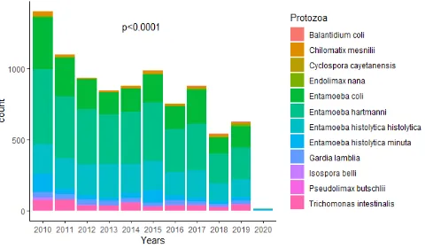
(9.4%) ranged from 21–40 years with a significant difference in distribution (p<0.0001) from one age group to another. The difference in the distribution of the age groups according to sex was statistically significant (p<0.001) (Table 1). The number of stool samples decreased significantly (p<0.0001) according to the years with the year 2010 having the largest number of samples infested with protozoans (Figure 1).
Table 1. Distribution of sociodemographic variables according to age groups
Age (years)
Variables
<21 (n=23.453)
No. (%)
21-40 (n=37.497) No.
(%)
41-60 (n=26.912) No.
(%)
61-80 (n=13.383) No.
(%)
≥81 (n=2586)
No. (%)
Total (n=106.846)
No. (%)
P-value
Age
Mean (SD) 12.7 (4.9) 31.9 (5.3) 50.0 (5.9) 69.2 (5.6) 85.5 (4.6) 42.6 (19.4) <0.001*
Sex
Men 508 (2.2) 912 (2.4) 986 (3.7) 541 (4.0) 105 (4.1) 3052 (2.8) <0.001
Women 532 (2.3) 2618 (7.0) 1518 (5.6) 804 (6.0) 225 (9.9) 5697 (5.3)
Years
2010 137 (0.6) 496 (1.3) 408 (1.5) 255 (1.9) 70 (2.7) 1366 (1.3) <0.001**
2011 143 (0.6) 390 (1.0) 329 (1.2) 162 (1.2) 52 (2.0) 1076 (1.0)
2012 90 (0.4) 357 (1.0) 272 (1.0) 136 (1.0) 53 (2.0) 908 (0.8)
2013 106 (0.5) 286 (0.8) 250 (0.9) 144 (1.1) 43 (1.7) 829 (0.8)
2014 113 (0.5) 363 (1.0) 227 (0.8) 132 (1.0) 30 (1.2) 865 (0.8)
2015 117 (0.5) 425 (1.1) 270 (1.0) 133 (1.0) 25 (1.0) 970 (0.9)
2016 86 (0.4) 344 (0.9) 189 (0.7) 99 (0.7) 17 (0.7) 735 (0.7)
2017 96 (0.4) 382 (1.0) 246 (0.9) 122 (0.9) 15 (0.6) 861 (0.8)
2018 79 (0.3) 235 (0.6) 144 (0.5) 55 (0.4) 15 (0.6) 528 (0.5)
2019 72 (0.3) 246 (0.7) 167 (0.6) 101 (0.8) 10 (0.4) 596 (0.6)
2020 1 (0.0) 6 (0.0) 2 (0.0) 6 (0.0) 0 (0.0) 15 (0.0)
Overall prevalence 1040 (4.4) 3530 (9.4) 2504 (9.3) 1345 (10.1) 330 (12.8) 8958 (8.4) <0.0001
P-value : p-value of Pearson's Chi-squared test *: p-value of Kruskal-Wallis rank sum test
**: p-value of Pearson's Chi-squared test with Yate's continuity correction.
Table 2. Distribution of isolated protozoa from stool samples
Protozoa isolate Number % for
n = 8958
% for n = 106846
Entamoeba hartmanni** 3549 39.6 3.3
Entamoeba coli** 2020 22.5 1.9
Entamoeba histolytica histolytica* 1893 21.1 1.8
Trichomonas intestinalis* 456 5.1 0.4
Entamoeba histolytica minuta* 451 5.0 0.4
Gardia duodenalis* 296 3.3 0.3
Chilomatix mesnilii** 168 1.9 0.2
Endolimax nana** 49 0.5 0.1
Isospora belli* 46 0.5 0.0
Balantidium coli* 21 0.2 0.0
Cyclospora cayetanensis* 6 0.1 0.0
Pseudolimax butschlii* 3 0.0 0.0
Total 8958 100.00 8.4
*: pathogenic protozoa **: nonpathogenic protozoa.
3.2. Distribution of Identified Protozoa
Among the positive protozoa infested stool samples, 7965 (7.5%) were positive for protozoa of the rhizopod class, 920 (0.9%) to those of the flagellate class, 52 (0.05%) to those of the sporozoa and 21 (0.02%) were positive to the protozoa of the ciliate branch. Identification of the germs after concentration and staining
showed that Entamoeba hartmanni was the most represented parasitic species, 3549 (3.3%) positive samples for this germ. Next, Entamoeba coli (E. coli) followed by E. histolytica histolytica were the two other species to be highly represented with 2020 (1.9%) and 1893 (1.8%) samples respectively. The proportion of the other identified protozoa are presented in Table 2.
Table 3. Distribution of identified protozoa according to sex
Sex
Isolated protozoa Men (n=40.935)
No. (%)
Women (n=63.058) No. (%)
Total (n=106.846)
No. (%) P-value
Balantidium coli 5 (0.0) 15 (0.0) 20 (0.0) 0.3531
Chilomatix mesnilii 50 (0.1) 113 (0.2) 163 (0.2) 0.255
Cyclospora cayetanensis 4 (0.0) 2 (0.0) 6 (0.0) 0.1922*
Endolimax nana 14 (0.0) 33 (0.1) 47 (0.0) 0.4623
Entamoeba coli 650 (1.6) 1329 (2.1) 1979 (1.9) 0.0305
Entamoeba hartmanni 1156 (2.8) 2302 (3.7) 3458 (3.3) 0.021
Entamoeba histolytica histolytica 712 (1.7) 1144 (1.8) 1856 (1.8) <0.001
Entamoeba histolytica minuta 175 (0.4) 264 (0.4) 439 (0.4) 0.0247
Gardia duodenalis 122 (0.3) 160 (0.3) 282 (0.3) 0.0027
Isospora belli 23 (0.1) 23 (0.0) 46 (0.0) 0.0424*
Pseudolimax butschlii 1 (0.0) 2 (0.0) 3 (0.0) 1.000*
Trichomonas intestinalis 140 (0.3) 310 (0.5) 450 (0.4) 0.0847
Overall prevalence 3052 (7.5) 5697 (9.0) 8958 (8.4) <0.0001
P-value : p-value of Pearson's Chi-squared test * : p-value of Fisher's exact test.
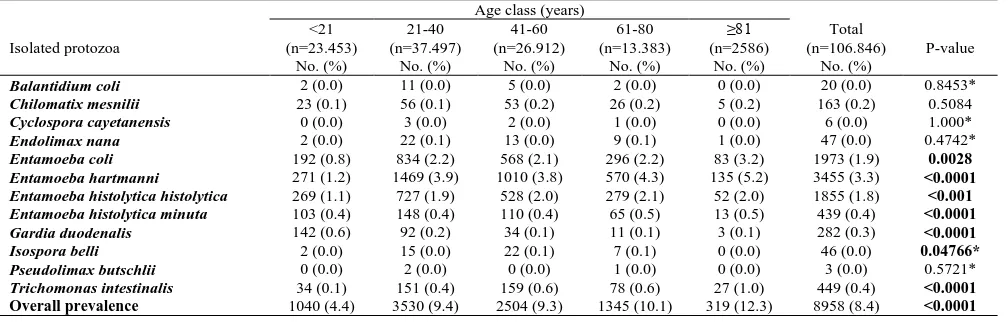
Table 4. Distribution of identified protozoa according to age groups
Age class (years)
Isolated protozoa
<21 (n=23.453)
No. (%)
21-40 (n=37.497)
No. (%)
41-60 (n=26.912)
No. (%)
61-80 (n=13.383)
No. (%)
≥81 (n=2586)
No. (%)
Total (n=106.846)
No. (%)
P-value
Balantidium coli 2 (0.0) 11 (0.0) 5 (0.0) 2 (0.0) 0 (0.0) 20 (0.0) 0.8453*
Chilomatix mesnilii 23 (0.1) 56 (0.1) 53 (0.2) 26 (0.2) 5 (0.2) 163 (0.2) 0.5084
Cyclospora cayetanensis 0 (0.0) 3 (0.0) 2 (0.0) 1 (0.0) 0 (0.0) 6 (0.0) 1.000*
Endolimax nana 2 (0.0) 22 (0.1) 13 (0.0) 9 (0.1) 1 (0.0) 47 (0.0) 0.4742*
Entamoeba coli 192 (0.8) 834 (2.2) 568 (2.1) 296 (2.2) 83 (3.2) 1973 (1.9) 0.0028
Entamoeba hartmanni 271 (1.2) 1469 (3.9) 1010 (3.8) 570 (4.3) 135 (5.2) 3455 (3.3) <0.0001
Entamoeba histolytica histolytica 269 (1.1) 727 (1.9) 528 (2.0) 279 (2.1) 52 (2.0) 1855 (1.8) <0.001
Entamoeba histolytica minuta 103 (0.4) 148 (0.4) 110 (0.4) 65 (0.5) 13 (0.5) 439 (0.4) <0.0001
Gardia duodenalis 142 (0.6) 92 (0.2) 34 (0.1) 11 (0.1) 3 (0.1) 282 (0.3) <0.0001
Isospora belli 2 (0.0) 15 (0.0) 22 (0.1) 7 (0.1) 0 (0.0) 46 (0.0) 0.04766*
Pseudolimax butschlii 0 (0.0) 2 (0.0) 0 (0.0) 1 (0.0) 0 (0.0) 3 (0.0) 0.5721*
Trichomonas intestinalis 34 (0.1) 151 (0.4) 159 (0.6) 78 (0.6) 27 (1.0) 449 (0.4) <0.0001
Overall prevalence 1040 (4.4) 3530 (9.4) 2504 (9.3) 1345 (10.1) 319 (12.3) 8958 (8.4) <0.0001
3.3.Association of Identified Protozoan
Species with Age and Sex
An association between parasitic infestation and gender has been observed. In fact, the proportion of parasitic infestations was higher in women with 5697 (9.0%) samples compared to 3052 (7.5%) in men, a proportion difference which was statistically significant (p<0.0001). The number of stool samples infested with rhizopods
E. hartmanni, E. coli, E. histolyticahistolytica and
E.histolytica minuta was significantly higher in women (2302, 1329, 1144 and 264 respectively) compared to men (1156, 650, 712 and 175 respectively). The p-values were respectively: p=0.021, p=0.0305, p<0.001 and p=0.0247
(Table 3). Among the flagellated protozoa, only G.
duodenalis was associated with the genus (p=0.0027). The sporozoan Isospora belli (I. belli) was also the only one to be associated with the genus (p=0.0424) (Table 3).
In relation to the age groups, Table 4 gives a detailed description of the distribution of the identified protozoa. As with the genus, E. hartmanni was the most common protozoan in all age groups and the difference in proportion of this germ from one age group to another was statistically significant (p<0.0001). E. coli was the second most represented protozoan in people aged at least 21 years (≥ 21 years) and the third in patients whose age was strictly less than 21 years (<21 years) with a p-value of significant Chi-square test (p=0.0028). Thespecies E. histolytica hystolytica was thethird to be represented in the
≥ 21 years old age group and the second in the <21 years old age group with a significant difference in distribution (p<0.001). Trichomonas intestinalis (T. intestinalis) was the fourth most common germ in those ≥ 21 years of age followed by E. histolytica minuta which was the fifth most represented at all age groups with a significant difference in distribution (p<0.0001). The age group most affected by the infestation was that of 21–40 years followed by that of 41–60 years with a significant difference in distribution from one age group to another (p<0.0001).
4. Discussion
In this retrospective observational study, which took place in Yaounde at CPC, 8958 (8.4%) stool samples were positive for the presence of a pathogenic or nonpathogenic intestinal protozoan with a significant decrease in the number of sample depending on the year (p<0.0001). This frequency of IPIs is higher than that of previous studies
[17,18,19,20] where the prevalence of IPIs were 3.39%,
5.4%, 5.75% and 5.93% respectively. Regarding the situation in Cameroon, the prevalence of intestinal protozoa in this study was lower than that found by Abange et al. [1] in Yaounde with 20.2% of parasitic infestations in HIV-infected patients and 15.8% in people not-infected with HIV, or that found in the same region at the Central Hospital by Vouking et al.[21], with 57.58% of parasitic infestations. In other regions, the results of other studies also show a higher proportion of these infestations compared to the present study [22,23,24,25]. This difference in the prevalence of parasitic infestations could be justified by the identification method used from one study to another as shown by Nsagha et al. [25] or
even Nkenfou et al. [24]; the immune status of the target population as reported by Nkenfou et al. [22], with a higher risk in PLHIV and whose CD4 count is relatively low; the environmental conditions of the study environment and risk behaviors as mentioned in certain studies [22,26,27,28]; as well as the density of the target population [20,29], and their nationality [30].
The significant decrease in the number of parasitic infestations over the years (p<0.0001) in the present study is in contradiction with the study by Diongue et al.[31], where the prevalence of parasitic infestations increased significantly between 2011 and 2015 (p<0.001) due to the persistence of risk behaviors in populations; and in agreement with the research of Abu-Madi et al. [20], where the prevalence of combined protozoa in Qatar decreased significantly from 2004 to 2014 (𝝌𝝌𝟐𝟐=167.4, p<0.001) due to progress in the prevention of intestinal infestations brought by public health and success in the social integration of immigrants who have come to work in this city, or in the study by Faria et al.[29], in which we note a decrease in the number of parasitic infestations between 2012 and 2015. In our context, this significant decrease in intestinal infestations could be justified by the improvement in the hygienic conditions of populations over the last ten decades and increase in awareness and preventive measures implemented by actors of public health.
In this study, the minimum age of patients was 1 year and the maximum age was 105 years for an average age of 42.6 years ± 19.4 SD. The distribution of the age variable in groups showed that the age group 21-40 years with an average of 32 years ± 5.3 SD was the most affected by IPIs, with a prevalence of 9.4%. This result is similar to that of Abu-Madi et al. [20] where the peak prevalence of infestations due to combined protozoa is around an average age of 30 years. The proportion of stool samples in female patients (9.0%) was greater than that of samples in male patients (7.5%) for a sex ratio of 0.5, therefore 1 man for 2 women. This difference in proportion was statistically significant (p<0.0001). This higher proportion of women compared to men obtained in this study is close to other previous studies carried out in Cameroon [21,22,23,25] and in the world, especially in the DRC [26], in Brazil [29] with a higher proportion in favor of men, or even in the South of Iran [32].
The profile of isolated protozoa from stool samples showed that the three most represented parasitic species in this study were E. hartmanni (3.3%) followed by E. coli
protozoa is due to the presence of environmental fecal contamination, high population density and regular consumption of contaminated water. This last proposition being in agreement with the studies of Vouking et al.[21] and Nkenfou et al. [24] which show that the quality of the water consumed is an important risk factor for the occurrence of IPIs.
A statistically significant association between identified protozoa and sex was found in this study (p<0.0001) with women (9.0%) who were more represented than men (7.5%). Significant differences were observed for E. coli
(p=0.0305), E. hartmanni (p=0.021), E. histolytica histolytica (p<0.001), E. histolytica minuta (p=0.0247),
G. duodenalis (p=0.0027) and I. belli (p=0.0424). These results are close to those of Abu-Madi et al. [20] where the difference in the distribution of the two identified species E. histolytica and G. duodenalis was statistically significant according to sex (E. histolytica/dispar, 𝝌𝝌𝟐𝟐=5.2, p=0.022 et G. duodenalis, 𝝌𝝌𝟐𝟐=10.5, p=0.001). Similarly, in the study by Ramos et al.[28], E. histolytica/dispar was significantly more represented in women compared to men (p=0.035). For the other protozoa B. coli, C. mesnilii,
C. cayetanensis, E. nana, E. gingivalis, P. butschlii and T. intestinalis differences in distribution were observed by sex but these were not significant. The fact that gender is a risk factor for intestinal infestations with a predominance in favor of the female could be explained by the fact that women in our context are the only ones to be the most engaged in domestic chores and farming [25], activities which exposes them to IPIs. On the other hand, in certain countries especially in Libya [30], Brazil [29,35] and Qatar [20], where the proportion of intestinal infestations is significantly higher in men compared to women because of the difference of behavior and activities that are much more devolved to the male gender.
Age is also an important risk factor for IPIs as mentioned by Faria et al. [29], with a higher intestinal infestation rate in children compared to adults. In our study, a difference in the distribution of protozoa according to age group was also observed and this was statistically significant from one age group to another (p<0.0001). The protozoa species for which the difference in distribution according to age group were: E. coli
(p=0.0028), E. hartmanni (p<0.0001), E. histolytica histolytica (p<0.001), E. histolytica minuta (p<0.0001),
G. duodenalis (p<0.0001), I. belli (p=0.04766) and
T. intestinalis (p<0.0001). Our finding is in agreement with the study carried out in Qatar [20], where the difference in distribution of the identified species was significant according to age group (p<0.001). As in the present study, the isolated species G. duodenalis, the nonpathogenic amoebae and E. histolytica/dispar were differently distributed according to the age groups considered (with all p-values <0.001). For G. duodenalis, the peak prevalence was among the age groups of the youngest people as is the case in our study where people of the 1st age group (<21 years) were more represented in terms of intestinal infestations with G. duodenalis. Likewise, the most nonpathogenic rhizopods (E. hartmanni
and E. coli) were in the middle age group (age group 21-40 years with an average age of 32 years). Result close to that of Abu-Madi et al. [20] where the average age of the middle group was 34.8 years. In the study by Ramos et
al. [28], a significant association between the age groups and the identified germs was found (p<0.001) with a significant decrease in the presence of G. intestinalis with age. A similar result to that found in our study where the prevalence of G. duodenalis was decreasing from the youngest to the oldest. In the study carried out in Rio de Janeiro (Brazil) [29], a significant difference between age groups and intestinal infestations caused by protozoa was also found (p<0.0001) as is the case with this study. In other studies, especially that carried out in Senegal by Diongue et al. [31], no significant difference was found between intestinal infestations and age groups although the infestations were more frequent in adults (67.4%), lower in the elderly (6.1%) and between two groups in children (26.5%).
Despite the significant decrease in IPIs in the Yaounde region between 2010 and 2020 (p<0.0001), the prevalence of protozoa in stool samples remains high. In order to ensure better control of the pathways of contamination of intestinal infestations which until now constitute a health problem in Cameroon, in Africa and in the world, we first of all propose the realization of more in-depth studies focused on lifestyle habits, such as those already achieved in Cameroon [24,25] and elsewhere [29,36,37,38,39,40,41], in order to clarify the predisposing factors for IPIs in humans and particularly in women who are most at risk in our context as shown by the results of our study. Furthermore, be interested in the parameters associated with a high prevalence of commensal intestinal parasites. Then, it would be more than necessary to implement in our medical analysis laboratories and research centers, the use of molecular diagnostic techniques as proposed by certain studies [8,42,43,44], in order to improve the identification of morphologically identical parasites (E. histolytica/E. dispar/E. moshkovskii) which are difficult to distinguish with the concentration techniques used routinely this in order to allow better diagnosis of IPIs and better treatment for infested people. Finally, the actors of the Ministry of Public Health must intensify the implementation of the broadened programs of sensitization of the populations on the hygienic measures to be respected in order to be safe from contamination pathways and ensure better prevention of intestinal infestations in our context.
5. Conclusion
List of Abbreviations
CPC Centre Pasteur of Cameroon DALYs Disability-Adjusted Life Years GBD Global burden of disease
HIV/AIDs Human Immunodeficiency Virus/Acquired Immunodeficiency syndrome
IPIs Intestinal parasitic infestations PLHIV People living with HIV WHO World Health Organization
Acknowledgements
Thanks are due to all the individuals who participated in this study.
Conflicts of Interest
The authors declare that there are no conflicts of interest regarding the publication of this paper.
References
[1] Abange WB, Nkenfou CN, Gonsu Kamga H, Nguedia CA,
Kamgaing N, Lozupone C, et al. Intestinal Parasites Infections among HIV Infected Children Under Antiretrovirals Treatment in Yaounde, Cameroon. J Trop Pediatr. 2020; 66(2): 178-86.
[2] Santos HLC, Martins LAF, Peralta RHS, Peralta JM, Werneck de Macedo H. Frequency of amoebiasis and other intestinal parasitoses in a settlement in Ilhéus City, State of Bahia, Brazil. Rev Soc Bras Med Trop. 2014; 47(1): 101-4.
[3] Ortega YR. Protozoan Parasites. In: Food Microbiology: Fundamentals and Frontiers, Fourth Edition. American Society of Microbiology; 2013. p. 713-33.
[4] Calegar DA, Nunes BC, Monteiro KJL, Santos JP dos, Toma HK, Gomes TF, et al. Frequency and molecular characterisation of Entamoeba histolytica, Entamoeba dispar, Entamoeba moshkovskii, and Entamoeba hartmanni in the context of water scarcity in northeastern Brazil. Mem Inst Oswaldo Cruz. 2016; 111(2): 114-9.
[5] Al-Jawabreh A, Ereqat S, Dumaidi K, Al-Jawabreh H, Abdeen Z, Nasereddin A. Prevalence of selected intestinal protozoan infections in marginalized rural communities in Palestine. BMC Public Health. 2019; 19(1): 1667.
[6] Association française des enseignants de parasitologie. Parasitoses et mycoses des régions tempérées et tropicales. 6e ed. France: Elsevier Masson; 2019.
[7] Espinosa Aranzales AF, Radon K, Froeschl G, Pinzón Rondón ÁM, Delius M. Prevalence and risk factors for intestinal parasitic infections in pregnant women residing in three districts of Bogotá, Colombia. BMC Public Health. 2018; 18(1): 1071.
[8] Gomes T dos S, Garcia MC, Souza Cunha F, Werneck de Macedo H, Peralta JM, Peralta RHS. Differential Diagnosis of Entamoeba spp. in Clinical Stool Samples Using SYBR Green Real-Time Polymerase Chain Reaction. The Scientific World Journal. 2014; 2014: 1-8.
[9] Kostopoulou D, Claerebout E, Arvanitis D, Ligda P, Casaert S, Sotiraki S. Identifying human enteric parasitic infections in Greece, with focus on Giardia and Cryptosporidium. Exp Parasitol. 2020; 211: 107864.
[10] Bond A, Vernon A, Reade S, Mayor A, Minetti C, Wastling J, et al. Investigation of Volatile Organic Compounds Emitted from Faeces for the Diagnosis of Giardiasis. JGLD. 2015; 24(3): 281-6.
[11] Caner A, Zorbozan O, Tunalı V, Kantar M, Aydoğdu S,
Aksoylar S, et al. Intestinal Protozoan Parasitic Infections in Immunocompromised Child Patients with Diarrhea. Jpn J Infect Dis. 2019.
[12] Iyer RN, Rekha Rao J, Venkatalakshmi A, Nahdi FB. Clinical and Microbiology Profile and Outcome of Diarrhea by Coccidian Parasites in Immunocompetent Children: The Pediatric Infectious Disease Journal. 2015; 34(9): 937-9.
[13] Bailenger J. Les méthodes diphasiques de concentration parasitaire en Coprologie: Explication de leurs divergences par l’énoncé de leur principe. Ann Parasitol Hum Comp. 1966; 41(6): 607-22.
[14] Sow D, Dieng Y, Haouchine D, Niang K, Niang T, Sylla K, et al. Comparison of Para-Selles Bailenger/Kop-Color Fumouze, Para-Selles-Iodésine/Kop-Color II Fumouze diagnostic kits with conventional microscopic methods in identifying intestinal parasitic diseases in Senegal. J Parasit Dis. 2017; 41(3): 814-22.
[15] R. Core Team. R: A Language and Environment for Statistical Computing. R Foundation for Statistical Computing, Vienna, Austria, 2019. https://www.R-project.org/.
[16] Harrison E, Drake T, Ots R, finalfit: Quickly Create Elegant Regression Results Tables and Plots when Modelling, 2019. https://CRAN.R-project.org/package=finalfit.
[17] Rui-Min Z, Su-Hua L, Ya-Lan Z, Yan D, Wei-Qi C, Cheng-Yun Y, et al. [Investigation on human intestinal parasitic diseases in ecological regions of Qinba Mountains in Henan Province in 2015]. Chinese Journal of Schistosomiasis Control. 2019; 31(2): 148-54.
[18] Shahnazi M, Sadeghi M, Saraei M, Alipour M, Hajialilo E. Prevalence of Parasitic Intestinal Infections Among Food Handlers in Qazvin, İran. Turkiye Parazitol Derg. 2019; 43(1): 16-20.
[19] Alasvand Javadi R, Kazemi F, Fallahizadeh S, Arjmand R. The Prevalence of Intestinal Parasitic Infections in Ahvaz, Southwest of Iran, during 2007-2017. Iran J Public Health. 2019; 48(11): 2070-3. url:
[20] Abu-Madi MA, Behnke JM, Boughattas S, Al-Thani A, Doiphode SH. A decade of intestinal protozoan epidemiology among settled immigrants in Qatar. BMC Infect Dis. 2016; 16(1): 370.
[21] Vouking MZ, Enoka P, Tamo CV, Tadenfok CN. Prevalence of intestinal parasites among HIV patients at the Yaoundé Central Hospital, Cameroon. Pan Afr Med J. 2014; 18.
[22] Nkenfou CN, Nana CT, Payne VK. Intestinal parasitic infections in HIV infected and non-infected patients in a low HIV prevalence region, West-Cameroon. PLoS ONE. 2013; 8(2): e57914.
[23] Lehman LG, Kangam L, Mbenoun M-L, Nguepi EZ, Essomba N, Tonga C, et al. Intestinal parasitic and candida infection associated with HIV infection in Cameroon. J Infect Dev Ctries. 2013; 7(02): 137-43.
[24] Nkenfou CN, Tchameni SM, Nkenfou CN, Djataou P, Simo UF, Nkoum AB, et al. Intestinal Parasitic Infections in Human Immunodeficiency Virus-Infected and Noninfected Persons in a High Human Immunodeficiency Virus Prevalence Region of Cameroon. Am J Trop Med Hyg. 2017; 97(3): 777-81.
[25] Nsagha DS, Njunda LA, Assob NJC, Ayima CW, Tanue EA, Kibu OD, et al. Prevalence and Predisposing Factors to Intestinal Parasitic Infections in HIV/AIDS Patients in Fako Division of Cameroon. American Journal of Epidemiology and Infectious Disease. 2017; 5(3): 42-9.
[26] Kyambikwa Bisangamo C, Jabari Mutwa P, Mulongo Mbarambara P. Profile of intestinal parasitosis among school-aged children in Kiliba (eastern DR Congo). Med Sante Trop. 2017; 27(2): 209-13.
[27] Azim A, Ahmed S, Paul SK, Nasreen SA, Sarkar SR, Ahmed MU, et al. Prevalence of Intestinal Parasites in Raw Vegetables Consumed by Inhabitants of Mymensingh City. Mymensingh Med J. 2018; 27(3): 440-4. PMID: 30141429
[28] Ramos JM, Rodríguez-Valero N, Tisiano G, Fano H, Yohannes T, Gosa A, et al. Different profile of intestinal protozoa and helminthic infections among patients with diarrhoea according to age attending a rural hospital in southern Ethiopia. Trop Biomed. 2014; 31(2): 392-7. PMID: 25134911
[29] Faria CP, Zanini GM, Dias GS, da Silva S, de Freitas MB, Almendra R, et al. Geospatial distribution of intestinal parasitic infections in Rio de Janeiro (Brazil) and its association with social determinants. PLoS Negl Trop Dis. 2017; 11(3): e0005445.
[30] Hussain A, Younis EZ, Elamami AH, Jelodar M, Mishra T, Shivaramaiah G. Prevalence of Intestinal Parasitic Infestation Among Expatriate Workers. Cureus. 2019; 11(6): e4894.
Samples of Patients in Le Dantec University Hospital of Dakar, Senegal, from 2011 to 2015. J Trop Med. 2017; 2017: 8296313.
[32] Heydari-Hengami M, Hamedi Y, Najafi-Asl M, Sharifi-Sarasiabi K. Prevalence of Intestinal Parasites in Food Handlers of Bandar Abbas, Southern Iran. Iran J Public Health. 2018; 47(1): 111-8.
[33] Hawash YA, Ismail KA, Almehmadi M. High Frequency of Enteric Protozoan, Viral, and Bacterial Potential Pathogens in Community-Acquired Acute Diarrheal Episodes: Evidence Based on Results of Luminex Gastrointestinal Pathogen Panel Assay. Korean J Parasitol. 2017; 55(5): 513-21.
[34] Hernández PC, Morales L, Chaparro-Olaya J, Sarmiento D, Jaramillo JF, Ordoñez GA, et al. Intestinal parasitic infections and associated factors in children of three rural schools in Colombia. A cross-sectional study. PLoS ONE. 2019; 14(7): e0218681.
[35] Barbosa CV, Barreto MM, Andrade R de J, Sodré F, d’Avila-Levy CM, Peralta JM, et al. Intestinal parasite infections in a rural community of Rio de Janeiro (Brazil): Prevalence and genetic diversity of Blastocystis subtypes. PLoS ONE. 2018; 13(3): e0193860.
[36] Jeske S, Bianchi TF, Moura MQ, Baccega B, Pinto NB, Berne MEA, et al. Intestinal parasites in cancer patients in the South of Brazil. Braz J Biol. 2017; 78(3): 574-8.
[37] Panti-May JA, Zonta ML, Cociancic P, Barrientos-Medina RC, Machain-Williams C, Robles MR, et al. Occurrence of intestinal parasites in Mayan children from Yucatán, Mexico. Acta Trop. 2019; 195: 58-61.
[38] Ihejirika OC, Nwaorgu OC, Ebirim CI, Nwokeji CM. Effects of intestinal parasitic infections on nutritional status of primary children in Imo State Nigeria. Pan Afr Med J. 2019; 33: 34.
[39] Choi B, Kim B. Prevalence and Risk Factors of Intestinal Parasite Infection among Schoolchildren in the Peripheral Highland Regions of Huanuco, Peru. Osong Public Health Res Perspect. 2017; 8(5): 302-7.
[40] Ubhayawardana N, Gammana Liyanage I, Herath HMJCB, Amarasekera U, Dissanayake T, de Silva S, et al. Direct Microscopy of Stool Samples for Determining the Prevalence of Soil-Transmitted Helminthic Infections among Primary School Children in Kaduwela MOH Area of Sri Lanka following Floods in 2016. J Environ Public Health. 2018; 2018: 4929805.
[41] Khodabakhsh Arbat S, Hooshyar H, Arbabi M, Eslami M, Abani B, Poor Movayed R. Prevalence of intestinal parasites among food handlers in Kashan, central Iran, 2017-2018. J Parasit Dis. 2018;42 (4): 577-81.
[42] Hannet I, Engsbro AL, Pareja J, Schneider UV, Lisby JG, Pružinec-Popović B, et al. Multicenter evaluation of the new QIAstat Gastrointestinal Panel for the rapid syndromic testing of acute gastroenteritis. Eur J Clin Microbiol Infect Dis. 2019; 38(11): 2103-12.
[43] Matsumura T, Hendarto J, Mizuno T, Syafruddin D, Yoshikawa H, Matsubayashi M, et al. Possible pathogenicity of commensal Entamoeba hartmanni revealed by molecular screening of healthy school children in Indonesia. Trop Med Health. 2019; 47: 7.
[44] Bonner M, Amard V, Bar-Pinatel C, Charpentier F, Chatard J-M, Desmuyck Y, et al. Detection of the amoeba Entamoeba gingivalis in periodontal pockets. Parasite. 2014; 21: 30.