SOME GENERAL ANATOMCAL ASPECTS OF
FICUS BENJAMINA
L. CV
DANIELLE (MORACEAE) LEAF
RODICA BERCU Faculty of Natural and Agricultural Sciences “Ovidius” University, Constantza, Roumania E-mail: [email protected] Key words: anatomy, cystolith, laticifers, leaf, Ficus benjamina
ABSTRACT
The paper presents an anatomical study concerning the leaf structure (petiole and blade) of a well-known Ficus species with ornamental value Ficus benjamina L. cv Danielle. The outermost layers of cells are rectangular, followed by a differentiated cortex and a fascicular vascular system, represented by phloem and xylem elements. The bifacial and hipostomatic blade upper epidermis is followed by a one-layered hypodermis with lithocysts and cystoliths to the upper and lower epidermis. The mesophyll is heterogeneous. The lower epidermis continuity is interrupted by anisocytic stomata. The mid rib possesses arcs I and III of conductive tissues of the vascular system. Remarkable is the presence of the apparently medullary leptocentric bundles.
Laticifers and oxalate crystals are present in the petiole and around the mid rib vascular elements. The thick cuticle with epicuticular wax, the lignification elements and the presence of hypodermis are probably anatomical features of the plant adaptation to xerophytic environments.
INTRODUCTION
Ficus species are trees or shrubs belonging to the Moraceae family which also include Ficus carica (the fig tree) and Ficus elastica (the Indian rubber tree). This genus includes over 800-2000 species originating in tropical regions of America, Asia, Africa and Australia. In cold and temperate regions are grown as houseplants (Henley and Poole, 1989).
Ficus benjamina L. (syn. Ficus benjaminii) (known as Benjamin's fig or Ficus tree) is an evergreen tree or shrub, native to India, Burma southern China, Malayezia and northern Australia, being one of the most demanding and showy Ficus species. It is evergreen shrub, known as a decorative species in Europe and can grow up to 15 m in these areas. (Mioulane, 2004; Della Beffa, 2007; Fox et al., 2005).
Ficus benjamina cv ‘Danielle’ (Weeping fig) is a tropical evergreen species of tree or large shrub. It can
reach 30 m in height outside in its original habitat and 2-4 m indoors. The plant has shiny, pointed, oval leaves which are toxic and its sap is an irritant (Fig. 1). Usually, this species is not flourishes. The fruits, solitary fleshy synconium, aresmall, round, green and insignificant (Dressler et al., 2014).
Metcalfe & Chalk (1983). Recent and remarkable studies of Ficus leaf epidermis and the presence of cystolith in a number of Ficus species belongs to Ummu-Hani and Noraini,, 2013 and Klimko and Truchan, 2006 and so on.
In Romania, studies concerning the leaf lamina structure of ornamental Ficus
leaves, with special reference to the presence of litocysts, cystoliths and laticifers in F. elastica andF. carica occur sporadically in some textbooks of Morphology and anatomy of plants (Tarnavschi et al., 1974). Leaf morphology and anatomy of Ficus carica, F. elastica
and F. lyrata belong to Bercu and Popoviciu (2014) and Bercu (2015a, b). Biometric and morphologic study of some species ofFicusleveas belongs to Bercu and Bavaru (2003b),
The aim of this paper is to highlight the anatomical features of Ficus benjamina cv
‘Danielle’ leaf and to contribute with more information concerning the knowledge of this species and its adaptation to the arid or semi-arid regions.
MATERIAL AND METHODS
The plant leaves were collected form S.C. Bricostore Romania S.A., Constantza in February, 2014. Small pieces of petiole and blade were fixed in FAA (formalin: glacial acetic acid: alcohol 5:5:90). Cross sections of the petiole and blade were performed by the freehand made technique (Bercu and Jianu, 2003a; Niculescu, 2004). The samples were stained with alum-carmine and iodine green. Anatomical observations and micrographs were performed with a BIOROM–T bright field microscope, equipped with a TOPICA 6001A video camera. It was measured the lithocysts length and width and the cystholiths length. The stomatal type was determinate by Dilcher (1974). The density of stomata/mm2
was determined by the classical method used in Plant Physiology (Boldor et al. 1960; Peterson et al., 2008). The stomatal index was calculated by Stace (1965) formula – (S/E+S)100. In this paper we used the scientific name of Ficus benjaminaspecies used by Roger Spenser et al. (2007), in accordance with The International Code of Nomenclature for Cultivated Plants (Trehane, 2004).
RESULTS AND DISCUSSION
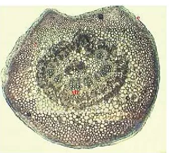
Cross section of the petiole discloses a semicirculaar shape with a slightly concave adaxial surface. Externally, the petiole is protected by a layer of more or less rectangular shaped cells with lateral and inner cutinized cells. The epidermal external walls are covered by a thick cuticle, supplemented with vegetal wax. The epidermis is followed by cortex. The cortex is differentiated into two zones. Just below the epidermis is an external zone of angular collenchyma tissue (7- layers of cells), followed by a parenchyma one of 6-7 layers of cellulosic-walled cells, the later consisting a number of crystal cells (rhombic calcium oxalate crystals and druses.) and non-articulated laticifers as well. A number of periphloemic fibers are present(Fig. 3, a).
Fig. 2.Cross section of the petiole – ensemble(x 80): c- cortex, e- epidermis, vb- vascular bundle.
Fig. 3. Transections of the petiole. Portion with epidermis and cortex (a, x 180). Portion with vascular bundles (b, x 140): co- collenchyma, coc- calcium oxalate crystals, d- druse, e- epidermis, lb-leptocentric bundles, l – laticifer, ph- phloem, pc- parenchyma cells, scl- sclerenchyma cells,
x-xylem.
The blade ofFicus lyratais bifacial and hypostomatic. The upper epidermal (adaxial epidermis layer) cells are rectangular in shape and covered by a visible cuticle covered by epicuticular wax (Fig. 4, a, b) such as Berg (2003a, b, c), Metcalfe and Clarck (1950b) and Rogwer (1993) reported for many Ficus species, mainly for abaxial blade surface. In the mid rib zone the epidermal cells are smaller and tangnetial elongated.
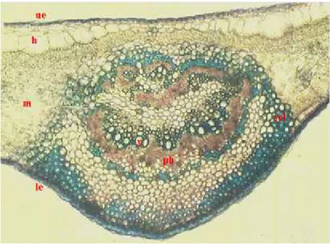
Fig. 5. Cross section of the blade in the mid rib zone - ensemble (x 100): h- hypodermis, le- lower epidermis, m- mesophyll, ph- phloem, scl- sclerenchyma, ue- upper epidermis, x- xylem.
It is followed by a sub-epidermal cell layer - hypodermis - formed by large cells with here and there large lithocysts (giant epidermal cell that protrudes into the mesophyll). In the mid rib zone the hypodermal cells are smaller. Inside the litocysts, as Metcalfe and Chalk (1950a) underline for Ficus genus and other Moraceae genus species, are the veritable cystoliths. Inside Ficus benjamina cv Danielle adaxial litocysts are almost oblong-shaped cystoliths (calcium carbonate deposes) with stalk (peg) and rounded solitary cystoliths, hanged by a shorter stalk to the abaxial epidermis. (Fig. 4, a, b). As Esau (1965) and Foster (1949) reported, the Ficus cystoliths appear in the cells of a multiple epidermis but in our findings they may apper in the unistratose epidermal hypodermis as well.
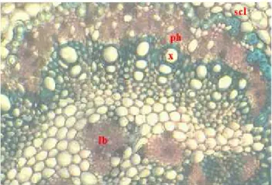
oxalate crystals and laticiferous elements are present in the vicinity of the mid rib zone (Fig. 6).
Fig. 6. Cross section of the blade with mid rib – detail (x 220): ph- phloem, lb- leptocentric bundle, scl- sclerenchyma, x – xylem.
The lower epidermis (abaxial epidermis) has rectangular cells smaller in comparison with those of the upper one, covered by cuticle and wax. The epidermis continuity is interrupted by the presence of stomata (Fig. 4, b; 5).
Fig. 7. Paradermal section of the abaxial epidermis(x 200): ec- epidermal cell, s- stoma.
Paradermal section of the lower epidermis discloses polygonal epidermal cells with straight walls regularly and anisocytic stomata (figure 7).
The stomatal density is 158 stomata/mm2and the stomatal index (SI) avergae 0.67
± 2,87.
epidermis of the blade is covered by a thick cuticle, supplementedwith wax, followed by a hypodermis with lithocysts and cistolyths inside such as the lower one which is interrupted by the presence of anisocytic stomata. The bifacial blade has a heterogeneous mesophyll. Notable is the presence of the apparently medullary leptocentric bundles in the petiole and mid rib vein.
The secretive elements are represented by laticifers and oxalate crystals both present in petiole and blade.
The anatomical features highlighted in this paper such as the thick cuticle with epicuticular wax, heavy lignification and the presence of hypodermis do Ficus benjamina
cv Danielle to be adaptable as an ornamental plant for the semi-arid and arid Romanian regions.
BIBLIOGRAPHY
1. Ali B., Mujeeb M., Aeri V., Mir S.R, Faiyazuddin M., Shakeel F., 2012 -
Anti-inflammatory and antioxidant activity of Ficus carica Linn. Leaves, Nat. Prod. Res., 26 (5): 460-465.
2. Bercu R., Jianu D.L., 2003 - Practicum of Morphology and anatomy of plants. Edit.
“Ovidius”University Press, Constanţa.
3. Bercu R., Bavaru E., 2003 - Morphological and morphometrical characterisation of some Ficus L. species leaf. Analele Universităţii „Ovidius”, Ser. Biologie-Ecologie,vol. 7: 9-16.
4. Bercu R., Popoviciu D.R., 2014 - Anatomical study of Ficus carica L. leaf, In: C. Crăciun A., Ardelean (coord.) Annals of the Romanian Society for Cell Biology, vol. 19(1): 33-36.
5. Bercu R., 2015a -Anatomical aspects of Ficus lyrataWarb., Annals of West University
of Timişoara,ser. Biology, 18(2): 107-114.
6. Bercu R., 2015b - Histological study of Ficus elastica Roxb. (Moraceae) leaf, 2015,
Natura, Biologie,Ser. III, vol. 58(2): 21-32.
7. Berg C.C., 2003a - Flora Malaesiana precursor for the tratament of Moraceae 1: The main subdivision of Ficus: the subgenera,Blumea, 48: 167-178.
8. Berg C.C., 2003b -Flora Malaesiana precursor for the tratament of Moraceae 3: Ficus:
subgenus Ficus, Blumea, 48: 529-550.
9. Berg C.C., 2003c -Flora Malaesiana precursor for the tratament of Moraceae 4: Ficus:
subgenus Synoecia,Blumea, 48: 551-571.
10. Boldor, O., Trifu M., Raianu O., 1981 - Fiziologia plantelor, Edit. Did. şi Ped., Bucureşti.
11. Cabrera C. N., Gelsi G. A., Albornoz P. L., Arias, M. E., 2009 - Leaf anatomy of Ficus maroma [Moraceae], and analyze of exposed leaves to environmental pollution in the province of Tucumán (Argentina),Lilloa, 46(1/2): 34-42.
12.Della Beffa M.T., 2007 -Plante de apartament, Edit. All, Bucureşti.
13 Dilcher D.L., 1974 - Approaches to the identification of angiosperms leaf remains. Bot. Rev. (New York),40(1):1-157.
14. Dressler S., Schmidt M., Zizka G. (Hrsg., 2014 - African plants - A Photo Guide. Senckenberg, Frankfurt/Main.
15.Esau K., 1965 -Plant Anatomy, John Wiley and Sons, Inc., New York.
16.Foster A.S., 1949 -Practical Plant Anatomy, D. Van Nostrand Co., New York.
17. Fox A.M., Gordon D. R., Dusky J.A, Tyson L., Stocker R.K., 2005 - IFAS
Assessment of the Status of Non-Native Plants in Florida's Natural Areas. Cited from the Internet (2006), http://plants.ifas.ufl.edu/assessment.html. Accesed 28.08.2015.
18 Henley, R. W., Poole R.T., 1989 - Evaluation of Selected Ornamental Figs for Interior
19. Niculescu N.,2004 - Practicum de Botanică sistematică – Partea I, Edit. Universitaria Craiova.
20. Klimko M., Truchan M., 2006 - Morphological variability of the leaf epidermis in selected taxa of genus Ficus L. (Moraceae) and its axonomicl implications, Acta Societatis Botanicorum Poloniae, 75(4): 308-324.
21. Lazreg-Aref H., Mars M., Fekih A., Aouni M., Said K., 2012 - Chemical composition
and antibacterial activity of a hexane extract of Tunisian Capri fig latex from the unripe fruit of Ficus carica, Pharm. Biol., 50(4): 407-412.
22. Mamoucha S., Fokialakis N., Christodoulakis S.N., 2016 - Leaf structure and
histochemistry of Ficus carica (Moraceae), the fig tree, Flora – Morphology, Distribution, Functional, Ecology of Plants,vol. 218: 24-34.
23. Metcalfe C.R.; Chalk L., 1983 - Secretory structures: Cells, cavities and canals. In: Anatomy of the dicotyledons. 2nd volume, Wood structure and conclusions of the general introduction, pp. 70–81, Clarendon Press, Oxford.
24. Metcalfe C.R., Chalk L., 1950a - Anatomy of the Dicotyledons, Vol. I, Clarendon
Press,Oxford:
25. Metcalfe C.R., Chalk L., 1950b - Anatomy of the Dicotyledons, Vol. II, Clarendon Press, Oxford.
26. Mioulane P., 2004 - Enciclopedia Truffaut. Grădini şi plante de interior, Edit. Grupul Editorial RAO, Bucureşti.
27. Peterson, R.L., Peterson A.C., Melville, L.H., 2008 - Teaching Plant Anatomy
through creative laboratory exercises, NRC Press, Ottava, Ontario, p. 26.
28. Rohwer J.G., 1993 – Moraceae. The Families and Genera of Vascular Plants. In
Kubidzki K, Rohwer J.G. and Bittrich V. (eds), II Flowering Plants – Dycotiledons Magniliid, Hamamelid and Caryophylid Families,Springer-Verlag, pp. 438-453.
29 Sonibare M.A., Jayeola A.A., Egunyomi,A., 2006 - Comparative Leaf Anatomy of
Ficus Linn. Species (Moraceae) from Nigeria, Journal of Applied Sciences, 6(15): 3016-3025.
30. Spencer R., Cross R., Lumley P., 2007 - Plant Names: A Guide to Botanical
Nomenclature,3rdedn.,CSIRO Publishing,Collingwood, Victoria:
31. Stace C. A. 1965 - Cuticular studies as an aid to plant taxonomy. Bulletin of British Museum (Natural History),Botany, 4: 3-78.
32.Tarnavschi T. I., Şerbănescu-Jitariu G., Rădulescu-Mitroiu N., Rădulescu D., 1974
- Practicum de morfologie şi anatomie vegetală, Tipografia Universitatii Bucureşti, Bucureşti.
33. Tomlinson P.B., 1956 - Studies in the systematic anatomy of the Zingiberaceae. J. Linn. Soc. (Bot.),55: 547-592.
34. Trehane, P., 2004 -50 years of the International Code of Nomenclature for Cultivated
Plants: Future prospects for the Code, Acta Hort,634: 17-27.
35. Ummu-Hani B.; Noraini T., 2013 - The structure of cystoliths in selected taxa of the