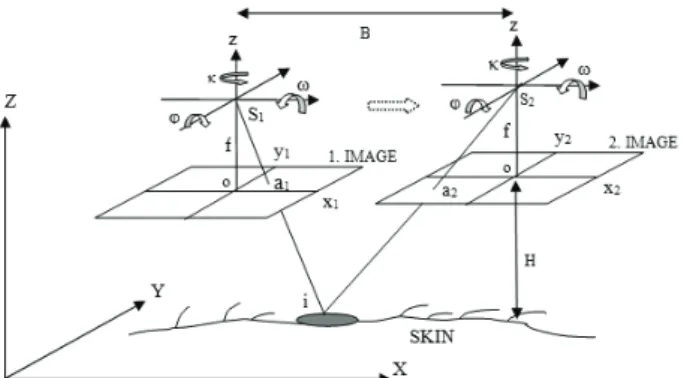
3D NON-INVASIVE INSPECTION OF THE SKIN LESIONS BY CLOSE-RANGE AND LOW-COST PHOTOGRAMMETRIC TECHNIQUES
Full text
Figure


Related documents
Mutant vac8 ⌬ cells contained fragmented vacuoles, and minimal vacuolar material was inherited by daughter cells in hyphal or budding forms.. Normal rates of growth and hyphal
Existing research regarding criteria used to make the final decision regarding university selection has a focus at undergraduate level (Maringe, 2006), with some
The objectives for Phase 1 were to (a) formulate a list of core clinical competencies for second-year occupational therapy students, to be assessed via e-OSPEs; (b) introduce clinical
We conclude that the combination of transthyretin, RBP and C-reactive protein showed good diagnostic performance in assessing vitamin A deficiency and has great potential to
A number of studies investigating the effect of mobile phone on human body have been done by number of investigators by examining heart rate variability, BP, when
Nelson and Stolterman (2012) claim that the skill of making such appreciative judgements is fundamental to design judgements. In one of the interviews a good example of how
On comparing representative serum protein electropherograms obtained in this study (Figs. 1 and 2 ) with a standard electrophoretic pattern of sea turtles found in literature
