A
cute coronary syndrome encom-passes a spectrum of coronary artery diseases, including unstable angina, ST-elevation myocar-dial infarction (STEMI; often referred to as “Q-wave myocardial infarction”), and non-STEMI (NSTEMI; often referred to as “non–Q-wave myocardial infarction”). The term “acute coronary syndrome” is useful because the initial presentation and early management of unstable angina, STEMI, and NSTEMI frequently are similar.Differentiating acute coronary syndrome from noncardiac chest pain is the primary diagnostic challenge. The initial assessment requires a focused history (including risk fac-tor analysis), a physical examination, an elec-trocardiogram (ECG) and, frequently, serum cardiac marker determinations (Table 1).1 Clinical Evaluation
Symptoms of acute coronary syndrome include chest pain, referred pain, nausea, vomiting, dyspnea, diaphoresis, and light-headedness. Some patients may present with-out chest pain; in one review,2
sudden dyspnea was the sole presenting feature in 4 to 14 percent of patients with acute myocardial infarction. Pain may be referred to either arm, the jaw, the neck, the back,
or even the abdomen. Pain radiating to the shoulder, left arm, or both arms somewhat increases the likelihood of acute coronary syndrome (likelihood ratio [LR]: 1.6).3
Typical angina is described as pain that is substernal, occurs on exertion, and is relieved with rest. Patients with all three of these features have a greater likelihood of having acute coronary syndrome than patients with none, one, or even two of these features. Chest pain that occurs suddenly at rest or in a young patient may suggest acute coronary vasospasm, which occurs in Prinzmetal’s angina or with the use of cocaine or methamphetamine. Only about 2 percent of patients with cocaine-associated chest pain have acute coronary syndrome.4
Atypical symptoms do not necessarily rule out acute coronary syndrome. One study5 found the syndrome in 22 percent of
596 patients who presented to emergency departments with sharp or stabbing pain. However, a combination of atypical symp-toms improves identification of low-risk patients. The same study5 demonstrated that
patients presenting with sharp or stabbing pain, pleuritic pain, and positional chest pain had only a 3 percent likelihood of hav-ing acute coronary syndrome.
The physical examination in patients with acute coronary syndrome frequently is nor-mal. Ominous physical findings include a new
The term “acute coronary syndrome” encompasses a range of thrombotic coronary artery diseases, including unstable angina and both ST-segment elevation and non–ST-segment elevation myocardial infarction. Diagnosis requires an electrocardiogram and a careful review for signs and symptoms of cardiac ischemia. In acute coronary syndrome, common electrocardiographic abnormalities include T-wave tenting or inversion, ST-segment elevation or depression (including J-point elevation in multiple leads), and pathologic Q waves. Risk stratification allows appropriate referral of patients to a chest pain center or emergency department, where cardiac enzyme levels can be assessed. Most high-risk patients should be hospitalized. Intermediate-high-risk patients should undergo a structured evaluation, often in a chest pain unit. Many low-risk patients can be discharged with appropriate follow-up. Troponin T or I generally is the most sensitive determinant of acute coronary syndrome, although the MB isoenzyme of creatine kinase also is used. Early markers of acute ischemia include myoglobin and creatine kinase–MB subforms (or isoforms), when available. In the future, advanced diagnostic modalities, such as myocardial perfusion imaging, may have a role in reducing unnecessary hospitalizations. (Am Fam Physician 2005;72:119-26. Copyright© 2005 American Academy of Family Physicians.)
Diagnosis of Acute Coronary Syndrome
SURAJ A. ACHAR, M.D., University of California, San Diego, School of Medicine, La Jolla, CaliforniaSURITI KUNDU, M.D., San Diego, California
WILLIAM A. NORCROSS, M.D., University of California, San Diego, School of Medicine, La Jolla, California
Only about 2 percent of patients with cocaine-associated chest pain have acute coronary syndrome.
mitral regurgitation murmur, hypotension, pulmonary rales, a new third heart sound (S3
gallop), and new jugular venous distention. Chest-wall tenderness reduces the likelihood of acute coronary syndrome (-LR: 0.2).3
The likelihood of silent ischemia tradition-ally has been thought to be greater in patients with diabetes. The “silent myocardial infarc-tion” hypothesis is based on the relatively high incidence of ischemic changes noted on screening ECGs in patients with
diabe-tes. However, in a prospective observational study6 of 528 patients with symptoms
sug-gestive of coronary artery disease on pre-sentation to the emergency department of a cardiac referral center, symptoms did not dif-fer significantly in patients with and without diabetes. The increased frequency of ischemic changes noted on screening ECGs in patients with diabetes simply may reflect their greater baseline risk of coronary artery disease.
Any patient with a history suggestive of
table 1
Risk Stratification to Determine the Likelihood of Acute Coronary Syndrome
Assessment
Findings indicating HIGH likelihood of ACS
Findings indicating INTERMEDIATE likelihood of ACS in absence of high-likelihood findings
Findings indicating LOW likelihood of ACS in absence of high- or intermediate-likelihood findings
History Chest or left arm pain or discomfort as chief symptom
Reproduction of previous documented angina Known history of coronary artery disease,
including myocardial infarction
Chest or left arm pain or discomfort as chief symptom
Age > 50 years
Probable ischemic symptoms Recent cocaine use
Physical examination
New transient mitral regurgitation, hypotension, diaphoresis, pulmonary edema or rales
Extracardiac vascular disease
Chest discomfort reproduced by palpation
ECG New or presumably new transient ST-segment
deviation (> 0.05 mV) or T-wave inversion (> 0.2 mV) with symptoms
Fixed Q waves
Abnormal ST segments or T waves not documented to be new
T-wave flattening or inversion of T waves in leads with dominant R waves Normal ECG Serum cardiac
markers
Elevated cardiac troponin T or I, or elevated CK-MB
Normal Normal
ACS = acute coronary syndrome; ECG = electrocardiogram; CK-MB = MB isoenzyme of creatine kinase.
Adapted from Braunwald E, et al. Unstable angina: diagnosis and management. Rockville, Md.: U.S. Dept. of Health and Human Services, Public Health Service, Agency for Health Care Policy and Research, National Heart, Lung, and Blood Institute, 1994; Clinical practice guideline no. 10; AHCPR publica-tion no. 94-0602.
The likelihood of acute coronary syndrome (low, intermediate, high) should be determined in all patients who present with chest pain.
C 9
A 12-lead ECG should be obtained within 10 minutes of presentation in patients with ongoing chest pain.
C 9
Cardiac markers (troponin T, troponin I, and/or creatine kinase–MB isoenzyme of creatine kinase) should be measured in any patient who has chest pain consistent with acute coronary syndrome.
C 9
A normal electrocardiogram does not rule out acute coronary syndrome. B 12
When used by trained physicians, the Acute Cardiac Ischemia Time-Insensitive Predictive Instrument (a computerized, decision-making program built into the electrocardiogram machine) results in a significant reduction in hospital admissions of patients who do not have acute coronary syndrome.
B 26, 27
A = consistent, good-quality patient-oriented evidence; B = inconsistent or limited-quality patient-oriented evidence; C = consensus, disease-oriented evidence, usual practice, opinion, or case series. For information about the SORT evidence rating system, see page 15 or http://www. aafp.org/afpsort.xml.
Acute Coronary Syndrome
acute coronary syndrome should be evalu-ated in a facility that has ECG and cardiac monitoring equipment.7 Patients with
sus-pected acute coronary syndrome who have chest pain at rest for more than 20 minutes, syncope/presyncope, or unstable vital signs should be referred to an emergency depart-ment immediately.7 The diagnosis of acute
myocardial infarction, which includes both STEMI and NSTEMI, requires at least two of the following: ischemic symptoms, diagnos-tic ECG changes, and serum cardiac marker elevation.8,9
The likelihood of acute myocardial infarc-tion is extremely low in patients with a normal or nearly normal ECG who are younger than 60 years and do not have pain described as “pressure” or pain radiating to the arm, shoulder, neck, or jaw. The likeli-hood of acute infarction is 1.1 percent or less with a normal ECG and 2.6 percent or less with nonspecific ECG changes.10
ECg interpretation
The ECG provides information that assists in stratifying the patient’s risk of having acute
coronary syndrome, establishing the diag-nosis, and determining the treatment strat-egy. Accuracy is enhanced when the ECG is obtained in a patient with ongoing chest pain. The characteristics of common ECG abnormalities in specific anatomic locations are presented in Table 2.11
DiAgnOStiC ACCuRACy
The predictive value of the ECG varies mark-edly, depending on the baseline risk (pretest probability) for coronary artery disease in a given patient. The number and magnitude of ECG abnormalities also affect sensitivity and specificity.
In a study12 of 775 consecutive patients
with chest pain who were admitted to a car-diac care unit, acute myocardial infarction was diagnosed in 10 percent of patients with normal ECG findings (11 of 107 patients) in the emergency department, 8 percent of patients with “minimal changes” (six of 73 patients), and 41 percent of patients with “frankly abnormal” ECG findings (245 of 595 patients).
The magnitude of an ECG abnormality
table 2
ECg findings for the Diagnosis of Acute Coronary Syndrome
ECG findings Lesion
Sensitivity (%) Specificity (%) Positive predictive value (%) Negative predictive value (%)
ST-segment elevation greater in lead III than in lead II plus ST-segment depression of > 1 mm in lead I, lead aVL, or both
Right coronary artery
90 71 94 70
Absence of the above findings plus ST-segment elevation in leads I, aVL, V5, and V6 and ST-segment
depression in leads V1, V2, and V3
Left circumflex coronary artery
83 96 91 93
ST-segment elevation in leads V1, V2, and V3 plus any
of the features below:
ST-segment elevation of > 2.5 mm in lead V1, right
bundle branch block with Q wave, or both
Proximal LAD coronary artery
12 100 100 61
ST-segment depression of > 1 mm in leads II, III, and aVF
Proximal LAD coronary artery
34 98 93 68
ST-segment depression of ≤ 1 mm or ST-segment elevation in leads II, III, and aVF
Distal LAD coronary artery
66 73 78 62
ECG = electrocardiogram; LAD = left anterior descending.
showed ST-segment depression in three or more leads or ST-segment depressions that were greater than or equal to 0.2 mV.
DiAgnOStiC guiDELinES
Subendocardial ischemia classically results in ST-segment depression and T-wave inver-sion.14 Approximately 25 percent of patients
with ST-segment depression and elevated creatine kinase–MB isoenzyme (CK-MB) levels eventually develop STEMI, and 75
per-cent have NSTEMI. Transmu-ral myocardial ischemia results in ST-segment elevation with the vector shifted toward the involved epicardial layer, and without treatment typically results in STEMI. Occasion-ally, “reciprocal” ST-segment depression occurs in leads that are electri-cally opposite to the area of injury.
Based on Marriott’s criteria,15 epicardial
injury is diagnosed when the J point (origin of the ST segment at its junction with the QRS complex) is (1) elevated by 1 mm or more in two or more limb leads or
precor-diac marker determinations confirm myo-cardial injury or infarction in more than 90 percent of patients with J-point elevation in the limb leads.9
Significant Q waves (greater than 0.04 seconds in duration and at least one quar-ter of the height of the corresponding R wave) suggest myocardial infarction. Iso-lated small Q waves in leads II, III, and aVF (in the electrically vertical heart) and leads I and aVL (in the electrically horizontal heart) frequently are normal. These small Q waves are known as “septal Q waves” because of the origin of the initial vector in ventricular depolarization.
Although the ECG may be completely normal in a patient with myocardial isch-emia and evolving infarction, classic ECG changes occur in STEMI.14 Within minutes,
there is J-point elevation, and tall, peaked, “hyperacute” T waves develop; ST-segment elevation and reciprocal-lead ST-segment depression also occur. Abnormal Q waves usually develop within the first day, and T-wave inversion and normalization of ST segments occur within hours to days.
SERum CARDiAC mARkERS
Serum cardiac marker determinations play a vital role in the diagnosis of acute myocardial infarction. Serum markers such as aspartate transaminase, lactate dehydrogenase, and lactate dehydrogenase subforms no longer are used because they lack cardiac specific-ity and their delayed elevation precludes early diagnosis.9 Characteristics of the most
important serum cardiac markers are sum-marized in Table 3.16-19
CREAtinE kinASE
Creatine kinase (CK) is an enzyme that is found in striated muscle and tissues of the brain, kidney, lung, and gastrointestinal tract. This widely available marker has low sensitivity and specificity for cardiac damage. Furthermore, CK levels may be elevated in a
in one study, acute myo-cardial infarction was diagnosed in 10 percent of patients with normal electrocardiogram findings. the Authors
SURAJ A. ACHAR, M.D., is assistant clinical professor of family and preventive medicine and associate director of the primary care sports medicine fellowship at the University of California, San Diego (UCSD), School of Medicine, La Jolla. Dr. Achar received his medical degree from the State University of New York at Buffalo School of Medicine and Biomedical Sciences. He completed a family practice residency and a fellowship in sports medicine at the UCSD School of Medicine.
SURITI KUNDU, M.D., is a family physician in private practice in San Diego. She also holds a volunteer clinical faculty appointment at UCSD Medical Center. Dr. Kundu graduated from the University of California, Davis, School of Medicine and completed a family practice residency at the UCSD School of Medicine. WILLIAM A. NORCROSS, M.D., is professor of clinical family and preventive med-icine and director of the Physician Assessment and Clinical Education Program at the UCSD School of Medicine. Dr. Norcross received his medical degree from Duke University School of Medicine, Durham, N.C., and completed a family practice residency at the UCSD School of Medicine.
Address correspondence to Suraj A. Achar, M.D., University of California, San Diego, School of Medicine, 9350 Campus Point Dr., La Jolla, CA 92037-0968
(e-trauma, seizures, renal insufficiency, hyper-thermia, and hyperthyroidism.
The serum CK level rises within three to eight hours after myocardial injury, peaks by 12 to 24 hours, and returns to baseline within three to four days.16 A serum CK level
may be used as a screening test to determine the need for more specific testing. Although CK commonly was measured serially (along with CK-MB) at the time of hospital admis-sion and six to 12 hours after admisadmis-sion, this marker largely has been replaced by cardiac troponins and CK-MB.9,16
Ck-mb iSOEnzymE
CK-MB is much more cardiac specific than CK alone, and is useful for the early diagno-sis of acute myocardial infarction.9 CK-MB
typically is detectable in the serum four to six hours after the onset of ischemia, peaks in 12 to 24 hours, and normalizes in two to three days. The CK-MB mass assay is more sensitive than the CK-MB activity assay.20
Like the CK level, the peak CK-MB level does not predict infarct size; however, it can be used to detect early reinfarction.16 Serial
CK-MB levels commonly are obtained at admission to the emergency department and are repeated in six to 12 hours, depending on the assay that is used.20
Ck-mb SubfORmS
CK-MB may be further characterized into subforms (or isoforms). CK-MB2 is found in myocardial tissue, and CK-MB1 is found in plasma. The CK-MB subform assay takes about 25 minutes to perform.21 A CK-MB2
level greater than 1 U per L in combination with a subform ratio greater than 1.5 sug-gests myocardial injury.9,22 One large study23
involving 1,110 patients with chest pain found that CK-MB subform analysis is 96 percent sensitive and 94 percent specific when the marker is measured six hours after symptom onset. However, the CK-MB subform assay is not yet widely available.
CARDiAC tROpOninS
Troponins (T, I, C) are found in striated and cardiac muscle. Because the cardiac and skeletal muscle isoforms of troponin T and I differ, they are known as the “cardiac
table 3
Characteristics of Serum Cardiac markers for the Diagnosis of Acute myocardial infarction*
Serum cardiac marker
Test first becomes positive (hours) Peak level (hours) Sensitivity (%) Specificity (%) Positive predictive value (%)† Negative predictive value (%)† Ck Single assay 3 to 8 12 to 24 35 80 20 90 Serial assays 95 68 30 99 Ck-mb Single assay 4 to 6 12 to 24 35 85 25 90 Serial assays 95 95 73 99 troponin i and t
Measured 4 hours after onset of chest pain
4 to 10 35 96 56 91
Measured 10 hours after onset of chest pain
8 to 28 89 95 72 98
CK = creatine kinase; CK-MB = MB isoenzyme of CK.
*—ST-segment elevation myocardial infarction or non-ST-segment elevation in patients presenting to emergency departments with chest pain.
†—Given a 12.5 percent overall likelihood of acute myocardial infarction. Information from references 16 through 19.
dial injury. Unlike troponin I levels, troponin T levels may be elevated in patients with renal disease, polymyositis, or dermatomyositis.
The cardiac troponins typically are mea-sured at emergency department admission and repeated in six to 12 hours.20 Patients
with a normal CK-MB level but elevated troponin levels are con-sidered to have sustained minor myocardial damage or microin-farction, whereas patients with elevations of both CK-MB and troponins are considered to have had acute myocardial infarc-tion. The cardiac troponins may remain ele-vated up to two weeks after symptom onset, which makes them useful as late markers of recent acute myocardial infarction.9
An elevated troponin T or I level is helpful in identifying patients at increased risk for death or the development of acute myocar-dial infarction.16 Increased risk is related
quantitatively to the serum troponin level. The troponins also can help identify low-risk patients who may be sent home with close follow-up.17 In a study17 of 773 patients
presenting to an emergency department with acute chest pain, those with a nor-mal or nearly nornor-mal ECG and a nornor-mal troponin I test six hours after admission had a very low risk of major cardiac events (0.3 percent) during the next 30 days. Bed-side troponin assays are being developed.
myOgLObin
Myoglobin is a low-molecular-weight pro-tein that is present in both cardiac and skel-etal muscle. It can be detected in the serum as early as two hours after myocardial necrosis begins. Myoglobin has low cardiac specificity but high sensitivity, which makes it most use-ful for ruling out myocardial infarction if the level is normal in the first four to eight hours after the onset of symptoms.9
Time changes in the myoglobin value also can be extremely helpful. Combining
Myoglobin should be used in conjunc-tion with other serum markers, because its level peaks and falls rapidly in patients with ischemia.
A practical plan of Action
No assessment protocol or constellation of tests is totally accurate in diagnosing acute coronary syndrome. From 1 to 4 percent of patients ultimately proven to have acute coronary syndrome are sent home from the emergency department.24 Patients with acute
coronary syndrome who are sent home with-out further evaluation are more likely to be women, to be nonwhite, to present without chest pain, or to have ECGs that are normal or show nonspecific changes.18
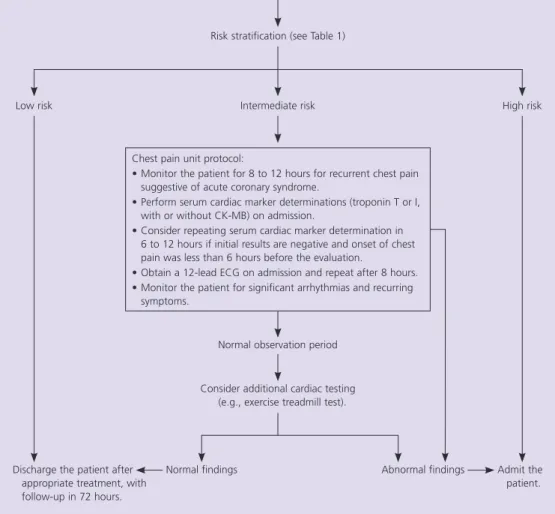
A suggested approach to the evaluation of patients with chest pain or symptoms consistent with acute coronary syndrome is provided in Figure 1. When a patient presents with chest pain or symptoms suggestive of acute coronary syndrome, vital signs should be obtained, the patient should be monitored, and a focused but careful history should be obtained. A 12-lead ECG should be obtained within 10 minutes of presentation.7
Risk stratification then should be performed using the criteria in Table 1.1 Alternatively,
the Acute Cardiac Ischemia Time-Insensitive Predictive Instrument can be used.26 This is
a computerized decision-making program that is built into the ECG machine. Use of this instrument in an emergency department resulted in no change in appropriate admis-sion of patients who had acute coronary syn-drome. The benefit of its use was a significant reduction in hospital admissions of patients who did not have acute coronary syndrome.26
However, a subsequent study27 suggested that
this benefit is not seen unless physicians have been trained in the use of the instrument.
Patients who are at high risk for acute coronary syndrome should be admitted to a coronary care unit. Patients at intermediate risk may be monitored in a telemetry bed in
from 1 to 4 percent of patients ultimately proven to have acute coronary syn-drome are sent home from the emergency department.
chest pain unit is a specialized unit within an emergency department or a medical cen-ter; the unit is dedicated to careful moni-toring and aggressive implementation of diagnostic protocols (clinical guidelines) for the evaluation of acute coronary syndrome. Most low-risk patients may undergo early exercise testing or can be discharged with careful outpatient follow-up.
Although protocols for chest pain units may vary somewhat, one protocol28 that has
been shown to be safe and cost-effective in an intermediate-risk population consists of the following:
1. Event monitoring and continuous ST-segment monitoring;
2. Measurement of troponins I and T and/ or CK-MB at admission and six to eight hours after admission;
3. Four patients staffed by one full-time nurse;
4. Admission to the cardiac care unit or a telemetry bed on the cardiology service for patients with elevated cardiac enzyme levels, recurrent chest pain consistent with unstable angina, or significant ventricular arrhythmias;
5. An exercise treadmill test for patients without abnormal findings on the initial tests, or a nuclear stress test or echocardio-graphic stress test;
6. Admission of patients with an equivocal or positive result.
Use of this type of systematic approach has the potential to improve the ability of physicians to care for patients with pos-sible acute coronary syndrome, as well as reduce the likelihood of medical error. In
Evaluation of patients with Chest pain or Symptoms Suggesting ACS
figure 1. Suggested approach to the evaluation of patients with chest pain or symptoms sug-gestive of aCS. (aCS = acute coronary syndrome; CK-Mb = Mb isoenzyme of creatine kinase; eCG = electrocardiogram.)
Patient presenting with chest pain or symptoms consistent with ACS
Low risk Intermediate risk High risk
Risk stratification (see Table 1)
Admit the patient. Discharge the patient after
appropriate treatment, with follow-up in 72 hours.
Normal observation period
Abnormal findings Consider additional cardiac testing
(e.g., exercise treadmill test).
Normal findings Chest pain unit protocol:
• Monitor the patient for 8 to 12 hours for recurrent chest pain suggestive of acute coronary syndrome.
• Perform serum cardiac marker determinations (troponin T or I, with or without CK-MB) on admission.
• Consider repeating serum cardiac marker determination in 6 to 12 hours if initial results are negative and onset of chest pain was less than 6 hours before the evaluation.
• Obtain a 12-lead ECG on admission and repeat after 8 hours. • Monitor the patient for significant arrhythmias and recurring
the future, advanced diagnostic modali-ties, such as myocardial perfusion imaging, may have a role in reducing unnecessary hospitalizations.
Author disclosure: Nothing to disclose.
REfEREnCES
1. Braunwald E, et al. Unstable angina: diagnosis and management. Rockville, Md.: U.S. Dept. of Health and Human Services, Public Health Service, Agency for Health Care Policy and Research, National Heart, Lung, and Blood Institute, 1994; Clinical practice guideline no. 10; AHCPR publication no. 94-0602.
2. McCarthy BD, Wong JB, Selker HP. Detecting acute car-diac ischemia in the emergency department: a review of the literature. J Gen Intern Med 1990;5:365-73. 3. Goodacre S, Locker T, Morris F, Campbell S. How useful
are clinical features in the diagnosis of acute, undiffer-entiated chest pain? Acad Emerg Med 2002;9:203-8. 4. Feldman JA, Fish SS, Beshansky JR, Griffith JL, Woolard
RH, Selker HP. Acute cardiac ischemia in patients with cocaine-associated complaints: results of a multicenter trial. Ann Emerg Med 2000;36:469-76.
5. Lee TH, Cook EF, Weisberg M, Sargent RK, Wilson C, Goldman L. Acute chest pain in the emergency room. Identification and examination of low-risk patients. Arch Intern Med 1985;145:65-9.
6. Funk M, Naum JB, Milner KA, Chyun D. Presentation and symptom predictors of coronary heart disease in patients with and without diabetes. Am J Emerg Med 2001;19:482-7.
7. Pollack CV Jr, Gibler WB. 2000 ACC/AHA guidelines for the management of patients with unstable angina and non-ST-segment elevation myocardial infarction: a practical summary for emergency physicians [published corrections appear in Ann Emerg Med 2001;38:468 and Ann Emerg Med 2002;39:100]. Ann Emerg Med 2001;38:229-40.
8. Nomenclature and criteria for diagnosis of ischemic heart disease. Report of the Joint International Society and Federation of Cardiology/World Health Organiza-tion Task Force on StandardizaOrganiza-tion of Clinical Nomen-clature. Circulation 1979;59:607-9.
9. Braunwald E, Antman EM, Beasley JW, Califf RM, Cheitlin MD, Hochman JS, et al. ACC/AHA guidelines for the management of patients with unstable angina and non-ST-segment elevation myocardial infarction. A report of the American College of Cardiology/Ameri-can Heart Association Task Force on Practice Guidelines (Committee on the Management of Patients With Unstable Angina) [published correction appears in J Am Coll Cardiol 2001;38:294-5]. J Am Coll Cardiol 2000;36:970-1062.
10. Rouan GW, Lee TH, Cook EF, Brand DA, Weisberg MC, Goldman L. Clinical characteristics and outcome of acute myocardial infarction in patients with initially normal or nonspecific electrocardiograms (a report from the Multi-center Chest Pain Study). Am J Cardiol 1989;64:1087-92. 11. Zimetbaum PJ, Josephson ME. Use of the electrocar-diogram in acute myocardial infarction. N Engl J Med 2003;348:933-40.
12. Slater DK, Hlatky MA, Mark DB, Harrell FE Jr, Pryor DB, Califf RM. Outcome in suspected acute myocar-dial infarction with normal or minimally abnormal admission electrocardiographic findings. Am J Cardiol 1987;60:766-70.
13. Lloyd-Jones DM, Camargo CA Jr, Lapuerta P, Giugliano RP, O’Donnell CJ. Electrocardiographic and clinical predictors of acute myocardial infarction in patients with unstable angina pectoris. Am J Cardiol 1998;81: 1182-6.
14. Goldberger AL. Clinical electrocardiography: a simpli-fied approach. 6th ed. St. Louis: Mosby, 1999:81-100. 15. Wagner GS, Marriott HJ. Marriott’s Practical
electrocar-diography. 10th ed. Philadelphia: Lippincott Williams & Wilkins, 2001:165.
16. Karras DJ, Kane DL. Serum markers in the emergency department diagnosis of acute myocardial infarction. Emerg Med Clin North Am 2001;19:321-37.
17. Hamm CW, Goldmann BU, Heeschen C, Kreymann G, Berger J, Meinertz T. Emergency room triage of patients with acute chest pain by means of rapid testing for cardiac troponin T or troponin I. N Engl J Med 1997; 337:1648-53.
18. Pope JH, Selker HP. Diagnosis of acute cardiac ischemia. Emerg Med Clin North Am 2003;21:27-59.
19. Balk EM, Ioannidis JP, Salem D, Chew PW, Lau J. Accu-racy of biomarkers to diagnose acute cardiac ischemia in the emergency department: a meta-analysis. Ann Emerg Med 2001;37:478-94.
20. Serum marker analysis in acute myocardial infarction. American College of Emergency Physicians. Ann Emerg Med 2000;35:534-9.
21. Puleo PR, Guadagno PA, Roberts R, Perryman MB. Sensitive, rapid assay of subforms of creatine kinase MB in plasma. Clin Chem 1989;35:1452-5.
22. Bock JL, Brogan GX Jr, McCuskey CF, Thode HC Jr, Hollander JE, Gunther T. Evaluation of CK-MB isoform analysis for early diagnosis of myocardial infarction. J Emerg Med 1999;17:75-9.
23. Puleo PR, Meyer D, Wathen C, Tawa CB, Wheeler S, Hamburg RJ, et al. Use of a rapid assay of subforms of creatine kinase-MB to diagnose or rule out acute myocardial infarction. N Engl J Med 1994;331:561-6. 24. Scirica BN, Morrow BA. Troponins in acute coronary
syndromes. Semin Vasc Med 2003;3:363-74. 25. Clinical policy: critical issues in the evaluation and
man-agement of adult patients presenting with suspected acute myocardial infarction or unstable angina. Ameri-can College of Emergency Physicians. Ann Emerg Med 2000;35:521-5.
26. Selker HP, Beshansky JR, Griffith JL, Aufderheide TP, Ballin DS, Bernard SA, et al. Use of the acute cardiac ischemia time-insensitive predictive instrument (ACI-TIPI) to assist with triage of patients with chest pain or other symptoms suggestive of acute cardiac ischemia. A multicenter, controlled clinical trial. Ann Intern Med 1998;129:845-55.
27. Lee TH, Goldman L. Evaluation of the patient with acute chest pain. N Engl J Med 2000;342:1187-95. 28. Farkouh ME, Smars PA, Reeder GS, Zinsmeister AR,
Evans RW, Meloy TD, et al. A clinical trial of a chest-pain observation unit for patients with unstable angina. Chest Pain Evaluation in the Emergency Room (CHEER) Investigators. N Engl J Med 1998;339:1882-8.