THE STRUCTURAL AND FUNCTIONAL CONSEQUNCES OF THE INTERACTION OF THE VINCULIN TAIL DOMAIN WITH F-ACTIN AND PIP2
Peter Matthew Thompson
A dissertation submitted to the faculty of the University of North Carolina at Chapel Hill in partial fulfillment of the requirements for the degree of Doctor of Philosophy in the Department of
Biochemistry and Biophysics.
Chapel Hill 2015
© 2013
ABSTRACT
Peter Matthew Thompson: The Structural and Functional Consequences of the Interaction of the Vinculin Tail Domain with F-actin and PIP2
(Under the direction of Sharon L. Campbell)
Vinculin is an essential, highly-conserved eukaryotic scaffolding protein. It localizes
to focal adhesions and adherens juctions, where it assists in physically linking the actin
cytoskeleton to the adhesive structure. Loss of vinculin causes embryonic lethality with
observable cardiovascular and neural defects. Vinculin is comprised of three domains: a
large helical head domain, an unstructured proline-rich linker, and a helical tail. Vinculin
functions as a scaffold, alternating between an autoinhibited conformation in which it
cannot bind ligands, and an open conformation in which it is free to bind ligands at all three
domains.
The tail domain binds a variety of ligands, two of which are F-actin and
phosphatidylinositol 4,5-bisphosphate (PIP2). Both interactions lack good structural models,
which has hindered the development of tools to understand the specific biological functions
these interactions play. Using a variety of biochemical and biophysical techniques and tools,
we have developed new structural models for these interactions. The PIP2 model shows how
Additionally, we characterize the backbone dynamics of Vt by nuclear magnetic
resonance. These experiments reveal the presence of μs-ms motions that reside where the
C-terminal arm interacts with the helix bundle. These motions may be involved in the
ACKNOWLEDGEMENTS
In some ways, this is the most difficult part of the dissertation to compose. It is impossible to, in a few paragraphs, adequately recognize and thank the numerous individuals who have supported and encouraged me in my development as a scientist.
I would like to start by thanking the taxpayers of North Carolina, who have funded, in part, my education since the 2nd grade. Their investment in my scientific training is sincerely appreciated.
I would also like to acknowledge my mentor, Dr. Sharon Campbell for how she has continually encouraged creativity and allowed me to pursue my own hypotheses. Sharon’s own enthusiasm and creativity have challenged me to continually push myself and my ideas.
In addition to Sharon, the other members of my thesis committee have each played a part in my development as a scientist. They encouraged me when I changed projects, offered both scientific and professional advice, and refused to hold me to anything but a high
standard. Their support will definitely be missed.
I must also acknowledge the past and current members of the Campbell Lab. They provided excellent advice, encouragement, friendship, and a gym partner during my graduate career.
Additionally, I want to thank the members of my Biophysics class at UNC,
particularly Dr. Bryan Der and Dr. Keith Miller. We pushed each other to be better and were there for each other when we needed a break.
I wish to thank my parents, brothers, and grandparents for the ways in which they pushed me to grow. While my parents and grandparents have always stressed the
importance of an education and encouraged me to test my limits, they, along with my brothers, pushed me to develop my social skills. While I am not an expert in human interactions, the social skills I developed have been invaluable in my research. I would not have been as successful without them.
Lastly, I want to thank my wife, Dr. Courtney Thompson. Her patience with the rigorous demands of graduate school and sacrifices for our family during the past few years have been inspirational. She has been strong, funny, empathetic, and firm when the
TABLE OF CONTENTS
LIST OF FIGURES ... x
LIST OF TABLES ... xiii
LIST OF ABBREVIATIONS AND SYMBOLS ... xiii
Chapter 1: Introduction and Background ... 1
Introduction ... 1
Focal adhesion architecture and signaling ... 3
Metavinculin is a tissue-specific splice variant of vinculin ... 4
Vinculin in health and disease ... 5
Chapter 2: Characterization of the Interaction Between the Vinculin Tail Domain and PIP2-containing Membranes ... 10
Introduction ... 10
Materials and Methods ... 12
Results ... 14
Discussion ... 20
Chapter 3: Structural, Biochemical, and Functional Characterization of the Actin-binding Site on Vinculin Tail ... 33
Introduction ... 33
Materials and Methods ... 35
Results ... 38
Introduction ... 55
Materials and Methods ... 56
Results ... 60
Discussion ... 63
Chapter 5: Characterization of the Role of Vt Backbone Dyanmics and Proline 989 in Vt Function ... 75
Introduction ... 75
Materials and Methods ... 76
Results ... 81
Discussion ... 89
Chapter 6: Conclusions and Future Directions ... 117
LIST OF FIGURES
Figure 1.1. The structure of vinculin. ... 7
Figure 1.2. Diagram of vinculin activation. ... 8
Figure 1.3. Comparison of vinculin and metavinculin tails. ... 9
Figure 2.1. The basic collar and basic ladder of Vt. ... 24
Figure 2.2. Modeling of the PIP2:Vt interaction. ... 25
Figure 2.3. Actin-binding and crosslinking activity of Vt LD-CT (R1060Q/K1061Q). ... 27
Figure 2.4. Mutations at the basic collar disrupt binding to PIP2 and actin crosslinking functions. ... 28
Figure 2.5. Mutation of basic collar residues to glutamine does not rescue F-actin crosslinking activity. ... 29
Figure 2.6. Mutations to the basic ladder disrupt PIP2 binding, but do not significantly impair F-actin binding or crosslinking. ... 30
Figure 2.7. CD characterization of basic collar and basic ladder mutants. ... 31
Figure 2.8. Comparative lipid co-sedimentation assays for Vt BC-4Q and Vt BL-4A. ... 32
Figure 3.1. Mutations at and near I997 and V1001 disrupt F-actin binding. ... 44
Figure 3.2. CD spectra of Vt variants. ... 45
Figure 3.3. 1H-15N HSQC spectra of Vt, Vt I997A, and Vt V1001A. ... 46
Figure 3.4. The manual fit and DMD models for the binding of Vt to F-actin. ... 47
Figure 3.5. Loss of actin-binding function in vinculin results in fewer, but larger, FAs... 48
Figure 3.6. Actin-binding by vinculin is required for reinforcement at FAs. ... 49
Figure 3.7. Cell spreading of Vin -/- MEFs expressing various vinculin constructs. ... 50
Figure 3.8. The cryo-EM model of Vt bound to F-actin. ... 51
Figure 3.9. The Vt helix bundle changes conformation upon binding F-actin. ... 52
Figure 4.2. Sedimentation curves for Vt. ... 67
Figure 4.3. Sedimentation curves for Vt Q1018K. ... 68
Figure 4.4. Size exclusion chromatography of Vt and Vt Q1018K. ... 69
Figure 4.5. HSQC titration of Vt Q1018K. ... 70
Figure 4.6. CD of Vt and Vt Q1018K. ... 71
Figure 4.7. 1H-15N HSQC overlay of Vt and Vt Q1018K. ... 72
Figure 4.8. Actin binding of Vt Q1018K. ... 73
Figure 4.9. Assignment of Vt Q1018K. ... 74
Figure 5.1. Vt helix 4 is kinked by the presence of proline 989. ... 102
Figure 5.2. Hydrogen exchange rates for Vt Q1018K. ... 103
Figure 5.3. R1, R2, and {1H}-15N NOE values for Vt Q1018K at two separate fields. ... 104
Figure 5.4. The R1R2 product for Vt Q108K at two fields. ... 105
Figure 5.5. Analysis of relaxation data by mode-free formalism. ... 106
Figure 5.6. NMR CPMG results for Vt Q1018K. ... 108
Figure 5.7. CD data for Vt P989A. ... 109
Figure 5.8. NMR of Vt P989A. ... 111
Figure 5.9. R1, R2, {1H}-15N NOE, and R1R2 values for Vt P989A/Q1018K. ... 112
Figure 5.10. Actin binding and bundling by Vt P989A is similar to Vt. ... 113
Figure 5.11. Fluorescent microscopy of F-actin bundles formed by Vt and Vt P989A... 114
Figure 5.12. Negative-stain EM of F-actin crosslinked by Vt and Vt P989A. ... 115
LIST OF TABLES
Table 2.1. Denaturation midpoints of Vt variants as assessed by CD thermal melts. ... 23
Table 4.1. Kd values for Vt and Vt Q1018K self-association as determined by AUC. ... 65
Table 5.1. Times of the relaxation periods used in R1 and R2 NMR experiments. ... 93
Table 5.2. Hydrogen exchange times. ... 94
Table 5.3. Protection factors for Vt Q1018K derived from NMR hydrogen exchange data. ... 95
Table 5.4. Models used for Lipari-Szabo model-free fitting of R1, R2, and {1H}-15N NOE datasets. ... 97
LIST OF ABBREVIATIONS AND SYMBOLS
2D two-dimensional
3D three-dimensional
AIC Akaike’s information criterion
AJ adherens junction
Akt protein kinase B
AUC analytical ultracentrifugation
BME β-mercaptoethanol
CD circular dichroism
CLEANEX CLEAN chemical exchange spectroscopy
CPMG Carr-Purcell-Meiboom-Gill
cryo-EM cryo-electron microscopy
CSA chemical shift anisotropy
DMD discrete molecular dynamics
DTT dithiothreitol
ECM extracellular matrix
EM electron microscopy
FA focal adhesion
HSQC heteronuclear single quantum coherence
IPTG β-D-1-thiogalactopyranoside
Kd dissociation constant
kDa kilodalton
kex exchange rate
krc exchange rate of a residue in an unfoled peptide
LB lysogeny broth
MEF mouse embryonic fibroblast
μs-ms microsecond/millisecond
NMR nuclear magnetic resonance
NOE Nuclear Overhauser Effect
PDB Protein Data BAnk
Pf protection factor
PI3K phophoinositide 3-kinase
PIP2 phosphatidylinositol 4,5-bisphosphate
PIPKIγ phosphatidylinositol phosphate kinase type 1 gamma
PKCα protein kinase C alpha
POPC 1-palmitoyl-2-oleoyl-sn-glycero-3-phosphocholine
PS phosphatidylserine
ps-ns picosecond/nanosecond
R1 longitudinal relaxation rate constant
R2 transverse relaxation rate constant
RT real-time
RTCA real-time cell analyzer
S2 Lipari-Szabo order parameter
SDS sodium dodecyl sulfate
src tyrosine-protein kinase CSK
SUVs small unilamellar vesicles
τe internal correlation time
TEV tobacco etch virus
Tm denaturation midpoint temperature
τm rotational correlation time
UV ultraviolet
VASP vasodilator-stimulated phosphoprotein
Vh vinculin head domain
Vin -/- MEFs vinculin-null mouse embryonic fibroblasts
Vt vinculin tail domain
Chapter 1: Introduction and Background
Introduction
Vinculin is an essential scaffolding protein, ubiquitously expressed in higher
eukaryotes, that localizes to focal adhesions (FAs) and adherens junctions (AJs). Initially
discovered in chicken gizzard in 1979 (1), vinculin is large (117 kDa) and highly conserved (2,
3). Vinculin plays a key role in signaling at sites of adhesion, and has been implicated in
regulating cell morphology, cell migration (4), cell stiffness (5, 6), adhesion strength (7),
adhesion turnover, and adhesion morphology (8, 9). It regulates these processes by binding
to multiple biomolecules, creating physical linkages between proteins at precise times and in
precise subdomains of focal adhesions and adherens junctions. Vinculin’s structure makes it
uniquely suited for this role.
Vinculin contains two distinct domains: an N-terminal head domain (Vh) and a
C-terminal tail domain (Vt), which are connected by a proline-rich linker (Figure 1.1). Vh and
Vt are alpha-helical in nature, while the proline-rich linker lacks defined structure (2). Vh
can be further subdivided into four subdomains (D1-D4), each of which is made up of one or
two helix bundles. Vt is a five-helix bundle with an N-terminal strap and a C-terminal
extension (10). While neither the strap or C-terminal extension have a defined secondary
structure, they make interactions with the helix bundle and are thought to be somewhat
ordered. Each vinculin domain is capable of binding to a variety of ligands, each with unique
cellular consequences. Vh can bind talin (11), α-catenin (12, 13), α-actinin (14), and MAPK
phosphatidylinositol-4,5-bisphosphate (PIP2) (3), Hic-5 (24), protein kinase C alpha (PKCα)
(25), tyrosine-protein kinase CSK (src) (26), and α-synemin (27).
In addition to the ligands mentioned above, Vh and Vt interact at high affinity (11, 28)
in the absence of ligands (2, 28). This conformation is referred to as the “inactive”
conformation, as vinculin is unable to bind ligands in this state. However, vinculin becomes
activated by binding multiple ligands, which compete with autoinhibitory contacts between
Vh and Vt (29), forming the “active” conformation (Figure 1.2). This exposes even more
binding sites on vinculin, allowing binding of more biomolecules. In addition to regulation of
vinculin conformation by the presence or absence of ligands, phosphorylation and other
post-translational modifications may also regulate the equilibrium between the active and
inactive conformations (30, 31).
While much is known about what ligands bind to vinculin, less is known about how
these ligands bind, especially to Vt. As of the writing of this manuscript, there are
co-structures in the PDB for vinculin with talin (32), α-actinin (33), α-catenin (34), ponsin/CAP
(35), vinexin (36), two bacterial ligands that exploit vinculin (from Shigella flexneri and
Rickettsia rickettsii) (37, 38), raver1 (39), and PIP2 (40). Because the Vt is able to bind to so
many ligands and because these binding sites are poorly characterized, it has been difficult
to study the cellular consequences of these specific interactions. Frequently, multiple
mutations (41, 42) or large deletions (7, 42) are made to vinculin constructs in cells, which
result in the unintended consequences of disrupting Vt structure and/or disrupting multiple
Focal adhesion architecture and signaling
While vinculin is localized to both FAs and AJs and has essential roles in each
structure, its roles at FAs have been better characterized, primarily due to the development
of vinculin-null fibroblasts and their ease of use. For this reason, much of the work described
in this thesis has been conducted in collaboration with cell biologists who study FAs, and a
brief description of FAs is provided.
FAs are large (up to 200 nm in length (44, 45)), multi-protein substructures in cells
that are responsible for mechanically linking the cytoskeleton to the extracellular matrix
(ECM). Of the proteins that comprise FAs, the vast majority are cytsoplasmic, save for the
protein integrin, a transmembrane protein. Integrins are receptors, comprised of an α and β
chain, that physically contact fibronectin, collagen, vitronectin, or other ligands present in
the ECM (46-48). Syndecan 4 (49) is another transmembrane protein that has been
identified in FAs, though its role seems to be less integral.
The cytoplasmic side of FAs is much larger and contains a greater variety of proteins.
Integrins span the plasma membrane, and so part of the protein does reside in the
cytoplasm. These tails change conformation upon binding (outside-in signaling), and can
also receive internal signals that are passed to the integrin receptors (inside-out signaling)
(50). The intracellular integrin tail domain can bind to talin, another protein localized to
focal adhesions. Talin, like vinculin, is a scaffolding protein that exists in an autoinhibited
state (51). It unfurls to bind actin, linking the actin cytoskeleton to integrin, though it does
this somewhat poorly, as its actin-binding affinity is somewhat weak (52). The unfurling of
talin exposes multiple vinculin binding sites, and it is thought that the recruitment of
vinculin by talin stabilizes the actin linkage to integrins (53).
Like vinculin, paxillin is a scaffolding protein that localizes to FAs. Much less is known
from the plasma membrane to the actin cytoskeleton, paxillin is found very near the
membrane (55). A variety of other proteins are localized to FAs. VASP, profilin, Arp2, and
Arp3 are involved in promoting actin polymerization and branching, and have all been
identified at FAs.
Many of these proteins are regulated by phosphorylation. Indeed, a common way to
visualize FAs in cells is to use a phosphotyrosine antibody coupled to a fluorophore (56).
Vinculin, paxillin, and focal adhesion kinase (FAK) are all targets of tyrosine kinases. FAK,
src, csk, and fyn are the primary tyrosine kinases responsible for tyrosine phosphorylation at
FAs (57). Additionally, PKCα, a serine kinase, is a key FA component and its ability to
phosphorylate Vt is dependent on PIP2 signaling (58). PKCα-mediated phosphorylation of
vinculin has been implicated in vinculin activation and subsequent FA maturation (30),
though direct validation of this mechanism has remained elusive.
All of this organization and signaling is designed to regulate attachment to the actin
cytoskeleton, which resides approximately 80+ nm from the plasma membrane (55). While
talin and vinculin are responsible for physically linking actin to the FA, other proteins can
bind actin and crosslink individual filaments, such as α-actinin (59). Vinculin likely
crosslinks filaments right as they reach the FA, while α-actinin continues to crosslink
filaments further from the FA (55). These crosslinked filaments form stress fibers, through
which the majority of the contractile forces are initiated. This contractility is generated by
myosin II and is regulated by the Rho family of small GTPases (60).
(63). This extra exon codes for residues between helices 1 and 2 in the Vt primary sequence
and confers unique functions to metavinculin (64). Structurally, these new residues replace
the residues 879-915, which code for the N-terminal strap and helix 1 in Vt (65) (Figure
1.3). Functionally, this helix swap confers higher affinity for raver1 (23) and weaker affinity
for PIP2 (66) in metavinculin compared to vinculin. Metavinculin interactions with F-actin
appear more complex. The tail domain of metavinculin can sever actin filaments, though this
function appears to be regulated by the proline-rich linker an the head domain and such
activity has not been observed in vivo (67). Metavinculin tail also appears to be less a
less-capable actin crosslinking protein (68), though these results require further study, as the
potential actin-severing properties may have skewed crosslinking analyses.
While the immediate cellular consequences of these structural and biochemical
differences requires further study, it is currently believed that metavinculin is specialized for
mechanotransduction, as its expression levels positively correlate with the force exerted on
cells (44, 66, 69). These levels are high in the smooth muscle of the aorta and uterus (66),
tissues that experience substantial amounts of force. However, when smooth muscle cells
from human aorta (44, 62, 70) and chicken gizzard (71) are subcultured, they exhibit a
marked decrease in metavinculin expression levels. This occurs as they are cultured, during
which they experience weaker forces and tension than in vivo. This suggests that there is a
feedback mechanism between the forces a cell experiences and the amount of metavinculin it
expresses.
Vinculin in health and disease
Because of its roles in regulating adhesions, vinculin is required for embryogenesis and
cardiovascular development, processes that require tight regulation of cell movement and
dilated cardiomyopathy (72). Mice with hemizygous expression of vinculin fare better under
normal conditions but will expire weeks after undergoing cardiac pressure loading (73).
Additionally, mutations in metavinculin are associated with the development of
cardiomyopathies in humans (65, 68, 74-77). These mutations are believed to disrupt the
interactions with of metavinculin with F-actin (68), though other possible mechanisms for
these mutants have not been thoroughly explored.
With a role in cell migration, many have hypothesized that vinculin plays a significant
role in cancer, specifically in metastasis (78). However, a clear link has been difficult to
establish. The first clues came from vinculin-null murine embryonic fibroblasts, which are
resistant to apoptosis and anoikis (15) and exhibit increased motility, weakened adherence,
and a rounded morphology (4, 9). These results suggest that vinculin is a tumor suppressor.
Indeed, studies have correlated lower expression levels in both transformed cell lines (79)
and in tumors (80) with a more tumorigenic phenotype. Activation of vinculin in M21
melanoma cells conferred greater susceptibility to chemotherapies (81). However, greater
vinculin expression was found in castration-resistant prostate tumors, while decreased
expression was observed in benign hyperplasias (82). Additionally, a transcriptome study of
colon cancer showed that, for primary tumors and metastases, vinculin expression is
increased, while metavinculin expression is decreased (83). Another study showed an
increase in vinculin expression in lung cancer metastases (84). Increased vinculin
localization to FAs in stiffer substrates correlates with increased phosphoinositide 3-kinase
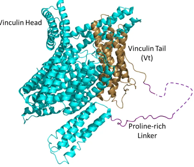
Figure 1.1. The structure of vinculin.
The crystal structure of chicken vinculin (PDB code 1ST6) is shown in a cartoon
representation. The vinculin head domain is shown in cyan, the proline-rich linker in purple,
Figure 1.2. Diagram of vinculin activation
In the absence of ligands, vinculin exists in an autoinhibited conformation, mediated
by a strong interaction between the head and tail domains. Activation of vinculin requires
the presence of both a head ligand, such as talin, and a tail ligand, such as F-actin. When
both ligands are available, the head domain and tail domain release their autoinhibitory
interaction and bind to their respective ligands. This conformational change allows for
binding of additional ligands on vinculin surfaces protected by the autoinhibited
Figure 1.3. Comparison of vinculin and metavinculin tails.
The overlaid structures of human Vt and metavinculin tail domains (structures used
are from PDB code 1RKE and Rangarajan et al. (65), respectively). Vinculin is shown in sand
and metavinculin in lavender. The structures are very similar, especially in the helix bundle.
Below is an alignment of the metavinculin 68-residue insert with vinculin residues 848 to
915. The second half of the insert shows high sequence similarity and homology to the
Chapter 2: Characterization of the Interaction Between the Vinculin
Tail Domain and PIP
2-containing Membranes
Introduction
Vinculin tail (Vt) can bind to many different ligands, including acidic phospholipids
(86), specifically phosphatidylinositol 4,5-bisphosphate (PIP2) (43). This interaction may
activate vinculin by disrupting the vinculin head:tail interaction (10, 32, 87). PIP2 is an
important signaling lipid for numerous cellular processes, including membrane ruffle
formation (88), exocytosis (89), and phagocytosis (90). In the case of focal adhesions (FAs),
PIP2 is generated by phosphatidylinositol phosphate kinase type 1 gamma (PIPKIγ) (91),
which is recruited to focal adhesions by talin (92, 93). Talin recruits vinculin, meaning that
vinculin colocalizes with and PIP2, which regulates the interaction of vinculin with talin (11,
94). PIPKIγ regulates FA dynamics (95, 96), is required for FA formation (97), and is
thought to be involved in recruitment and activation of vinculin at FAs (96, 98). While much
is known about the general role of PIP2 at FAs, the consequences of the vinculin:PIP2
interaction are much less understood, in part because of the lack of understanding for how
Vt binds PIP2. The interaction has been implicated in cell spreading, cell migration, FA
turnover, and force transduction (41, 42, 99), though the vinculin constructs used in these
(2, 58, 99), the basic ladder (41), or deleting the C-terminus (41, 42, 58, 99, 100). Other
groups have expressed peptide fragments of Vt and found that the basic ladder (3) or the
C-terminus (101) are sufficient for PIP2 binding.
However, the use of multiple mutations, deletions, and fragments of Vt likely alter its
structure and perturb other Vt functions (43). Hence, their use in studying the vinculin:PIP2
interaction in cells provides potentially misleading results. For example, the use of two
separate vinculin variants with reported PIP2 defects of comparable severity,
vinculinK952Q/K956Q/R963Q/R966Q and vinculin R1060Q/K1061Q, resulted in two
distinct cellular phenotypes; the double mutant disrupted the ability of the cells to exert
force on their substrate by 50%, the quadruple mutant did not (42). These disparate and
confusing results result largely from the lack of a good structural model for the interaction of
Vt with PIP2 and the use of poorly characterized PIP2-defective vinculin variants. This
problem is not unique to the interaction of vinculin with PIP2, as similar approaches have
been used to study the interaction of vinculin with actin (Chapter 3).
Recently, a crystal structure of a Vt mutant bound to a soluble, short-chain PIP2 was
solved by Chinthalapudi et al., providing the first structural model for how PIP2 binds to Vt
(PDB file 4PR9) (40). In the crystal structure, Vt R1060A forms trimers upon associating
with PIP2. Two of the Vt molecules recognize the PIP2 headgroup via residues in the basic
collar whereas the third Vt molecule interacts with the PIP2 headgroup through residues
K944 and R945. To test their model, mutations were made to residues in the basic collar (Vt
K1061Q) or the basic ladder (K944Q/R945Q), and were found to disrupt PIP2-binding by
lipid co-sedimentation. The authors posit that these mutations support their model for how
Vt binds PIP2 and that their model accurately depicts the oligomerization state of Vt upon
binding to PIP2. Vt has been shown, by crosslinking, to dimerize and trimerize in the
PIP2, these models do not explain how Vt would insert into a lipid bilayer (3). Additionally,
vinculin-null mouse embryonic fibroblasts expressing vinculin showed phenotypes effects
for two different vinculin constructs with weak PIP2 binding. The K944Q/R945Q double
mutation, as evaluated by fluorescent recovery after photobleaching (FRAP), prevents
exchange of vinculin at FAs, while the K1061Q mutant only mildly disrupts exchange of
vinculin at FAs. We applied biochemical and computation methods to this system to address
these discrepancies.
Materials and Methods
Expression and purification of Vt
Vt constructs expression constructs were a gift from Susan Craig. The pET15b Vt
expression vector encoded a His-tag followed by a thrombin cleavage site and then chicken
Vt residues 884-1066. Vt was expressed by transforming BL21 E. coli with the vector,
growing to an OD at 600 nm of 0.6, dropping the temperature to 18 C°, and inducing
expression with 0.25 mM isopropyl β-D-1-thiogalactopyranoside (IPTG). Cells were grown
overnight, centrifuged, resuspended in lysis buffer (20 mM Tris, pH 7.5, 150 mM NaCl, 5
mM imidazole, 5 mM β-mercaptoethanol), and frozen until sonication. Vinculin tail protein
was purified as previously described (43).
Lipid co-sedimentation assays
Vt binding to PIP2 was evaluated by lipid co-sedimentation assays using small,
(Avanti Polar Lipids) to produce the SUVs. Relative protein amounts were quantified using
ImageJ (104).
Binding to PS was evaluated similarly, with the only difference being the ratio of lipids
used. When assembling SUVs, the reported percentage of PS was used, and the remaining
lipids were a 3:1 mixture of phoshphatidlyethanolamine to phosphatidylcholine.
Actin co-sedimentation assays
Actin co-sedimentation assays for both actin binding and actin crosslinking
(bundling) properties of Vt were performed as previously reported (6, 103).
Mutagenesis of DNA constructs
Vt variants were generated using QuikChange site-directed mutagenesis (Stratagene)
and sequences were verified by DNA sequencing (Genewiz).
Computational modeling
Modeling of the interaction of the headgroup of PIP2 with Vt (residues 896-1055) was
performed with MedusaDock (105, 106). MedusaDock utilizes a library of rotamers for the
sidechains of Vt and a separate library for rotamers of the PIP2 headgroup. Docking
simulations are performed using these libraries, while the secondary structure remains
fixed, and the results are ranked by MedusaScore. The lowest energy pose is reported.
Modeling of the interaction of the full PIP2 molecule,
1-stearoyl-2-arachidonoyl-sn-glycero-3-phospho-(1'-myo-inositol-4',5'-bisphosphate), with Vt in the absence of a lipid
bilayer was performed with discrete molecular dynamics (DMD) simulations (107-109). The
initial docking pose was taken from the MedusaDock simulations. In these all-atom DMD
simulations, Gō constraints were used to maintain intramolecular Cβ/Cα contacts between
Vt residues during the simulation. These constraints were applied for all residues in Vt or
allows for greater sampling of potential Vt:PIP2 interactions while maintaining the structural
integrity of Vt. The assumption that Vt retains its fold upon binding to PIP2 is supported by
the lack of large conformational changes in the helix bundle in the crystal structure of Vt
bound to a short-chain PIP2 (40).
Modeling of the interaction of PIP2 within a lipid bilayer was performed using
all-atom, explicit solvent molecular dynamics simulations. A lipid bilayer of
1-palmitoyl-2-oleoyl-sn-glycero-3-phosphocholine (POPC) with a single PIP2 molecule was generated, and
an initial pose of Vt (residues 896-1055) bound to PIP2 using the model generated from
MedusaDock. Simulations were done with GROMACS (110) using a CHARMM27 force field
(111) and performed for 150 nanoseconds. A control experiment with a bilayer consisting
entirely of POPC was also conducted.
Circular dichroism spectroscopy
Circular dichroism (CD) spectra were collected as reported (43). Spectra were
collected on a Jasco J-815 CD Spectrometer at 25°C in 10 mM potassium phosphate, pH 7.5,
50 mM Na2SO4, and 1 mM DTT.For spectral scans, Vt was concentrated to 5 μM for 260-190
or 450 μM for 350-250 nm. For thermal melts, 5 μM Vt was used and ellipticity at 222 nm
was monitored as the temperature was increased from 25 C° to 95 C° at a rate of 2 C°min-1.
Results
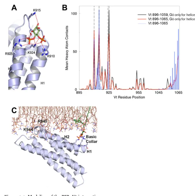
Computational modeling predicts distinct roles for the basic collar and basic ladder
unreasonable. MedusaDock identified the basic collar, specifically residues R910, K915,
K924, and R925, as the site on Vt with the highest affinity for the PIP2 headgroup. R910
interacted primarily with the hydroxyl of C4, K915 with the hydroxyl of C6 (which would
place it near the phosphoryl group on C1 in the full PIP2 molecule), K924 with the hydroxyl
of C5, and R925 with the phosphoryl group on C4 (Figure 2.2). This finding is in agreement
with the general geometry of the basic collar compared to the basic ladder, in which the
positive charges within the basic collar are clustered in a smaller surface area more
compatible with headgroup coordination relative to the basic ladder.
Next, DMD simulations (107-109) were performed with a complete PIP2 molecule,
1-stearoyl-2-arachidonoyl-sn-glycero-3-phospho-(1'-myo-inositol-4',5'-bisphosphate).
Simulating the interaction with the entire PIP2 molecule was the logical next step from the
initial simulations with just the inositol headgroup. Simulations were performed using Gō
constraints on Cb/Ca contacts (with the interaction square-well depth 0.5ε, where ε is the
DMD energy unit (108, 112)) for all residues in Vt (residues 896-1065) or Gō constraints
only for residues in alpha-helices (using Vt constructs 896-1065 or 896-1059). After
selecting 2000 snapshots with a binding energy less than -10 kcal/mol (105), the residues in
contact with PIP2 were tallied (Figure 2.2). In all cases, the contacts at the basic collar
remained high. When using Gō constraints only for helical residues, some contacts were
observed at the basic ladder, though contacts at the basic collar were more frequent.
While both MedusaDock and DMD simulations suggested that the basic collar was
the primary determinant for PIP2 headgroup recognition, these simulations were done only
with the PIP2 molecule or headgroup, not in the context of a lipid bilayer. To better mimic
the physiological interaction at the membrane, all-atom, explicit solvent molecular dynamics
simulations of Vt bound to PIP2 in a 1-palmitoyl-2-oleoyl-sn-glycero-3-phosphocholine
roughly parallel to the lipid bilayer at the start of the simulation. However, by the end of the
simulation, the side chains of K944, R945, K952, and K956, all in helix 3, show lipid
interactions resulting in insertion in the lipid bilayer (Figure 2.2). Throughout the
simulation, the basic collar maintained contacts with PIP2. A control simulation was
performed without PIP2, and no specific interaction with the POPC membrane was observed.
Results from these simulations suggest that, while the basic collar is responsible for PIP2
recognition, the basic ladder is required for insertion into and full association with lipid
bilayers.
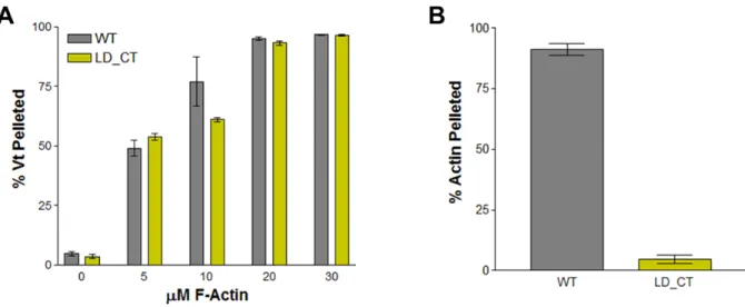
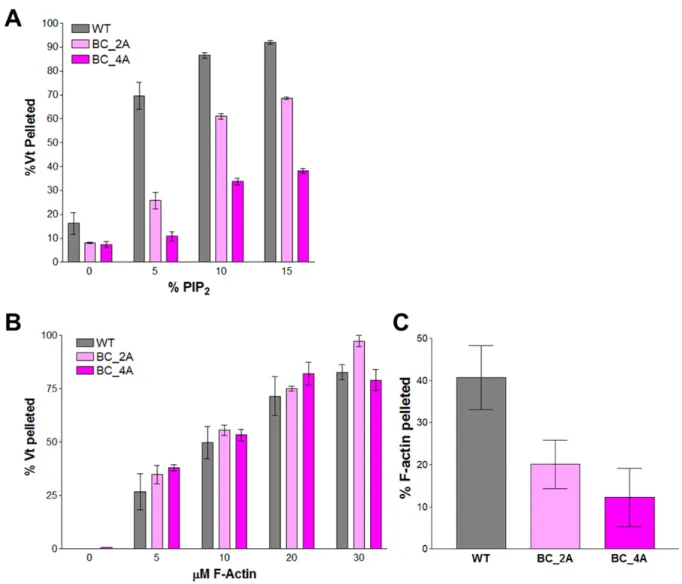
A common Vt variant deficient in PIP2-binding is a poor actin crosslinker
The vinculin variant, LD-CT (vinculin R1060Q/K1061Q), has been used in a number of
studies as a lipid-binding deficient vinculin construct (41, 42, 58). While Ziegler et al. (58)
observed no defects in actin binding or bundling for Vt LD-CT, Diez et al. reported that this
construct may have “impaired actin-vinculin binding” that would explain the lower strain
energy cells generate when expressing vinculin Vt LD-CT (42). To address this controversy,
we performed actin co-sedimentation assays with Vt LD-CT. High-speed co-sedimentation
assays revealed that Vt LD-CT has no observable actin-binding defect (Figure 2.3).
However, low-speed actin co-sedimentation assays show that Vt LD-CT is deficient in
F-actin crosslinking (Figure 2.3). These results further highlight the importance of the
C-terminus in actin crosslinking and the ability of vinculin to regulate mechanical force
through actin crosslinking (6). This can explain the decreased strain energy seen by Diez et
residues for PIP2 headgroup recognition are R910, K915, K924, and R925. The T6 construct
(Vt T6) construct, reported by Cohen et al. (28), consists of a K924A/R925A double
mutation. We measured the PIP2-binding activity of Vt T6 and the quadruple mutant
R910A/K915A/K924A/R925A (Vt BC-4A) using a PIP2 co-sedimentation assay (43). While
Vt T6 exhibited a modest decrease in binding to PIP2, with roughly half as much Vt binding
at 5% PIP2 (Figure 2.4), Vt BC-4A showed an even greater drop in PIP2 binding, with a
6-fold decrease in binding at 5% PIP2. Fitting the curves to a one-site binding model resulted
in an approximate KD of 43 ± 12 μM for Vt, 191 ± 29 μM for Vt T6, and 551 ± 66 μM for Vt
BC-4A. These findings suggest that the basic collar, specifically residues R910, K915, K924,
and R925, plays a key role in PIP2 binding.
To assess the viability of these variants as tools for study of a PIP2-specific defect in
cells, the basic collar Vt variants were tested for their ability to bind F-actin and to dimerize
to bundle F-actin. Using an actin co-sedimentation assay, no significant change in binding to
F-actin for any basic collar Vt variant was observed (Figure 2.4). These results are not
surprising, given that the basic collar is separate from the actin-binding surface of Vt.
However, mutation of the basic collar did result in a significant decrease in the ability of Vt
to bundle F-actin filaments, as measured by a low-speed co-sedimentation assay (Figure
2.4). This decrease was not rescued by using the more conservative Vt variant, Vt BC-4Q
(R910Q/K915Q/K924Q/R925Q) (Figure 2.5).
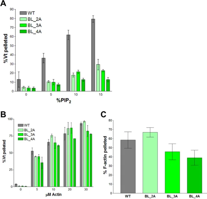
Mutation of residues in the basic ladder impairs binding to PIP2-containing liposomes but
not Vt dimerization
Since mutations to the basic collar disrupt the ability of Vt to bundle F-actin, vinculin
variants at this site would be poor tools to study lipid-binding by vinculin in cells. Observed
disrupting PIP2-binding. Three constructs were generated to test the importance of helix 3 in
binding to PIP2-containing liposomes: Vt K944A/R945A (BL-2A), Vt K944A/R945A/K952A
(BL-3A), and Vt K944A/R945A/K952A/K956A (BL-4A). These constructs all show
decreased binding to PIP2-containing liposomes (Figure 2.6). Vt BL-4A exhibited the most
severe defect, with an effective Kd 13-fold weaker than Vt WT. Vt BL-2A and Vt BL-3A have
Kd values roughly 6.5-fold weaker than Vt WT.
Actin binding and crosslinking activities for these variants were evaluated to test the
specificity of the PIP2-binding defect. Vt BL-2A, Vt BL-3A, and Vt BL-4A do not have
significantly different effective Kd values for binding actin (Figure 2.6). Additionally, slow
speed actin co-sedimentation assays show that there is no significant difference in the ability
of these variants to crosslink F-actin (Figure 2.6). These data, in conjunction with the lipid
co-sedimentation data, support the use of these basic ladder variants in studying the
biological function of the vinculin:PIP2 interaction.
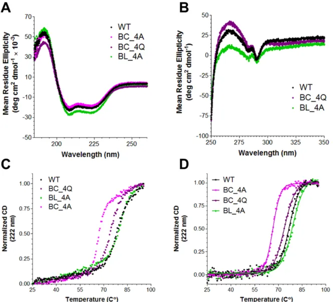
Mutations to the basic collar decrease Vt stability
To evaluate potential alterations in Vt structure by mutations to the basic collar and
basic ladder, CD data were collected. Far-UV data show a slight increase in helical secondary
structure for Vt BL-4A (Figure 2.7). This increase in helical character may result from the
K944A and R945A mutations, which may extend helix 3 by incorporating some of the helix
2-helix 3 loop residues into helix 3. Near-UV CD data, which report on the packing of W912
with W1058 and act as a proxy for tertiary structure, show that Vt BC-4Q and Vt BL-4A
has a 10 C° decrease in Tm that is only partially rescued with the more conservative Vt
BC-4Q. Vt BL-4A is slightly more stable than Vt WT, but only by 2.2 C°. These results suggest
that the basic collar, in addition to mediating Vt dimerization and actin crosslinking activity,
is important for Vt stability.
Mutation of residues in the basic ladder result in a larger defect in PS binding relative to
mutation of residues in the basic collar
As mutations at both the basic collar and basic ladder impair Vt binding to
PIP2-containing liposomes, it is difficult to determine which site is responsible for PIP2 specificity
by lipid co-sedimentation. Co-sedimentation experiments conducted with increasing
amounts of PIP2 did not show a large difference in the ability of Vt BC-4Q or Vt BL-4A to
bind PIP2-containing liposomes (Figure 2.8). However, since our computational modeling
and biochemical data suggest that the basic ladder is responsible for insertion into the lipid
membrane and the basic collar provides PIP2 headgroup specificity, but that both functions
are important for lipid binding in our co-sedimentation assays, we tested the hypothesis that
the basic ladder in critical for insertion by using lipid co-sedimentation assays with
increasing concentrations of PS and no PIP2. While both Vt BC-4Q and Vt BL-4A bound
PS-containing liposomes weaker than Vt WT, Vt BC-4Q retained binding, with 15% of Vt BC-4Q
bound when liposomes were 60% PS (Figure 2.8). Vt BL-4A, however, did not show
significant binding, even at high levels of PS. These data suggest that the basic ladder
residues K944, R948, K952, and K956 drive the non-specific interactions with
negatively-charged lipid membranes. Because mutations at the top of the basic ladder impair lipid
association but do not impair actin interactions, these variants are reasonable tools to study
Discussion
While it is known that vinculin binds PIP2 with high specificity (43) and facilitates
membrane association (3), the biological role of this interaction remains unclear. The study
of the vinculin:PIP2 interaction has been frustrated by a number of factors. The lack of a
good structural model and seemingly contradictory biochemical data resulted in the use of
vinculin variants with multiple ligand-binding defects, not solely PIP2 (41, 42, 99, 113, 114).
The use of these tools has resulted in publication of seemingly contradictory data, even
within the same manuscript (40, 42)! The recent structure from Chinthalapude et al. (PDB
file 4PR9) provides a good start to developing a structural model of the interaction of
vinculin with PIP2, but it falls short of explaining how this interaction takes place at a
biological membrane (40).
The biochemical data presented here are in agreement with those published by
Chinthalapudi et al., who observed that mutating K944 and R945 or mutating K1061 weaken
PIP2-binding activity by Vt (40). Both the basic collar and the basic ladder play important
roles in binding to PIP2-containing membranes. However, while Chinthalapudi et al.
attribute the effects of mutating K944 and R945 to the presence of a second PIP2-binding
site for the PIP2 headgroup on Vt that also assists in oligomerization, our results suggest that
these residues interact with the lipid bilayer in a PIP2-independent manner; the larger defect
in binding to PS-containing liposomes and our computational model point to the role of
these residues as critical for insertion into the plasma membrane. This interpretation of the
The structural and biochemical data also explain the lower affinity of metavinculin tail
for PIP2 (66). Metavinculin has a threonine at position 978, which replaces R910 in Vt. The
loss of one of the key residues for binding of PIP2 at the basic collar explains why
metavinculin still binds PIP2, though weaker than Vt.
The data presented here also support our recent structural model of the Vt-F-actin
interaction, which highlights the role of helices 4 and 5, not helix 3. Further, actin
co-sedimentation assays reveal that mutation of the basic collar and basic ladder do not
significantly affect actin binding. However, mutations at the basic collar do disrupt the
ability of Vt to crosslink actin filaments into bundles (Figures 2.3, 2.4, 2.5). Even the use
of conservative mutations to glutamine did not preserve Vt’s crosslinking activity. The
failure of Vt LD-CT to crosslink F-actin further highlights the importance of the C-terminus
in F-actin bundling (6, 115). The weaker crosslinking activity of Vt BC-2A and the nearly
absent crosslinking activity Vt BC-4A suggest that residues R910, K915, K924, and R925 are
integral in forming the Vt dimer. The failure of Vt BC-4Q to improve crosslinking activity
over Vt BC-4A indicates that the positive nature of the helix 1-helix 2 loop is likely required
for Vt to dimerize and crosslink actin filaments. It also provides new data to help develop a
model for the actin-induced Vt dimer.
These data provide new insights with which to develop better structural models for the
vinculin:PIP2 interaction and, ultimately, better tools to study the biological function of this
interaction. There remain further challenges to an accurate structural model, including
understanding the conformational changes that take place when Vt binds to PIP2 (10) and
the formation of higher order oligomers at membranes (10, 43, 66, 102). The Vt:PIP2
structure by Chinthalapudi et al. identifies an oligomeric species that coordinates PIP2, but
this model is unclear regarding how vinculin inserts into the membrane (40). Similarly, it
structural information derived from crystallography, NMR, or even cryo-electron
microscopy of vinculin associated with PIP2 in lipid bilayers will aid in further
understanding vinculin’s insertion into the membrane. Of the mutants described here, Vt
BL-3A and Vt BL-4A are likely the best mutants to use in cells to study the biological role of
the vinculin:PIP2 interaction. They exhibit a strong PIP2-binding defect, but, unlike the basic
collar mutants, retain the ability to crosslink actin filaments. Cellular studies with these
vinculin variants will be conducted to study the effects of the vinculin:PIP2 interaction of
T
m(C°)
WT
76.8 ± 0.1
BC-4A
66.5 ± 0.1
BC-4Q
73.8 ± 0.1
BL-4A
79.0 ± 0.2
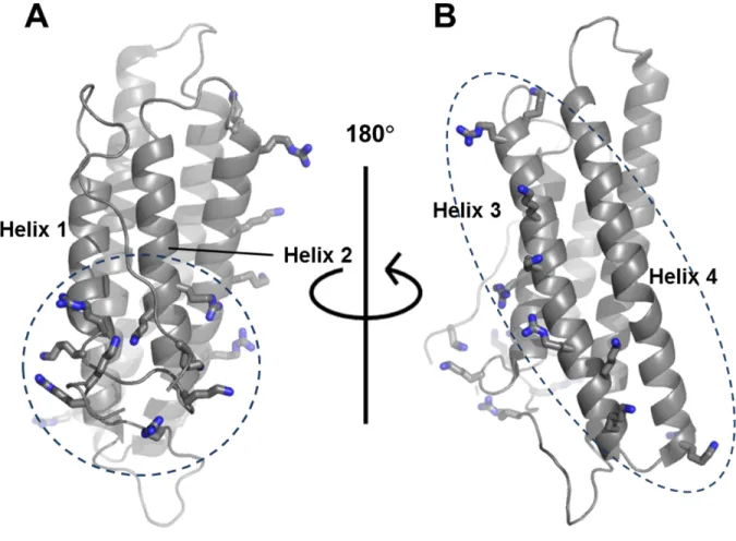
Figure 2.1. The basic collar and basic ladder of Vt.
Shown on Vt (PDB file 1ST6) are the basic collar (A) and basic ladder (B) residues. The
basic collar is comprised of K881, R910, K911, K915, K924, R925, R1057, R1060, and K1061,
which are found in the N-terminal strap, helix 1, the helix 1-helix 2 loop, helix 2, and the
C-terminal hairpin. The basic ladder is comprised of K944, R945, K952, K956, R963, K966,
K970, K975, and R976, which are found in helixes 3 and 4. Various studies have claimed that
the basic ladder (3), the basic collar (2, 99, 101), or both (40-42) are responsible for
Figure 2.2. Modeling of the PIP2:Vt interaction.
The results of MedusaDock (105, 106) (A), DMD (B), and GROMACS (C) simulations
are shown. The MedusaDock simulations, using the PIP2 headgroup, identified residues
R910, K915, K924, and R925 as residues that mediate PIP2 binding. These results were
followed by DMD simulations with the full PIP2 molecule (B). The average number of heavy
atom contacts with PIP2 is reported for each Vt residue in the three simulations. R910, K915,
amount of contact. The final pose of the GROMACS molecular dynamics simulation (C)
shows PIP2 (green) still bound to the basic collar and residues K944 and R945 inserting into
Figure 2.3. Actin-binding and crosslinking activity of Vt LD-CT (R1060Q/K1061Q).
Shown are the results from actin co-sedimentation assays evaluating the ability of Vt
LD-CT to bind (A) and crosslink (B) F-actin. Vt LD-CT binds to F-actin well, but is
Figure 2.4. Mutations at the basic collar disrupt binding to PIP2 and actin crosslinking
functions.
Shown are results from PIP2 co-sedimentation (A) and actin co-sedimentation (B, C)
assays. Vt BC-2A and Vt BC-4A exhibit decreased binding to PIP2-containing liposomes (A).
Figure 2.5. Mutation of basic collar residues to glutamine does not rescue F-actin
crosslinking activity.
Shown are the results of actin co-sedimentation assays measuring the ability of Vt
BC-4Q to bind (A) and bundle (B) F-actin. Vt BC-BC-4Q contains more conservative mutations
(basic to polar) than Vt BC-4A. Vt BC-4Q binds F-actin with a similar affinity to Vt WT.
Figure 2.6. Mutations to the basic ladder disrupt PIP2 binding, but do not significantly
impair F-actin binding or crosslinking.
Shown are results from PIP2 co-sedimentation (A) and actin co-sedimentation (B, C)
Figure 2.7. CD characterization of basic collar and basic ladder mutants.
Far-UV (A) and near-UV (B) spectra of Vt WT and Vt mutants. All constructs exhibit
the characteristic signature of an α-helical protein (A). The near-UV spectra for Vt BL-4A
suggests the packing interaction between W912 and W1058 has been altered, but not
abolished, as characteristic peaks are present at 283 and 290 nm. The normalized thermal
denaturation profiles for the Vt proteins (C) were adjusted by subtracting the buffer
contributions and fit with a sigmoidal curve (D, Table 2.1). Mutations to the basic collar
Figure 2.8. Comparative lipid co-sedimentation assays for Vt BC-4Q and Vt BL-4A.
Lipid-cosedimentation assays with PIP2-containing (A) or PS-containing liposomes.
Chapter 3: Structural, Biochemical, and Functional Characterization
of the Actin-Binding Site on Vinculin Tail
1Introduction
Focal adhesions (FAs) and adherens junctions (AJs) are essential structures in
eukaryotic cells that link cells to their environment: the extracellular matrix (ECM) and
other cells, respectively. These structures serve both structural and signaling purposes, both
of which are mediated through interactions with the actin cytoskeleton. Proper signaling
between these adhesions and the actin cytoskeleton is essential for cell migration, cell
survival, and the biological generation of force as reviewed by Gardel et al. (116). To ensure
efficient signaling, a large number of proteins present at FAs and AJs can bind to and/or
remodel the actin cytoskeleton.
Vinculin is one such protein, though defining its interaction with actin has remained a
difficult and somewhat contentious endeavor for scientists. The ability of vinculin bind
F-actin was debated for years after vinculin’s discovery until it was determined that the Vh:Vt
interaction masks actin binding and must be disrupted for vinculin to bind actin (117).
Subsequently, it has been shown that vinculin can cap actin filaments at the barbed end
(inhibiting polymerization) (118), nucleate actin polymerization in low ionic strength buffer
(119), and crosslink with other vinculin molecules to bundle F-actin filaments (120). While
1A significant portion of this chapter previously appeared in an article in Structure. The original citation is as follows:
these interactions have been studied biochemically, the lack of structural models for the
interactions has hindered our understanding of their roles in a cellular context.
Within the past eight years, a number of studies have been published attempting to
describe, at greater resolution and with techniques other than random mutagenesis and
deletions, the interaction of Vt with F-actin. The first such study was published by Janssen et
al. and used mutageneis data from Cohen et al. (28) and negative-stain electron microscopy
(EM) to propose two patches on Vt that bind two longitudinally adjacent F-actin protomers
within the filment (121). These two patches, termed the “upper” and “lower” sites, were
evaluated by Golji and Mofrad with molecular dynamics simulations (122). They identified
interactions between Vt helix 4 not identified by Janssen et al., slightly shifting the perceived
upper site. Additionally, they simulated Vt binding to the barbed end of the actin filament.
Not long after, Thompson et al. used negative stain EM and new mutagenesis data to further
support the role of helix 4, specifically its hydrophobic patch, in binding to F-actin (103).
Their mutagenesis data provided two new vinculin mutants with the most significant
actin-binding defects reported (I997A and V1001A). Another point mutant, R1049E, was
identified as being a weak actin binder, especially in low ionic strength solutions (123). This
mutant disrupts binding at the lower site. These mutants, when used in cells, show that
vinculin binding to actin is required for cell reinforcement, cell-spreading (103), cell
migration, and generation of traction forces (123). Vinculin does this by binding to F-actin,
slowing its retrograde flow, and altering the size, density, and growth rate of focal adhesions
of the Campbell and Alushin labs is beginning to identify the conformational changes using
cryo-EM. Some of that work, as well as work published previously (103), is described below.
Materials and Methods
Protein expression and purification
The plasmids for expression of vinculin and Vt were generated by the Liddington lab
(2, 10) . The gene for full-length chicken vinculin (residues 1-1066) was placed in a pET15b
vector. The gene for chicken Vt (residues 879-1066) was also placed into a pET15b vector for
protein expression. All mutagenesis was performed using QuikChange site-directed
mutagenesis (Stratagene, La Jolla, CA, USA) and the DNA sequence was verified by
sequencing (Genewiz, RTP, NC, USA).
To express either protein, chemically competent BL21 DE3 E. coli with the Rosetta2
plasmid (EMD Millipore, Billerica, MA, USA) were transformed with the vector and plated
on lysogeny broth (LB) plates containing ampicillin and chloramphenicol. A single colony
was selected and grown overnight. The overnight growth was used to inoculate flasks with 1
L LB. Cells were grown at 37C until OD reached 0.6. For full-length vinculin, expression
was induced with 1 mM isopropyl β-D-1-thiogalactopyranoside (IPTG) and expression
proceeded for three hours. For Vt, expression was induced with 250 μM IPTG and the
incubator temperature was cut to 16-18 C, and cells were harvested after 16-20 hours. To
harvest, cells were pelleted via centrifugation and then resuspended in lysis buffer (20 mM
Tris, 150 mM NaCl, 5 mM imidazole, and 2 mM β-mercaptoethanol (BME)). Cells were lysed
either with sonication or homogenization and the lysate was subsequently clarified by
centrifugation at 25,000 × g for 45 minutes. Both full-length vinculin and Vt were purified
using Ni-NTA beads (Qiagen, Germantown, MD, USA). The lysate was applied to the beads,
with lysis buffer with 60 mM imidazole. The protein was eluted with lysis buffer with 500
mM imidazole.
After purification by nickel column, proteins were dialyzed into thrombin cleavage
buffer (20 mM Tris pH 7.5, 500 mM NaCl, 2 mM CaCl2, and 5 mM BME) and their histidine
tags cleaved by addition of thrombin (Sigma-Aldrich, St. Louis, MO, USA). Proteins were
then further purified by ion exchange chromatography (Q-column for vinculin, S-column for
Vt) and size exclusion chromatography (S-200 superdex for both vinculin and Vt).
IpaA peptide (Ac-NNIYKAAKDVTTSLSKVLKNIN-NH2) was provided by both the
UNC Hight-Throughput Synthesis and Array Facility (Chapel Hill, NC, USA) and LifeTein
(Hillsborough, NJ, USA). The lyophilized peptide was resuspended in actin
co-sedimentation buffer (10 mM Tris pH 7.5, 100 mM KCl, 2.5 mM MgCl2, and 2 mM
dithiothreitol (DTT)) and centrifuged to remove aggregates and insoluble contaminants.
Peptide concentration was determined by absorbance at 280 nm using an extinction
coefficient of 1200 M-1 cm-1.
Actin co-sedimentation
Actin co-sedimentation assays were performed as previously described (6, 103).
G-actin was polymerized by addition of 2X and 1X G-actin co-sedimentation buffer so that the
final concentration of actin is 45.6 μM and the buffer is at 1X. Polymerization was carried
out for 30 minutes. Ten μL of 100 μM Vt or vinculin was then mixed with enough actin and
buffer to give the reported actin concentration in a total volume of 100 μL. When using the
intensities were compared using ImageJ (104). The percentage of protein pelleted was
calculated by dividing the intensity of the pellet band by the sum of the intensities of the
pellet and supernatant bands.
Circular dichroism
Circular dichroism (CD) spectra were collected on a Jasco J-815 CD Spectrometer
(JASCO, Easton, MD, USA). CD data were collected in 10 mM potassium phosphate, pH 5.5
or pH 7.5, 50 mM Na2SO4, and 1 mM DTT at 25°C unless otherwise specified. For near-ultraviolet (UV) measurements, protein was concentrated to 450 μM, while far-UV
measurements were collected on protein at 5 μM.
NMR spectroscopy
To make uniformly 15N-enriched Vt for NMR, expression was done in M9 media with 15NH4Cl as the sole nitrogen source. 15N-Vt was purified in the same manner as Vt from LB, exchanged into NMR buffer (10 mM potassium phosphate, pH 5.5, 50 mM NaCl, 0.1% NaN3,
2 mM DTT, and 10% D2O), and concentrated to 50 μM. Two-dimensional heteronuclear
single quantum coherence (HSQC) spectra were collected on a Varian INOVA 700 MHz
spectrometer at 37°C. Spectra were processed with NMRPipe (125) and analyzed with
NMRViewJ (126).
Generation of negative stain EM images, manual fit model, and discrete molecular
dynamics (DMD) model
Collection of the EM images and generation of both structural models have been
Cell culture, cell-spreading assays, and microscopy
Maintenance of vinculin-null mouse embryonic fibroblasts (Vin -/- MEFs), real-time
cell analyzer (RTCA) assays, and microscopy were performed as previously reported (103,
115).
Results
Mutation of the hydrophobic patch on Vt disrupts binding to F-actin
For eight years after the publication of the first structural model for the interaction of
Vt with F-actin (121), there were no specific mutations identified in vinculin that disrupted
the interaction. This hampered the ability of cell biologists to tease out the role of vinculin’s
interaction with F-actin without disrupting other interactions mediated by vinculin’s tail
domain. The best mutation identified for disrupting actin binding, at that point, was a triple
mutant, D959A/E960A/R963A, that disrupted binding to actin by 20% relative to wild type
Vt (28).
A former student in the Campbell lab, Sean Palmer, was trying to make mutations in
Vt to disrupt its propensity to self-associate (Chapter 4) (127). This self-association is
extremely weak (~330 μM) and is mediated through a hydrophobic patch on Vt that resides
on helices 4 and 5 (10, 127). Surprisingly, mutations at this interface disrupted binding to
F-actin, even though these mutations were not near the actin-binding surfaces identified in the
Janssen model. Specifically, Vt variants I997A and V1001A both significantly disrupted
protein. Without the addition of IpaA peptide, neither wild type vinculin nor the
actin-binding deficient variants I997A and V1001A were able to bind, F-actin (Figure 3.1). This
was expected, as vinculin must be activated by the presence of a head ligand to bind F-actin
(117). The addition of an activating IpaA peptide, however, rescued actin binding in vinculin
(103, 124). In I997A and V1001A vinculin, the addition of the IpaA peptide only partially
rescued binding to F-actin (103, 124), showing that the actin-binding mutations have a
similar effect in the context of the full-length protein.
To test the structural integrity of these Vt variants, CD spectra were collected from 185
nm to 350 nm. The far-UV spectra report on secondary structure, while the near-UV spectra
report on packing of W912 and W1058. These spectra were collected at both pH 5.5 and pH
7.5, as the charge on the sidechain of H906 has been reported to have a small effect on Vt
structure (127, 128). The CD spectra show a small change in the helicity of Vt I997A at pH
7.5, though the change is quite modest, suggesting that these mutations do not significantly
impact the structure of Vt (Figure 3.2) (103). To further test the structural integrity, 1H-15N HSQC NMR spectra were collected. For both Vt I997A and V1001A, only a few residues show
changes in their chemical shift values, and these residues are near to the sites of mutation,
suggesting that any change in Vt structure caused by these mutations is small and localized
(Figure 3.3) (103). The NMR and CD data suggest that both I997A and V1001A mutations
do not significantly affect Vt structure and that the actin-binding defect of these Vt variants
are due to disruption of the actin-binding site.
Development of a new structural model for the interaction of Vt with F-actin
Using the information from these mutations, new EM images of Vt-decorated F-actin,
and computational modeling, two possible structural models for the interaction of F-actin
reassign the upper site to helix 4, not helix 2. These models place the hydrophobic patch on
helix 4 of Vt in a hydrophobic groove in the actin filament (103). This groove is a canonical
binding site for actin-binding proteins (129).
While both the manual fit and DMD models place helix 4 in the hydrophobic groove in
the actin filament, the orientation of Vt is quite different. In the manual fit model, the
C-terminal end of helix 4 faces the barbed end of the actin filament, while in the DMD model,
the C-terminal end of helix 4 faces the pointed end (Figure 3.4). In this manner, the DMD
model has Vt rotated 180° relative to the manual fit model. Because of the resolution
limitations from the negative stain EM images, both models fit well into the
three-dimensional reconstruction and explain the effective disruption of actin binding by the
I997A and V1001A mutations.
Two additional mutations, T993A and E1015A, were made to further test these models.
In both models, T993 faces F-actin, though only in the DMD model does the sidechain of
E1015 face F-actin (Figure 3.4). Both of these mutations decrease actin binding, though the
effects are weaker than the I997A and V1001A mutations (Figure 3.1). These additional
data support choosing the DMD model over the manual fit model, though they are not
sufficient to eliminate consideration of the manual fit model.
Disruption of F-actin binding increases F-actin flow at focal adhesions, altering adhesion
size and function
With the development of point mutations in vinculin with specific defects in F-actin
To further examine the role of actin-binding, Thompson et al. expressed vinculin
I997A and vinculin V1001A in Vin-/- MEFs. MEFs expressing vinculin I997A or vinculin V1001A had slightly fewer, but larger FAs, similar to what Thievessen et al. observed
(Figure 3.5) (103, 124). To test the ability of these cells to respond appropriately to external
forces, three dimensional force microscopy (130) was used. MEFs expressing the various
vinculin constructs were plated and then magnetic beads coated in fibronectin (FN) were
added. After time passed to allow for FAs to develop between the MEFs and the beads,
successive pulses of magnetic force were applied. MEFs expressing wild type vinculin
underwent a strengthening of their focal adhesions, reinforcement, which decreased the
movement of beads upon successive pulses. However, MEFs expressing vinculin I997A or
vinculin V1001A failed to reinforce (Figure 3.6) (103).
Vinculin has also been implicated as having a role in cell spreading, though the role
of actin-binding in this function is unknown (4). Vin-/- MEFs and Vin-/- MEFs expressing vinculin, vinculin I997A, and vinculin V1001A were allowed to spread on FN and monitored
with the xCELLigence real-time cell analyzer system (131). Vin-/- MEFs exhibited a spreading defect, as evident by the lower cell index readout, which was only partially rescued by
vinculin I997A or vinculin V1001A (Figure 3.7) (103). This suggests that, while
actin-binding by vinculin plays a role in cell-spreading, it is not the only vinculin function involved
in regulation of cell spreading.
Cryo-EM modeling reveals details of Vt conformational change
Vt undergoes a conformational change when it binds to F-actin, though the structural
details of the conformational change are relatively unknown (10, 120). Bakolitsa et al.
showed that actin-binding by Vt leads to an increase in susceptibility to proteolytic cleavage
Collaboration with the Alushin Lab at the National Heart, Lung, and Blood Institute
was initiated to address some of these concerns. Using cryo-EM and Vt ΔC5, a Vt variant
defective in actin bundling (6), an 8.5 Å structural model for Vt bound to F-actin was
developed (Figure 3.8) (132). This model shows two key conformational changes that take
place upon actin binding: the straightening of helix 4, which is kinked by the presence of
P989, and the displacement of helix 1 (Figure 3.9). Whether helix 1 unfolds or remains a
helix and whether it has any interactions with F-actin is unclear, though there is some
evidence that it may bind to the barbed-end actin protomer (unpublished). However, it is
clear that binding to F-actin converts Vt structure from a five-helix bundle to a four-helix
bundle.
While the 8.5 Å resolution of the cryo-EM model limits the detail with which
conformational changes in Vt can be described, it does provide insight to the mode of actin
binding. In comparing the cryo-EM model to the published models (103, 121, 122), it is clear
that, while none of the previous models could predict the Vt conformational changes, the
DMD model published by Thompson et al. comes the closest to predicting the actin-binding
surface on Vt (Figure 3.8) (103). Though the resolution limitation precludes the
identification of specific contacts made by sidechains, those closest to F-actin are more likely
to interact. Thus, to further test the validity of the cryo-EM model, two Vt residues that had
not been mutated, R987 and M1022, previously were mutated to alanine. Actin
co-sedimentation assays show that these mutants disrupt binding to F-actin (Figure 3.10).
bound to F-actin has long hampered a better understanding of the biological role of this
interaction. This has been further complicated by the conformational change that vinculin
undergoes upon binding to F-actin and by the intrinsic difficulties F-actin poses to structural
biologists.
Using some fortuitous mutagenesis results, a combination of EM, modeling, and
biochemical techniques, the highest-resolution model for the interaction of vinculin with
F-actin has been developed. This model confirms that the upper F-F-actin binding site on Vt is
localized to the hydrophobic patch on helices 4 and 5 (103). Additionally, the Vt helix bundle
loses helix 1, becoming a four-helix bundle upon binding to F-actin, explaining the limited
proteolysis data suggesting an increase in flexibility around helix 1 (10). The predictive
power of this structural model in disrupting actin binding further supports its validity.
The development of these structural models has allowed for rational use of specific
point mutations in cells to determine the biological role of actin binding by vinculin.
Actin-binding by vinculin regulates FA growth and maturation, affecting both cell spreading and
mechanotransduction in mouse fibroblasts. Further experiments using vinculin mutants
selectively deficient in F-actin binding showed that, by binding to actin, vinculin serves as a
mechanical clutch at FAs to regulate retrograde actin flow, which is an important factor
regulating FA development (124).
With the tools now in place, future studies should be able to determine the role of actin
binding by vinculin at adherens junctions and in cellular migration. Additionally, the role of
actin binding by metavinculin can now also be evaluated using the tools reported here. As
vinculin’s role in cancer becomes more defined, the role of actin binding by vinculin in both
tumor growth and in metastasis will be of interest. If actin binding by vinculin is important
for cancer progression, perhaps the Vt conformational change can be targeted as a