SHEAR BOND STRENGTH OF ORTHODONTIC BRACKETS WITH VARYING BONDING PROTOCOLS
Jeremy J. Grabouski
A thesis submitted to the faculty at the University of North Carolina at Chapel Hill in partial fulfillment of the requirements for the degree of Master of Science in the School of Dentistry
(Orthodontics).
Chapel Hill 2016
Approved by:
Lorne Koroluk
Ching-Chang Ko
ABSTRACT
Jeremy J. Grabouski: Shear bond strength of orthodontic brackets with varying bonding protocols
(Under the direction of Lorne Koroluk)
Objectives: To assess in vitro shear bond strength (SBS), modified adhesive remnant index (ARI), and treated enamel surface textures of various bracket bonding techniques. Methods: Extracted human premolars (n=97) were randomly assigned to six groups and bonded using
assigned protocols. SBS was measured using a universal testing machine. Modified ARI was
measured via optical stereomicroscopy. Etched enamel surface textures were qualitatively
described using scanning electron microscopy (SEM). Results: There was a statistically significant interaction between bonding technique and bracket placement. Various significant
differences were found between groups. All groups exhibited clinically acceptable means SBS.
ARI measurements differed between bonding techniques for both maxillary and mandibular
brackets. Conclusions: All tested methods exhibited clinically acceptable shear bond strengths. Pre-etching prior to the application of SEP may enhance the strength of brackets compared to SEP
alone. Pretreatment of normal enamel surfaces with NaOCl for 1 minute may affect the mode of
ACKNOWLEDGEMENTS
Thank you to the members of my thesis committee, Dr. Lorne Koroluk, Dr. Ching-Chang
Ko, Dr. Lee Boushell, for your mentorship, patience, and guidance throughout this project.
Thank you to Dr. Ceib Phillips for your statistical expertise. Thank you to Adam Danze, John
Whitley, and Wallace Ambrose for your technical and laboratory assistance. Thank you to 3M
Unitek for the donation of various bonding materials required for the success of this project.
Thank you to the Southern Association of Orthodontists for the generous research grant. Thank
you to NIH/NIDCR R01DE022816 for additional support. A very special thanks to my wife,
TABLE OF CONTENTS
LIST OF TABLES ... vii
LIST OF FIGURES ... viii
LIST OF ABBREVIATIONS ... ix
LIST OF SYMBOLS ...x
A REVIEW OF THE LITERATURE...1
The Beginnings of Orthodontic Bonding ...1
Biologic Components of Orthodontic Bonding ...4
Enamel ...4
Salivary Pellicle ...5
Chemical Components of Orthodontic Bonding...6
Phosphoric Acid Etch & Pumice ...6
Adhesives ...9
Self-Etching Adhesives (Self-Etching Primers, SEPs) ...9
Sodium Hypochlorite ...11
Composite Resin ...12
Testing Extracted Teeth ...13
Shear Bond Strength ...13
Storage ...13
SHEAR BOND STRENGTH OF ORTHODONTIC BRACKETS WITH VARYING BONDING
PROTOCOLS ...20
Introduction ...20
Materials and Methods ...23
Statistical Analysis ...27
Results ...27
Discussion ...29
Conclusions ...32
References ...33
APPENDIX ...20
Tables ...37
LIST OF TABLES
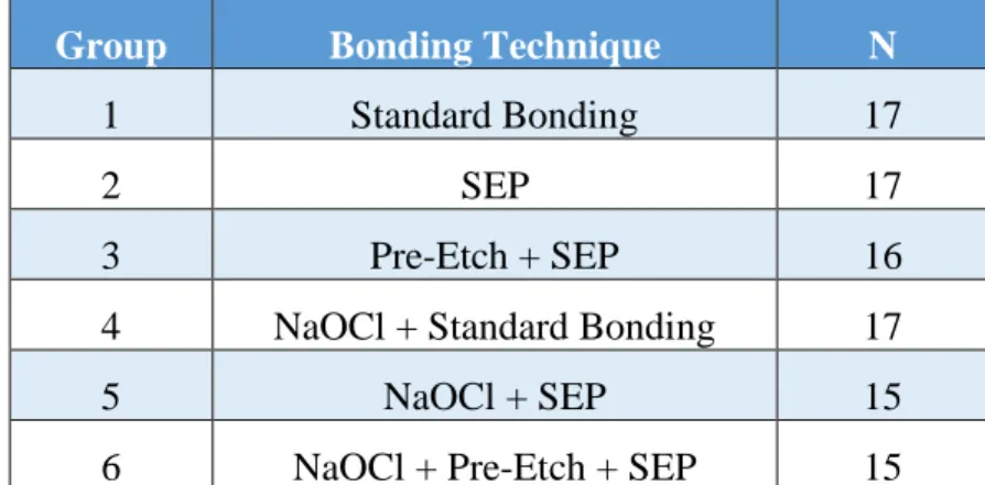
Table 1 – Distribution of extracted premolars to bonding groups ...37
Table 2 – Descriptive Statistics of Various Bonding Techniques and Bracket Types ...38
LIST OF FIGURES
Figure A – Bis-GMA molecular structure ...40
Figure B – Author illustration of enamel extracellular matrix after extraction of the ameloblasts ...41
Figure C – Modern bracket base with undercuts for mechanical retention to composite resin ...42
Figure 1 – Transbond™ Plus Self Etching Primer and Transbond™ Plus Self Etching Primer Easy Roller...43
Figure 2 – Five-sample Mounting Jig ...44
Figure 3 – Boxplot of Means and Standard Deviations of Peak SBS ...45
Figure 4 – Mandibular Combined ARI Score Frequency ...46
Figure 5 – Maxillary Combined ARI Score Frequency ...47
LIST OF ABBREVIATIONS
ARI Adhesive Remnant Index
cm Centimeter
MPa Megapascal
µm Micrometer
mW Miliwatt
mm Millimeters
min Minute
nm Nanometer
NIDCR National Institute of Dental and Craniofacial Research
NIH National Institutes of Health
N Newton
PE Pre-Etch
SEM Scanning Electron Microscope
SEP Self-Etching Primer
SBS Shear Bond Strength
NaOCl Sodium Hypochlorite
LIST OF SYMBOLS
© Copyright Symbol
® Registered Trademark
A REVIEW OF THE LITERATURE
The Beginnings of Orthodontic Bonding
In the 18th century, malocclusions were referred to as “irregularities,” and the process of correcting these “irregularities” was known as “regulating.” The Father of Orthodontia, Pierre
Fauchard, determined that the best way to “regulate” the teeth was to fix appliances to them that
would apply force. An advancement made in 1871 by William E. Magill allowed for better
attachment of appliances with bands. Several decades later, in the early 1900s, Edward Angle
included bands on the first molars and several other teeth for his E-arch. Angle’s appliances
would go through several iterations, ultimately culminating in the fully-banded Edgewise
appliance in 1925.1
Exclusively banded appliances were common practice and universally used in
orthodontics until Dr. Michael Buonocore laid the foundation for adhesive dentistry. In 1955,
Buonocore reported that applying phosphoric acid to enamel could render a tooth more
“receptive to adhesion.” He demonstrated this claim attaching a polymethylmethacrylate button
to both an unetched control tooth and a tooth etched with 85% phosphoric acid for 30 seconds.
The etched tooth showed a 100-fold increase in bond strength when compared to the control.2 Five years later, Swanson and Beck evaluated all current techniques for intraoral bonding to
enamel and determined that all current adhesive materials were not strong enough for clinical
with an epoxy-based resin and demonstrated successful dental bonding of plastic orthodontic
brackets.4
Dental adhesives shifted radically with the publication of the 1963 Landmark article
“Properties of a silica-reinforced polymer for dental restorations” by Rafael Bowen. Dr. Bowen
demonstrated that silica could be coupled with polymer during a curing process to make a very
strong bonding material that we know today by the word “composite.”5,6 The problem was that
the silica particles required a coating in order to bond with the polymer. Bowen found that a
coating traditionally used in making glass-reinforced polyester laminates known as
tris(2-methoxyethoxy) vinyl silane solved the problem and so was born bisphenol A-glycidul
methacrylate, or “bis-GMA” (Figure A) for short.
This self-cure two-paste composite demonstrated many attractive qualities for dental
bonding and was soon combined with the acid-etch technique. 5 According to Proffit, there are several advantages to bonding brackets over traditional banding. These advantages include: ease
of bracket placement, no interproximal extension thus no need for separation of the teeth, easier
to remove, more hygienic, more esthetic, and less irritation to the gingiva.7 With overwhelming advantages to bonded brackets, especially anterior brackets, and a new clinically effective
adhesive, bonding became commonplace in orthodontics. In 1979, Dr. Leonard Gorelick
conducted a survey of 7000 practicing orthodontists produced 2000 respondents. Of the 2000
respondents, 93% indicated that they were using composite bonding in some capacity in their
practices.8
The two-paste, self-cure composite popularized by Bowen required an initiator in order to
begin the polymerization process known as “curing.” Once the two pastes were mixed, the
N,N-Dimethyl-amino-p-toluidene. Mixing two different pastes and waiting for the mixture to cure presented
several limitations. Oxygen air bubbles could be incorporated during the mixing process and
inhibit polymerization leading to worsening of physical properties. The longer working time was
also problematic.9 In the 1980’s, the benzoyl peroxide initiator was replaced with a
camphorquinone photoinitiator. Camphorquinone is a unique initiator because it is activated by
visible light between 400nm and 500nm with a peak absorption at 460-480nm. Clinically, a
photoinitiator offers distinct advantages over a traditional initiator because it allows for extended
manipulation of the composite resin and an immediate on-demand cure. While certain clinical
situations may warrant a self-cure or dual-cure composite, a vast majority of the composite resin
used today contains camphorquinone and is activated by focused blue light.
According to Reynolds, an essential feature of an orthodontic resin is the ability to resist
masticatory forces and remain firmly attached to the teeth during the treatment of malalignment.
He determined that the shear bond strength an orthodontic resin must have to meet this clinical
expectation was 5.88-7.84MPa.10,11 Dental bonding techniques continued to be refined
throughout the 1980s and 1990s. As different bonding chemicals and techniques were improved,
each significant improvement was referred to in terms of “generations.” In generations 1-4, the
bond strength of enamel and dentin bonds was improved and the performance was increased.
The fourth generation system consisted of a total-etch with phosphoric acid followed by multiple
bottles of primer and adhesive prior to the addition of composite. This system demonstrated
Reynold’s acceptable orthodontic clinical bond strength and so began attempts to simplify the
process. Research and product development shifted toward increasing clinical efficiency.
Fifth-generation bonding decreased the number of steps by combining the primer and adhesive into a
and prime/adhesive step into one single chemical application. The self-etching adhesive (often
called self-etching primer) was born. 12
Biologic Components of Orthodontic Bonding
Enamel:
Dental bonding includes not only bonding to enamel, but also bonding to dentin for
restorative purposes. These two processes are slightly different due to the differences in physical
composition between enamel and dentin. Orthodontic brackets are placed almost exclusively
onto enamel, making dentin bonding techniques unnecessary for the treatment of malocclusion.
Enamel is the hardest and most highly mineralized substance in the human body. The
makeup of enamel (by weight) consists of 96% minerals, 3% water, and 1% organic material,
such as proteins. The majority of the mineral content is calcium phosphate in carbonated
hydroxyapatite crystals.13 These highly oriented crystals are extremely long and contain over 1000 times the volume of similar crystals in bone, dentin, and cementum. The crystals are
organized into bundles known as prisms, about 4µm in diameter, and extend outward from the
dentin surface.14
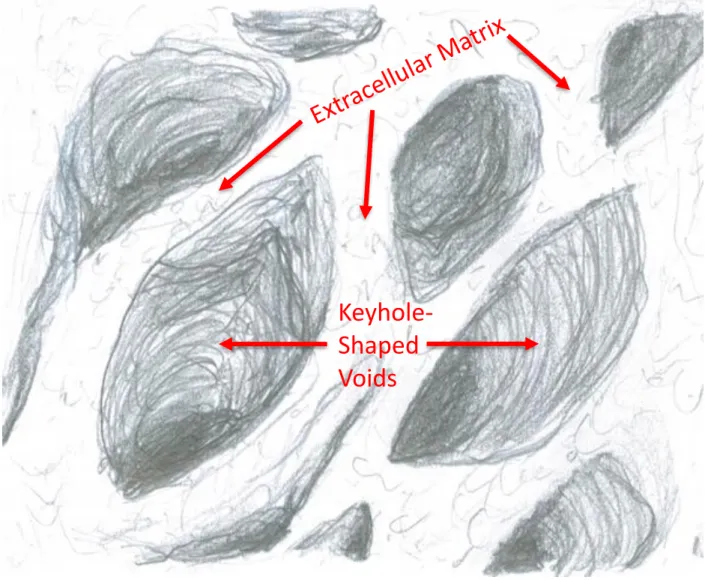
The creation of enamel, amelogenesis, is genetically controlled and the size, shape, caries
susceptibility, and even shade can vary from person to person. Included in the 1% of organic
material that makes up enamel are two classes of proteins, known as amelogenins and enamelins,
which help establish the enamel framework for mineral deposition during amelogenesis. This
formative process begins at the dentinoenamel junction for dentin and enamel simultaneously.
On the enamel side, crystal nuclei extend into long and evenly spaced ribbons and grow in length
as ameloblasts secrete enamel proteins into an extracellular matrix (Figure B). This matrix is
Tomes processes of ameloblasts and eventually mineralize. If this process is interrupted the
resulting enamel crystals can be too thin or hypoplastic. After a certain length is reached,
proteinases arrest the growth of crystal length and begin the mineralization process. As minerals
are deposited for 3-4 years, the enamel crystals grow in width and thickness and begin to harden.
If this process is interrupted, the enamel can be pathologically soft or porous.14
After the development of the tooth is complete, the tooth is ready to erupt. Upon eruption
into the oral cavity, the enamel surfaces immediately acquire a salivary pellicle made up of
salivary proteins among other things.15
Salivary Pellicle:
Human saliva serves several different roles: lubrication, digestion, and protection. Saliva
protects the dentition because it serves as an acid buffer to prevent decay of enamel structure, it
adds volume and dilutes the potency of potentially harmful chemicals, and salivary flow allows
for clearance of destructive agents. Additionally, saliva has a high content of proteins such as
glycoproteins and proline-rich proteins that aid in the creation of the dental salivary pellicle.
In 1963, Dawes et al. described the acquired enamel pellicle as a structureless and
bacteria-free film covering the teeth immediately after eruption.16 Meckel took things one step further and measured this film, concluding that the immediate pellicle ranges from 1-10µm in
thickness.17 Furthermore, Armstrong determined that the composition of pellicle differed from that of dental plaque and his component analysis concluded that acquired salivary pellicle
consisted of mainly salivary mucoprotein and bacterial cell wall material.18 Further research has shown that enamel adsorbs specific salivary biopolymers and this dynamic biofilm modulates all
interactions between the teeth and oral cavity. The pellicle begins to form on the tooth surface
electron microscope (SEM) examination, this pellicle appears to be a complex network of
relatively even thickness. Unfortunately for the orthodontist, the acquired pellicle becomes
thicker on the buccal surface of the teeth when compared to the lingual surface of the teeth.
When mature, the pellicle lubricates dental contact, thereby reducing abrasion and attrition while
providing protection from acidic attack.19
Chemical Components of Orthodontic Bonding
Phosphoric Acid Etch & Pumice:
The creation of the enamel etch pattern rendering a tooth more susceptible to adhesion
requires a strong acid. The acid removes a small amount of interprismatic enamel creating a
porous surface, thus increasing the total bonding surface area and allowing adhesion promoters
to penetrate into enamel pores and ultimately results in secure micromechanical retention.7 Buonocore began in 1955 with a solution of 85% phosphoric acid etch that he placed onto
enamel for 30 seconds to demonstrate his proof of concept.2 Since that time, the type and concentration of the acid has been analyzed as well as the duration that the acid is left in contact
with the enamel. Retief used 50% concentration in his studies and Gorelick and Silverstone
determined that 30-50% was ideal.8,20,21 While acceptable bond strengths were obtained from as low as 2% phosphoric acid, Chow and Brown found that concentrations under 27% produce the
insoluble byproduct of dicalcium phosphate dihydrate while concentrations over 27% produce
the soluble byproduct of monocalcium phosphate monohydrate.22,23 The soluble product could be washed away when rinsing the tooth of acid while the insoluble byproduct could present
troublesome complications with the bonding chemicals. Multiple studies have examined how
long to leave acid etch in contact with the enamel surface. Kinch et al. determined that there was
60 seconds. Since more enamel is dissolved and hence more damage is done to the teeth over
the course of 60 seconds, he recommended using 15 seconds as the clinical guideline.24 Olsen et al. similarly found no significant differences in shear bond strength when etching for 10 seconds
versus 30 seconds.25 Gilpatrick et al. even found that an adequate bond strength could be obtained by etching for only 5 seconds.26
Etching dissolves hydroxyapatite crystals and provides micromechanical retention by
allowing penetration of adhesion promoters and development of resin tags during bonding.27 As the concentration of phosphoric acid decreases so does the length of the resin tags (22 microns
for 35% phosphoric acid, 12 microns for 20%, 9 microns for 10% and 5%, 5 microns for 3%).
Interestingly, increasing the concentration of phosphoric acid above 35% also decreased the
length of resin tags although it increased the depth of the total etch and presumably increased
enamel damage (9 microns for 65%). There may be no correlation, however, between bond
strength and resin tag length and this relationship may be mainly attributable to the ability of the
monomer to penetrate the enamel rods and crystallites.28
Though it has been demonstrated that a high bond strength does not depend on an ‘ideal
etch pattern,’ the etch patterns of enamel surfaces have been classified into three major types,
though additional types have been proposed15,29-31:
Type 1: The enamel rods are dissolved and there is preferential prism core etching
(thought to be most favorable for bonding).
Type 2: The area around the enamel rods is dissolved and there is preferential prism
periphery etching.
There has been some discussion that anterior and posterior teeth behave differently to
etching of the enamel surfaces. Similar studies have identified differences in etch quality for
maxillary and mandibular teeth. These studies concluded that the etch quality is less favorable in
mandibular teeth and becomes progressively less favorable posteriorly. Reasons for these
differences may include bracket adaptation, increased force levels, increased humidity, and
increase in ‘prismless’ enamel in posterior teeth.32
In 1973, Miura et al. concluded that a pumice prophy was absolute necessity prior to
bonding orthodontic brackets to remove dental pellicle and increase bracket retention.33 A decade later in 1983 however, Main et al. concluded that the pumice was wholly unnecessary
because etching the tooth with phosphoric acid and rinsing it completely removed the acquired
salivary pellicle, provided that the tooth remain isolated until bonding.34 Further research supported Main’s findings, showing no significant differences in bond strength, surface
characteristics, or bracket retention rates in pumice vs no-pumice preparations of conventionally
bonded brackets.35,36
This debate has surfaced yet again with the advent of self-etching primer. Since there is
no etch-and-rinse step in the preparation of the enamel surface with a self-etching primer, and the
pH of the SEP is substantially higher than phosphoric acid, the pellicle is never entirely removed.
Clinical studies have once again concluded that a pumice prophy is an absolute necessity prior to
bonding orthodontic brackets with SEP.37,38 Other studies argue that pre-etching with phosphoric acid to remove the pellicle before applying SEP is a more simplistic step that can
result in higher bond strengths.39
Though once thought to have an impact, fluoride applied to the enamel surface does not
Adhesives:
The interface between composite resin and tooth structure has been referred to by several
different names including ‘primer,’ ‘adhesive,’ ‘bond enhancer,’ and others. All of these can be
grouped under what Ray et al. called “adhesion promoters.”41 The adhesion promoter is
responsible for penetrating the etched enamel and acting as a wetting agent, which subsequently
forms resin tags that create micromechanical adhesion to the enamel surface. The adhesion
promoter offers a strong chemical bond with the composite resin. Traditional adhesives are very
sensitive to contamination with saliva since they are hydrophobic and saliva promotes the
immediate remineralization of the etched enamel surface, resulting in poor bond strength.7
Self-Etching Adhesives (Self-Etching Primers, SEPs):
SEPs are the result of research into the sixth and seventh generations of adhesive dental
bonding. They allow the etch and primer to be completed in one single step, decreasing the
number of steps involved for bonding. This saves time clinically and is very convenient for the
clinician, though the bonding agent is more expensive.12 Self-etching primers also offer the distinct advantage of being hydrophilic and are subsequently much less sensitive to moisture and
more forgiving in the hostile oral environment.7 In fact, one of the major solvent used in most SEPs is water.42 Some authors have concluded that the shear bond strength (SBS) of SEPs is significantly lower compared to conventional bonding materials but most have agreed that there
is no difference in survival time.43-45 Other studies have suggested that the SBS of SEPs is no different or even slightly better than traditional techniques.46,47 Most studies seem to agree that the SBS of orthodontic bracket using SEPs have been acceptable for use clinically.48 Failure rates in-vivo for SEPs are wildly variably with some reports of failure rates as low as 1.6% and
in the 2012 Fleming meta-analysis resulted in 5.9% for SEPs and 4.5% with conventional etch
and rinse methods.49 This variability may be related to the enamel surface preparation prior to applying the SEP.
Chemical engineers had a tall order creating the SEP. They were tasked with finding a
way to etch the enamel surface, much like phosphoric acid, but leave the acid in place and have it
also act as a semi-permanent adhesion promoter. 50 The methacrylate group and phosphoric acid were combined into a clever methacrylated phosphoric acid ester in order to etch and prime at
the same time. Though this acid has a higher pH than traditional phosphoric acid, the pH does
not seem to be a primary determinant in the bond strength that a SEP is able to attain.50 Calcium is dissolved by the acid and removed from the hydroxyapatite crystal structure. Instead of
rinsing the acid and byproducts away, like with traditional phosphoric acid etching, the calcium
complexes with the phosphate group and effectively neutralizes it. The elegant chemical
reaction actually uses the waste product to alter the pH of the acid and solves two problems at the
same time. Since the acid is neutralized with the calcium byproduct, the SEP must be agitated
on the enamel surface to expose unreacted acid ester with hydroxyapatite crystals and continue
the etching process. As the water and other solvents evaporate from the SEP, the fluid becomes
more viscous, slowing the transport of unreacted acid to the enamel surface. One advantage of
the SEP is that every possible etched portion of the enamel surface is simultaneously coated with
an adhesion promoter, the primer. The neutral phosphates in contact with enamel then becomes
a permanent and safe part of the polymer when cured with light and activating the
camphorquinone initiator. After curing, unreacted phosphate cannot be transported to the enamel
surface because it has been permanently locked within the polymer and cannot continue the
Bishara described the resin tags provided by SEPs as being thin in width and less uniform
when compared to traditional bonding techniques.51 Shinchi et al. argues that there is no correlation between length of resin tags or etch pattern and bond strength.28 It has also been hypothesized that the decrease in mechanical retention with little change in bond strength
indicates an additional mechanism of adhesion in the form of a chemical bond not currently
recognized.47 SEPs result in decreased depth of etch and subsequently less damage to the enamel surface. It has also been recognized that less composite/adhesive remains on the enamel surface
after debonding and therefore easier to remove residual composite.51 As of 2008, SEPs were routinely being used by 30% of practitioners in the United States. 49 A meta-analysis comparing conventional orthodontic bonding to self-etching primers concluded that while SEP may result in
a modest time savings, there is weak but insignificant evidence that SEP may increase the risk of
bond failure, and in the absence of clear evidence that one is superior the technique used remains
at the digression of the clinician.49
Sodium Hypochlorite:
A 1994 case report at the University of North Carolina Chapel Hill described difficulties
in attaching a bracket to a hypocalcified tooth using all known techniques. 52 The author was finally successful after using a 1 minute application of 5% sodium hypochlorite (NaOCl) prior to
acid etching. 52 The rationale for using the NaOCl was that hypocalcified amelogeneisis imperfecta teeth can have as much as an eight-fold increase in the amount of enamel protein
compared to normal enamel and this might be affecting the bonding properties of the enamel.52 This profound effect of NaOCl increasing bond strength to hypocalcified enamel was supported
proteins from the enamel surface and increases the effective bonding surface.54 Ramakrishna et al. attempted to use NaOCl after acid etching rather than the generally accepted order of applying
NaOCl before acid etching and the procedure had no significant effect on bond strength.55 Espinoza compared etch patterns using three groups: no NaOCl, 30 sec pretreatment of NaOCl,
and 60 sec pretreatment of NaOCl. The researcher found that the 60 second pretreatment made a
significant difference in the amount of surface conditioned and quality of the etch pattern when
compared to no NaOCl and the 30 second NaOCl pretreatment.54 The endodontic literature has also reported increased bond strength of composite resin after teeth had been cleaned with
NaOCl.56 When examining the bond strength of a resin-modified glass ionomer, Justus et al. determined that conditioning the enamel surface with 1 minute of NaOCl can significantly
increase the SBS.57
Composite Resin:
Present day composite resins contain inert fillers, such as silica, and resin monomers. The resin
can be cured and polymerized by light, chemical, or both. A light cured resin is convenient
because no mixing is required and it has better initial properties. However, light must be able to
penetrate to the resin’s location sometimes restricting its use. Proffit defines a successful
bonding material as dimensionally stable, fluid enough to penetrate enamel, strong, and easy to
use clinically.7 Resin monomers do not contain many carboxyl groups and do not chelate to dentin, enamel, or dentin surfaces. The majority of the adhesion is regulated by
micromechanical retention.58 Therefore, optimal resin adhesion requires adequate preparation of an enamel surface.59 Similarly, mechanical retention is required within the metal bracket base. Manufacturers design clever undercuts in the bracket bases to provide this mechanical interlock
Testing Extracted Teeth
Shear Bond Strength:
Oxford Dictionaries defines shear as: “A strain in the structure of a substance produced
by pressure when its layers are laterally shifted in relation to each other.”60 A majority of studies regarding orthodontic bond strength use ‘shear’ bond strength rather than peel, tension, torsion,
or cleavage because it is the most reproducible. It is important to note, however, that the shear
bond strength can be very significantly affected based on the location of the blade applying the
force during debond. Ideally, the blade of the debonding instrument should be placed at the
bracket base where it meets the tooth enamel. In this way, the entire layer of the bracket based is
being evenly shifted laterally in relation to the enamel surface. As Klocke et al. demonstrated, a
shift of the blade toward the ligature groove of the bracket wings can generate significant peel
forces and decrease the bond failure point by over 50%. In the in-vitro study, when the blade of
the universal testing machine was placed at the bracket base in contact with enamel, the peak
debonding force 22.70±4.23 MPa, when the blade was tested again in the ligature groove the
peak debonding force fell to 11.52±2.74 MPa and fell again to 9.44±2.96 MPa when the blade
was moved out to the tie-wings.61 Klocke also showed that the direction of force against the bracket can significantly affect the shear bond strengths measurements and called for
standardization of all samples when doing orthodontic shear bond testing.62
Storage:
Extracted teeth can be stored in a variety of mediums prior to testing. Though extracted teeth are
drier than vital teeth in-vivo and more prone to enamel fractures, they can be very useful for
research if stored in a proper solution. Some possible storage mediums include 70-96% ethanol,
not a concern, a buffered 0.9% saline solution, 2% glutaraldehyde, or standard tap water are also
possible options.63 Formalin and glutaraldehyde have to potential to initiate protein cross-linking reactions and affect the bonding surface of extracted teeth64,65. Several studies indicated that ethanol and formalin led to progressively increased dehydration of the specimen and decreased
REFERENCES
1. Wahl N. Orthodontics in 3 millennia. Chapter 1: Antiquity to the mid-19th century. Am J Orthod Dentofacial Orthop. 2005;127(2):255-259.
2. Buonocore MG. A simple method of increasing the adhesion of acrylic filling materials to enamel surfaces. J Dent Res. 1955;34(6):849-853.
3. Swanson LT and Beck JF. Factors affecting bonding to human enamel with special reference to a plastic adhesive. Journal of the American Dental Association. 1960;61(5):581-586.
4. Newman GV. Bonding plastic orthodontic attachments to tooth enamel. J. New Jersey D. Soc.
1964(35):346.
5. Bayne SC. Beginnings of the dental composite revolution. J Am Dent Assoc. 2013;144(8):880-884.
6. Bowen RL. Properties of a silica-reinforced polymer for dental restorations. J Am Dent Assoc. 1963;66(1):57-64.
7. Proffit WR, Fields HW, Sarver DM, Ackerman JL. Contemporary orthodontics. St. Louis, MO: Elsevier/Mosby; 2013.
8. Gorelick L. Bonding/the state of the art. A national survey. J Clin Orthod. 1979;13(1):39-53.
9. Caughman WF and Rueggeberg FA. Shedding new light on composite polymerization. Oper Dent. 2002;27(6):636-638.
10. Reynolds I. A review of direct orthodontic bonding. Br J Orthodont. 1975;2:171-178.
11. Reynolds IR, von Fraunhofer JA. Direct bonding in orthodontics: A comparison of attachments. Br J Orthod. 1977;4(2):65-69.
12. Strassler HE. Self-etching resin adhesives. Inside Dentistry. 2007;3(2).
13. Powers JM and Sakaguchi RL. Resin composite restorative materials. In: Craig's restorative dental materials. 12th ed. St. Louis, MO: Mosby Elsevier; 2006:189.
14. Simmer JP and Hu JC. Dental enamel formation and its impact on clinical dentistry. J Dent Educ. 2001;65(9):896-905.
15. Powers JM and Sakaguchi RL. Bonding to dental substrates. In: Craig's restorative dental materials. 12th ed. St. Louis, MO: Mosby Elsevier; 2006:213.
1963;8:653-17. Meckel AH. The formation and properties of organic films on teeth. Arch Oral Biol. 1965;10(4):585-598.
18. Armstrong WG. Origin and nature of the acquired pellicle. Proc R Soc Med. 1968;61(9):923-930.
19. Hannig M, Fiebiger M, Guntzer M, Dobert A, Zimehl R, Nekrashevych Y. Protective effect of the in situ formed short-term salivary pellicle. Arch Oral Biol. 2004;49(11):903-910.
20. Retief DH. The use of 50 per cent phosphoric acid as an etching agent in orthodontics: A rational approach. Am J Orthod. 1975;68(2):165-178.
21. Silverstone LM. Fissure sealants. laboratory studies. Caries Res. 1974;8(1):2-26.
22. Chow LC and Brown WE. Phosphoric acid conditioning of teeth for pit and fissure sealants.
J Dent Res. 1973;52(5):1158.
23. Barkmeier WW, Gwinnett AJ, Shaffer SE. Effects of reduced acid concentration and etching time on bond strength and enamel morphology. J Clin Orthod. 1987;21(6):395-398.
24. Kinch AP, Taylor H, Warltler R, Oliver RG, Newcombe RG. A clinical trial comparing the failure rates of directly bonded brackets using etch times of 15 or 60 seconds. American Journal of Orthodontics and Dentofacial Orthopedics. 1988;94(6):476-483.
25. Olsen ME, Bishara SE, Boyer DB, Jakobsen JR. Effect of varying etching times on the bond strength of ceramic brackets. Am J Orthod Dentofacial Orthop. 1996;109(4):403-409.
26. Gilpatrick RO, Ross JA, Simonsen RJ. Resin-to-enamel bond strengths with various etching times. Quintessence Int. 1991;22(1):47-49.
27. Sakaki T, Fukushima T, Kawai S, Matsumoto M. Effect of physical properties of direct bonding adhesives on bonding to etched enamel. J Prosthet Dent. 1994;71(6):552-559.
28. Shinchi MJ, Soma K, Nakabayashi N. The effect of phosphoric acid concentration on resin tag length and bond strength of a photo-cured resin to acid-etched enamel. Dent Mater.
2000;16(5):324-329.
29. Hobson RS and McCabe JF. Relationship between enamel etch characteristics and resin-enamel bond strength. Br Dent J. 2002;192(8):463-468.
31. Galil KA and Wright GZ. Acid etching patterns on buccal surfaces of permanent teeth.
Pediatr Dent. 1979;1(4):230-234.
32. Mattick CR and Hobson RS. A comparative micro-topographic study of the buccal enamel of different tooth types. J Orthod. 2000;27(2):143-148.
33. Miura F, Nakagawa K, Ishizaki A. Scanning electron microscopic studies on the direct bonding system. Bull Tokyo Med Dent Univ. 1973;20(3):245-260.
34. Main C, Thomson JL, Cummings A, Field D, Stephen KW, Gillespie FC. Surface treatment studies aimed at streamlining fissure sealant application. J Oral Rehabil. 1983;10(4):307-317.
35. Lindauer SJ, Browning H, Shroff B, Marshall F, Anderson RH, Moon PC. Effect of pumice prophylaxis on the bond strength of orthodontic brackets. Am J Orthod Dentofacial Orthop. 1997;111(6):599-605.
36. Barry GR. A clinical investigation of the effects of omission of pumice prophylaxis on band and bond failure. Br J Orthod. 1995;22(3):245-248.
37. Burgess AM, Sherriff M, Ireland AJ. Self-etching primers: Is prophylactic pumicing necessary? A randomized clinical trial. Angle Orthod. 2006;76(1):114-118.
38. Lill DJ, Lindauer SJ, Tufekci E, Shroff B. Importance of pumice prophylaxis for bonding with self-etch primer. Am J Orthod Dentofacial Orthop. 2008;133(3):423-6; quiz 476.e2.
39. Fitzgerald I, Bradley GT, Bosio JA, Hefti AF, Berzins DW. Bonding with self-etching primers--pumice or pre-etch? an in vitro study. Eur J Orthod. 2012;34(2):257-261.
40. Damon PL, Bishara SE, Olsen ME, Jakobsen JR. Effects of fluoride application on shear bond strength of orthodontic brackets. Angle Orthod. 1996;66(1):61-64.
41. Ray NJ. Aspects of adhesion in dentistry - part III: Adhesion promoters. Journal of the Irish Dental Association. 1983;29(4):56-61.
42. Cinader D. Chemical processes and performance comparisons of transbond plus self etching primer. Orthodontic Perspectives. 2001(8):5-6.
43. Aljubouri YD, Millett DT, Gilmour WH. Laboratory evaluation of a self-etching primer for orthodontic bonding. Eur J Orthod. 2003;25(4):411-415.
45. Basaran G, Ozer T, Devecioglu Kama J. Comparison of a recently developed nanofiller self-etching primer adhesive with other self-self-etching primers and conventional acid self-etching. Eur J Orthod. 2009;31(3):271-275.
46. Bishara SE, Oonsombat C, Ajlouni R, Laffoon JF. Comparison of the shear bond strength of 2 self-etch primer/adhesive systems. American Journal of Orthodontics and Dentofacial
Orthopedics. 2004;125(3):348-350.
47. Buyukyilmaz T, Usumez S, Karaman A. Effect of self-etching primers on bond strength - are they reliable? Angle Orthod. 2003;73(1):64-70.
48. Bishara SE, VonWald L, Laffoon JF, Warren JJ. Effect of a self-etch primer/adhesive on the shear bond strength of orthodontic brackets. Am J Orthod Dentofacial Orthop. 2001;119(6):621-624.
49. Fleming PS, Johal A, Pandis N. Self-etch primers and conventional acid-etch technique for orthodontic bonding: A systematic review and meta-analysis. Am J Orthod Dentofacial Orthop. 2012;142(1):83-94.
50. Ostby AW, Bishara SE, Denehy GE, Laffoon JF, Warren JJ. Effect of self-etchant pH on the shear bond strength of orthodontic brackets. American Journal of Orthodontics and Dentofacial Orthopedics. 2008;134(2):203-208.
51. Bishara SE, Gordan VV, VonWald L, Olson ME. Effect of an acidic primer on shear bond strength of orthodontic brackets. Am J Orthod Dentofacial Orthop. 1998;114(3):243-247.
52. Venezie RD, Vadiakas G, Christensen JR, Wright JT. Enamel pretreatment with sodium hypochlorite to enhance bonding in hypocalcified amelogenesis imperfecta: Case report and SEM analysis. Pediatr Dent. 1994;16(6):433-436.
53. Saroglu I, Aras S, Oztas D. Effect of deproteinization on composite bond strength in hypocalcified amelogenesis imperfecta. Oral Dis. 2006;12(3):305-308.
54. Espinosa R, Valencia R, Uribe M, Ceja I, Saadia M. Enamel deproteinization and its effect on acid etching: An in vitro study. J Clin Pediatr Dent. 2008;33(1):13-19.
55. Ramakrishna Y, Bhoomika A, Harleen N, Munshi A. Enamel deproteinization after acid etching-is it worth the effort. Dentistry. 2014;4(200):2161-1122.
56. Ari H, Yasar E, Belli S. Effects of NaOCl on bond strengths of resin cements to root canal dentin. J Endod. 2003;29(4):248-251.
57. Justus R, Cubero T, Ondarza R, Morales F. Fluoride-releasing resin-modified glass ionomer cements: Comparing shear bond strength of two adhesives. Seminars in orthodontics.
58. Ewoldsen N and Herwig L. Decay-inhibiting restorative materials: Past and present.
Compend Contin Educ Dent. 1998;19(10):981-4, 986, 988 passim; quiz 992.
59. Cacciafesta V, Bosch C, Melsen B. Clinical comparison between a resin-reinforced self-cured glass ionomer cement and a composite resin for direct bonding of orthodontic brackets. part 2: Bonding on dry enamel and on enamel soaked with saliva. Clin Orthod Res.
1999;2(4):186-193.
60. Oxford dictionaries: Definition of shear.
http://www.oxforddictionaries.com/us/definition/american_english/shear. Accessed February 14, 2016.
61. Klocke A and Kahl-Nieke B. Influence of force location in orthodontic shear bond strength testing. Dent Mater. 2005;21(5):391-396.
62. Klocke A and Kahl-Nieke B. Effect of debonding force direction on orthodontic shear bond strength. Am J Orthod Dentofacial Orthop. 2006;129(2):261-265.
63. Gittner R, Muller-Hartwich R, Jost-Brinkmann P. Influence of various storage media on shear bond strength and enamel fracture when debonding ceramic brackets: An in vitro study.
Semin Orthod. 2010;16(1):49-54.
64. Galembeck F, Ryan DS, Whitaker JR, Feeney RE. Reaction of proteins with formaldehyde in the presence and absence of sodium borohydride. J Agric Food Chem. 1977;25(2):238-245.
65. Monsan P, Puzo G, Mazarguil H. Mechanism of glutaraldehyde-protein bond formation.
Biochimie. 1975;57(11-12):1281-1292.
SHEAR BOND STRENGTH OF ORTHODONTIC BRACKETS WITH VARYING BONDING PROTOCOLS
Introduction
Dental bonding was developed in 1955 when Buonocore demonstrated a 100-fold
increase in bond strength of polymethylmethacrylate buttons to teeth when first etched with 85%
phosphoric acid.1 Zachrisson saw the utility of being able to attach metal bracket to teeth for orthodontics and Reynolds defined the desire of orthodontists as, “the ability to keep accessories
firmly attached to the teeth and resist masticatory forces during treatment of malalignment.”2,3 Additionally, Reynolds gave meaning to the words “firmly attached” when he defined a
clinically acceptable minimum shear bond strength of orthodontic brackets as 5.88MPa to
7.84MPa.3,4 This firm attachment is made possible by phosphoric acid etch and primer to form resin tags within the enamel surface resulting in adequate micromechanical retention for the resin
and subsequently the bracket.3 Since its inception, orthodontic bonding has continually been modified and improved resulting increased bond strength, time savings for the clinician to carry
out the bonding procedure, and successful bonding in non-ideal environments. The continual
re-tooling of bonding materials and techniques has largely focused on the micromechanical and
chemical preparation of the enamel surface.
Enamel is the hardest and most highly mineralized substance in the human body. The
makeup of enamel (by weight) consists of 96% minerals, 3% water, and 1% organic material,
such as proteins. The enamel surfaces of erupted teeth are also covered with a salivary pellicle
phosphoric acid etch have traditionally been used to remove this pellicle and condition the
enamel surface for orthodontic bonding. In 1973, Miura et al. concluded that a pumice
prophylaxis was absolutely necessary prior to bonding. 7 Ten years later, however, Main et al. determined that phosphoric acid etchant alone was sufficient to remove the salivary pellicle prior
to bonding and that pumice was an unnecessary step.8 These findings were supported by additional benchtop and clinical studies that found no significant difference in bond strength,
surface characteristics, or bracket retention rates in pumice vs no-pumice conventional
orthodontic bracket bonding.9,10 The discussion of pellicle removal became important once again with the advent of self-etching primers (SEPs).
The introduction of SEP allowed the combination of a self-limiting acid and primer into a
single liquid and single clinical step. As of 2008, 29.5% of American orthodontists reported
routinely using SEPs in practice.11 SEPs can simplify the clinical procedure of bonding brackets and reduce chair time because there are fewer steps and may not be as dependent on optimal
isolation technique. SEPs have also been shown to be less technique sensitive than traditional
etch-and-rinse bonding.12 Contamination with saliva does not seem to significantly decrease the bond strength of SEPs; however, SEPs have significantly lower bond strength than traditional
primers. 13,14 Though the bond is characterized by decreased length of resin tags compared to conventional etch-and-rinse bonding, acceptable clinical bond strength is attained.15,16
SEP alone, however, does not remove the salivary pellicle with the same efficiency as the
phosphoric acid etch. SEPs lack the etch-and-rinse step responsible for removing salivary
pellicle in traditional bonding, a shortcoming that has led manufacturers to recommend a pumice
additional step. Pre-etching with phosphoric acid etch prior to applying SEP not only removes
this salivary pellicle, but also enhances the bond strength of SEP to values similar to traditional
etch and rinse systems. This increase in shear bond strength may be due to not only a
pellicle-free surface, but also longer resin-tags at the site of bonding.19-21 The advantages of using pre-etch with SEP are removal of pellicle from the bonding surface, increased SBS to level of
conventional bonding, less technique sensitivity, and increased tolerance to moisture and
contamination in the field of bonding.15,22 While SEPs and traditional bonding methods are reliable methods of attaching brackets, there are situations where bonding techniques are
ineffective, such as teeth with hypomineralized enamel.23
In 1994, Venezie et al. published a case report in which pretreatment of hypomineralized
enamel with sodium hypochlorite (NaOCl) for 1 minute increased orthodontic bracket bond
strength.23 The endodontic literature has also reported increased bond strength of composite resin after tooth exposure to sodium hypochlorite used during irrigation of root canals.24 NaOCl removes organic matter and proteins from the enamel surface and increases the amount of
effective bonding surface. In 2008, Espinoza found that conditioning of the enamel surface with
NaOCl for 1 minute prior to etching increased the amount of surface conditioned and the quality
of the etch pattern when compared microscopically to unbleached samples and samples bleached
for 30 seconds.25 Similarly in 2010, Justus et al. found that treatment of enamel surfaces with NaOCl for 1 minute prior to etching significantly increased the shear bond strength (SBS) of a
resin-modified glass ionomer.26
The purpose of this study is to identify bonding techniques of SEP and NaOCl which
result in acceptable shear bond strengths and study the underlying mechanisms.
This study was evaluated by the University of North Carolina Office of Human Research
Ethics and was determined to be exempt (IRB # 14-1492).
Ninety-eight human premolars (46 maxillary, 52 mandibular) were collected from the
University of North Carolina Department of Oral and Maxillofacial Surgery, Chapel Hill, North
Carolina. Exclusion criteria for specimens included amorphous or unusual anatomy, large
enamel cracks or craze lines, enamel defects on buccal surface, decalcification on buccal surface,
presence of caries, evidence of facial restorations, and presence of residual composite from
orthodontic treatment.27 Qualifying samples with intact buccal enamel surfaces were stored in a 0.2% solution of thymol and sterile distilled water to minimize enamel fractures during
testing.28,29 Samples were identified as ‘maxillary’ or ‘mandibular’ and given a reference number. Block randomization using an online random number generator (random.org) assigned
specimens, in blocks of twelve, to one of six groups. Groups were individually assigned a
unique bonding preparation technique (Table 1). Each sample was removed from its container
and pumiced with a rubber cup for 10 seconds using Whip Mix® Preppies™ Flour of Pumice
non-fluoride pumice paste. The samples were rinsed and air dried thoroughly. All bonding
protocols were carried out according to manufacturer recommendations.
Group 1 (n=17) was assigned to be the control group using the standard, or traditional,
three-step bonding protocol. Buccal surfaces of the teeth were etched with 35% phosphoric acid
etch (Unitek™ Etching Gel, 3M Unitek, Monrovia, Calif) for a period of 15 seconds. Samples
were rinsed and dried thoroughly. Effectiveness of acid etch was confirmed by observation of
the ‘frosty’ enamel appearance. Transbond™ XT primer (3M Unitek, Monrovia, Calif) was
applied thinly and evenly with a microbrush to the etched enamel surface followed by bracket
Group 2 (n=17), the self-etching primer (SEP) group, was prepared with Transbond™
Plus Self Etching Primer (3M Unitek, Monrovia, Calif). Individual SEP applicators were placed
into the Transbond™ Plus Self Etching Primer Easy Roller (Figure 1) and mixed according to
the instructions of the manufacturer. Successful mixing of the product was confirmed when the
applicator brush exhibited a yellow color. The buccal enamel surface of the sample was rubbed
for 5 seconds with moderate pressure using the supplied microbrush. A two second, gentle
stream of air adequately dried primer into thin film prior to bracket application.
Group 3 (n=16), the pre-etch group, was prepared with a 35% phosphoric acid etch
(Unitek™ Etching Gel, 3M Unitek, Monrovia, Calif) for a period of 15 seconds. Samples were
rinsed and dried thoroughly followed by the protocol used for the SEP in group 2.
Sodium hypochlorite (NaOCl), better known as household bleach, was diluted from
8.25% to 5.25% with sterile distilled water. The resulting solution was applied to the surface of
the samples in group 4-6, reapplying and agitating for a period of 1 minute. The samples were
rinsed and dried thoroughly.
Group 4 (n=17), after 1 minute application of 5.25% NaOCl, was assigned an identical
bonding protocol to the control group (Group 1). Group 5 (n=15), after 1 minute application of
5.25% NaOCl, was assigned an identical bonding protocol to the SEP group (Group 2). Group 6
(n=15), after 1 minute application of 5.25% NaOCl, was assigned an identical protocol to the
pre-etch group (Group 3).
Precoated orthodontic premolar brackets (Victory Series, 3M Unitek, Monrovia, Calif)
were used in the current study to control for the amount of composite applied to the mesh of the
bracket as well as the force with which composite was applied to the pad. Brackets differed in
All brackets were ‘universal’ and could be applied equally well to right and left, as well as first
and second premolars. Mesh bonding pads of both maxillary and mandibular brackets had a
surface area of 0.0141in² (9.096756mm²) according to the manufacturer. Brackets were
individually sealed at the factory and opened immediately prior to bonding. Brackets were
placed on the tooth surface using a bracket placement plier and a scaler was used for positioning
and removal of excess composite. Bracket placement was considered acceptable when the
vertical line on the bracket face was parallel with the long axis of the tooth root, the bracket
horizontal slot was parallel with the central groove and the buccal cusp ridge of tooth, and the
majority mass of bracket was approximately in the center of the clinical crown of the tooth.
After appropriately positioned, brackets were pressed evenly onto tooth surface until fully seated
and excess composite was expressed equally from all sides of bracket. Excess composite was
removed immediately before curing with a scaler tip perpendicular to tooth surface.30
Brackets were cured with an Ortholux™ Luminous Curing Light (3M Unitek, Monrovia,
Calif) with a wavelength of 430-480nm (peak 455±10nm).31 The light intensity of this device was 1600mW/cm² according to the manufacturer which was verified using a dental radiometer
(Bluephase® Meter, Ivoclar Vivadent). As per the manufacturer recommendations, the brackets
were cured >3 seconds on the mesial and >3 seconds on the distal.
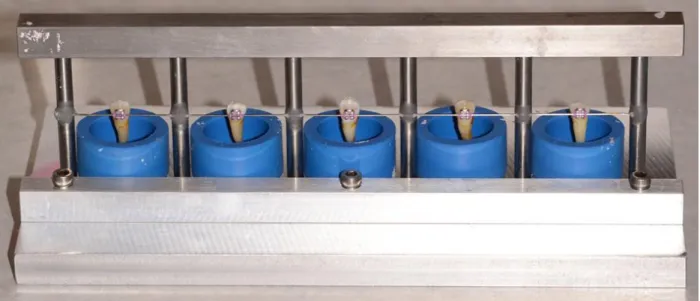
A mounting jig was constructed that allowed simultaneous mounting of up to 5 samples
(Figure 2). Bracketed teeth were ligated with elastomeric ligatures to a fixed 0.021”x0.025”
stainless steel wire and placed into silicon cups of chemical-cure acrylic resin. The jig ensured
that the buccal surface of the tooth and bracket base would be parallel to the applied shear force
and perpendicular to the acrylic mounting base. Acrylic was not allowed to touch the bracket or
teeth. When the acrylic was firm to the touch and beginning to feel warm, samples were
removed from the silicon cups and placed into individual containers of thymol solution in order
to re-hydrate the teeth and minimize the thermal side effects of the setting acrylic on the samples.
Samples did not undergo shear bond testing for at least 24 hours after mounting.32
An Instron® Model 4411 (Instron®, Canton, MA) screw-driven universal testing
machine with TestWorks® software was configured for shear bond strength testing. Machine
was properly calibrated with crosshead speed of 0.5mm/min and tested prior to data sampling.
Samples were secured in a custom aluminum Instron® base and bracket bases were aligned
parallel with the blade tip attachment of Instron® 500N load cell. The test was started with zero
load and finished when debond of the bracket occurred. The peak load, measured in newtons of
force, was recorded by the software. Newton measurements were divided by the area of the
bracket base (9.096756mm²) to obtain the megapascal (MPa) values used for analysis and
comparison. Brackets were collected and labeled after the debonding procedure was completed.
In 1984, Artun and Bergland introduced the Adhesive Remnant Index (ARI) as a means
of identifying the specific site of bond failure.33 This index has since been modified from a three option measurement to a five option measurement as a more complex means of identifying the
site of bond failure.34 After shear bond testing, each tooth and bracket combination of the present study was examined under stereomicroscopy (Nikon® SMZ18). Modified ARI scores
were recorded for each sample and photographs of each bracket base were saved via
NIS-Elements®. The modified ARI scoring system categories are: (1) all adhesive remains on tooth,
(2) more than 90% of adhesive remains on tooth, (3) more than 10% but less than 90% of
adhesive remains on tooth, (4) less than 10% of adhesive remains on tooth, (5) no adhesive
Lingual cusps were removed from eight of the samples with cutting disks after shear
bond testing was completed. The enamel surfaces were rinsed, dried, and conditioned as
follows: (1) no preparation, (2) 15 seconds 35% phorphoric acid etch, (3) 15 seconds 35%
phosphoric acid etch + SEP, (4) SEP, (5) 1 minute 5.25% sodium hypochlorite, (6) 1 minute
5.25% sodium hypochlorite + SEP, (7) 1 minute 5.25% sodium hypochlorite + 15 seconds 35%
phosphoric acid etch, (8) 1 minute 5.25% sodium hypochlorite + 15 seconds 35% phosphoric
acid etch + SEP. Samples were dehydrated and coated with 8nm of gold/palladium. A Hitachi
SEM allowed visualization at magnifications up to 100,000x.
Statistical Analysis
Significance for all statistical testing was set at p≤0.05. A factorial ANOVA, with bond
group and bracket type and the interaction term as explanatory variables, was used to analyze
peak shear bond forces. A Cochran-Mantel-Haenszel row mean score test was used to determine
if significant differences existed between bonding techniques and mode of failure. For statistical
analysis, the ARI scores of 1-2 and 4-5 were combined, resulting in three groups. These three
groups represent the primary modes of failure as being between the enamel and composite,
within the composite, and between the composite and the bracket base.
Results
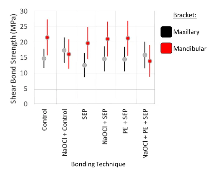
Descriptive statistics for each group are displayed in Table 2. The mean SBS values for
maxillary teeth were 14.79±3.08 MPa, 12.60±3.97 MPa, and 14.53±3.96 MPa for brackets
bonded with the control method, SEP, and Pre-etch + SEP, respectively. Maxillary teeth
pre-conditioned with sodium hypochlorite exhibited mean SBS values of 17.43±4.08 MPa,
14.58±4.03 MPa, and 15.89±4.29 MPa for brackets bonded with the control method, SEP and
19.68±5.19 MPa, and 21.30±5.62 MPa for brackets bonded with the control method, SEP, and
Pre-etch + SEP, respectively. Mandibular teeth pre-conditioned with sodium hypochlorite
exhibited mean SBS values of 16.14±4.75 MPa, 21.03±5.64 MPa, and 13.89±5.12 MPa for
brackets bonded with the control method, SEP and Pre-etch + SEP, respectively. The means and
standard deviations are illustrated with boxplot (Figure 3).
A factorial ANOVA was used to analyze differences in peak shear bond strength with
group and bracket placement as main effects. There was a statistically significant interaction
(P<0.01) between bonding technique and bracket placement. Tukey pairwise comparisons
indicated the presence of several statistical significant interactions between groups. The
mandibular control differed from the mandibular NaOCl + Pre-etch + SEP (P<0.05).
Additionally, maxillary SEP differed from mandibular control (P<0.01), mandibular Pre-etch +
SEP(P<0.02), and mandibular NaOCl + SEP(P<0.03).
When compared with their similar counterparts, groups that had NaOCl applied to the
surface prior to bonding exhibited a statistically insignificant increase in bond strength except for
the mandibular NaOCl + Control and the mandibular NaOCl + PE +SEP.
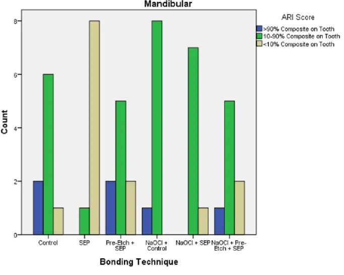
The frequency distribution of the Adhesive Remnant Index (ARI) measurements is
illustrated in Table 3. No samples of maxillary or mandibular teeth were measured with an ARI
score of 1. Specimens bonded with SEP (n=7) and Pre-Etch + SEP (n=3) were the only samples
where an ARI score of 5 was measured. Additionally, SEP and NaOCl + SEP were the only two
groups to not have an ARI score of ‘2’ recorded. Condensed ARI scores are illustrated in bar
graphs for mandibular samples (Figure 4) and maxillary samples (Figure 5). A
significantly different among bonding techniques for both the maxillary and mandibular
brackets.
SEM examination of prepared enamel samples showed erosive surfaces in Figure 6.
Phosphoric acid resulted in larger, irregular opening holes than SEP treatment that created a
small but uniform porous structure. The enamel surface treated by NaOCl was relatively flat and
non-porous.
Discussion
As illustrated in Figure 4, the mandibular SBS values are consistently higher than the
maxillary SBS values until the sodium hypochlorite was added. The addition of bleach created
an unexpected interaction between maxillary and mandibular brackets. Because of this
unexpected interaction, the authors believed it was appropriate to keep maxillary and mandibular
tests separate with ‘bracket’ as an additional explanatory variable for SBS and ARI statistical
analysis. The results of this study may have been affected by the small sample size.
With an identical mesh pad and identical preparation techniques, it was not expected that
significant differences would exist between maxillary and mandibular brackets. This finding
decreased sample sizes within each group because the maxillary and mandibular brackets could
not be combined into single groups. Additionally, the difference between brackets may have
shed some light on a factor that is rarely mentioned in the existing literature analyzing SBS of
orthodontic brackets on extracted human premolars. It is reasonable to suggest that differences
in bracket base adaptation, differences in Instron blade alignment, or enamel quality differences
may be the cause of this finding35-37.
Figure 3 validates previous studies that found relative differences between Control, SEP,
conventional etch-and-rinse bonding 15,39,40. In addition, it has been established that applying phosphoric acid prior to using SEP results in higher bond strengths approaching that of
conventional bonding, a pattern that was followed with both maxillary and mandibular samples
in the present study19-21,41. Figure 3 also indicates that NaOCl mildly increased the SBS of SEP without pre-etching for both maxillary and mandibular brackets. No clear conclusions can be
drawn from the using NaOCl prior to applying phosphoric acid (NaOCl + Control and NaOCl +
Pre-etch + SEP). It appeared that the bleach decreased the bond strength of mandibular samples,
significantly so with the NaOCl + Pre-etch + SEP group (p<0.05) and approaching significance
with the NaOCl + Control group (p=0.37), while doing the exact opposite for the maxillary
samples. The mean shear bond strength of all tested samples met or exceeded the minimally
clinically acceptable shear bond strength for orthodontic brackets (≥8MPa) as set forth by
Reynolds3,4.
This in-vitro study may have a number of limitations. Extracted premolars become more
brittle with time which may result in increased enamel fractures during testing.29 Lab tests also ignore the effects of polymerization shrinkage, tooth flexure, and PDL compression but can be
valuable in establishing rough measurements of relative strengths between bonding agents as
well as forecasting clinical effectiveness. Though the samples in the present study were
designed to represent forces that may be encountered clinically, the extent of forces involved
with debonding a bracket (thermal, peel, shear, torsion, tension, wedge) are exceedingly
complicated and cannot be accurately reproduced in the laboratory. Furthermore, the
load-to-failure test does not reproduce the cyclic load experienced in the clinical setting.42
The significant differences found by the Cochran-Mantel-Haenszel analysis between
5-6). It appears that SEP groups tended to leave less composite on the tooth than any other group.
This finding agrees with the results of the split mouth study by Murfitt et al. and the in-vitro
study by Bishara et al. that found the predominant mode of failure for the SEP group was
between the composite-enamel interface while the conventional etch-and-rinse group was within
the composite.16,43 The addition of NaOCl before applying SEP altered this trend in both maxillary and mandibular groups. It is possible that the NaOCl altered the interaction between
the SEP and the enamel surface, tending to increase the SBS (though not significantly) and leave
more composite attached to the tooth surface after debonding.
For ARI score 1 criteria to be met, all composite must remain on the tooth and none of
the composite can remain on the bracket34. With the mechanical retention offered in the micro-mesh of modern brackets, it is virtually impossible to have 100% of the composite remain on the
tooth unless one of the following conditions are met: (1) mesh is displaced to allow passage of
mechanically locked composite during debonding, (2) composite retains enough formability to
bypass mechanical undercuts during debonding, (3) composite was initially absent from
mechanical undercuts, (4) the shape of the micro-mesh provides a clear path of draw for
composite resin during debond, lacking mechanical undercuts. It is of no surprise to the authors
of this paper that an ARI score of 1 was not measured on any of the samples. In every sample, at
least some composite remained in the mesh of the bracket after debonding, necessitating an ARI
score >1. For this reason, we believed that it was appropriate to combine the ARI scores of 1-2
and 4-5 during statistical analysis for an accurate representation of where a majority of the bond
failure occurred.
Careful visual examination of Figure 6 indicates some differences in pattern of etch
NaOCl, no immediate differences are evident. This finding agrees with a previous study by
Ahuja et al (2010) which concluded that there are not statistically significant visual surface
differences when enamel is treated with sodium hypochlorite.44 SEP alone has a relatively homogenous pattern of etch with small holes and no major surface features. The application of
NaOCl prior to using SEP appears to leave a surface with larger features, grooves, and
irregularities that are similar to that of 35% phosphoric acid etch. The differences in etch pattern
observed involving SEP could potentially contribute to the mode of failure differences found in
this study.
Conclusions
We can conclude within the limitations of this study that:
All tested methods exhibited clinically acceptable (>8MPa) mean bracket shear bond
strengths.
Pre-etching prior to the application of SEP had no deleterious effect on shear bond
strength.
Pre-etching prior to the application of SEP may increase the shear bond strength of
orthodontic brackets compared to SEP alone, but not in a statistically significant way.
Pretreatment of normal enamel surfaces with 5.25% NaOCl for 1 minute may affect the
REFERENCES
1. Buonocore MG. A simple method of increasing the adhesion of acrylic filling materials to enamel surfaces. J Dent Res. 1955;34(6):849-853.
2. Zachrisson BJ. A posttreatment evaluation of direct bonding in orthodontics. American Journal of Orthodontics and Dentofacial Orthopedics. 1977;71(2):173-189.
3. Reynolds I. A review of direct orthodontic bonding. Br J Orthodont. 1975;2:171-178.
4. Reynolds IR and von Fraunhofer JA. Direct bonding in orthodontics: A comparison of attachments. Br J Orthod. 1977;4(2):65-69.
5. Powers JM and Sakaguchi RL. Bonding to dental substrates. In: Craig's restorative dental materials. 12th ed. St. Louis, MO: Mosby Elsevier; 2006:213.
6. Armstrong WG. Origin and nature of the acquired pellicle. Proc R Soc Med. 1968;61(9):923-930.
7. Miura F, Nakagawa K, Ishizaki A. Scanning electron microscopic studies on the direct bonding system. Bull Tokyo Med Dent Univ. 1973;20(3):245-260.
8. Main C, Thomson JL, Cummings A, Field D, Stephen KW, Gillespie FC. Surface treatment studies aimed at streamlining fissure sealant application. J Oral Rehabil. 1983;10(4):307-317.
9. Barry GR. A clinical investigation of the effects of omission of pumice prophylaxis on band and bond failure. Br J Orthod. 1995;22(3):245-248.
10. Lindauer SJ, Browning H, Shroff B, Marshall F, Anderson RH, Moon PC. Effect of pumice prophylaxis on the bond strength of orthodontic brackets. Am J Orthod Dentofacial Orthop. 1997;111(6):599-605.
11. Fleming PS, Johal A, Pandis N. Self-etch primers and conventional acid-etch technique for orthodontic bonding: A systematic review and meta-analysis. Am J Orthod Dentofacial Orthop. 2012;142(1):83-94.
12. Grubisa HSI, Heo G, Raboud D, Glover KE, Major PW. An evaluation and comparison of orthodontic bracket bond strengths achieved with self-etching primer. American Journal of Orthodontics and Dentofacial Orthopedics. 2004;126(2):213-219.
14. Bishara SE, Oonsombat C, Soliman MM, Warren JJ, Laffoon JF, Ajlouni R. Comparison of bonding time and shear bond strength between a conventional and a new integrated bonding system. Angle Orthod. 2005;75(2):237-242.
15. Bishara SE, VonWald L, Laffoon JF, Warren JJ. Effect of a self-etch primer/adhesive on the shear bond strength of orthodontic brackets. Am J Orthod Dentofacial Orthop. 2001;119(6):621-624.
16. Bishara SE, Gordan VV, VonWald L, Olson ME. Effect of an acidic primer on shear bond strength of orthodontic brackets. Am J Orthod Dentofacial Orthop. 1998;114(3):243-247.
17. Burgess AM, Sherriff M, Ireland AJ. Self-etching primers: Is prophylactic pumicing necessary? A randomized clinical trial. Angle Orthod. 2006;76(1):114-118.
18. Lill DJ, Lindauer SJ, Tufekci E, Shroff B. Importance of pumice prophylaxis for bonding with self-etch primer. Am J Orthod Dentofacial Orthop. 2008;133(3):423-6; quiz 476.e2.
19. Erickson RL, Barkmeier WW, Kimmes NS. Bond strength of self-etch adhesives to pre-etched enamel. Dent Mater. 2009;25(10):1187-1194.
20. Devarasa GM, Subba Reddy VV, Chaitra NL, Swarna YM. Self-etching adhesive on intact enamel, with and without pre-etching. Microsc Res Tech. 2012;75(5):650-654.
21. Fitzgerald I, Bradley GT, Bosio JA, Hefti AF, Berzins DW. Bonding with self-etching primers--pumice or pre-etch? an in vitro study. Eur J Orthod. 2012;34(2):257-261.
22. Rotta M, Bresciani P, Moura SK, et al. Effects of phosphoric acid pretreatment and
substitution of bonding resin on bonding effectiveness of self-etching systems to enamel. J Adhes Dent. 2007;9(6):537-545.
23. Venezie RD, Vadiakas G, Christensen JR, Wright JT. Enamel pretreatment with sodium hypochlorite to enhance bonding in hypocalcified amelogenesis imperfecta: Case report and SEM analysis. Pediatr Dent. 1994;16(6):433-436.
24. Ari H, Yasar E, Belli S. Effects of NaOCl on bond strengths of resin cements to root canal dentin. J Endod. 2003;29(4):248-251.
25. Espinosa R, Valencia R, Uribe M, Ceja I, Saadia M. Enamel deproteinization and its effect on acid etching: An in vitro study. J Clin Pediatr Dent. 2008;33(1):13-19.
26. Justus R, Cubero T, Ondarza R, Morales F. Fluoride-releasing resin-modified glass ionomer cements: Comparing shear bond strength of two adhesives. Seminars in orthodontics.
2010(16):66-75.