THE EFFECT OF VIBRATORY STIMULI ON MEASURES OF NEUROMUSCULAR FUNCTION
Derek N. Pamukoff
A dissertation submitted to the faculty at the University of North Carolina at Chapel Hill in partial fulfillment of the requirements for the degree of Doctor of Philosophy in the Curriculum
of Human Movement Science in the College of Arts & Sciences.
Chapel Hill 2015
ABSTRACT
Derek N. Pamukoff: The Effect Of Vibratory Stimuli On Cortical And Spinal Neuron Excitability
(Under the direction of J. Troy Blackburn)
Context: Muscle vibration enhances neuromuscular function, but the mechanism of improvement is unclear. Heightened motor neuron excitability within the spinal cord could be responsible for improved muscle function following vibration. However, the response of supraspinal structures – such as the motor cortex – to vibration is unclear. Vibratory treatments could benefit individuals with quadriceps dysfunction, such as patients with knee pathologies. Whole body (WBV) and local muscle vibration (LMV) improve quadriceps function but the efficacy of treatment may vary. Objective: To compare the effects of whole body and local muscle vibration on measures of quadriceps function. Participants: Sixty recreationally active young adults, and twenty individuals with anterior cruciate ligament reconstruction.
Interventions: Healthy subjects were randomized to one of three groups (WBV, LMV and control) and completed three testing sessions. Subjects completed testing of quadriceps spinal neuron excitability, corticomotor excitability, or a maximal voluntary isometric contraction and then completed an intervention based on group assignment. Subjects repeated the assessment immediately, ten minutes, and twenty minutes following the intervention. Subjects completed the remaining two assessments in separate sessions. Injured subjects completed testing of
ACKNOWLEDGEMENTS
This project could not have been completed without the support of my committee, research team, funding sources, friends, family, and colleagues.
To my advisor Dr. Troy Blackburn – I sincerely thank you for your guidance and patience over the past four years. I admire your daily effort, kindness, and professional attitude. You have brought out the best in me during my time at Carolina, and I thank you for all of the
opportunities. Thank you for always challenging and supporting me, and for always having my best interests in mind. As a colleague and friend, I wish you the best in the future and hope to cross paths again.
To Dr. Mike Lewek – Thank you for all of the assistance over the past four years in classes, seminars, and lab projects. Your instruction has guided me through the comprehensive exam, dissertation, and grant writing process. Thank you for continuously challenging me to think from new perspectives and develop as a scientist.
To Dr. Brian Pietrosimone – Thank you for your constant support in the laboratory on a daily basis. Your enthusiasm and passion for research is infectious. Thank you for inspiring me to do my best work.
To Dustin Lee and Stuart Gupton – I simply would not have been able to finish this project without your support. Thank you for your dedication, patience, and punctuality over the past year. The future is very bright for both of you!
To my classmates in HMSC – Thank you for all of the laughs and memories during my time at Carolina. I never felt far from home with all of you around every day.
TABLE OF CONTENTS
LIST OF TABLES ... xii
LIST OF FIGURES ... xiii
LIST OF ABBREVIATIONS ... xiv
CHAPTER 1: INTRODUCTION ... 15
CHAPTER 2: REVIEW OF LITERATURE ... 22
INTRODUCTION ... 22
ANTERIOR CRUCIATE LIGAMENT INJURY ... 22
Epidemiology ... 22
Anterior Cruciate Ligament Anatomy ... 23
Injury Risk Factors ... 24
KNEE OSTEOARTHRITIS EPIDEMIOLOGY ... 26
Articular Cartilage Anatomy ... 27
Changes in Afferent Discharge ... 32
Spinal Reflex Pathways ... 33
Supraspinal Centers ... 35
POST TRAUMATIC OSTEOARTHRITIS FOLLOWING ACL INJURY ... 36
VIBRATION TRAINING ... 39
Effects on Muscle Function ... 44
Whole Body Vibration vs. Local Muscle Vibration ... 45
MEASUREMENT TOOLS AND METHODOLOGICAL CONSIDERATIONS ... 47
Electromyography ... 47
Hoffman’s Reflex ... 48
Transcranial Magnetic Stimulation ... 52
SUMMARY ... 56
CHAPTER III: EXPERIMENTAL DESIGN AND METHODS ... 57
Subjects ... 57
Aims 1-3... 57
Aim 4 ... 57
Experimental Design ... 58
Aims 1-3... 58
Aim 4 ... 59
Hoffman Reflex ... 61
Corticospinal Excitability ... 62
Intervention Procedures ... 63
Data Reduction ... 64
Data Analysis ... 65
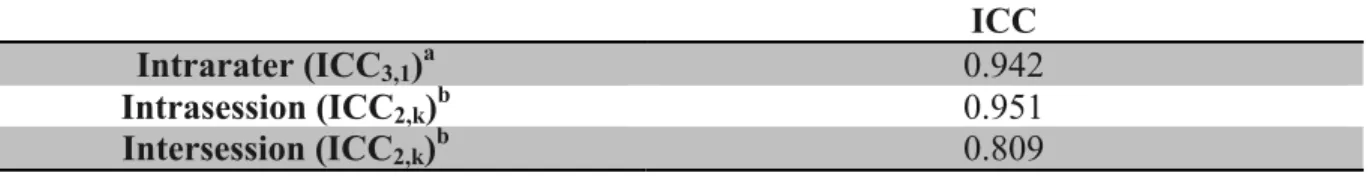
Reliability ... 66
CHAPTER 4: RESULTS AND DISCUSSION SUMMARY ... 68
Results – Healthy Cohort ... 68
Discussion – Healthy Cohort ... 70
Discussion – ACLR Cohort ... 71
CHAPTER 5: MANUSCRIPT 1 ... 72
Overview ... 72
Introduction ... 73
Methods... 75
Experimental Design ... 75
Electromyography ... 76
Maximal Voluntary Isometric Contraction (MVIC) procedures ... 76
Interventions ... 77
Data Reduction ... 77
Statistical Analyses ... 78
Results ... 78
Discussion ... 80
Conclusion ... 84
CHAPTER 6: MANUSCRIPT 2 ... 85
Overview ... 85
Introduction ... 86
Methods... 88
Experimental Design ... 88
Subjects ... 88
Electromyography ... 89
Intervention ... 90
Data Reduction ... 91
Statistical Analyses ... 92
Results ... 92
Discussion ... 96
Conclusion ... 99
CHAPTER 7: MANUSCRIPT 3 ... 100
Overview ... 100
Introduction ... 101
Methods... 103
Experimental Design ... 103
Subjects ... 103
Hoffmann Reflex ... 104
Corticomotor Excitability ... 105
Voluntary Activation ... 106
Intervention ... 106
Statistical Analyses ... 107
Results ... 107
Discussion ... 109
Conclusion ... 112
CHAPTER 8: MANUSCRIPT 4 ... 113
Overview ... 113
Methods... 116
Experimental Design ... 116
Subjects ... 117
Hoffmann Reflex ... 117
Voluntary Activation ... 118
Statistical Analyses ... 120
Results ... 120
Discussion ... 125
Conclusions ... 128
LIST OF TABLES
Table 1: Possible Outcomes ... 21
Table 2: EMG - Intersession Reliability ... 66
Table 3: EMG - Intrasession Reliability ... 66
Table 4: Hmax/Mmax Reliability ... 66
Table 5: AMT Reliability ... 67
Table 6: MEP Reliability ... 67
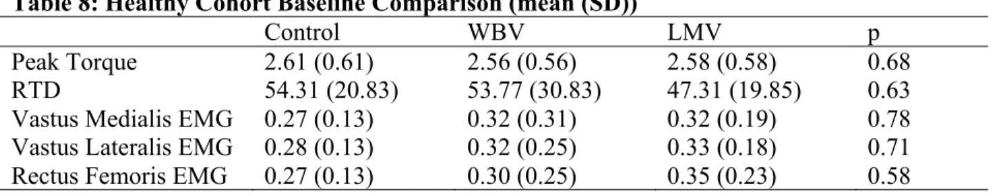
Table 7: Healthy Cohort Descriptive Statistics ... 79
Table 8: Healthy Cohort Baseline Comparison ... 79
Table 9: Healthy Cohort Peak Torque Results ... 80
Table 10: Healthy Cohort Rate of Torque Development Results ... 80
Table 11: Healthy Cohort Quadriceps EMG Results ... 80
Table 12: ACLR Cohort Baseline Characteristics ... 92
Table 13: Healthy Cohort AMT Results ... 108
Table 14: MEP Results ... 108
Table 15: CAR Results ... 108
Table 16: H-Reflex Results ... 108
LIST OF FIGURES
Figure 1: Arthrogenic Muscle Inhibition………...31
Figure 2: Pathway to OA………...36
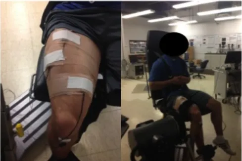
Figure 3: EMG Sensors and Dynamometer.………..60
Figure 4: Lycra Swim Cap……….62
Figure 5: WBV and LMV Interventions………63
Figure 6: CAR Position………..90
Figure 7: ACLR Peak Torque………93
Figure 8: ACLR EMG………...94
Figure 9: ACLR RTD………95
Figure 10: CAR Calculation………106
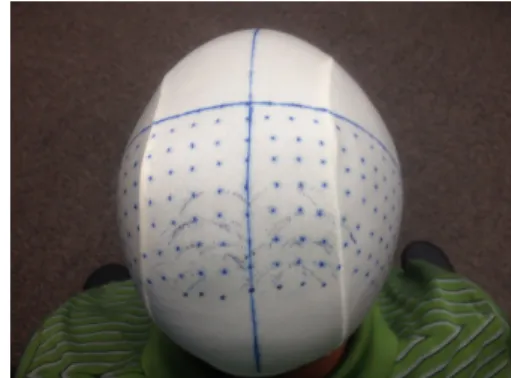
Figure 11: TMS Mapping………119
Figure 12: ACLR AMT………...122
Figure 13: ACLR CAR………123
Figure 14: ACLR MEP………124
LIST OF ABBREVIATIONS ACL Anterior Cruciate Ligament
ACLR Anterior Cruciate Ligament Reconstruction OA Osteoarthritis
AMI Arthrogenic Muscle Inhibition
VT Vibration Therapy
WBV Whole Body Vibration LMV Local Muscle Vibration CAR Central Activation Ratio EMG Electromyography
TMS Transcranial Magnetic Stimulation AMT Active Motor Threshold
CHAPTER 1: INTRODUCTION
As many as 250,000 anterior cruciate ligament (ACL) injuries occur each year in the United States.3 ACL reconstruction is costly, amounting to nearly $50,000 per procedure in direct medical costs.4,5 However, patients with ACL injury and reconstruction are at much greater risk for developing osteoarthritis (OA),6-10 which drastically elevates the cost associated
The pathway from ACL injury to tibiofemoral OA is multifactorial and can in part be explained by alterations in neuromuscular function following injury.20-24 Quadriceps weakness is common following ACL reconstruction due to arthogenic muscle inhibition (AMI).21-24
Alterations in afferent input to the central nervous system decrease the excitability of the
quadriceps, ultimately leading to a compromised ability to activate the quadriceps voluntarily.20
This altered afferent input stems from joint effusion, excessive joint laxity, pain, or deafferentation of the native ACL from reconstruction or deficiency, or a combination
thereof.25,26 Lesser quadriceps activation leads to reductions in knee extensor strength, which may contribute to the development of OA. Several studies suggest that baseline quadriceps function is a predictor of OA progression.23,24,27-30 Furthermore, OA is considered to be a
mechanically driven disease, meaning that alterations in joint loading influence OA
improving neuromuscular function by addressing lingering quadriceps weakness is an important strategy to prevent or delay the development of OA in patients with ACL injury.
Rehabilitation strategies aimed to improve quadriceps strength are largely ineffective because they do not address underlying deficits in neuromuscular function (i.e. AMI).38,39
Patients with inhibition respond poorly to traditional rehabilitation protocols and display minimal improvements in knee extensor strength.39 Therefore, novel strategies are needed to enhance the efficacy of current rehabilitation programs. Vibration therapy (VT) is an increasingly popular mode of exercise due to reports of improved muscle strength, power, EMG amplitude, and physical function.40-49 When a muscle is vibrated, a reflexive contraction known as the tonic vibratory reflex (TVR) is evoked.50-52 Essentially, there is a discharge of the primary endings of
the muscle spindle (Ia afferent) via repeated rapid lengthening of the muscle from vibration.53 This excites the alpha motor neurons, resulting in muscle contraction. However, the TVR only accounts for increases in muscle activity during VT, and does not account for improvements that are observed following cessation of VT.54,55 Therefore, it is likely that there are other
mechanisms that explain why improvements in muscle function persist following VT. The intent of VT is to increase the number and frequency of excitatory stimuli sent to α-motorneurons in an attempt to override the inhibitory afferents coming from the damaged joint. Therefore, VT may be an effective method for improving quadriceps function following knee joint injury to prevent or delay the onset of OA.
Despite reports of enhanced muscle strength and power following VT,40,41,56 there is
paucity in the literature regarding the mechanisms by which VT alters neuromuscular function. Early studies suggest that enhanced motor neuron excitability within the spinal cord is
excitatory and inhibitory influences on spinal reflex activity. For example, some studies suggest a suppression of spinal neuron excitability following VT in healthy individuals57-59 and in
patients with spinal cord injury,58 potentially due to presynaptic inhibition.60 These findings suggest that any improvement in muscle function following VT cannot be attributed to gains in spinal neuron excitability. What remains unclear is the role of supraspinal structures, such as the motor cortex following VT. Cortical areas also receive and process afferent signals and produce cortical potentials in response to VT.61 Furthermore, muscle afferent input to the cerebral cortex is a major contributor in motor control.62 Limited evidence using transcranial magnetic
stimulation (TMS) suggests that brief exposure to VT enhances corticospinal excitability.63-65 This is particularly relevant, as studies show that patients with knee pathology have lesser corticospinal excitability.66,67 As such, if VT elicits improvements in cortical and spinal excitability, it could be a potent adjunct treatment to improve quadriceps function. However, studies evaluating the effect of VT on corticospinal excitability have used small samples, and no study, to my knowledge, has concurrently evaluated spinal and cortical neuron excitability following VT.
The majority of studies have reported enhanced muscle function following whole body vibration (WBV).40,47,48,56,68-73 However, WBV platforms are cost prohibitive (up to $10,000) and provide limited portability. Local muscle vibration (LMV) also improves muscle
than indirectly via WBV. We recently demonstrate that LMV improves quadriceps activity for a sustained period of time post-application in healthy individuals55 and that WBV and LMV produce similar increases in voluntary quadriceps activation following experimental knee joint effusion.75 However, no studies have compared the effects of WBV and LMV on cortical and spinal neuron excitability.
Patients with ACL injury have deficits in corticospinal excitability that contribute to AMI and reduce quadriceps function.66,76,77 Reduced quadriceps function may contribute to the
development of OA, and current rehabilitation strategies are largely ineffective in individuals with AMI. Novel strategies are needed to address AMI, and VT may provide an adjunct
treatment to improve quadriceps function. However, it is unclear how VT alters neuromuscular function. Therefore, the purpose of this study is to evaluate the effects of VT on measures of neuromuscular function. The specific aims are as follows:
1) To determine the effects of WBV and LMV on corticospinal excitability, spinal neuron excitability, electromyography (EMG) amplitude, and voluntary muscle activation during a maximal isometric contraction in healthy adults. I hypothesize that WBV and LMV will increase EMG amplitude and corticospinal excitability, but will suppress spinal neuron
excitability, and that these changes will be greater than those observed in a control
group receiving no treatment.
corticospinal excitability and EMG amplitude, but attenuate spinal neuron excitability,
relative to the control group receiving no treatment, but the magnitude of these
improvements will not differ between WBV and LMV.
3) To determine the duration of the effect of WBV and LMV on corticospinal excitability, spinal neuron excitability, EMG amplitude, and voluntary muscle activation during a maximal isometric contraction in healthy adults. I hypothesize that the effect of LMV and WBV on corticospinal, spinal neuron excitability, and EMG amplitude will persist for at
least 10 minutes following cessation of treatment.
4) To compare the effects of WBV and LMV on EMG, corticospinal excitability, spinal neuron excitability, voluntary muscle activation, and EMG during a maximal isometric contraction in a subset of patients with ACL injury. I hypothesize that LMV and WBV will enhance corticospinal excitability and EMG amplitude, but attenuate spinal neuron
excitability, relative to the control condition receiving no treatment, but the magnitude of
these improvements will not differ between WBV and LMV.
If VT enhances corticospinal excitability and quadriceps function, it would be a particularly potent treatment for individuals with knee pathologies who experience AMI. Furthermore, LMV represents a portable and less expensive alternative to WBV, and
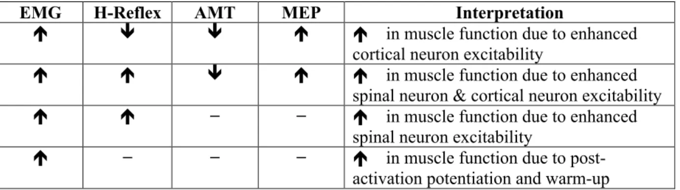
Table 1: Possible Outcomes
EMG H-Reflex AMT MEP Interpretation
é ê ê é é in muscle function due to enhanced cortical neuron excitability
é é ê é é in muscle function due to enhanced spinal neuron & cortical neuron excitability é é − − é in muscle function due to enhanced
spinal neuron excitability
CHAPTER 2: REVIEW OF LITERATURE
INTRODUCTION
The purpose of this literature review was to review pertinent studies and deficiencies in understanding. Specifically, this review focuses on short-term and long-term neuromuscular alterations following anterior cruciate ligament injury, such as impaired quadriceps muscle function, and altered gait kinematics and kinetics. This review addresses how deficiencies in quadriceps function alter knee joint loading during gait, and contribute to joint degeneration and tibiofemoral osteoarthritis development. Second, this review identifies areas of deficiency in rehabilitation for ACL injuries, and why novel strategies to improve quadriceps function are needed. Thirdly, this review provides evidence for the use of vibration therapy to improve muscle function, and how it may specifically apply to patients with ACL injury. Additionally, this literature review illustrates how and why LMV may provide a cost-effective and portable alternative to WBV. Lastly, results of this review are summarized and proposed aims are suggested.
ANTERIOR CRUCIATE LIGAMENT INJURY
Epidemiology
$7.6 billion annually in the United States when treated with reconstruction, and $17 billion annually when treated with rehabilitation.4,5 Following ACL injury, alterations in muscle function and gait biomechanics elevate the risk of developing tibiofemoral osteoarthritis (OA).
6-10 Patients with ACL injury are between three and five times more likely to develop OA
compared to individuals with no injury. 6-11 Interestingly, patients with ACL injury are at greater
risk for OA development regardless of reconstruction procedures. Patients with ACL injury that undergo reconstruction are 3-5 times more likely to develop OA compared to healthy
individuals, and patients with ACL injury that do not undergo reconstruction are equally likely to develop OA compared to those with reconstruction.10 Overall, OA is seen in up to 13% of all knees with no concomitant meniscal injury, and up to 48% of knees with a concomitant meniscal injury at ten years or less of follow-up.8 Therefore, while ACL reconstruction may be effective for restoring near normal knee joint function, long-term consequences on joint health following injury are unchanged, and rehabilitation programs are ineffective.38,39 Importantly, the majority of ACL injuries occur in youth populations,15 which greatly elevates the lifetime cost of an ACL injury when considering the increased risk of OA development.
Anterior Cruciate Ligament Anatomy
The ACL is a 2-4 centimeter long dense band of connective tissue between the femur and tibia. It is an important structure in the knee joint and serves primarily to resist and limit anterior tibial translation and rotational loads.78-82 The ACL is divided into two bundles largely
posterolateral bundle originate along the postero-distal aspect of the femoral attachment and insert along the posterolateral aspect of the tibial attachment.78 These bundles are not isometric, and the anteromedial bundle tightens as the knee moves into flexion, while the posterolateral sbundle becomes slack. 81
The ACL receives innervation from the posterior articular branches of the tibial nerve, and also has several mechanoreceptors (Ruffini, Pacini, free nerve endings, and golgi-like receptors). 84-86 These mechanoreceptors have a role in proprioception and provide afferent information regarding knee position. For instance, Ruffini receptors are sensitive to changes in length. Deformation of the ACL influences output of the spindles in muscles surrounding the joint. 87,88 Furthermore, free nerve endings act as nociceptors for pain. If the ACL is damaged or
ruptured, loss of afferent information from these receptors contributes to diminished quadriceps function.88,89 Additionally, the ACL provides information regarding joint position sense. Therefore, patients with ACL injury would have diminished proprioceptive function.
Reconstructive procedures are aimed to restore structural stability to the knee joint, and make no attempt at restoring function of the mechanoreceptors, which partially explains why quadriceps weakness may persist in patients with ACL injury. Lastly, the ACL is also vascularized with blood supply from the middle gennicular artery.81 However, the distal area of the ACL that is subject to compressive loads has poor vascularity, which may explain why the ACL does not heal on its own.
Injury Risk Factors
The majority of ACL injuries (~70%) are the result of non-contact mechanisms. Despite a vast body of literature regarding ACL injury, no single causative mechanism has been
physiological. Firstly, sex hormone concentration may influence collagen synthesis of the ACL thus contributing to its integrity and function.90,91 Furthermore, ACL function may vary
depending on the phase of the menstrual cycle. For instance, greater joint laxity is observed in the periovulatory and luteal phases of the menstrual cycle.92,93 Fluctuations in sex hormone concentration during the menstrual cycle may contribute to the high prevalence of ACL injuries among females compared to males. Secondly, anatomical risk factors for ACL injury include smaller ACL size, greater quadriceps angle, and smaller intracondylar notch size. Interestingly, females have smaller ACLs, greater quadriceps angles, and smaller intracodylar notches
compared to males, and this likely increases their risk for ACL injury. Thirdly, there are biomechanical factors that influence risk for ACL injury. For example, landing with greater vertical impact force increases the risk for ACL injury.94,95 Furthermore, kinematic and kinetic differences during athletic tasks may influence ACL loading such as anterior tibial shear force, knee valgus position, internal knee extension moment, and knee flexion angle. Females have greater anterior tibial shear force, greater knee valgus, greater knee extension moments, and lesser knee flexion during landing tasks compared to males.94,96,97 Importantly, while anatomical
and hormonal risk factors are not modifiable characteristics, biomechanical factors may be modified through supplementary training.
Lastly, current rehabilitation programs often leave individuals at risk for re-injury.
Patients with ACL injury are 12% more likely to tear their ACL compared to healthy individuals. Therefore, it is likely that rehabilitation programs are not effective in restoring neuromuscular
38,39 and biomechanical function. Patients with ACL reconstruction have abnormal landing
reconstruction have alterations in gait that influence risk for knee OA.2,36,98,99 These alterations are detailed later in this summary.
KNEE OSTEOARTHRITIS EPIDEMIOLOGY
Osteoarthritis (OA) is a gradual reduction of articular cartilage within a joint. Clinical diagnosis of OA is typically guided by symptoms and physical examination. However, radiographic evidence is used to assess OA severity and progression, and is defined by joint space narrowing and presence of osteophytes. The Kelgren-Lawrence scale is a common metric of OA progression (grade 1 – doubtful narrowing of joint space and possible osteophytic lipping, grade 2 – definite osteophytes and definite narrowing of joint space, grade 3 – moderate multiple osteophytes and definite narrowing of joint space, grade 4 – large osteophytes and marked narrowing of joint space). OA affects approximately one third of adults older than 65, and is a leading cause of physical disability.16 Knee OA is the most common kind of OA, affecting 9
million Americans at an annual cost of $51 billion.100 Specifically, patients with ACL injury are three to five times more likely to develop knee OA compared to those without injury, regardless of whether they undergo reconstruction.6-11
Knee OA can be classified as post-traumatic (e.g. following ACL injury) or idiopathic (as a result of other non-specific causes). Patients with ACL injury who develop knee OA account for nearly 10% of all knee OA cases.4 The direct cost of ACL when considering knee OA development is estimated at approximately $7.6 billion annually when patients undergo ACL reconstruction.4 The cost of knee OA also increases with severity. Mild knee OA incurs a direct annual cost of $9,801 per patient, moderate knee OA has a direct annual cost of $14,761 per patient, and severe knee OA has a cost of $22,111 per patient.4 Therefore, slowing the
knee OA. Lastly, knee OA contributes to lower extremity disability, sedentary behavior, and comorbidities such as cardiovascular disease.17 Additional risk factors that may predispose one to post traumatic knee OA include concomitant meniscal injury,15 obesity,101 joint
alignment,102,103 age, gender, sedentary lifestyle, and muscle weakness.104
Articular Cartilage Anatomy
Articular cartilage is a connective tissue that provides a smooth and lubricated surface for articulation, and attenuates joint loading.105 Articular cartilage is made of hyaline cartilage and
is between two and four millimeters thick.106 Interestingly, articular cartilage does not receive blood supply or innervation. Therefore, it has limited intrinsic healing capabilities. Despite a lack of innervation and blood supply to articular cartilage, patients with OA still experience pain from nociceptors in other areas of the knee joint, bone marrow lesions, joint effusion, and
inflammation. Articular cartilage is composed of a dense extracellular matrix and distribution of specialized cells called chondrocytes. Chondrocytes are the dominant cell type in articular cartilage. They are highly specialized metabolically active cells that play an important role in maintenance and repair of the extracellular matrix. 106 Chondrocytes respond to many stimuli such as growth factors, mechanical loads, and hydrostatic pressures.107 However, chondrocytes cannot replicate, and therefore have limited healing ability in response to injury. The
extracellular matrix is made primarily of water, collagen, proteoglycans, and glycoproteins. 106 The contents of the extracellular matrix assist in retaining water, which is essential to proper function.
layer contains a high number of chondrocytes, and protects deeper layers from sheer stress. The superficial zone is in contact with synovial fluid, and is responsible for providing resistance to sheer, tensile, and compressive forces from joint articulation. The middle zone provides a transition between the superficial and deep zones and represents up to sixty percent of total cartilage volume. It is comprised of proteoglycans and thicker collagen fibers. These fibers are arranged obliquely, and the middle layer contains a low number of chondrocytes. The middle layer functions to resist compressive force. The deep zone represents nearly thirty percent of articular cartilage volume, and has the largest concentration of proteoglycans, but lowest water concentration compared to the other zones. Collagen fibers are arranged perpendicular to the articular surface and provide the greatest resistance to tensile forces compared to the other zones. Lastly, the calcified zone secures the cartilage to the bone by anchoring the collagen fibers of the deep zone to the subchondral bone.
In addition to zones, the extracellular matrix consist of distinct regions based on proximity to the chondrocytes, composition, and collagen fiber diameter. The extracellular matrix can be divided into pericellular, territorial, and interterritorial regions. Firstly, the pericellular matrix is a thin layer adjacent to the cell membrane and completely surrounds the chondrocytes. It is composed of proteoglycans and glycoproteins, and serves to initiate signal transduction within cartilage during load bearing.109 Secondly, the territorial matrix surrounds the pericellular matrix and is composed of fine collagen forming a network around the cells. This region is much thicker and protects cartilage against mechanical stresses. 110 Lastly, the
Articular cartilage has unique viscoelastic properties that facilitate the transmission of loads to the underlying subchondral bone. Articular cartilage is able to undergo high cyclic loading while demonstrating little or no evidence of damage under normal circumstances.112,113 Application of joint loading causes an increase in interstitial fluid pressure. The increase in pressure causes fluid to flow out of the extracellular matrix, and create frictional drag on the matrix.114 When the load is removed, the interstitial fluid flows back into the tissue. As stated above, articular cartilage is viscoelastic and displays a time-dependent behavior when subject to constant loading due to flow-dependent, and flow-independent mechanisms. Firstly, the
permeability of articular cartilage is very low, and thus there is frictional drag on the matrix as fluid slowly flows in and out of the tissue from changes in pressure. Additionally, the amount of frictional drag on the matrix increases with higher loading rates, thus increasing the load placed on the articular cartilage. Secondly, the structure of the collagen-proteoglycan matrix provides intrinsic viscoelastic characteristics. Overall, these mechanisms provide support and reduce the stress that acts on the solid matrix. The viscoelastic nature of cartilage has implications for cartilage loading and development of knee OA, and is detailed later in this summary.
Articular cartilage also exhibits creep and stress-relaxation responses to loading. When a constant compressive stress is applied to cartilage, the cartilage will deform or creep until
same magnitude of impact load.37 Deficits in quadriceps function may contribute to greater loading rates during gait, and thus contribute to articular cartilage breakdown.
NEUROMUSCULAR ALTERATIONS FOLLOWING INJURY
Quadriceps weakness is common following ACL injury,20,115 despite surgical
reconstruction and subsequent rehabilitation.22,38,39 Muscle weakness is likely attributable to disuse atrophy. However, atrophy does not occur instantaneously, and thus does not acutely influence force production. Alternatively, arthrogenic muscle inhibition (AMI) occurs
immediately following injury causing an immediate reduction in force production.20 AMI also
contributes to ongoing quadriceps weakness associated with ACL injury.38 AMI is a form of reflexive neural inhibition that results in diminished motor drive to muscles associated with the injured joint.20 Although AMI functions as a protective mechanism to prevent pain and
excessive motion, the ultimate result is further weakness and wasting of the surrounding musculature. AMI is commonly measured using the central activation ratio (CAR), which estimates the percentage of motor units (recruitment and rate coding) that can be activated voluntarily. In patients with ACL injury, deficits in quadriceps activation are commonly
reported, but vary between 8 and 45%.104 Unfortunately, deficits in activation persist for several years following reconstruction and rehabilitation, and may contribute to joint degeneration and osteoarthritis. For instance, Urbach and colleagues 22 found that the quadriceps were still
approximately 20%.116 Interestingly, deficits in quadriceps activation are also observed in the uninjured limb in patients with ACL injury.77,117,118 Previous studies have found inhibition in the uninjured limb ranging from 7 to 24%, and some have reported nearly equal inhibition to that of the injured limb.39,118 Inhibition in the quadriceps of the contralateral limb suggest that AMI is caused by both local mechanisms surrounding the joint, and central mechanisms that control movement and muscle function throughout the body (Figure 1).
In addition to providing stability to the knee joint, the ACL also has mechanoreceptors that provide sensory
information to spinal and supraspinal regions
regarding joint movement, position, and loading.81
Following injury, altered
afferent information is transmitted to the central nervous system, resulting in a decrease in the excitability of the quadriceps alpha motoneuron pool.81 Abnormal afferent discharge can come from stimulation or damage to mechanoreceptors, joint effusion, joint laxity, or nociceptors in response to pain. The ACL also has projections to the muscle spindles in adjacent muscles, which are sensitive to changes in length, thus providing additional proprioceptive information. Lastly, alterations in spinal reflex and corticospinal function can contribute to AMI and are detailed below.
Changes in Afferent Discharge
Swelling is common following knee injury and surgery and contributes to AMI. Moreover, swelling persists for up to 3 months following initial ACL injury, and for up to 12 months following reconstruction.119 Swelling can also contribute to AMI independently of other factors such as inflammation, pain, and structural damage.1 For instance, injecting saline into a
knee joint to increase intra-articular pressure (IAP) in uninjured joints can artificially cause AMI.25,75 Swelling raises the IAP within the knee joint, thus increasing the firing frequency and recruitment of group 2 afferent fibers, which are sensitive to changes in stretch and
pressure.120,121 Therefore, greater discharge of group 2 afferents likely has a large inhibitory effect on quadriceps function. For instance, prior studies have also shown that swelling reduces quadriceps activation measured via electromyography,122-124 H-reflex amplitude,125,126 and force output.75,127,128
Inflammation also occurs following injury and surgery. Inflammation alters the sensitivity of articular nerve endings supplied by group 3 and 4 afferents via a process called peripheral sensitization.129 The activation threshold of these receptors is lowered following
injury, thus increasing afferent output to the central nervous system.129-131 Importantly, these types of fibers constitute the majority of afferent fibers within the knee joint. Group 3 and 4 afferents are nociceptive, and inflammation increases pain in association with afferent discharge, and pain may contribute to the magnitude of AMI.132 Also, the inflammatory process may increase the activity of otherwise silent free nerve endings.130,131 In response to inflammation,
Structural damage or joint degeneration results in greater translation of the knee joint surfaces (femur and tibia) during movement that increase activation of mechanoreceptors and nociceptors that signal the end ranges of joint motion.133 For example, animals with transected ACLs have greater afferent activity in the major nerves supplying the knee joint.134 Although ACL reconstruction reduces joint laxity and afferent discharge, alterations in afferent activity persist following reconstruction.135 These results suggest that ACLR is not entirely effective in restoring normal function, which may partially explain why AMI persists following
reconstructive procedures.
Lastly, acute injury may also damage the sensory endings located within the ACL, thus reducing afferent output.133,136 Although factors such as swelling, inflammation, and joint laxity
increase joint afferent discharge, different types of joint afferents may have contrasting effects (inhibitory vs. excitatory) on motor neuron excitability. Acute damage to articular receptors within the ACL or joint capsule may reduce excitatory afferent input to the alpha motoneuron pool, thus contributing to lower quadriceps activation.136 Overall, swelling, inflammation, pain, joint laxity, and damage to articular receptors contribute to alterations in afferent discharge to the central nervous system, which causes AMI.
Spinal Reflex Pathways
Abnormal afferent discharge from a damaged knee joint alters the excitability of reflexive pathways within the spinal cord.133,136,137 As a result, excitability of the quadriceps alpha
motoneuron pool is reduced, which prevents full activation of the corresponding muscle.133,136,137 Essentially, there are four main spinal pathways that may contribute to AMI: Ib inhibitory
Firstly, Ib inhibitory interneurons located within the spinal cord receive input from Ib afferent fibers and other joint afferents.138 Alterations in afferent discharge described above facilitate the Ib inhibitory pathway, thus contributing to AMI by inhibiting the agonist muscle.137 For
example, artificial knee joint effusion has been shown to increase Ib inhibition of the quadriceps H-reflex during voluntary muscle contractions.137
The flexion reflex is a polysynaptic pathway that produces a pattern of flexor facilitation and extensor inhibition.139,140 Therefore, any facilitation of the flexion reflex would contribute to knee extensor (quadriceps) AMI. The flexion reflex is mediated by a variety of different
interneurons that receive input from many peripheral afferents, such as the articular receptors.141 In response to joint inflammation, the activation threshold of afferent neurons associated with the flexion reflex is lowered and these neurons become hyperexcitable.142 In addition, lower flexion reflex thresholds are found in patients with knee pathology – such as those with knee
osteoarthritis and ACL injury – compared to healthy controls.143-145 Furthermore, activation of the flexion reflex also produces inhibition of the quadriceps during maximal isometric
contractions of the knee extensors. In summary, any activation or facilitation of the flexion reflex likely contributes to AMI of the quadriceps.
The gamma loop may also mediate quadriceps activation. Gamma motoneurons
this leads to a reduction in Ia afferent facilitation of the quadriceps alpha motoneuron pool, thus reducing overall quadriceps activation. Pre-synaptic inhibition may also influence excitability of the alpha motoneuron pool and thus contribute to AMI.26 Pre-synaptic inhibition excites
inhibitory interneurons that project to the synaptic terminal of the Ia afferent fibers. Excitation of an inhibitory interneuron causes a reduction in the quantity of neurotransmitter released following an afferent volley. In addition, this form of inhibition contributes to further gamma loop inhibition, which leads to AMI.
Supraspinal Centers
Joint afferents have projections to both spinal and supraspinal areas.149,150 Supraspinal centers are influenced by joint afferent activity and contribute to AMI. Transcranial magnetic stimulation (TMS) of the motor cortex provides a method to quantify changes in corticospinal excitability associated with knee pathology.66,151 Interestingly, some studies have found that corticospinal excitability measured via TMS (amplitude of motor evoked potential) is greater in individuals with knee pathology compared to healthy control subjects.151 This seems
counterintuitive since patients with knee pathologies would be expected to have lesser corticospinal excitability, and there is only limited evidence available suggesting lesser
corticospinal excitability among patients with knee pathologies. However, authors suggest that greater corticospinal excitability following injury may be indicative of a compensatory response in an attempt to overcome inhibition of the quadriceps alpha motoneuron pool. Heroux et al. 66 found lesser resting motor thresholds in patients with unilateral ACL injury compared to healthy controls, suggesting enhanced excitability of musculature surrounding the injured joint.
and the flexion reflex. Injury to the knee joint enhances descending input from the brainstem, which increases excitability of the flexion reflex and increase AMI. Last, studies show that changes in quadriceps activation rely on motivation. Any reduction in quadriceps strength or activation may be due to an adjustment in voluntary effort in response to fear of pain or eliciting further damage to an injured joint.
POST TRAUMATIC OSTEOARTHRITIS FOLLOWING ACL INJURY
Quadriceps weakness resulting from AMI is common following ACL injury despite reconstruction and rehabilitation, and AMI is found in up to 78% of patients with ACL reconstruction and up to 100% in patients who are ACL deficient.104 The quadriceps are essential to normal ambulation. In healthy individuals, the quadriceps act to attenuate shock during gait, and assist in evenly distributing load across the knee joint. However, deficits in quadriceps function from injury result in loads being transmitted in greater magnitude and at a faster rate to the lower limb.20 Specifically, patients with ACL injury have alterations in lower extremity kinematics and kinetics that may
elevate the risk of developing OA, and quadriceps dysfunction is thought to play a major role in the genesis of these biomechanical alterations (Figure 2).2
Osteoarthritis is a gradual breakdown of articular cartilage from repetitive joint loading. Moreover, articular cartilage is viscoelastic and is, therefore, sensitive to the rate at which it is
Figure 2: How gait mechanics can initiate osteoarthritis
loaded.152 When articular cartilage is loaded at faster rates it stiffens, thus elevating its risk for failure and breakdown. Previous studies in animal models demonstrate that repetitive impulsive loading of the limbs results in rapid degeneration of articular cartilage, regardless of the
magnitude of the load.152 Impulsive loading refers to the relationship of a force applied over time and can be mathematically expressed as the product of force and velocity (displacement over time). Therefore, reducing the time interval over which ground reaction force is absorbed would elevate the impulse experienced in the lower extremity.
Articular cartilage adapts to habitual loading that occurs during walking.153-155
Immobilization of the knee joint causes cartilage thinning, suggesting that functional loading is necessary to maintain cartilage health. Following acute loading of the limb, cartilage adjusts its metabolism, resulting in greater proteoglycan production and proliferation of chondrocytes – the functional unit of cartilage.99 Ultimately, these alterations result in thicker cartilage and
improved joint health. However, alterations in gait kinematics may shift the contact areas between bony surfaces that form joints to areas that are not typically loaded.156 Therefore, areas of thickest cartilage that are conditioned to frequent load bearing are no longer in contact with each other. Areas of pressure are shifted, resulting in lesser joint space and the initiation of osteoarthritis.
Patients with ACL injury and knee osteoarthritis have lesser quadriceps strength resulting from AMI as discussed above. Deficits in quadriceps function influence gait biomechanics in the sagittal plane.34,36,98,157 During weight acceptance, the limb must accept full support of the
has been labeled “quadriceps avoidance gait”. Lesser knee flexion excursion reduces the time interval over which ground reaction forces are absorbed, and therefore increase impulse at the knee joint. In brief, lesser quadriceps activation influences shock absorption during gait via lesser knee flexion, which elevates the impulsive load at the knee and likely increases the risk of osteoarthritis development.
While the quadriceps largely act to control loading at the knee joint in the sagittal plane, alterations in quadriceps function also influence frontal plane loading. The quadriceps have a small moment arm capable of contributing to adduction/abduction at the knee.31 Quadriceps and hamstrings co-contraction provides most of the support for the knee adduction moment during gait.31 The knee adduction moment is an indicator of osteoarthritis risk.158 Tibiofemoral
osteoarthritis most commonly affects the medial compartment of the knee joint. Excessive knee adduction places a greater load on the medial compartment, and increases the risk of joint degeneration.102,158 Furthermore, greater knee adduction moment is associated with greater disease incidence, progression, and severity.158-160 Importantly, patients with ACL injury have greater knee adduction moments compared to healthy individuals.161 However, the relationship
between quadriceps strength and adduction moment in patients with ACL injury is not clear. Nevertheless, improvement of quadriceps strength may contribute to lesser adduction moments and reduce the risk of further joint degeneration following knee injury.
Lastly, patients who are ACL deficient or ACL reconstructed commonly display an offset towards excessive internal tibial rotation.156,162 The thickest areas of cartilage are loaded when
excessive internal rotation potentially loads thin areas of cartilage that are more likely to break down.
VIBRATION TRAINING
Rehabilitation programs in patients with knee pathologies are largely ineffective in restoring quadriceps function due to AMI.38,39,163 Vibration therapy is a growing alternative modality for exercise due to reports of enhanced muscle strength, power, and
electromyography.164 Vibration also has positive effects on flexibility, bone and joint health, and many common measures of physical function.53 However, despite several studies that report improvements in markers of physical health, there are studies that report detrimental or equivocal results following vibration.46,70 Nonetheless, vibration may provide a novel treatment that could be used in conjunction with traditional rehabilitation programs to reduce AMI.
Vibration employs sinusoidal mechanical oscillations with periodic alterations of force and acceleration over time. Vibration provides a forced oscillation, and energy is transmitted from an actuator (the source of vibration, i.e. – vibrating platform) to a resonator (i.e. the human body or muscle) to produce a neuromuscular response. Essentially, the human body is
accelerated by vibration, which causes a reactive force within.53 This force is proportional to
example, posture can adjust the axial stiffness of the body to reduce the transmission of the vibratory stimulus as it travels from its source throughout the body. For example, standing in an erect posture compared to a squatting evokes a stronger transmission of vibration to the head during WBV. Likewise, shifting body weight to the forefoot rather than the mid or rear foot reduces vibration transmission. Attenuation of the signal increases as ankle, knee, and hip joint angle increases. Modulation of muscle activity is also relevant, and assuming a squat position will increase tension within the lower extremity musculature and increase signal transmission to these muscles.
There is also an internal response from muscle when vibration is applied to the body. Muscles go through a rapid series of eccentric (lengthening) and concentric (shortening) actions. Changes in muscle length trigger a neural reflex – the tonic vibratory reflex – due to stimulation of the muscle spindles.51,52,165 The muscle spindles are a specialized group of muscle fibers arranged in parallel with the contractile fibers of skeletal muscle.166 The functional unit of the muscle spindle is the intrafusal fiber, which is further categorized into three types: the nuclear chain fiber, the dynamic nuclear bag fiber, and the static nuclear bag fiber.167 Intrafusal fibers lie
muscle spindles following rapid increases in length from vibration will increase excitatory input to the alpha motoneuron pool. This causes a reflexive contraction in the homonymous muscle, which may facilitate increases in muscle function (strength, power, EMG etc.).
However, the tonic vibratory reflex only accounts for heightened muscle function during vibration, and there are studies that report enhancements of muscle function following vibration that persist for several minutes.54,55 Some authors suggest adaptations such as elevated muscle temperature and blood flow akin to traditional warm-up activities, increased gravitation force placed on a muscle producing a training effect, and enhanced corticospinal excitability and intracortical processes.69,168,169 Overall, it is unclear why muscle function improves following vibration, and additional research is necessary to evaluate other mechanisms that may be responsible for improvements in muscle function following vibration, such as changes in corticospinal excitability and intracortical processes.
As described above, vibration largely activates the Ia afferents of the muscle spindle system to produce a reflexive contraction. However, the spinal circuitry is the first stage within the motor feedback loop for generating fast efferent reactions to proprioceptive input. Moreover, supraspinal areas also receive and process proprioceptive information and generate evoked cortical potentials following vibration.61 Afferent input from muscles is a requirement for proper neuromuscular control, and muscle afferent facilitation accounts for a large proportion of central motor drive. Prior studies have shown that altered Ia afferent input influences the excitability of corticospinal pathways and activation of cortical motor areas.62 Vibration applied directly to a
Effects on Spinal and Supraspinal pathways
To my knowledge, there are only three studies that have evaluated the effects of vibration on the excitability of corticospinal pathways using TMS. Mileva et al. ,64 Kossev et al.,170 and Siggelkow et al. 65 found that vibration heightens the activity of the brain in healthy individuals during vibration, suggesting enhanced corticospinal excitability. Specifically, motor evoked potential (MEP) amplitude 64,65,170 and latency 65 measured using single-pulse suprathreshold transcranial magnetic stimulation were augmented and shortened, respectively. Additionally, increases in corticospinal excitability were found during LMV 65,170 and WBV.64 For example, Mileva et al.64 found that tibialis anterior MEP amplitude was augmented during WBV at 30Hz and amplitude of 1.5mm, and Siggelkow et al.65 found that MEP amplitudes were increased in
the extensor carpi radialis during 80Hz and 120Hz, 0.5mm peak-to-peak amplitude LMV. These findings suggest that adaptations following vibration are not restricted to the periphery, but also involve corticospinal processes. Moreover, these results suggest that vibration may improve corticospinal excitability. However, these studies used very small samples (n=7, n=10) thus warranting more comprehensive evaluations. Furthermore these studies largely evaluated MEP amplitude during the vibratory stimulus. Siggelkow et al.65 measured MEP 1 second after the offset of LMV, and Mileva et al.64 measured MEP characteristics for up to 110 seconds post WBV. Therefore, what also remains unclear is if the effect of vibration on corticospinal
used 7 healthy subjects and testing was completed and no testing was completed to determine if the change in recruitment threshold persisted following WBV.
Alpha motoneuron excitability within the spinal cord can also influence muscle function. However, there is inconsistent evidence regarding the effect of vibration on spinal reflexes. Some studies have found a suppression of the Hoffman (H) reflex following prolonged LMV and WBV, 57,58,172,173 whereas others have observed facilitated reflexes after WBV.169,174 The
duration of the vibratory stimulus can greatly influence the outcome. Following prolonged vibration (15-30 minutes), it is possible that the muscle becomes fatigued due to neurotransmitter depletion from rapid muscle contractions. Furthermore, prolonged vibration (several minutes to one hour) can temporarily dampen the transmission in Ia afferent fibers by increasing
Effects on Muscle Function
Surface electromyography (EMG) provides an easy method to measure activation of a muscle, and can be altered during and following vibration. Authors suggest that vibration causes a change in motor unit discharge, which is observable via EMG. However, there is discrepancy in the literature regarding the efficacy of vibration on EMG. For example, some studies report elevated EMG amplitude following vibration 55,68,178-181 However, others report reductions or equivocal findings in EMG following vibration.46,70 Ambiguous findings in the current literature could be the result of heterogeneous stimulation parameters. Muscle activity and function in the lower extremity may be modified as a response to changes in the frequency of stimulation. Greater damping of the vibration stimulus occurs when the stimulus frequency is close to the natural frequency of soft tissue. Therefore, a muscle’s electrical and mechanical responses to vibration could be related to the frequency of stimulation. For instance, Cardinale and Lim observed greater EMG amplitudes in the quadriceps during 30Hz WBV compared to 40Hz and 50Hz.178 Likewise, we previously demonstrated that LMV also acutely increases EMG of the quadriceps by 5-10% in healthy control subjects, and is most effective at 30Hz compared to 60Hz.55 Importantly, these effects were evident for several minutes following vibration treatment, which greatly enhances the utility of vibration as a treatment modality. These same stimulating parameters and protocol will be utilized in this study.
responsible for muscle power improvements. Similarly, there are studies that report increases in functional tasks such as vertical jump height 54,184 and one repetition maximum.56,185 However, some studies report equivocal findings on one repetition maximum 55,73,186 and rate of torque development,55 and detrimental effects on muscle strength.70,187,188 For example, De Ruiter et al.70 found that knee extensor strength was reduced by 7% following an acute bout of 5 by 1
minute exposures to WBV. However, this study used a vibration frequency of 30Hz and 8mm peak-to-peak amplitude, whereas Tihanyi et al.56 used the same frequency but smaller amplitude (2mm peak-to-peak), and found acute increases in peak knee extensor strength following WBV. Decreases in muscle strength may be a result of overstimulation of a muscle, which depletes the availability of excitatory neurotransmitters necessary for maximal muscle contraction. Cochrane et al. 168 suggests that the improvement in muscle power is from a warm-up effect and elevated muscle temperature following vibration. This study compared the effects of warm-up modality (stationary cycling for 10 minutes at 70W, hot water bath emersion, WBV – 26Hz, 6mm peak-to-peak amplitude for 6 minutes) on muscle power and counter movement jump performance and found similar gains across warm-up modalities. There is also evidence that vibration stimuli cause post-synaptic potentiation,68 which may be responsible for observed improvements in muscle strength and power. However, no study has examined concurrent changes in muscle function and corticospinal excitability following vibration, and altered spinal and supraspinal function may be responsible for the ascribed gains or losses in muscle strength.
Whole Body Vibration vs. Local Muscle Vibration
effective (~$200) and portable alternative to WBV platforms. For example, Iodice et al.190 showed that leg extension muscle strength and counter movement jump performance were increased following acute and chronic exposure to focused local vibration. Additionally, Pamukoff et al.55 found that EMG of the quadriceps was elevated following acute exposure to 30Hz local vibration. There are studies that show equivocal results following LMV.45,46
However, it is difficult to gain an overall interpretation of results due to differential stimulating parameters. For instance, Moran et al.46 and Luo et al.45 used a frequency of 65Hz with an amplitude of 1.2mm, whereas Pamukoff et al.55 used a frequency of 30Hz and acceleration of 2 g, and Iodice et al.190 used a frequency of 300Hz and amplitude of 2mm. Additionally, Moran et al.46 studied the biceps brachii and found no significant results. Therefore, optimal treatment
parameters (frequency, amplitude, duration) are unclear, and may vary by delivery method and muscle of interest. While WBV and LMV provide similar stimuli, the efficacy of adaptation may be different due to differential damping characteristics.
During WBV, the vibratory stimulus is damped by the musculature surrounding the ankle and knee joints, which reduces the magnitude of the vibration stimulus delivered to the
MEASUREMENT TOOLS AND METHODOLOGICAL CONSIDERATIONS
Electromyography
A motor unit is comprised of an alpha motoneuron and all of the muscle fibers that it innervates. To produce force, muscle fibers must receive an impulse from their alpha motoneuron. Once a motoneuron is activated by spinal or supraspinal inputs, an electrical impulse propagates down to the motoneuron to each motor endplate. At this point, a muscle fiber’s membrane permeability to sodium increases and sodium begins to rapidly enter the cell. Eventually, action potential will be generated at the point where threshold is exceeded due to a shift in membrane polarity from sodium ion transport. EMG refers to the experimental technique concerned with the development, recording, and analysis of myoelectric signals that are formed by the physiologic variations in the state of muscle fiber membranes.191 Furthermore, surface EMG provides an easy way to estimate overall motor unit activation.
The nervous system can modulate muscle force via two mechanisms: motor unit recruitment and frequency of motor unit action potentials/rate coding. In other words, an individual can create more force by recruiting more motor units, or recruiting motor units at a higher frequency. At any point in time, the EMG signal is a composite electrical sum of all of the active motor units. The EMG signal is observed by placing an electrode directly over a superficial muscle, and a second electrode over an electrically neutral site. A differential amplifier detects the difference between the two recording electrodes, and attenuates any signal common to both sites. Surface EMG must be interpreted with caution, and it can be influenced by several confounding factors. Firstly, it is very important that the site of recording is carefully prepared to reduce signal impedance by removing dead skin cells and skin oils. Affixing
placement is extremely important. Electrodes must be placed in areas from which action potentials from the underlying muscle fibers can be recorded. Therefore, they should be placed away from highly tendinous areas and motor end points, and the orientation of the electrode must be parallel to muscle fibers. The EMG signal can also be influenced by blood flow, muscle length, muscle depth, and crosstalk from adjacent synergist or antagonist muscles.
I have previously demonstrated that exposure to LMV applied to the patellar tendon causes acute increases in EMG amplitude of the quadriceps during maximal voluntary isometric contractions.55 However, no study has compared the effects of WBV, and LMV on quadriceps EMG. Furthermore, it is unclear how long the effect lasts. My previous data showed significant increases in EMG amplitude five minutes following treatment (p<0.01), and that seven additional subjects were needed to observe a significant increase up to thirty minutes following treatment.
Hoffman’s Reflex
of the alpha motoneurons located in the spinal cord when presynaptic inhibition and intrinsic excitability are held constant.194,195
The H-reflex in the quadriceps can be measured using surface EMG on the vastus medialis. The electrical stimulus of the femoral nerve results in clear deflections of the EMG signal. The first deflection represents the stimulus itself (i.e. the stimulus artifact). The second fluctuation is the M-wave, which occurs ~5 milliseconds following the electrical stimulus. The M-wave results from direct stimulation of motoneuron axons in the nerve being stimulated and a subsequent action potential. Lastly, the H-reflex in the quadriceps appears approximately 15 milliseconds following electrical stimulation. The order of the M and H waves are determined by the order of neural fiber recruitment and distribution of alpha motoneurons. For example, fibers with larger diameters have less resistance to stimulation compared to smaller fibers, and can thus be stimulated at lower intensities.196 Additionally, Ia afferent fibers have larger cross sectional areas than efferent alpha motoneurons, and respond to lower stimulating intensities. At very large stimulating intensities, both the afferent and efferent fibers are excited, which results in the M-wave because the alpha motoneurons have been directly stimulated.192 However, the
H-reflex is controlled by monosynaptic pathways, and thus the M-wave will always occur before the H-reflex even when efferents and afferents are stimulated together. When the stimulating intensity is small, initial recruitment only involves the largest alpha motoneurons and a small M-wave is produced. As the stimulus increases, smaller fibers are progressively recruited due to their higher activation thresholds, and an increase is seen in M-wave amplitude.192
synapses with Ia afferents and are not directly stimulated. The recruitment curves of the M-wave and H-reflex differ which make them distinguishable. The M-M-wave is S-shaped, and the H-reflex is U-shaped. The stimulus intensity threshold needed to stimulate the Ia afferent, the maximal H-reflex amplitude, and its decline with increasing intensity determine the shape of the H-reflex. The decline observed in the H-reflex corresponds with an increase in the amplitude of the M-wave. When the M-wave is maximal, the H-reflex is depressed and not observable. The H-reflex will increase as stimulus intensity gradually increases until a maximal amplitude at which the M-wave is initially produced. The reduction in H-reflex amplitude can be explained by antidromic collision.197 At this stimulus intensity, both the Ia afferent and the alpha
There are several factors that may confound or influence the H-reflex. Firstly, the testing position while eliciting the H-reflex will influence the amplitude. Specifically, any change in joint position that influences muscle length will affect the amplitude of the H-reflex. For
instance, the H-reflex amplitude is larger when muscles are tested in shorter positions. At longer lengths, greater background EMG activity is observed due to autogenic inhibition of the
agonist.198 Secondly, background EMG activity in a muscle will influence H-reflex amplitude. The H-reflex increases if the background EMG of the agonist increases. Therefore, muscle contraction, which increases EMG, would increase H-reflex amplitude.199-201 Likewise, similar H-reflex amplitudes can be elicited using smaller stimulus intensities if there is greater agonist background EMG. However, the opposite is observed if there is excessive antagonist
background EMG.200,202 Overall, baseline activity of a muscle should be kept minimal, and subject position should remain consistent to allow a clear interpretation of spinal reflex excitability.
There are limitations and assumptions of the H-reflex that must be considered when interpreting findings. Firstly, the H-reflex does not occur naturally in human movement since it’s an electrically induced response. Although it is considered the analog of the mechanically-evoked spinal stretch reflex, it ignores the contribution of the muscle spindle system, which modulates reflex output during human movements.203 Secondly, the H-reflex precludes the influence of presynaptic inhibition. Presynaptic inhibition influences neurotransmitter release at the synapse of the Ia afferent and the alpha motoneuron. Therefore, the H-reflex can be modified independently of changes in motoneuron membrane potential. Importantly, alterations in
evidence to suggest that direct tendon vibration suppresses the H-reflex. This could be due to disfacilitation and autogenic inhibition causing withdrawal of Ia afferent activation, and increased selectivity of Ib afferent fiber stimulation 204.
Transcranial Magnetic Stimulation
Transcranial Magnetic Stimulation (TMS) provides a non-invasive and safe way to assess human motor control. TMS was developed nearly 30 years ago by Barker et al.205 who showed that the corticospinal tract could be activated by a short lasting magnetic stimulus applied over the scalp of a human subject. Corticospinal activation is demonstrated as a contraction of muscles on the contralateral side of the body by measuring the latency and amplitude of the evoked potential using surface electromyography (EMG).206 This EMG response is referred to as a motor-evoked potential (MEP). Characteristics of MEPs can be used to quantify
corticospinal excitability and intracortical inhibition and facilitation.206
contralateral muscles. TMS stimulates motoneurons indirectly and directly.207 Firstly, a direct (D) wave can also be created through direct stimulation of the axonal segment of the
corticospinal neuron.208 The D-wave represents direct activation of the upper motoneurons.208 The D-wave is observed first and its short latency suggests that it originates from direct
activation of corticospinal axons just below the gray matter of the brain.208 Secondly, TMS
produces descending volleys of the pyramidal neurons via presynaptic neurons that initially create an indirect (I) wave that stimulates the most superficial layers of the motor cortex, which propagates to the deep layers and eventually down to the spinal cord.207 Each I-wave is
generated from the depolarization of an axon synapsing directly onto a corticospinal neuron.207 I-waves on their own are very small, but continue to grow in size and number with increasing stimulus intensity. Therefore, the propagation of I-waves can be considered dose-dependent.
Operationally, corticospinal excitability is commonly measured in two ways using single pulse TMS: the motor threshold (MT) and MEP amplitude.206 Firstly, MT reflects the minimum amount of magnetic energy needed to excite neural tissue and cause a measurable MEP. This measure represents the excitability of motor neurons in the motor cortex, spinal cord,
excitability and integrity of the corticospinal tract. TMS excites both inhibitory and excitatory pathways, and the MEP reflects the balance between these pathways. An MEP of larger amplitude reflects greater excitability and vice versa. MEP latency represents the time interval between the TMS stimulus and observable MEP. A shorter latency reflects greater excitability and vice versa. Interestingly, MEP amplitude 64,65,170 and latency 65 measured using single-pulse
suprathreshold TMS can be augmented and shortened in the extensor carpi radialis following brief LMV 65,170 and WBV.64 These findings suggest that the effects of vibration are not restricted to the periphery, but also involve corticospinal processes.
MT and MEP amplitude are commonly assessed using single pulse TMS. However, paired pulse TMS can also be used to assess intracortical inhibitory and facilitatory processes. During paired-pulse testing, the two pulses termed the conditioning stimulus (CS) and testing stimulus (TS) are delivered separated by a specified stimulus interval. The conditioning stimulus is subthreshold when measuring intracortical inhibitory processes, and suprathreshold when measuring intracortical facilitatory processes. The testing stimulus occurs several milliseconds following the CS. The length of the interstimulus interval (ISI) between the CS and TS
determines whether the effect of TMS is excitatory or inhibitory.
Cortical inhibition refers to the attenuation of cortical output from gamma-Aminobutyric acidreceptor (GABA) interneurons.209 GABA is an inhibitory neurotransmitter and influences interneurons that can be divided into two types: GABAA and GABAB. The GABAA interneurons
contain ligand-gated channel receptors and are responsible for fast acting inhibition (short interval intracortical inhibition – SICI),209 whereas the GABAB interneurons contain G-protein
inhibitory post-synaptic potentials mediated by GABAA and GABAB, respectively. As stated
above, the CS to measure SICI and LICI is subthreshold (60-80% of MT). The immediate effect of the CS is synchronous activation of a small number of cortical pre-synaptic fibers. The mild depolarization causes excitation of inhibitory neurons. However, because the CS is below MT, yet still suppresses the MEP following the TS, it is thought that inhibitory interactions occurred at the cortical level and the CS suppressed further recruitment of descending volleys by the testing stimulus. The TS is always suprathreshold. A suprathreshold stimulus determines corticospinal output leading to an MEP, but a subthreshold stimulus will only excite local cortical neruons. Therefore, a subthreshold CS and suprathreshold TS combined can assess the effects of interneurons on cortical output. The overall result is a reduced MEP amplitude.
Intracortical facilitation (ICF) occurs following a CS that is suprathreshold (110-120% MT) and an ISI lasting 7-20ms.208 These ISI’s correspond with intervals between the first indirect wave and the following I wave.208 Since the peaks of the I-waves are in phase, the input from the TS comes during epochs, which increases the firing probability (facilitation).
Essentially, the peak of the second I-wave from the CS is in phase with the peak of the I-wave from the TS. The effect of a facilitatory CS is synchronous activation of a large number of cortical fibers. The CS may raise the excitability of spinal neurons that are more readily discharged, resulting in an increased MEP amplitude.