The Effect of Estradiol on IL-6 Concentration in Response to an Acute Bout of Exercise
Kristen J. Koltun
A thesis submitted to the faculty of the University of North Carolina at Chapel Hill in partial fulfillment of the requirements for the degree of Master of Arts in the Department of Exercise and
Sport Science (Exercise Physiology).
Chapel Hill 2012
Approved by: Anthony C. Hackney, Ph.D., D.Sc.
ii ABSTRACT
KRISTEN J. KOLTUN: The Effect of Estradiol on IL-6 Concentration in Response to an Acute Bout of Exercise
(Under the direction of Anthony C. Hackney, Ph.D., D.Sc.)
This study attempted to find the effect of differing estradiol (E2) concentrations on the IL-6 response to an acute bout of exercise. Ten eumenorrheic women (n=10) who were not using oral
contraceptives completed two 60 minute running sessions at 61% VO2 peak, once during the follicular phase (low E2) and once during the luteal phase (high E2) of the menstrual cycle. Resting, immediately post exercise, and 30 minute recovery blood samples were assessed for IL-6. Resting E2 concentrations confirmed the appropriate hormonal status for each experimental session. Neither the
absolute concentration of IL-6 nor the percent change in IL-6 from rest was significantly different
between phases (main effect and interaction effect; responses approached significance; p=0.07). In
conclusion, there is no relationship between E2 and IL-6 in response to exercise; although IL-6 trends towards being lower with high E2 concentration. Based on this, it cannot be suggested that female athletes should alter training levels based on hormonal status, however, additional research is
iii
ACKNOWLEDGEMENTS
This thesis would not have been possible without the help and support of several individuals.
First, I would like to thank my advisor, Dr. Anthony Hackney, for his guidance, assistance, and
motivation throughout the course of the study. I would also like to thank my committee members,
Dr. Troy Blackburn and Dr. Eric Ryan, for their added insights along the way. I thank my subjects
for their generosity and willingness to participate in the study. And lastly, but certainly not least, I
want to thank my family, friends, and classmates who were there for me throughout this entire
iv TABLE OF CONTENTS
LIST OF FIGURES………...vii
LIST OF TABLES………...viii
LIST OF ABBREVIATIONS………ix
Chapter I. INTRODUCTION...………1
Statement of the problem……….4
Research questions………...4
Research hypotheses………5
Definition of terms...………..5
Assumptions……….6
Limitations………...6
Delimitations………6
Significance………..7
II. REVIEW OF LITERATURE………..8
Introduction………..8
Cytokine response………8
IL-6 response to exercise………...9
Menstrual cycle phases………..11
Estrogens and estradiol………..11
Estrogen response to exercise………12
Interaction of IL-6 and estrogen………13
Summary………17
III. METHODOLOGY………18
v
Instrumentation………..19
Protocol………..19
Pre-Screening………..19
Orientation session………..20
Sessions I and II………..21
Blood procedures………...22
Hematocrit………...22
Hemoglobin……….23
Plasma volume shift………23
Cytokine (IL-6), estradiol and lactate.……….……23
Data analysis………..23
IV. RESULTS………..25
Subject characteristics……….………...25
VO2 peak testing……….………...25
Hormonal status determination………..………26
Extended running session………..………26
Cytokine (IL-6) response to running trials……….28
Estradiol-IL-6 relationship………..………...29
V. DISCUSSION………30
Study design……….………..30
Hormonal conditions………..……….30
Prolonged running bout………..…….31
Cytokine response……….……….33
Exercise effects……….………...33
Menstrual phase effects……….………..35
Conclusions………36
Limitations……….37
VI. SUMMARY, CONCLUSIONS, AND RECOMMENDATIONS………38
Summary………38
Hypotheses (Accept or Reject)………..39
Future research………..……….39
APPENDICES………...40
vi
Appendix B: Medical History Form………52
Appendix C: Ovulation Kit Instructions………..56
Appendix D: IL-6 ELISA Instructions………57
Appendix E: Orientation Session Data Collection Form……….62
Appendix F: Experimental Session Data Collection Form……….……….64
Appendix G: Estradiol Assay Instructions………...66
vii
LIST OF FIGURES
Figure
1. Cytokine response to inflammation………2
2. Estrogen levels across the menstrual cycle………2
viii
LIST OF TABLES Table
1. Physical characteristics of subjects………..………25
2. VO2, HR and RPE during extended running bout………28
ix
LIST OF ABBREVIATIONS
ACSM American College of Sports Medicine
bpm Beats per minute
EDTA Ethylenediaminetetraacetic Acid
FFA Free fatty acids
Hb Hemoglobin
Hct Hematocrit
HR Heart rate
IL-6 Interleukin-6
Km/h Kilometers per hour
L Liter
µg Micrograms
mL Milliliters
mm Millimeters
ng Nanogram
pg Picograms
pmol Picomoles
RER Respiratory Exchange Ratio
x
RPE Rating of Perceived Exertion
VO2 max Maximal oxygen consumption
CHAPTER I
Introduction
The inflammatory response is a localized tissue response to injury,1 which consists of a series of non-specific events.2,3 This local response to infection or injury involves the production of
cytokines, which are released at the site of inflammation. Cytokines then act as intracellular signals,
facilitating the arrival of other immune cells, such as lymphocytes, neutrophils, and monocytes, which
work to clear any antigens and facilitate the healing process.3,4
Cytokines are released in two phases. Those released early in the process include Tumor
Necrosis Factor-alpha (TNF-α) and Interleukin-1 (IL-1). These are pro-inflammatory cytokines
which initiate and progress the inflammatory response.3 The second phase of cytokine release consists primarily of Interleukin-10 (IL-10) and Interleukin-1 Receptor Antagonist (IL-1ra), which
are anti-inflammatory and lessen inflammation.3 Interleukin-6 (IL-6), which is released throughout the inflammatory response is an interesting cytokine because it is produced in larger amounts than
any other cytokine and, at different points in the inflammation response, IL-6 may have different functions. Early in the inflammatory response, IL-6 concentration is correlated with TNF-α and IL-1
and acts as a pro-inflammatory cytokine.18 However, later in the response, IL-6 can upregulate IL-1ra
and function as an anti-inflammatory cytokine.2
Exercise has successfully been shown to result in local inflammation. Numerous studies have
demonstrated that IL-6 values increase in response to physical activity.2-8 Estrogen concentrations are also raised in response to acute exercise bouts.5, 9-11 Figure 1 demonstrates the timeline and
2
Figure 1. The cytokine response during inflammation. TNF-α and IL-1 are pro-inflammatory cytokines released early in the cascade. Later, the anti-inflammatory cytokines are released. These include IL-10 and IL-1ra.3 IL-6 peaks immediately post-exercise and has both pro- and anti-inflammatory effects.2,18
Pedersen, 2000
Figure 2. Estrogen levels are low early in the cycle during the follicular phase, but rise just prior to ovulation. They then decline and plateau during the luteal phase at a higher concentration than during menses. At the end of the luteal phase, levels decline until menses begins.1
3
Estrogens are the end product of the hypothalamus-pituitary-gonadal axis.12 Estrogen levels fluctuate in a regular pattern across the menstrual cycle, with most cycles ranging from 24-36 days
and lasting 28 days on average. The follicular phase is the time prior to ovulation, which occurs at
day 14, and the luteal phase is the second half of the cycle. The mid-follicular phase is characterized
by low estrogen levels, and the mid-luteal has higher estrogen levels. Figure two demonstrates the
cyclical pattern of estrogens during the menstrual cycle
The three estrogens that circulate in females are estradiol, estrone, and estriol. The most
prevalent and biologically active of these is estradiol.1 Estrogens have many functions in the body, including a link to the inflammatory response. Previous research suggests that estrogen, particularly
estradiol, may have immune effects and be able to alter the inflammatory response, acting in an
anti-inflammatory capacity.13, 14
Due to the immune and potentially anti-inflammatory effects of estrogen, the interaction
between estrogen and IL-6 has been studied. In vitro and murine studies have consistently shown that
estrogen inhibits IL-6 production and the inflammatory response.13, 15, 16 Puder et al.14 demonstrated that estrogen supplementation attenuates the IL-6 response in post-menopausal women. In studies
conducted in humans in response to exercise, there has been conflicting data. Estradiol
supplementation in men did not result in a decreased IL-6 response after exercise, but it is thought
that there are differences in the estrogen receptor or the inflammation pathway between the genders or
that the exposure time to estradiol was insufficient8. Timmons et al.17 conducted a similar study, involving eleven men, six women on oral contraceptives, and six women not using oral
contraceptives. This experiment found no significant differences in post-exercise IL-6 levels between
the groups. However, although there were no significant differences, the IL-6 concentration was
greater in each group when estradiol was at a lower concentration.17 It is possible that the sample size for this study was not large enough to find any significance between groups. Most recently Ives et
4
these women were on oral contraceptives and there were no significant differences in estrogen
concentration across the two sessions. Oral contraception minimizes the fluctuation in estrogen
concentration across the menstrual cycle, which would limit the effects on IL-6. Also, if the resting
estrogen concentration was not significantly different between the two experimental trials and the
exercise bouts were the same duration and intensity, it would not be expected that the change in IL-6
concentration would differ between the trials. Based on these results, there is mixed information on
whether estrogen has an effect on IL-6. There is a need for a study examining the effect of an acute
bout of exercise on local IL-6 concentration in women not using oral contraceptives at points during
the menstrual cycle when the estrogen concentration is significantly different.
Statement of the Problem
The purpose of this experiment was to determine the effect of estradiol concentration on IL-6
concentration in response to an acute bout of exercise in females who are not using oral
contraceptives.
Research Questions
1. What is the acute effect of exercise on plasma IL-6 concentration?
2. What is the effect of exercise and 30 minutes of recovery on plasma IL-6 concentration?
3. Is there a significant difference in plasma estradiol values at rest between the follicular and luteal
phases?
4. Is there a significantly different %Δ in IL-6 concentration from rest to immediately post-exercise
between the follicular and luteal phases?
5. Is there a significantly different %Δ in IL-6 concentration from rest to 30 minutes of recovery
5 Research Hypotheses
Hypothesis 1: Plasma IL-6 concentration will be significantly increased from rest immediately
post-exercise.
Hypothesis 2: Plasma IL-6 concentration will be significantly increased from rest after 30 minutes of
recovery from exercise.
Hypothesis 3: Pre-exercise plasma estradiol will be significantly higher during the luteal trials
compared to follicular trials.
Hypothesis 4: The %Δ in plasma IL-6 concentration will be significantly lower during luteal trials
compared to follicular trials from rest to immediately post-exercise.
Hypothesis 5: The %Δ in plasma IL-6 concentration will be significantly lower during luteal trials
compared to follicular trials from rest to 30 minutes of recovery.
Definition of Terms
Inflammation – Acute inflammation is an early post-injury response characterized by a rapid increase
in local blood flow and vascular permeability, and an influx of lymphocytes, such as neutrophils, and
cytokines.4
Cytokines – Intracellular signals that are released at the site of inflammation and attract immune
cells.4
Pro-inflammatory Cytokine – These are cytokines that enhance the inflammatory immune response,
such as TNF-α and IL-1.18
Anti-inflammatory Cytokine – These are cytokines that restrict the magnitude and duration of the
6
Interleukin-6 (IL-6) – This cytokine is released in large amounts in response to exercise and has both
pro- and anti-inflammatory effects.18 IL-6 peaks immediately post-exercise and is primarily pro-inflammatory at this time.3
Estradiol – The primary and most biologically active estrogen, which has a specific biological rhythm
based on the menstrual cycle.5 It is believed to have a strong anti-inflammatory effect.8
Eumenorrhea – Having a regular menstrual period for at least the previous six months.
Follicular Phase – The first phase of the menstrual cycle, which lasts from menses until ovulation at
approximately day 14. Estrogen levels remain low during this phase until there is a peak just prior to
ovulation.1
Luteal Phase – Lasts from ovulation until menses begins. It is characterized by estrogen levels that
plateau, approximately between days 20-25, at a higher concentration than the follicular phase.1
Assumptions
1. Subjects adhered to all diet and physical activity recommendations prior to the experimental trials.
Limitations
1. A pre-testing diet was recommended, but the subjects will be left to follow the guidelines on their
own.
Delimitations
1. Only healthy, recreationally active, college aged, eumenorrheic females who are not on birth
control were recruited for this study.
2. Diet was monitored, but not strictly controlled.
7
4. Menstrual cycle phase was determined by the forward counting method21 and confirmed with urinary ovulatory kits.
Significance
This study attempted to determine the relationship between inflammation and the menstrual
cycle, particularly how estradiol affects IL-6 levels in response to an acute bout of exercise.
Inflammation produces local swelling and pain,1 which can be uncomfortable and hinder future activities. If inflammation is more severe, characterized by higher IL-6 levels, at different times in
the menstrual cycle it may be a recommendation to decrease training levels or supplement with an
anti-inflammatory drug during that phase. If inflammation can be controlled, this may allow for a
CHAPTER II
Review of Literature
Introduction
This review of literature covers a variety of topics. First, the basic inflammatory response is
discussed, with a particular focus on the release of cytokines. Following that, the role of IL-6 and its
response to exercise is examined. Next, the fluctuation in estrogen resulting from the menstrual cycle
is introduced. Estrogen is then further reviewed with respect to its roles in the body as well as how it
is affected by an acute bout of exercise. Lastly, the interaction between IL-6 and estrogen is reviewed.
This last portion begins with in vitro studies, then animal models and moves toward human based
models ending with how this relationship is affected by exercise.
Cytokine Response
An acute bout of exercise can act as a physical stress on the body, often leading to
inflammation. The inflammatory response is a localized tissue response to injury.1 This response consists of a series of non-specific events and local inflammation can lead to the systemic acute phase
response.2,3 The local response to infection or injury involves the production of cytokines, which are then released at the site of inflammation. These cytokines act as intracellular signals, facilitating the
arrival of other immune cells, such as lymphocytes, neutrophils, and monocytes, which work to clear
any antigens and facilitate the healing process.3,4
In response to exercise, or any other inflammation inducing event, there is a well-documented
9
as pro-inflammatory because if they are injected, most, if not all, of the acute phase response will be
produced.3 IL-6 alone will not induce inflammation and can be classified as an inflammation responsive cytokine.3 IL-6 has both pro- and anti-inflammatory effects, but immediately post-exercise its role is primarily pro-inflammatory and it is highly correlated with TNF-α and IL-1.18 IL-1ra and IL-10 are released into recovery from exercise and are anti-inflammatory in nature.3 All of these cytokines work in conjunction with each other in order to balance the inflammatory response,
thereby allowing for recovery and healing of the damaged tissue.
IL-6 Response to Exercise
Numerous studies have been conducted in order to determine the relationship between IL-6
and an acute exercise bout. The exercise has ranged from a various types of aerobic exercise, to
eccentric leg contractions, and a later purpose was to determine what type of mode and intensity is
necessary to induce an IL-6 response to exercise.
The earlier study conducted by Ostrowski et al.2 had sixteen males complete the Copenhagen marathon. Blood was drawn one week prior to the race, immediately post-exercise, and two hours post-exercise and was used to measure plasma IL-6 and the mRNA for IL-6. Plasma IL-6 increased
from 1.5 ± 0.7 pg/mL to 94.4 ± 12.6 pg/mL immediately after the race. This study also found that
mRNA for IL-6 was detectable after the marathon. Ostrowski et al.3 followed up these findings with a similar study. Similar testing was conducted on ten male runners in the next year’s Copenhagen
marathon. This study found similar results to the previous one and reported that IL-6 peaked
immediately post-exercise, where it presented a 128 fold increase compared to pre-race values.
Later studies also used aerobic exercise to measure the IL-6 response to exercise, however
they were conducted in a laboratory setting with a different duration and intensity. Timmons et al.8 found a significant increase in IL-6 concentration after having eleven recreationally active men cycle
10
contraceptives to run on a treadmill for one hour at 65% of their VO2 max and also found a significant increase above baseline values. This shows that IL-6 will increase with sufficient exercise in both
genders.
Other studies utilized resistance exercise as the stimulus for increased IL-6. One particular
protocol significantly increased IL-6 in twelve males after completing 300 eccentric leg contractions.4 Steensberg et al.7 required subjects do five hours of knee extensions at 40% maximal workload. This study not only further confirms that IL-6 will increase in response to exercise, but it also helps to
target the source. Arterial plasma concentration of IL-6 increased from 0.74 ng/L at rest to 14.13
ng/L after five hours of one-legged knee exercise. By drawing blood from the femoral artery and
vein, it was possible to measure the IL-6 produced by the active muscle. During this exercise bout
net skeletal muscle IL-6 production increased significantly and this demonstrates that IL-6 can be
produced by working muscle.
Mendham et al.6 examined the effect of mode and intensity of exercise on the acute IL-6 response. Four different exercise protocols were used. Two were resistance exercises at either 60%
or 80% of their 1 repetition maximum (RM). The other two involved cycling at either 30% or 50% of
their maximal aerobic workload. All four protocols lasted 40 minutes to control for time. When examining mode, there were no significant differences between the moderate-vigorous intensity
exercise protocols. The other main result from this study was that the acute increase in IL-6 was
significantly larger in both moderate-vigorous intensity protocols compared to the low-intensity
protocols. In response to moderate-vigorous aerobic exercise the relative increase in IL-6 was 70.9 ±
4.5 %. Overall, this study suggests that when matched for time, moderate-vigorous resistance
exercise and aerobic activities will result in similar IL-6 increases post-exercise. All of these studies
support the concept that IL-6 is significantly increased from baseline values after an acute bout of
exercise in both men and women. Also, the magnitude of the rise in IL-6 can vary depending on the
11 Menstrual Cycle Phases
The sex hormone levels in females of reproductive age fluctuate in a cyclical pattern across
the menstrual cycle. A typical cycle ranges from 24-36 days. Menses occurs approximately the first
five days of the cycle and ovulation around day 14.12 The early days of the menstrual cycle, prior to ovulation, are referred to as the follicular phase and the days after ovulation are the luteal phase. By
knowing where an individual is during the menstrual cycle it is possible to predict whether estrogen
levels will be high or low.
Early in the follicular phase, estrogen levels are low until around day 8, when the levels begin
to rise.1 The baseline estradiol concentration during the follicular phase is 97.3 ± 19.0 pg/mL.11 Estrogen levels peak at about day 12, just prior to ovulation. After ovulation estrogen levels decline,
but reach a plateau that is of a higher concentration than during the follicular phase at approximately
days 20-25.1 During the luteal phase, baseline estradiol levels are 196.3 ± 20.0 pg/mL.11 After that, estrogen levels are lowered even further until menses when the cycle begins again.1
Estrogens and Estradiol
Estrogens are the end product of the hypothalamus-pituitary-gonadal axis. Gonadotropin
releasing hormone (GnRH) is released from the hypothalamus, which then stimulates the release of Follicle Stimulating Hormone (FSH) and Luteinizing Hormone (LH) from the anterior pituitary.
These hormones affect the follicles, which then alter the estrogen levels.12
There are three estrogens that circulate in females. These include estradiol (17β-estradiol),
estrone, and estriol. The most abundant of these and the one that has the largest effects on target
tissues is estradiol.1 These estrogens have multiple functions throughout the body, which include stimulating bone and muscle growth, forming secondary sex characteristics, altering central nervous
system activity, and maintaining the function of reproductive glands and organs as well as altering the
12
What makes estrogen so relevant to the inflammatory process is that some research has
indicated that this hormone, particularly estradiol, may have immune and anti-inflammatory effects.
Physiological levels of estradiol have been observed to boost the immune response in young adults
during the reproductive years.13 Similarly, cytokines can activate the hypothalamic-pituitary-adrenal axis and gonadal steroids can also affect this pathway, thereby altering the response to inflammatory
stimuli.14 These findings suggest that estrogen may have a significant effect on the inflammatory pathway and this interaction should be studied further.
Estrogen Response to Exercise
Previous studies have been conducted in order to examine the effect of exercise on estrogen
and estradiol. These experiments first sought to find if estrogen levels would be altered in response to
the physical stress of exercise. A later aim was to determine what type of stimulus would be
necessary to induce those changes as well as examining phase differences.
Jurkowski et al.11 studied the effect of exercise on ovarian and gonadotropic hormones, as well as the effect of intensity and menstrual phase on these hormones. The design of this study required nine healthy females with regular menstrual cycles, who were not on oral contraceptives, to
exercise at two different time points during the menstrual cycle. These time points were in the
follicular (six to nine days after menses), and in the luteal (six to nine days after ovulation) phases.
The exercise bout consisted of twenty minutes of cycling at 30-35% of their max power output,
twenty minutes at 60-66% and then to exhaustion at 85-95% of maximal power output. This study
determined that estradiol will increase with exercise. When comparing changes in concentration
between the two testing dates, estradiol was only significantly higher at exhaustion during the
follicular phase, but during the luteal phase estradiol increased with each rise in intensity. When
13
exercise. Light exercise did not significantly increase estradiol levels from rest, but it was
significantly higher in both the heavy and exhaustive exercise.
Bonen et al.9 further investigated the effects of an acute bout of exercise on the concentration of estradiol as well as on LH, FSH, and progesterone. This study consisted of ten healthy females
participating in thirty minutes of intense cycling with a mean intensity of 75% VO2 max. Based on records of their menstrual histories their phase was determined. These participants were mostly in the
luteal phase and during menses, but some had cycles that were too irregular to determine. Estradiol
was measured pre- and post-exercise and the percent change was calculated. Overall, estradiol
increased from 163.3 ± 45.7 pg/mL pre-exercise to 185.7 ± 50.5 pg/mL post-exercise for a percent
change of 13.5 ± 7.5%. This study also suggests that there is relationship between the rise in estradiol
and both the intensity and duration of exercise. With respect to intensity, estradiol concentrations
increased when exercise was at intensity greater than 60% VO2 max. Duration of thirty minutes was a long enough stimulus to bring about these changes as well.
More recent research has been conducted, showing similar rises in estradiol in response to
acute bouts of exercise. Forty minutes of cycling at 75% of maximal heart rate has produced a
significantly greater change in estradiol compared to control.10 One hour of treadmill exercise at 65% of VO2 peak has also caused increases in estradiol concentration. Estradiol increased significantly with exercise, and there was no difference in concentration between thirty minutes and sixty minutes.5 These studies add more evidence to the concept that estradiol concentrations will increase in response
to an acute bout of exercise.
Interaction of IL-6 and Estrogen
Many studies have been conducted in order to determine the effects of estrogen on the
14
cultures, animal models, and human models and many different types of stress were utilized to induce
inflammation.
One of the first studies on this topic used IL-1 and TNF-α to induce IL-6 production in
human bone derived cells, as well as rat and murine osteoblasts. Girasole et al.15 measured the inhibition of estradiol on IL-6 production. In this study, under basal conditions, cells produced very
little IL-6. Cells treated with IL-1 and TNF-α had much higher levels of IL-6 demonstrating that
these are effective ways of inducing an IL-6 response. The addition of estradiol to these cells did not
affect the basal production of IL-6 but it did significantly inhibit IL-6 when it was produced in
response to either IL-1, TNF-α, or both. Inhibition was in a dose-dependent fashion. This study
further examined if the inhibitory effect of estradiol on IL-6 was at the level of mRNA. It was
determined that pretreatment with estradiol caused a 49% decrease in IL-6 mRNA after exposure to
both IL-1 and TNF-α. These findings suggest that estrogens, such as estradiol, can have an inhibitory
effect on IL-6.
Pottraz et al.16 examined the molecular basis of how estradiol inhibits IL-6 production using a human cell model (HeLa) as well as a mouse cell line (MBA 13.2). These cells were treated with
17α-estradiol, 17β-estradiol, progesterone, testosterone and diethylstilbestrol (DES; synthetic estrogen). Some cells were transfected with an estrogen receptor plasmid and some were given a
control. In human HeLa cells that were both pretreated with17β-estradiol and had the estrogen
receptor, the stressor of phorbol 12-myristate 13-acetate (PMA) did not stimulate the promoter region.
The mean value of the reporting factor in cells given both the IL-6 plasmid and the estrogen receptor
plasmid and which were treated with estradiol was significantly less compared to cells treated with
only estradiol or those without the receptor. In murine bone marrow cells that were treated similarly,
cytokine stimulated production of IL-6 was also inhibited by estradiol. Overall, this study observed
15
estradiol. The hormone requires an estrogen receptor to be present in order to have an effect and the
inhibition of IL-6 production by estradiol is through a receptor-mediated action.
Rather than just using a particular human or mouse cell line, mice as a whole have also been
used as a model to study the interaction between estradiol and IL-6. Kovacs et al.13 used
burning/scalding of mice as a physical stress. Young mice that were injured showed a larger increase
in IL-6 levels compared to animals that were not burned. Part of this study required the mice to be
pretreated with estrogen. In the injured mouse group, estrogen attenuated IL-6 production. In the
presence of estrogen, IL-6 levels were lowered to nearly half of the value compared to when they
were injured but not treated with estrogen. The IL-6 value in mice that were injured and treated with
estrogen was not significantly greater than the value in mice that were not injured. This experiment
used a mouse model and burning as a stressor to induce an IL-6 response and it further demonstrates
that treatment with estrogen can lower the IL-6 response.
On a larger scale, IL-6 and estradiol were examined in humans. Puder et al.14 used an endotoxin to stimulate IL-6 production. Endotoxin is known to stimulate the synthesis and release of
inflammatory cytokines from peripheral monocytes and macrophages. This study consisted of six
females between the ages of 42-68 who were menopausal or had undergone an oophorectomy. All of these women had low estrogen levels and they were treated with a transdermal estradiol patch for one
month and injected with endotoxin. Endotoxin successfully increased IL-6 concentrations in both the
presence and absence of estradiol. However, in the estradiol treated group, mean IL-6 values peaked
at 341 ± 94 pg/mL compared with 936 ± 620 pg/mL in the control. The area under the curve (AUC)
of this response was also calculated. The AUC for IL-6 calculated for a five hour period after
endotoxin injection was 31.3% lower in the group treated with estradiol compared to the untreated
group. Although the mechanism is still unclear, this is another example of how the IL-6 response was
16
More recent studies have focused on this interaction in response to exercise. Timmons et al.8 used eleven healthy, recreationally active men and had them cycle at 65% VO2 max for 90 minutes. Each group was treated with estradiol for eight days and was left untreated for eight days. Both trials
resulted in an increase in IL-6, however no effect of estradiol supplementation was found. A
potential reason for there being no differences between groups could be based on gender differences
in the estrogen receptor or that eight days was not a sufficient exposure time.
Timmons et al.17 did further research on this topic. This time the subjects consisted of twelve women, six of which were on oral contraceptives, and eleven men. They underwent the same
exercise protocol consisting of 90 minutes of cycling at 65% VO2 max. Women were tested during the follicular (day 8 ± 3) and luteal (day 20 ± 2) phases. Resting levels of IL-6 were not significantly
different across any group or menstrual phase. Estradiol concentrations were lower in oral
contraceptive users compared to non-users, and they did not fluctuate significantly across the
menstrual cycle. The estradiol concentration in men was 134 ± 70 pmol/L. Estradiol in
non-contraceptive users in the follicular phase was 195 ± 188 pmol/L and in the luteal phase was 361 ±
263 pmol/L. In oral contraceptive users, estradiol was 42 ± 41 pmol/L in the follicular phase and 14
± 11 in the luteal phase. Post exercise IL-6 values were also different, but not significantly. Men had a post exercise IL-6 concentration of 6.7 ± 2.8 pg/mL. Non oral contraceptive users in the follicular
phase had values of 7.7 ± 2.4 pg/mL and in the luteal phase had values of 6.3 ± 0.8 pg/mL. Oral
contraceptive users in the follicular phase had concentrations of 5.5 ± 2.1 pg/mL and in the luteal
phase it was 6.8 ± 1.0 pg/mL. Although there were no significant differences, the IL-6 concentration
was greater in each group when estradiol was at a lower concentration.
Most recently, Ives et al.5 studied ten women on oral contraceptives and required them to run on a treadmill for one hour at 65% VO2 max. This study also suggests that estradiol and IL-6 were not significantly related across phase or time. However, in this study there were no significant
17
Many studies have examined the interaction between IL-6 and estrogen, particularly estradiol,
in response to a physical stress. Early in vitro and murine studies have suggested that there is a
relationship between these two variables, and that estradiol will attenuate the IL-6 response. Some
human based studies have also supported this finding. More recent experiments have used exercise as
the stressor and measured estradiol and IL-6 values. In these examples, there has been no significant
effect of estradiol on IL-6 levels.
Summary
This review of literature has provided information on numerous topics. First, the basic
inflammatory response to a stress was introduced. Next, the IL-6 response to the physical stress of
exercise was discussed. Then, the fluctuations in hormone levels across the menstrual cycle were
presented. Next, the physiology of estrogen release was mentioned and how it relates to the immune
response was introduced. After that, the estrogen response to exercise was discussed. Finally, the
CHAPTER III
Methodology
This study consisted of three visitations for each participant. The first session included an
orientation and maximal oxygen uptake (VO2 max) test. The next two sessions were the testing days, which occurred during the midfollicular and midluteal phases of the menstrual cycle. During the
orientation session the protocols were explained, participant characteristics were obtained, menstrual
history was recorded, and VO2 max was measured. Sessions I and II were the experimental tests which required participants to perform 60 minutes of treadmill running at 65% of their predetermined
VO2 max. Baseline, immediately post-exercise, and 30 minutes into recovery blood samples were taken to measure estradiol and IL-6 concentrations.
Participants
Ten healthy, recreationally active, pre-menopausal women were recruited for this study.
Sample size was estimated from previous research in this laboratory to have sufficient power based
on ten subjects. To be included in this study, participants had to be eumenorrheic for the past six
months and have not used oral contraceptives or any other hormone therapy in the six months prior to
participation. Also, subjects could not currently be taking any anti-inflammatory medications, such as ibuprofen, naproxen, or aspirin. Each participant was informed of the experimental protocol and
any risks and rewards. They signed an informed consent form in order to participate. All participants
refrained from strenuous physical activity and were asked to eat a diet similar in calories and
19 Instrumentation
Height was determined using a portable stadiometer (Perspectives Enterprises, Portage, MI).
Body mass was measured using a mechanical scale (Detecto, Webb City, MO). Skinfolds were
measured with a Lange skin caliper (Beta Technology, Inc., Santa Cruz, CA). Maximal oxygen
consumption was determined during a continuous, incremental treadmill test on a Quinton Q65
treadmill (Bothell, WA). Respiratory gases were obtained for the orientation session, as well as the
two experimental sessions using the Parvo Medics TrueMax 2400 Metabolic System(Parvo Medics,
Salt Lake City, UT). Heart rate was monitored continuously using the Polar telemetry system (Polar
Electro, Inc., Lake Success, NY). Ratings of perceived exertion were determined using Borg’s 20
point scale. Ovulation kits (Baby Hopes, Morrisville, NC) were used to determine menstrual cycle
phase. Hematocrit was determined using an Adams MHCT II microhematocrit centrifuge (Becton
Dickinson, Franklin Lakes, NJ) and an International Microcapillary Reader (International Equipment
Company, Needham Heights, MA). Hemoglobin was determined from a Stanbio Lab Hemopoint H2 analyzer (Boerne, TX). Whole blood samples were placed in an IEC Centra-8R refrigerated
centrifuge (International Equipment Company, Needham Heights, MA) and the resultant plasma was
stored. Plasma IL-6 was measured using BioLegend Legend MaxTM ELISA kits (San Diego, CA). Plasma estradiol was measured using the radioactive (125I) immunoassay technique, (Siemens Healthcare Technologies, Los Angeles, CA). Lactate was measuring using a NOVA Biomedical
Lactate+ Analyzer (Waltham, MA).
Protocol
Pre-Screening
Potential subjects emailed and made contact with the investigator. During that
correspondence, it was determined if they fit all inclusion and exclusion criteria. Upon being
20
Orientation Session
The participant arrived at the Applied Physiology Laboratory and was informed of the
experimental protocol, made aware of possible risks and rewards associated with the protocol, and
signed an informed consent form (Appendix A). After consenting to participate in the experiment,
the participant underwent a physical screening and a 12 lead electrocardiogram. Once the participant
filled out the medical history questionnaire (Appendix B) and passed the medical screening, physical
characteristics including height, weight, and age were obtained. Percent body fat was determined
from skinfold thicknesses, gluteal circumference and age using the Jackson, Pollock and Ward
method.19
Following this, VO2 max was determined using a continuous, incremental treadmill test. The participant began with a short warm up of her preferred pace followed by some light stretching. After
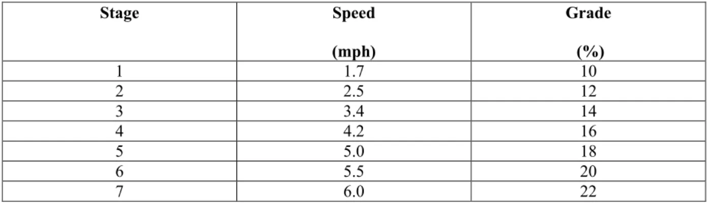
the warm up, resting oxygen consumption (VO2) was recorded for three minutes. The participant then began the graded exercise test. A Bruce treadmill protocol was used.20 Figure 3 lists the three minute stages of the Bruce protocol treadmill test. Heart rate (HR), rating of perceived exertion
(RPE), and respiratory gases were monitored constantly throughout the test (Appendix E). After the
test was completed, the participant recovered actively or passively and was able to leave the laboratory once their heart rate returned to less than 100 beats per minute (bpm). For the VO2 max test to be valid, a participant must have three of the following four criteria: a plateau in VO2 with increases in workload, respiratory exchange ratio (RER) greater than or equal to 1.1, RPE > 18, and
21
Figure 3. The speed (mph; miles per hour) and grade for each three minute stage of the Bruce protocol incremental exercise test.
Stage Speed
(mph)
Grade (%)
1 1.7 10
2 2.5 12
3 3.4 14
4 4.2 16
5 5.0 18
6 5.5 20
7 6.0 22
Sessions I and II
Participants arrived at the Applied Physiology Laboratory at one point during the mid-follicular phase of the menstrual cycle and at one point during the mid-luteal phase. Menstrual cycle
phase was determined and scheduled based on the forward counting method21 and confirmed by urinary ovulation kits (Appendix C). The two experimental trials were counterbalanced to prevent
any order effects. The menstrual cycle is being used to manipulate the estrogen levels between the
two trials. Early in the menstrual cycle, during the mid-follicular phase (approximately between days
3-7), estrogen levels are low. Later in the cycle, during the mid-luteal phase (approximately days
20-25), estrogen levels are significantly higher. Based on this natural fluctuation, the effect of differing
estrogen levels can be examined. The two sessions were scheduled during these two phases in order
to utilize the natural variation in estrogen concentrations; however, the exact days varied depending
on the actual length of the subject’s menstrual cycle. Twenty four hours prior to the experimental
trials participants were told to refrain from strenuous physical activity and completed a food diary so
that nutrient intake was relatively constant between the two trials. Both experimental trials were done
at similar times in the day in order to minimize any effects of circadian rhythms on hormone
22
Participants were asked if they complied with all guidelines and their responses were
recorded. Dietary logs were collected to ensure that participants were consuming adequate calories
and carbohydrates. After compliance was given, participants rested in the supine position for ten
minutes. After ten minutes resting, blood was obtained (3 mL) using the standard venipuncture
technique. The blood sample was placed in a sterile K2-EDTA (purple top) VacutainerTM tube and immediately put on ice. Next, participants completed five minutes of a warm up and stretching. The
participants then ran for 60 minutes at the previously determined workload of 65% of VO2 max (Appendix F). Sixty minutes at 65% intensity was chosen in order to ensure that both estradiol and
IL-6 would be elevated in response to the exercise.6,9,18 After completion of the 60 minutes of exercise, a blood sample (3 mL) was obtained and immediately put on ice. A third and final blood
sample draw (3 mL) was taken at 30 minutes into recovery. Plasma was separated from blood
samples and stored at -80° C until later analysis for estradiol, IL-6, and lactate.
Blood Procedures
Hematocrit
Resting and post-exercise Hematocrit (Hct) values for each of the experimental trials were
determined in triplicate from whole blood samples immediately at the time of exercise tests. Whole
blood was drawn into 75 mm Allied Corporation microcapillary tubes (Fisher Scientific, Pittsburgh,
PA) and sealed using Critoseal (Krackeler Scientific, Albany, NY). Capillary tubes were then spun
in a microhematocrit centrifuge for three minutes at 10,000 RPM. The capillary tubes were then
placed on a hematocrit wheel to determine the hematocrit values of each sample. A mean of the three
23
Hemoglobin
Resting, immediately post-exercise, and recovery hemoglobin (Hb) values were determined
in duplicate from the whole blood samples of the experimental trials using the Stanbio Lab
Hemopoint H2 analyzer (Boerne, TX) immediately at the time of exercise tests.
Plasma Volume Shift
Exercise induced plasma volume shifts were calculated from Hb and Hct values according to
the Dill and Costill method.22 Plasma volume shift was used as a potential explanation for changes in cytokine and estradiol concentrations.
Cytokine (IL-6), Estradiol and Lactate Assays
Whole blood was centrifuged at 3,000 x g for 10 minutes to separate the plasma. The
resultant plasma was transferred to storage tubes and stored at -80°C until cytokine and estradiol
analyses were conducted. Plasma estradiol was measured using the radioactive (125I) immunoassay technique (Siemens Healthcare Technologies, Los Angeles, CA) involving solid-phase antibody
procedures. The assay manufacturer reports a minimal detectable level of 2.0 pg/mL. Plasma levels
of IL-6 were measured using high-sensitivity enzyme-linked immunosorbent assay kits (BioLegend,
San Diego, CA). The assay manufacturer reports a minimum detectable concentration of 1.6 pg/mL (Appendix D). Lactate was measured using a NOVA Biomedical Lactate+ Analyzer (Waltham, MA)
with a reported minimum-detectable concentration of 0.5 mM/L. All blood assays (unknowns) were performed in duplicate while standards were in triplicate (For details, see Appendix).
Data Analysis
All data were analyzed using SPSS statistical software (version 18.0, Chicago, IL).
24
(SD). In addition, effect size was calculated for all significant measures. This later calculation was
used to determine practical vs. significant findings.
A 2x3 (estradiol level x time) totally within subjects repeated measures ANOVA with a
Bonferoni post-hoc test was used to evaluate the effects of estradiol on IL-6 concentration. A 2x2
(estradiol level vs. time) ANOVA was used to evaluate the percent change (%Δ) in IL-6 values
between pre- and post-exercise and recovery during each menstrual cycle phase (estradiol level).
Percent change was calculated using the equation:
% Δ = [(Post-exercise-Baseline)/Baseline] x 100
Resting levels of estradiol at midfollicular and midluteal phase experimental sessions were
CHAPTER IV Results Subject Characteristics
Twelve active female subjects were recruited for this study. Ten of the twelve subjects
recruited fully completed all aspects of the study protocol. Two subjects were forced to withdraw due to personal reasons. All subjects recruited met the inclusion criteria of being eumennorheic and not
using oral contraceptives for the previous six months. Ovulation kits confirmed normal menstrual
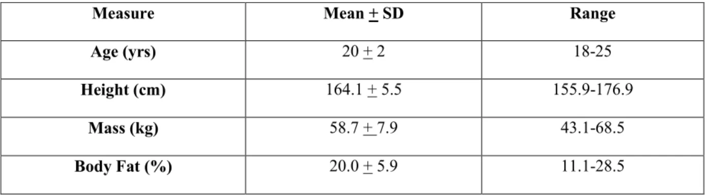
function for all subjects. Participant physical characteristics are reported in Table 1.
Table 1: Physical characteristics of the participants (n=10). Values are Means (+ SD).
Measure Mean + SD Range
Age (yrs) 20 + 2 18-25
Height (cm) 164.1 + 5.5 155.9-176.9
Mass (kg) 58.7 + 7.9 43.1-68.5
Body Fat (%) 20.0 + 5.9 11.1-28.5
VO2 Peak Testing
Only eight of the ten participants who completed the study met the criteria for a valid VO2 max test during the incremental exercise test (see Methods chapter). Because of this, all maximal
oxygen uptake results will be referred to as VO2 peak tests.
The absolute VO2 peak (mean + SD) obtained was 2.96 + 0.57 L/min (range 2.00-4.04 L/min) and the relative VO2 peak obtained was 50.4 + 8.2 mL/kg/min (range 38.4-64.6 mL/kg/min). The peak HR obtained was 195 + 9 bpm (range 184-209 bpm) and the peak RPE obtained was 18 + 1
26
(range 9.49-13.15 minutes) ending with an average stage of 4.25 + .45 of the Bruce protocol (range
4-5) corresponding to a speed of 11.4 + 1.0 km/h (range 10.9km/h-13.0 km/h) and an incline of 17 +
1% (range 16-18%). These responses suggest the subjects were at a high level of cardiovascular
fitness based upon the ACSM guidelines.18
Hormonal Status Determination
Menstrual cycle length for the subjects who completed all aspects of the protocol (n=10) was
30 + 3 days (range 25-35 days). Individual menstrual cycle phases were estimated using the forward
counting method21 and confirmed by urinary ovulation kits (see Methods chapter). The day on which subjects performed the running exercise during the midfollicular phase was 5 + 3 days (range 2-9
days) after the onset of menses. The day on which subjects performed the running exercise during the
midluteal phase was 24 + 4 days (range 18-29 days) after the onset of menses.
Hormonal analysis of resting blood samples for estradiol confirmed the appropriate menstrual
phase and hormonal condition of the subjects. The midfollicular estradiol concentration was 27.04 +
6.12 pg/mL and the midluteal estradiol concentration was 67.64 + 21.70 pg/ml. The difference in
estradiol concentration between the two phases was significantly different (p<.001).
Extended Running Session
Each participant completed two sixty minute running trials, one of which was during the
midfollicular phase of the menstrual cycle and the other after ovulation during the midluteal phase. All subjects complied with all pre-session guidelines (see Methods chapter) before each of the 60
minute trials. Each running trial was to consist of 60 minutes of running at 65% VO2 peak. The overall mean absolute running VO2 was 1.81 + 0.46 L/min and the relative running VO2 was 31.2 + 7.8 mL/kg/min. This corresponded to an actual running intensity of 61.3 + 7% (range 49.9-73.1%)
27
No significant difference in the relative running VO2 (p=.103) or absolute VO2 (p=.066) between the two phases was found. Although there was no difference in VO2 across hormonal conditions, relative VO2 did demonstrate an increase in effort over time. Analysis showed a higher VO2 at time points 30 and 60 minutes compared to minute 10 (p=.035, p=.016, respectively); i.e., main effect for time. However, there was no significant difference in VO2 between 30 minutes and 60 minutes (p=.187). Also, no interaction effect was found between phase and time point for VO2.
No significant main effect for menstrual cycle phase or interaction effect was found for HR.
However, ANOVA results did indicate a significant main effect for time during exercise (p<.001).
Post Hoc analysis indicates that HR at all time points was significantly higher than rest (p<.001) and
that HR was significantly higher at 60 minutes compared to 30 minutes (p=.049). Mean HR was not
significantly higher from minute 10 compared to minute 30 (p=.631) or minute 60 (p=.096).
Similarly to HR, there was no significant main effect for menstrual cycle phase with regards
to RPE (p=.111) although there was a significant main effect for time. Post Hoc analysis indicates
that at minute 30 and minute 60, RPE was significantly higher than at minute 10 (p=.019, p=.007,
respectively) and RPE was also significantly higher at minute 60 compared to minute 30 (p=.005).
Lactate immediately post-exercise was not significantly different between the two phases
(p=.160). The lactate concentration during the midfollicular phase was 2.95 + 1.80 mM/L. During
the midluteal phase it was 2.41 + 1.29 mM/L.
Plasma volume increased by +0.7 + 23.8% over the sixty minute running bout during the
midfollicular phase, whereas it decreased by -5.3 +10.7% during the midluteal phase. These plasma
28
Table 2: Mean (+ SD) VO2, HR, and RPE for prolonged running bouts for the midfollicular and midluteal menstrual phases.
Menstrual Cycle Phase
Measure Time
REST 10 min 30 min 60 min
Midfollicular VO2
(mL/kg/min)
4.51 +
0.65* 30.73 + 8.11€ 32.02 + 7.78 32.63 + 8.08
HR (bpm) 75 + 13¥ 153 + 25 161 + 19 170 + 19
RPE 10 + 2√ 11 + 2£ 13 + 3
Midluteal VO2
(mL/kg/min) 4.89 + 0.59* 29.74 + 8.07€ 30.91 + 8.02 31.42 + 7.95
HR (bpm) 66 + 12¥ 155 + 18 164 + 18 167 + 19
RPE 10 + 2√ 12 + 3£ 14 + 3
A “ ” indicates no value was recorded for that particular time point. * Indicates a significantly lower VO2 compared to 10,
30 and 60 minutes (p<.001). € Indicates a significantly lower VO2 compared to 30 and 60 minutes (p<.05). ¥ Indicates a
significantly lower HR compared to 10, 30, and 60 minutes (p<.001). √ Indicates a significantly lower RPE compared to 30 and 60 minutes (p<.05). £ Indicates a significantly lower RPE compared to 60 minutes (p<.05).
Cytokine (IL-6) Response to Running Trials
Resting, post exercise, and recovery IL-6 concentrations as well as the percentage change
from rest are reported in Table 3. There was no significant main effect for phase (p=.076) or the
interaction effect (p=.069) for the absolute concentration of IL-6. The only significant finding for
absolute IL-6 concentration was a main effect for time (p=.003). Post Hoc analysis indicates that
IL-6 was not significantly elevated from rest to immediately post exercise (p=.072), but that IL-IL-6
concentration did increase significantly from rest to 30 minutes of recovery after exercise (p=.003).
The effect size for this change was 0.963 (Cohen d) which is between a moderate (0.6 Cohen d) and
29
Since all IL-6 changes observed were substantially greater than the plasma volume shifts
detected, no fluid shift corrections of IL-6 concentrations were conducted.
The percent change of IL-6 from rest to immediately post-exercise and from rest to 30
minutes of recovery was also examined. The main effect for phase was not significant (p=.072).
When comparing the relative change from rest between immediately post-exercise and 30 minutes of
recovery there was also no significant difference (p=.301) and no significant interaction effect
between phase and time was found either (p=.768).
Table 3: Mean (+ SD) IL-6 response and percent change (relative to rest) during the prolonged running bout for midfollicular and midluteal menstrual phase.
Menstrual Cycle Phase Time Rest (pg/mL) Immediately Post Exercise (pg/mL) Immediately Post Change (%) 30 Minutes Recovery (pg/mL) Recovery Change (%) Midfollicular 6.25 + 6.84 35.85 + 50.69 517.85 +
415.16
41.11 + 30.65 684.88 + 318.24
Midluteal 8.46 + 9.65 16.40 + 12.40 266.33 + 348.56
12.73 + 14.59 361.51 + 928.89
Estradiol-IL-6 Relationship
As an exploratory analysis, correlations (Pearson) were calculated between the difference in
estradiol concentration between phases and the difference in IL-6 concentration between phases for
the immediately post-exercise and the 30 minutes into recovery samples. The correlation between
estradiol and IL-6 immediately post was r=0.168 (p=.643) and the correlation between estradiol and
CHAPTER V Discussion
The purpose of this study was to determine the effect of estradiol fluctuations during the
menstrual cycle on IL-6 concentration at rest and in response to an acute bout of exercise in females
who are not using oral contraceptives. It was hypothesized that the IL-6 concentration after exercise
would be significantly lower during the midluteal phase of the menstrual cycle when estradiol
concentrations are elevated. This chapter will address this purpose and report on whether the study
design was effective, describe the cytokine response to exercise and relate the findings to previously
published literature, and explain possible reasons for these outcomes.
Study Design
Hormonal Condition
In order to use the natural fluctuation of estradiol in females, subjects were tested once during
the midfollicular phase and once during the midluteal phase of the menstrual cycle. That is, after the
onset of menses, during the midfollicular phase, estradiol levels are low, whereas during the midluteal phase, estradiol levels plateau at a higher concentration. All subjects were able to perform the
prolonged running bout once during each phase. Because of the cyclical nature of female hormone
levels, having a subject tested during a particular menstrual phase it did not ensure that the estradiol
levels would change and be significantly different. For this study, however, statistical analysis
confirmed that the estradiol concentration was significantly different between the two menstrual cycle
phases and changed in the expected manner. Estradiol concentration during the midluteal phase was
31
respectively), thus all subjects demonstrated the correct hormonal changes between the two testing
sessions with midluteal estradiol concentration being higher than the midfollicular estradiol
concentration. These hormonal values fell within the physiologically accepted ranges for each phase
and the fact that the estradiol concentration was approximately doubled from the midfollicular phase
to the midluteal phase also agrees with numerous other studies.9,11,23 However, the absolute concentrations of estradiol were lower than that found in some studies.9,11,23,24 Jurkowski et al.11 reported a midfollicular estradiol concentration of 103.8 + 16.0 pg/mL and a midluteal concentration
of 199.5 + 16 pg/mL. Bonen et al.9 had even higher values of 163.3 + 45.7 pg/mL for the
midfollicular phase and 266.1 + 87.8 pg/mL for the midluteal phase. The most similar numbers to the
current were found by Fu et al.31 who reported follicular estradiol concentration as 34.1 + 12.3 pg/mL and luteal estradiol concentration as 55.3 + 20.4 pg/mL. The somewhat lower absolute
concentrations in the present study could be a result of the high physical activity levels of the current
subjects. That is, estrogen levels have been shown to decrease after aerobic training30 and all subjects in the present study were physically active individuals.
Prolonged Running Bout
All participants successfully completed 60 minutes of treadmill running on the two separate
occasions at an average intensity of 61% of VO2 peak. This intensity is slightly lower than the goal of 65% of VO2 peak intended, but research would suggest this is intense enough to illicit a rise in IL-6.6 For all subjects, the speed and grade of the treadmill during each run was identical between the two
trials. In order to limit the effect of natural circadian rhythms the two trials for each individual were
at approximately the same time of day. However, the time of day for all trials was not consistent for
all subjects. Furthermore, due to time constraints of the study, participants were not evenly
counterbalanced. Seven subjects performed the midfollicular trial first while three subjects performed
32
although there was no apparent differences in responses of the seven subjects tested in one order and
the three tested in the opposite order. Also, the performance characteristics of the exercise were not
different between sessions.
No difference in VO2, HR, or RPE values was found between phases for the 60 minute exercise trials (main effect for phase). This is consistent with other literature that has examined
exercise performance across the menstrual cycle.23,26,27 The one study that did find a significant difference in RPE between menstrual cycle phases was conducted in a population of sedentary
women.28 These less active subjects might experience a different level of exertion during exercise, as well as being less experienced in monitoring exercise exertion. The non-significant difference in
lactate concentration immediately after exercise also agrees with other studies that have examined this
relationship.23,25-27 Collectively, the similar VO
2, HR, RPE, and lactate data confirm the subjects’ exercise sessions were highly alike and suggest any IL-6 response differences were not due to
differing aspects of the exercise sessions.
In response to the 60 minute exercise, all values were elevated compared to the resting
values. Oxygen uptake increased at each time point and then plateaued from minute 30 until the end
of exercise. Thus, the second half of the exercise was at a steady state VO2. Heart rate increased from rest and remained relatively level from 10 minutes to 30 minutes and then increased slightly at
60 minutes. Based on this rise in HR the end of the exercise session was more physiologically
difficult compared to the beginning of the session. Based on the HR response, exercise was not at a
steady state for the entire 60 minutes of running. An increase in HR towards the end of a long
endurance exercise is, however, consistent with the cardiac drift phenomenon.29 During prolonged exercise plasma fluid can be lost from the blood, thereby reducing venous return and cardiac output.
33
Perceived exertion continued to rise at each measurement time point which demonstrates that
the exercise became progressively more difficult during the 60 minutes, in agreement with the HR
findings. Although the exercise was at a constant intensity, RPE increased throughout the prolonged
running bout which could be attributed to the extended length of the exercise bout and potential
subject fatigue.
Lactate concentration immediately post exercise was also not significantly different between
the two sessions. This is consistent with other studies that have measured this variable.25-27 These studies did not have the same exact exercise component (i.e., intensity, duration) but they all had the
same findings that lactate was the same between phases at all time points and intensities. Based on
the extended duration of the exercise bout, and the subjects’ need to run below their lactate threshold
(i.e., a moderate intensity), the observed lactate concentrations were similar to what was expected
based on the literature.25,27
Cytokine Response
Exercise Effects
The average resting concentration of IL-6 for all twenty exercise sessions was 7.35 + 2.43
pg/mL. This resting concentration is higher than most other exercise study reports. In male subjects Ostrowski et al.2 reported resting IL-6 concentration as 1.5 +0.7 pg/mL, while Steensberg et al.7 had
resting concentrations of 0.74 (range, 0.67-1.50) pg/mL, and Mendham et al.6 measured IL-6 at 1.31 + 0.28 pg/mL. In non-oral contraceptive using females pre-exercise IL-6 concentration was 2.0
pg/mL as reported by Timmons et al.8 Chaffin et al.24 also had resting values less than 2.00 pg/mL, both of which are lower than the results of this study. The finding of elevations in resting IL-6 in the
present subjects could be a result of excessive background interference in the assay outcomes due to a
limited practical experience by the investigator performing the assays; nonetheless, the levels were
34
validity in the data. An additional factor accounting for the elevated resting IL-6 may be related to
the study design pre-exercise protocol. Holmes et al.36 report that the level of resting IL-6 is inversely related to the level of circulating free fatty acids (FFA). At rest, the circulating level of
FFA is directly related to the duration of a post-prandial fast. The pre-exercise fast that the subjects
were asked to complete was relatively short compared to most studies being only two hours. This
means the subjects started the exercise trials with low FFA levels, and thus elevated IL-6. Holmes et
al.36 reported resting IL-6 levels as 6.05 + 0.89 pg/mL for their subjects having FFA levels at that expected for the present study subjects based upon their level of fasting. The IL-6 concentration of
Holmes et al.36match very similarly with that of the present study.
Interleukin-6 concentration increased from rest after the 60 minutes of running, but this
change was not significant (p=.072; main effect time). There was a concentration of 26.13 + 9.03
pg/mL immediately after exercise. This concentration continued to rise until 30 minutes of recovery
from exercise where it was 26.92 + 5.54 pg/mL, this concentration is significantly elevated compared
to rest (p<.05). Most studies have found that IL-6 peaks immediately after exercise2,3,6,18. However, there are various factors that can affect the cytokine response. These include the type of physical
activity, the type of contraction, as well as the intensity and duration of exercise. The rise in IL-6 is most closely related to the duration of the exercise bout18. The absolute IL-6 concentration after exercise in this study was higher than some previous studies4,6,5,8,18 but lower than others.2,3,6 The IL-6 response to aerobic endurance exercise in men has been reported by Ostrowski et al.2 as reaching a level of 94.4 +12.6 pg/mL after completion of a marathon. Timmons et al.8 found that IL-6 rose to approximately 7.0 pg/mL after cycling for 90 minutes at 65% of VO2. There is far fewer data with respect to females who were not using oral contraceptives and underwent aerobic exercise. One study
found that IL-6 rose to approximately 7.0 pg/mL after 90 minutes of cycling at 65% VO2max. The difference in IL-6 concentration between these studies could be attributed to the type of exercise that
35
immediate post-exercise value was not expected but the fact that the recovery values were
significantly greater than rest suggests that the exercise bout intensity and duration was sufficient to
illicit a rise in IL-6 (N.B., the slightly higher variable observed in the SD at the immediate versus
recovery concentration most certainly affected the lack of detection of a significant increase).
The increase in IL-6 following exercise was about 3.5 fold for the current study. Most other
studies do not report the percent change from rest but the fold increase is able to be determined.
While the marathon studies have reported that IL-6 rose as much as a 128-fold,3 the current findings are close to studies with similar aerobic exercise components. In two studies Timmons et al.8,17 found that IL-6 had a 3-fold increase in concentration. Ives et al.5 found about a 3.5 fold increase as well in oral contraceptive using females, while Chaffin et al.24 had approximately a 4-5 fold increase in non-oral contraceptive using females.
Menstrual Phase Effects
The primary research question of this study was to examine the relationship between estradiol
and IL-6 in active females who do not use oral contraceptives. The results of this study suggest that there is no significant difference in IL-6 concentration in response to varying estradiol concentrations
either at rest or in response to exercise. When examining the interaction effect between phase and
time there were no significant findings. Although the IL-6 concentration was lower in the midluteal
phase compared to the midfollicular phase at all time points, the difference was not large enough to
be significant (p=.074; interaction effect). With respect to percent change, this finding was also not
significant (p=.072). In both cases the effect nearly reached statistical significant and had there been
less variability in the IL-6 response significance might have been reached based on the critical
statistical criteria of 0.05.
In animal and in vitro studies, IL-6 concentration was consistently found to be lower in