REVIEW
Mechanisms of neuroimmune gene induction in alcoholism
Fulton T. Crews&Ryan P. Vetreno
Received: 17 November 2014 / Accepted: 24 February 2015 / Published online: 20 March 2015 #The Author(s) 2015. This article is published with open access at Springerlink.com
Abstract
RationaleAlcoholism is a primary, chronic relapsing disease of brain reward, motivation, memory, and related circuitry. It is characterized by an individual’s continued drinking despite negative consequences related to alcohol use, which is exem-plified by alcohol use leading to clinically significant impair-ment or distress. Chronic alcohol consumption increases the expression of innate immune signaling molecules (ISMs) in the brain that alter cognitive processes and promote alcohol drinking.
Objectives Unraveling the mechanisms of alcohol-induced neuroimmune gene induction is complicated by positive loops of multiple cytokines and other signaling molecules that con-verge on nuclear factor kappa-light-chain-enhancer of activat-ed B cells and activator protein-1 leading to induction of ad-ditional neuroimmune signaling molecules that amplify and expand the expression of ISMs.
ResultsStudies from our laboratory employing reverse tran-scription polymerase chain reaction (RT-PCR) to assess mRNA, immunohistochemistry and Western blot analysis to assess protein expression, and others suggest that ethanol in-creases brain neuroimmune gene and protein expression through two distinct mechanisms involving (1) systemic in-duction of innate immune molecules that are transported from blood to the brain and (2) the direct release of high-mobility group box 1 (HMGB1) from neurons in the brain. Released HMGB1 signals through multiple receptors, particularly Toll-like receptor (TLR) 4, that potentiate cytokine receptor re-sponses leading to a hyperexcitable state that disrupts neuro-nal networks and increases excitotoxic neuroneuro-nal death. Innate immune gene activation in brain is persistent, consistent with
the chronic relapsing disease that is alcoholism. Expression of HMGB1, TLRs, and other ISMs is increased several-fold in the human orbital frontal cortex, and expression of these mol-ecules is highly correlated with each other as well as lifetime alcohol consumption and age of drinking onset.
Conclusions The persistent and cumulative nature of alcohol on HMGB1 and TLR gene induction support their involve-ment in alcohol-induced long-term changes in brain function and neurodegeneration.
Keywords TLR4 . HMGB1 . Ethanol . Cytokines . Microglia . RAGE . Gut permeability . Amphoterin . Innate immune . Alcohol use disorder . Frontal cortex
Introduction: microglia and innate immune genes
Microglia are tissue-specific monocyte-like cells of mesoder-mal origin (Ginhoux et al.2010), whereas all other brain cells are derived from the neuroectoderm. Monocytes and tissue-specific monocyte-like cells (e.g., microglia) express innate immune signaling molecules originally characterized within the peripheral immune system. In the brain, microglia consti-tutively express Toll-like receptor (TLR) 4 and other innate immune receptors that are responsive to proinflammatory sig-nals like high-mobility group box 1 (HMGB1) but are also responsive to neurotransmitters (Kettenmann et al.2013).
Innate immune gene upregulation with rapid monocyte re-sponses to infection was first characterized in blood as acute phase response proteins that today are known to include mul-tiple cytokines, chemokines, proteases, cellular oxidases, and cytokine receptors. Acute phase responses and monocyte ac-tivation involve amplification in the expression of a large number of genes through kinase signaling pathways that F. T. Crews (*)
:
R. P. VetrenoBowles Center for Alcohol Studies, University of North Carolina at Chapel Hill, School of Medicine, CB# 7178, 1021 Thurston-Bowles Building, Chapel Hill, NC 27599-7178, USA
converge on two distinct transcription factors: nuclear factor kappa-light-chain-enhancer of activated B cells (NF-κB) and activator protein-1 (AP-1). Both NF-κB and AP-1 induction promote the expression of innate immune cytokines (Li and Verma2002; Valles et al.2004), including tumor necrosis factor-alpha (TNFα) and interleukin-1beta (IL-1β) as well as upregulation of TLRs and other cytokine receptors. In ad-dition, innate immune proteases and oxidases are induced, particularly cyclooxygenase (COX-2) and nicotinamide ade-nine dinucleotide phosphate oxidase (NADPH oxidase) as well as major histocompatibility (MHC) signaling molecules, such as beta-2 microglobulin. These monocyte-microglial-expressed proteins and their receptors are innate immune sig-naling molecules (ISMs) that are expressed in the brain (Blanco and Guerri2007; Guerri and Pascual2010; Valles et al.2004). This review will refer to these brain signaling molecules asBneuroimmune^due to their characterization in the immune system of the brain, while recognizing that sig-naling across multiple unique brain cells differs from immune inflammation in response to infection.
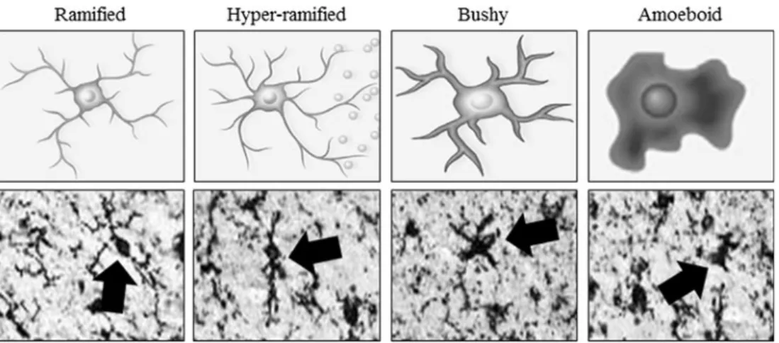
Brain neuroimmune signaling primarily involves monocyte-microglial innate immune signals and not adaptive immune antibodies. Although microglia are unique tissue-specific brain monocyte-like cells, similar to all monocytes, microglia undergo morphological changes that characterize stages of activation (Graeber2010) (Fig.1). Resting ramified microglia likely contribute trophic and other signals similar to the wound healing monocyte phenotype termed M2 that upon activation can become hyper-ramified, with secretion of
proinflammatory cytokine signals (Beynon and Walker
2012). However, activated microglia do not necessarily al-ways adopt an M1 phenotype as Marshall et al. (2013) found that young adult rats subjected to a 4-day binge model of alcohol led to partial microglial activation as evidenced by increased expression of OX-42 but not a fully activated phe-notype characterized by expression of OX-6 or ED-1. This partial microglial action was accompanied by an increase in the anti-inflammatory cytokine IL-10 and no increase in pro-inflammatory cytokines IL-6 or TNFα. Further proinflamma-tory activation, known as M1 monocyte phenotype, involves expansion of processes to aBbushy morphology^and finally a
Bphagocytic^rounded morphology (Colton2009). The rela-tionship between morphological changes in monocyte-like cells including microglia and the secretion of ISM is poorly understood, although increased severity of pathology is asso-ciated with greater ISM induction and activated morphology. Like all monocytes, microglial activation can lead to NF-κB transcription of proinflammatory genes, which signal to other microglia as well as astroglia, oligodendroglia, and neurons, amplifying neuroimmune gene induction within and across cells by induction of TLRs and cytokine receptors, many belonging to the IL-1βreceptor family that activate kinase cascades that converge on NF-κB (Blanco and Guerri
2007; Blanco et al.2004,2005; Fernandez-Lizarbe et al.2009,
2013; Pascual et al.2011b; Valles et al.2004). The amplifica-tion of ISMs across cells and tissue can lead to pathology, and understanding the processes of monocyte signaling provides insight into microglial signaling in brain. The most severe
Fig. 1 Activated morphology of microglia. Representative schematics a n d p h o t o m i c r o g r a p h s o f h u m a n b r a i n m i c r o g l i a ( I b a 1 immunohistochemistry) depicting morphological stages of microglial activation. Ramified orBresting^microglia are characterized by long, ramified processes with comparatively small cell bodies. Mildly activated hyper-ramified microglia are characterized by increased branching of processes as well as lengthening of processes and the secretion of proinflammatory cytokines (Beynon and Walker2012). Bushy morphology is intermediate activation and is characterized by swollen, truncated processes, and enlarged cell bodies. Amoeboid or
acute example of monocyte activation during infection is sep-sis. Sepsis and the systemic inflammatory response syndrome refer to aBcytokine storm,^ which involves a pronounced increase in multiple proinflammatory cytokines and other ISMs that cause a potentially fatal innate immune reaction consisting of positive feed-forward loops between cytokines and immune and tissue cells that result in highly elevated cytokine blood levels, multi-organ failure, and death (Osterholm2005). Models of sepsis that involve activation of an acute phase-like response lead to increased expression of multiple cytokines that are induced in distinct phases. During the initial phase, TNFαand IL-1βexpression is in-creased during the first several hours after innate immune activation and then subside. The second phase involves HMGB1, which is an agonist at multiple receptors that con-tribute to further activation of proinflammatory cascades (Fig.2). Disulfide-HMGB1 is a TLR4 agonist (Tang et al.
2012), and thiol-HMGB1 is an agonist at the receptor for advanced glycation end products (RAGE; Allette et al.
2014) and also dimerizes with proinflammatory molecules (Tang et al. 2012; Venereau et al. 2012), such as IL-1β
(Wahamaa et al.2011) that enhances IL-1βreceptor induction of proinflammatory molecules through NF-κB. HMGB1 in-creases in blood approximately 16 h after infection in models of sepsis and persists for several days during which mice die. Mortality is prevented by anti-HMGB1 antibody treat-ment (Wang et al.2001) consistent with HMGB1 induction of aBcytokine storm^by acting through multiple receptors that converge on proinflammatory NF-κB signaling. Survivors of sepsis models show prolonged increases in serum HMGB1 and cognitive deficits that are blunted with HMGB1 antibody treatment (Chavan et al.2012). To model alcoholic hepatitis and alcohol-induced release of gut endo-toxin, we systemically administer lipopolysaccharide (LPS) a n d p o l y i n o s i n i c : p o l y c y t u d y l i c a c i d ( p o l y I : C ) . Administration of these endotoxins systemically after eth-anol treatment exacerbates the innate immune response. Acute binge drinking also increases serum endotoxin levels albeit at a lower level observed under septic conditions. High binge drinking doses cause the gut to become perme-able or Bleaky^ (Ferrier et al.2006). Only high doses of ethanol, e.g., at least 2–3 g/kg ETOH intragastric doses (Ferrier et al.2006), potentiate gut innate immune signal-ing, disrupting gut tight junctions, and opening sites that allow the gut biome bacteria and their endotoxins to enter the portal circulation leading to the liver where they can initiate a proinflammatory response (Sims et al. 2010). Released LPS potentiates alcohol-induced liver inflamma-tion and secreinflamma-tion of proinflammatory cytokines, including the proinflammatory cytokine TNFα, which is released into the blood. Proinflammatory cytokines in the blood are transported across the blood–brain barrier (Banks and Erickson2010; Qin et al. 2007) such that both cytokines
and alcohol enter the brain where they induce neuroimmune activation.
Innate immune signaling molecules in the brain appear to contribute to both brain health and pathology. Indeed, recent studies find that MHC molecules contribute not only to most neurodegenerative diseases (Gage2002; Glass et al.2010) as well as alcohol and drug dependence (Crews2012) but are also critically involved in brain development (Huh et al.
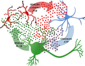
2000). Within the brain microglia, innate immune cytokines, such as TNFα, IL-1β, and HMGB1 as well as TLRs, purinergic receptors (e.g., P2X7), various cytokine receptors, and innate immune proteases and oxidases, all amplify Fig. 2 High-mobility group box 1 (HMGB1) is actively and/or passively released leading to multiple signaling pathways. Actively released HMGB1 from brain slice cultures found histochemical evidence of release from neurons by ethanol (Zou and Crews 2014), although HMGB1 release likely occurs from most brain cell types. Neurons and glia release HMGB1 during glutamate excitation (Maroso et al.2010, 2011). HMGB1 is also released during necrotic cell death activating innate immune signaling. HMGB1 has multiple signaling mechanisms regulated by oxidation of cysteines. Fully oxidized HMGB1 (blue,left) does not activate proinflammatory signaling, although it may contribute to resolution of the proinflammatory state. HMGB1 in the all-thiol form
(yellow,middle) is an agonist at the receptor for advanced glycation
through NF-κB and AP-1 loops that confound studies that are focused on studying a single neuroimmune signaling mole-cule (Fig.3). NF-κB regulates the transcription of proinflam-matory innate immune genes as well as many other genes (Perkins2007). Ethanol increases NF-κB–DNA binding and expression of multiple innate immune genes including flammatory cytokines, TNFα, IL-1β, and MCP-1, the proin-flammatory oxidase, iNOS, and proteases TACE and tPA (Zou and Crews2010). Previously, we found that ethanol increased NF-κB p65 nuclear immunohistochemistry consis-tent with NF-κB p50⁄p65 subunit nuclear translocation and transcription activation. Similarly, we found that antibodies to p50 or p65 super-shifted EMSA gels, suggesting that etha-nol increased brain NF-κB p65–p50 heterodimer–DNA bind-ing (Zou and Crews2006). Taken together, ethanol-induced NF-κB–DNA binding and target gene expression support eth-anol activation of NF-κB transcription of proinflammatory genes. However, using an ELISA-based DNA binding analy-sis, we found large increases in NF-κB subunit p50 protein but not NF-κB p65 protein. A similar finding has been reported for prefrontal cortex gene expression in the post-mortem hu-man alcoholic brain (Okvist et al.2007). Array analysis of gene expression in post-mortem alcoholic prefrontal cortex found 479 transcripts with NF-κB–DNA binding sites that were generally upregulated, with analysis of NF-κB subunit proteins indicating NF-κB p50 was the dominant subunit expressed in human alcoholic brain (Okvist et al.2007). Although homodimers of NF-κB p50 inhibit transcription (Perkins2007), increases in NF-κB p50 protein are often
as-sociated with increased transcription. Several mechanisms are involved in increased NF-κB p50 activation of transcription including protease processing of inhibitory NF-κB p105 to transcriptionally active NF-κB p50 (Hoffmann et al.2006) or through NF-κB p50 homodimer association with BCL3, atypical IκB, and other proteins that activate gene transcrip-tion involving NF-κB p50 (Ghosh and Hayden2008). Thus, an increase in NF-κB p50 is consistent with increased NF-κB gene transcription. Although transcription is complex, ethanol-induced progressive increases in NF-κB–DNA bind-ing and increased transcription likely represent one mecha-nism of ethanol induction amplification of brain innate im-mune genes. The mechanisms that regulate innate imim-mune gene induction in various brain cells that contribute in vivo to amplification of specific innate immune genes are poorly understood. In the current review, literature pertaining to al-coholism and innate immunity was reviewed using the PubMed search engine, and this review will focus on two mechanisms of ethanol sensitization of microglia and induc-tion of neuroimmune genes. The first involves a systemic mechanism whereby blood innate immune signaling mole-cules induce brain neuroimmune genes and a second local mechanism involving neuronal–glial signaling through neuroimmune genes that regulate neuronal excitability. It
should be noted that neuroimmune activation appears to be involved in the later stages of heavy drinking and that binge drinking is required to activate the innate immune system.
Ethanol-induced blood innate immune signals activate brain neuroimmune signaling
Acute binge drinking increases blood cytokines in normal healthy humans (Bala et al.2014). Recent studies indicate that ethanol in the gut releases HMGB1 (Ge et al.2014) which activates TLR4 causing the gut to leak endotoxin LPS-like bacterial products that stimulate proinflammatory cytokine in-duction in the liver (Ge et al.2014) thereby increasing circu-lating blood cytokines (see Gao et al. 2011for a review) (Fig.3). One mechanism involves ethanol causing the gut to become more permeable orBleaky.^Indeed, this occurs pri-marily with high doses of ethanol (i.e., at least 2–3 g/kg ETOH intragastric doses [Ferrier et al.2006]) that activate gut innate immune signaling through disruption of gut tight junctions that allows the gut microbiome bacteria and their endotoxins to enter portal circulation leading to the liver inducing an innate immune response in blood (Sims et al. 2010). Alcoholism is associated with liver disease resulting in many alcoholics having elevated levels of blood cytokines. However, in healthy adults, an acute alcohol binge dose also increases blood cytokines (Bala et al. 2014), and in human alcoholics without liver disease, gut permeability is increased during active drinking with increased blood endotoxin and proinflammatory cytokines (Leclercq et al. 2012). Interestingly, a 3-week period of abstinence led to a resolution of gut permeability, but levels of circulating proinflammatory cytokines (i.e., TNFαand IL-6) remained elevated across ab-stinence. Furthermore, the authors found that serum levels of circulating cytokines were correlated with levels of depression and alcohol craving. These data suggest that ethanol-induced gut permeability results in a peripheral blood innate immune response that impacts brain and behavior (Leclercq et al.
blood–brain barrier (e.g., TNF receptor [Banks and Erickson
2010; Qin et al.2007]) and also stimulate endothelial cells to release cytokines in brain (Watkins et al.1995) (see Fig.3). Thus, acute ethanol exposure can induce a blood cytokine response by increasing endotoxin leak from the gut that can lead to changes in the brain and behavior.
Induction of acute increases in blood levels of TNFα, other cytokines, and ISM can have long-lasting effects on the brain. For instance, Qin et al. (2007) found that a single systemic dose of LPS increased mRNA and protein levels of TNFαin parallel in the liver, blood, and brain, but the liver and blood response subsided after 12–24 h, whereas the brain response persisted for at least 10 months. However, in transgenic mice lacking blood–brain barrier TNF receptor transporters, a sin-gle systemic dose of LPS increased TNFαlevels in the blood but not in the brain suggesting that TNFα transport by its receptor across the blood–brain barrier is necessary for acti-vating proinflammatory neuroimmune responses in the brain. As mentioned above, proinflammatory gene expression in blood and brain parallels each other at early time points. Surprisingly, the increase in proinflammatory gene expression in the brain persisted for months leading to degeneration of dopaminergic neurons of the substantia nigra that could con-tribute to dopamine hypofunction leading to sensitization of the response to the rewarding effects of ethanol (Blednov et al.
2011; Qin et al.2007). Although the binge ethanol-induced liver and blood responses are not as pronounced as LPS,
chronic ethanol pretreatment sensitizes systemic and brain proinflammatory cytokine responses to a single systemic dose of LPS following the conclusion of ethanol treatment through TLR4 (Qin et al.2008) and to a single systemic dose of poly I:C through TLR3 (Qin et al.2013). Ethanol appears to sen-sitize microglia by upregulating multiple innate immune sig-naling molecules, including TLR receptors (Crews et al.
2013). Chronic ethanol treatment of mice for 10 consecutive days (5.0 g/kg, i.g.) increases brain expression of the proin-flammatory cytokine monocyte chemotactic protein-1 (MCP-1) that persists for at least 1 week of abstinence following ethanol treatment (Qin et al. 2008). Exposure of C57BL/6J mice to 10 daily doses of ethanol followed by a single system-ic dose of LPS resulted in a potentiation of proinflammatory cytokine induction in the liver, blood, and brain, resulting in persistently higher brain expression long into abstinence rela-tive to LPS alone-treated animals (Qin et al.2008). In these studies, ethanol sensitized mice to the LPS-induced neuroimmune response that resulted in sustained increases in multiple proinflammatory cytokines (e.g., TNFα, IL-1β, and MCP-1) in the brain but not in the liver. While the mecha-nism(s) of the sustained brain response and transient liver response are not clear, we found that IL-10, an anti-inflammatory factor that inhibits NF-κB, is increased in the liver 1 week after alcohol treatment but decreased in the brain (Qin et al. 2008). This finding is consistent with anti-inflammatory mechanisms contributing to the reversal of the Fig. 3 Ethanol in the gut causes leakage of bacterial products into the
portal vein increasing hepatic TNFαrelease into the blood which induces neuroimmune gene expression in the brain. High doses of consumed alcohol in the gut (i.e., at least 2–3 g/kg ETOH intragastric doses [Ferrier et al.2006]) increases permeability allowing bacterial products such as endotoxin-lipopolysaccharide (LPS) to enter portal circulation. Alcohol and LPS enter portal circulation leading to induction of liver tumor necrosis factor-alpha (TNFα) and other proinflammatory cytokines that are released into the blood and enter the brain through cytokine-specific receptor transport (e.g., the TNFαreceptor) (See Qin et al.2007for details). This activates positive loops of proinflammatory
liver response. Bacterial products enter portal circulation acti-vating Kupffer cells (i.e., liver monocytes) that produce cyto-kines, including TNFα, that can be transported into the brain where they activate brain neuroimmune signaling that persists for long periods (Qin et al.2007). Thus, the spread of a sys-temic innate immune response to brain represents a mecha-nism of brain neuroimmune gene induction.
Microglia and alcohol in the brain
Ethanol-induced hyper-ramified microglia have been impli-cated in alcohol-induced activation of neuroimmune signaling pathways in the brain. In rats, intermittent and chronic ethanol exposure sensitize microglia, priming them for further activa-tion and increasing expression of proinflammatory cytokines providing indirect evidence for the role of microglia in alcohol-induced neuroinflammation and neurotoxicity (Alfonso-Loeches and Guerri 2011; Zhao et al. 2013). Alcohol activates microglia via TLRs (Alfonso-Loeches and Guerri2011). Specifically, TLR4 expression and TLR4/TLR2 association on cultured microglia appear to be necessary for alcohol-induced microglia activation with the production of inflammatory mediators and cortical neuronal apoptosis (Fernandez-Lizarbe et al. 2009,2013). In an in vitro study (Boyadjieva and Sarkar2013b), application of microglial-conditioned media enhanced ethanol-induced apoptosis of cultured hypothalamic neurons. Interestingly, neutralization of TNFα abolished the neuronal cell death induced by microglial-conditioned media, suggesting that microglial pro-duction of TNFαplays a key role in ethanol-induced neuro-toxicity in developing neurons. Additionally, they found that ethanol induces oxidative stress in neurons by increasing the cellular production of O2−, reactive oxygen species (ROS), and nitrite while decreasing levels of the antioxidant glutathi-one (GSH) and the cellular activity of other antioxidative en-zymes, including glutathione peroxidase, catalase, and super-oxide dismutase. Furthermore, treatment with either a synthet-ic superoxide dismutase/catalase mimetsynthet-ic (EUK-134) or a water-soluble analog of vitamin E (Trolox), both of which are well-known antioxidants, protected developing hypotha-lamic neurons from oxidative stress and cellular apoptosis caused by ethanol-treated microglia media. These findings are consistent with proinflammatory loops of positive feed-forward amplification of neuroimmune signaling with multi-ple components, including TLR receptors and proinflamma-tory cytokines that contribute to neurodegeneration (Fig.3). Further, the P2X7R, which is a member of the purinergic P2X family of ATP-gated ion channels, is highly expressed on microglia and activation of these receptors is associated with release of the proinflammatory cytokines IL-1β(Ferrari et al.
1997; Lister et al.2007) and TNFα(Hide et al.2000; Lister et al.2007). The functional responses of P2X7R activation by
ATP are associated with ongoing cellular damage and chronic brain inflammation. Indeed, recent experimental evidence in-dicates that stimulation of P2X7Rs mediate ATP-induced ap-optosis through microglial production of superoxide (Parvathenani et al. 2003; Raouf et al. 2007). Interestingly, the P2X7 receptor might also play a role in microglial prolif-eration since TNFα application to hippocampal-entorhinal cortex slice culture led to an increased in proliferating microg-lia (Zou et al. 2012). Thus, there are numerous processes through which alcohol induces reactive oxygen species.
The systemic increases in proinflammatory signals broadly activate neuroimmune signals across the brain (Sugama et al.
2009). In addition to neuroimmune cytokines, oxidases such as COX-2, nitric oxide synthetasem and NADPH oxidase, which includes phargocytic oxidase (e.g., gp91PHOX, which was classically found increased in monocyte with phagocytic morphology) form ROS that can contribute to neurotoxicity (Takeuchi2010). Reactive oxygen species are oxidative mol-ecules that oxidize proteins, break down cell membranes, in-duce cell death, and activate NF-κB as a component of proin-flammatory amplification within and across cells (Fig. 3). NADPH oxidase, a multi-subunit enzyme that catalytically makes superoxide, is increased in the frontal cortex by both a single systemic dose of LPS and 10 days of ethanol treat-ment (5.0 g/kg, i.g.), particularly gp91PHOX, which is the su-peroxide forming subunit (Qin and Crews2012). These find-ings are consistent with oxidative stress, through innate im-mune gene induction, contributing significantly to alcoholic brain damage in the orbitofrontal cortex. Qin et al. (2013) found that a single systemic dose of LPS induces microglial activation, NADPH oxidase, and oxidative stress that persist for at least 20 months and leads to persistent neuroimmune gene induction and a progressive persistent neurodegenera-tion. The prolonged and persistent induction of NADPH oxi-dase and oxidative stress in the brain could contribute to the long-lasting increases in NF-κB transcription since oxidative free radicals can activate NF-κB.
exposed to chronic 10-day ethanol treatment (5.0 g/kg, i.g.) and post-mortem human alcoholic orbitofrontal cortex (Qin and Crews2012) consistent with neuroimmune signaling am-plifying across brain cell types. In agreement with the in vivo findings, in vitro studies reveal that ethanol (25, 50, and 100 mM ethanol) dose-dependently increased induction of oxidative stress (i.e., O2−, ROS, and nitrite) and apoptotic cell death in neuronal hypothalamic primary cultures. Interestingly, ethanol-activated microglial-conditioned media potentiated ethanol-induced production of ROS and oxidative stress in cultured hypothalamic neuronal cells leading to in-creased apoptotic cell death (Boyadjieva and Sarkar2013b). Therefore, ethanol-activated neuroimmune signaling may pro-duce ROS and nitrite that decrease the cellular activity of anti-oxidative enzymes and increasing expression of neuroimmune molecules in neurons that contribute to neurodegeneration.
The mechanisms of alcohol-induced neurodegeneration are complex involving multiple neuroimmune signals as well as alterations in trophic signals (Crews and Nixon
2009; Guadagno et al. 2013). Ethanol induces IL-1βand IL-6 as well as transforming growth factor-β1 (TGF-β1; Alfonso-Loeches et al. 2010; Chen et al. 2006). One of the cellular signaling mechanisms by which alcohol in-duces neuronal apoptosis involves increased neuronal re-lease of TGF-β1. It has been shown that alcohol-induced increases of TGF-β1 levels in neuronal cells is accompa-nied by increased expression of transcription factor E2F1 (overexpression sensitizes cells to apoptosis), reduced ex-pression of cyclin D1 and cyclin-dependent kinase-4 (key regulator of cell cycle progression), elevated levels of mi-tochondrial pro-apoptotic proteins bak, bad, and bcl-xs, lowered levels of the anti-apoptotic protein bcl-2, in-creased production of apoptotic enzyme caspase-3, and increased neuronal cell death (Chen et al. 2006; Kuhn and Sarkar 2008). Hypothalamic neuronal cell cultures following treatment with ethanol-activated microglial-conditioned media showed decreased production levels of cyclic adenosine monophosphate (cAMP) and brain-derived neurotropic factor (BDNF; Boyadjieva and Sarkar2013a). Further, treatment with BDNF or dibutyryl cAMP decreased ethanol-activated microglial-conditioned medium-induced changes in intracellular free radicals, ROS, and O2 as well as nitrite, GSH, and catalase. Therefore, ethanol by increasing the production of microglial-derived factors reduces cellular levels of cAMP and BDNF leading to an increase in cellular oxi-dative status and apoptosis of neuronal cells. Although ethanol-induced sensitization of microglia leads to an in-crease in the production of ISMs while also decreasing trophic support that clearly contributes to altered neuronal vitality and increase neuronal death, further studies are needed to identify all the mechanisms by which ethanol-activated signaling induces neurodegeneration.
Ethanol and neuronal excitation release HMGB1 triggering neuroimmune activation
A second mechanism of neuroimmune activation in brain in-volves neuronal activation of adjacent microglia and astro-cytes (Crews and Vetreno 2014; Sugama et al. 2009). This form of neuroimmune activation in specific neuronal nuclei may represent a form of neuroplasticity. Experience-induced neuroimmune gene induction can lead to long-lasting changes in neuronal excitability that could contribute to neuroplasticity similar to synaptic long-term potentiation. Pathological in-creases in excitability could contribute to mental diseases as well as increasing sensitivity to excitotoxic neuronal death. Excited neurons release HMGB1, also known as amphoterin (Huttunen and Rauvala 2004), which stimulates microglia thereby increasing NF-κB transcription of proinflammatory cytokines (Crews et al.2013; Zou and Crews2014) as well as increasing neuronal excitability (Maroso et al. 2010). Microglia and astrocytes release cytokines increasing neuro-nal excitability in part due to reduced glial uptake of glutamate (Zou and Crews2005) and increased glial release of glutamate (Fig. 4) as well as neuronal HMGB1-TLR4 signaling that enhances neuronal sensitivity to glutamate (Maroso et al.
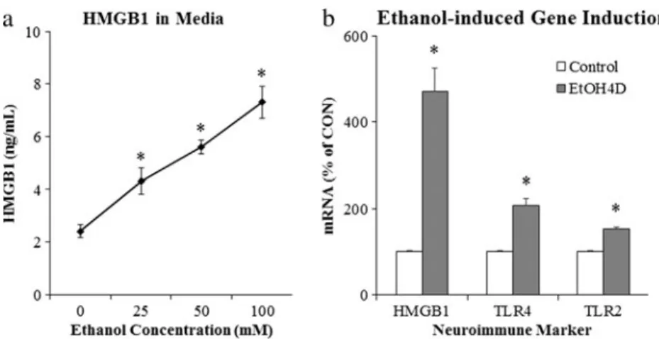
2010). Ethanol-induced release of HMGB1 (Fig. 5) likely contributes to adaptive changes in the brain that contribute to alcoholic neuropathology, both the behavioral pathology
as well as neurodegeneration. However, it is still a matter of debate whether neurons express the TLR4 receptor as our group and others report evidence of neuronal TLR4 (Okun et al.2009; Vetreno et al. 2014; Vetreno and Crews 2012) using immunohistochemistry, but other studies employing fluorescence-activated cell sorting techniques suggest that they are mostly expressed by glial cells (Schwarz and Bilbo
2013). Ethanol-induced release of HMGB1 results in the in-duction of multiple neuroimmune molecules including TLR4 and TLR3 receptors as well as increased HMGB1 expression (Fig.5). The induction of agonists and receptors is character-istic of innate immune responses that likely contribute to am-plification and persistence of innate immune gene induction in the brain. Actively released HMGB1 is acetylated, and we discovered that ethanol increases HMGB1 acetylation through altered histone deacetylases. Acetyl-HMGB1 is found in cy-tosolic vesicles that is released during neuronal activation or treatment with ethanol (Zou and Crews2014). Release of acetyl-HMGB1 by ethanol is consistent with active neuronal release since cell death released HMGB1 is not acetylated. The importance of ethanol-induced release of HMGB1 and activation of TLR4 became apparent in large part due to the elegant experiments of Consuelo Guerri’s laboratory. These studies found that ethanol treatment induces neuroimmune genes in microglia and astrocyte cultures as well as in vivo in mice but not in transgenic cells or mice that lack TLR4 receptors (Alfonso-Loeches et al.2010; Blanco et al.2005; Fernandez-Lizarbe et al.2009; Pascual et al. 2011a; Valles et al.2004). The TLR4 receptor is constitutively expressed on microglia, making them key components of drug-induced neuroimmune activation (Fernandez-Lizarbe et al. 2009; Schwarz and Bilbo2013). More recent studies by Guerri’s laboratory found that TLR4 is also integral to ethanol-induced cortical neuronal death (Alfonso-Loeches et al.
2010), dopamine release (Pascual et al.2009), damage to white matter (Alfonso-Loeches et al.2012), and other pa-thologies associated with chronic alcohol-induced changes in the brain (Pascual et al. 2011a). In hippocampal-entorhinal cortex slice culture studies, ethanol treatment increases innate immune gene expression in a time-dependent fashion similar to responses to LPS or IL-1β administration, although ethanol induces a much smaller response in comparison (Crews et al. 2013; Zou and Crews 2012). Primary culture studies allow for cell type-specific analysis of the effects of ethanol on neuroimmune gene induction. However, these culture techniques are lim-ited in that they do not allow for assessment of the interac-tion of neurons with glia. Indeed, by using hippocampal-entorhinal cortex slice culture, which include neurons as well as glial cells, we found that HMGB1 activity through TLR4 is critically involved in the modulation of the innate immune response to ethanol (Zou and Crews2014). These studies support the hypothesis that HMGB1-TLR4 signal-ing underlies many of the effects of alcohol on the brain. Although the culture studies indicate that ethanol can in-duce neuroimmune genes in glial (Blanco et al. 2005; Fernandez-Lizarbe et al. 2009) and brain slice cultures (Zou and Crews2010b,2014), the in vivo studies in trans-genic mice lacking TLR4 receptors (Alfonso-Loeches et al.
2010; Pascual et al. 2011a) likely blunt both ethanol-induced systemic blood and the local neuronal responses. These studies support HMGB1-TLR4 signaling in neuroimmune gene induction by neuronal excitation and ethanol. The loss of ethanol-induced dopamine responses in mice lacking TLR4 receptors is consistent with HMGB1-TLR4 induction of neuroimmune genes contributing to the development of alcoholism as well as alcoholic neurode-generation (Crews et al.2011).
Fig. 5 Ethanol releases high-mobility group box 1 (HMGB1) leading to activation of neuroimmune signaling. Ethanol causes the release of HMGB1 into the media from hippocampal-entorhinal cortex (HEC) slice culture.aEthanol causes dose-dependent increase of HMGB1 release into culture media, relative to controls (*p< 0.05, n= 3). b Western blot analysis of the whole cell lysate revealed that HMGB1
Ethanol induction of HMGB1-TLR signaling in the brain
Numerous studies support a significant role for neuroimmune genes in contributing to alcoholism (Blednov et al.2012; Crews and Vetreno2014; Crews et al. 2011; Osterndorff-Kahanek et al.2013). Human genetic studies find that poly-morphisms of genes encoding IL-1βand other neuroimmune genes are associated with susceptibility to the development of alcoholism (Crews2012; Marcos et al. 2008; Pastor et al.
2005). Neuroimmune gene expression is increased in human alcoholic brains and correlates across rodent lines bred for high and low alcohol consumption as models of alcoholism (Flatscher-Bader et al.2005; Liu et al.2008; Mulligan et al.
2006). Further, LPS treatment increases brain immune gene expression, and LPS treatment of high alcohol drinking C57BL/6J mice results in a further increase in alcohol con-sumption and preference that lasts for months (Blednov et al.
2011). The mechanism of increased alcohol drinking could involve neuroimmune-induced dopamine hypofunction in the ventral tegmental area sensitizing the brain to ethanol re-ward (Blednov et al.2011). Transgenic knock down of certain neuroimmune genes in mice (Blednov et al.2005,2012) and targeted disruption of TLR4 in the central amygdala (Liu et al.
2011) reduced alcohol consumption. In addition, pharmaco-logical suppressions of various neuroimmune signaling path-ways reduce alcohol intake in different animal models (Bell et al.2013; Mayfield et al.2013). These studies suggest that neuroimmune gene induction contributes to increased alcohol drinking and alcoholism.
Studies investigating the mechanism of ethanol induction of proinflammatory genes in the brain have led to the discov-ery that chronic ethanol increases expression of TLRs as well as the TLR4 receptor agonist HMGB1 (see Fig.5). Ethanol treatment of mice modeling 10 days of binge drinking (Crews et al.2013) and in vitro treatment of rat brain slice cultures with ethanol for 4 days (Zou and Crews2014) leads to in-creased expression of HMGB1, TLR2, TLR3, and TLR4 mRNA, with immunohistochemistry suggesting that the in-duction occurs largely in neurons (Crews et al. 2013). Studies in post-mortem human alcoholic brain also find in-creased expression of HMGB1, TLR2, TLR3, and TLR4 (Crews et al.2013). Increased expression of receptors and agonists are common in innate immune signaling and contrib-ute to amplification within and across cells. Ethanol induction of HMGB1 and TLR likely contributes to amplification of neuroimmune gene induction in concert with neuronal excit-ability (Fig.4). Ethanol treatment of brain slice cultures finds that HMGB1 antagonists, siRNA TLR4 knockdown, and an-tagonists block ethanol induction of proinflammatory genes (Crews et al.2013; Zou and Crews2014). These studies sug-gest that HMGB1-TLR signaling is central to ethanol induc-tion of neuroimmune genes. However, TLRs and many cyto-kine receptors are within the IL1-receptor family and share
kinase signaling cascades in the brain and glial cells that all converge upon NF-κB (Blanco et al.2005,2008) confound-ing which signals might be first. Interleukin-1β-IL1 receptor signaling is linked to HMGB1-TLR signaling (Maroso et al.
2011). Interleukin-1β induction involves formation of the inflammasome, unique intracellular multi-protein organelles involved in the synthesis and secretion of IL-1β. We found that ethanol treatment of hippocampal brain slice cultures in-creased expression of IL-1βand NLRP-inflammasome pro-teins as well as increased expression in post-mortem alcoholic human hippocampus that with HMGB1-TLR signaling con-tributed to ethanol-induced inhibition of neurogenesis (Zou and Crews 2012). Although the mechanisms underlying ethanol-induced innate immune gene induction in the brain are complex involving many neuroimmune genes, HMGB1, TLR, and other neuronal–glial neuroimmune signaling mole-cules contribute to the neurobiology of alcoholism (Vetreno and Crews2014).
Persistence of neuroimmune gene induction in the brain
Although innate immune signaling in monocytes has many similarities with neuroimmune signaling in microglia, as men-tioned earlier, one difference appears to be that neuroimmune action persists for long periods once activated. LPS treatment of mice, which models the gut release of endotoxins (e.g., LPS) and human alcoholic hepatitis, results in rapid increases in proinflammatory cytokines in the liver, blood, and brain, with liver and blood levels returning within 24 h, whereas brain microglial activation persists for months that after 7 and 10 months results in a progressive degeneration of substantia nigra (SN) tyrosine hydroxylase (TH) expressing dopamine neurons (Qin et al.2007). Another study comparing male and female C57BL/6J mice found that males show de-layed (7 months) loss of SN dopaminergic neurons after a single LPS dose. However, females required multiple monthly LPS treatments that after 7 and 20 months later showed an approximate 40 to 50 % loss of SN TH-IR DA neurons and reduced rotor-rod ability that was transiently restored by L -dopa/carbidopa treatment (Liu et al. 2008). Neuroimmune signaling across and within brain cells likely contributes to loops of neuroimmune-NF-κB activation that include NADP H oxidase, the enzyme that makes reactive oxygen species described earlier (Qin and Crews2012).
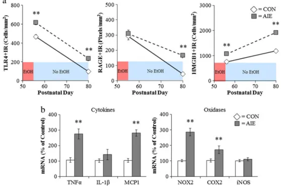
findings of in vivo chronic ethanol exposure modeling binge drinking that involve cycles of ethanol exposure and absti-nence that cause progressive induction of HMGB1-TLR-RAGE expression and sensitization to neuroimmune activa-tion. Indeed, models of moderate drinking find that multiple cycles cause transient neuroimmune induction in the brain (Whitman et al.2013), whereas binge drinking models that utilize doses consistent with binge levels of ethanol (e.g., 5.0 g/kg, i.g.) induce persistent increases in the brain neuroimmune genes and proteins including HMGB1, TLR, and RAGE in adolescent rats (Vetreno and Crews 2012; Vetreno et al.2013) and adult mice (Qin and Crews2014; Qin et al.2007,2008). Similarly, studies of post-mortem hu-man brain find that expression levels of HMGB1 and TLR in orbital frontal cortex (OFC) correlate with lifetime alcohol consumption across normal and alcoholic humans (Crews et al.2013). This interesting correlation could only occur if ethanol induction of HMGB1-TLR receptors was persistent and cumulative with binge drinking episodes. Interestingly, treatment of adolescent rats modeling underage binge drink-ing induces HMGB1, TLR4, and RAGE that continue to un-dergo developmental changes in the brain (Fig.6). HMGB1,
TLR, and RAGE induction persists into abstinence, with TLR4 and RAGE showing developmental decreases in the frontal cortex and HMGB1 showing a developmental in-crease. Multiple other neuroimmune genes remain induced in adulthood (Fig.6). Risk of alcohol dependence increases with a younger age on drinking onset (Fig. 7), and post-mortem prefrontal cortex expression also correlates with age of drinking onset (Fig.7) (Vetreno et al.2013). Together, these studies suggest that the persistent increase in brain HMGB1-TLR4 and neuroimmune signaling contribute to the chronic relapsing nature of alcoholism and the slow progressive de-generation found in alcoholism (Crews and Nixon2009).
While induction of the innate immune system through NF-κB signaling cascades has been implicated in alcohol-induced neurodegeneration, recent evidence suggests that a shift in neurotrophic/innate immune signaling might also be involved. Cyclic AMP-responsive element binding protein (CREB) and many of its target genes, including neuropeptide Y (NPY) and brain-derived neurotrophic factor (BDNF), pro-mote neuronal survival and protect neurons from excitotoxicity and apoptosis (Lonze and Ginty2002). Levels of CREB-DNA binding, phosphorylated CREB, and BDNF
Fig. 6 Adolescent intermittent ethanol (AIE) treatment leads to a persistent induction of neuroimmune genes in the adult brain.aMale Wistar rats were treated with ethanol (5 g/kg/day, i.g.,w/v, 2 days on/ 2 days off) or comparable volumes of water from postnatal day (P)25 to P55. Brain tissue was collected either 24 h (P56) or 25 days after the last ethanol treatment (P80) to assess the persistent expression of neuroimmune markers. Toll-like receptor 4 (TLR4) immunoreactivity was upregulated 24 h after ethanol treatment and remained elevated for 25 days following the conclusion of ethanol treatment. In contrast, there was no change in receptor for advanced glycation end-product (RAGE) expression immediately following the conclusion of ethanol treatment but was elevated 25 days after the conclusion of ethanol treatment.
are decreased in the rat frontal cortex following a 24-h with-drawal from chronic ethanol exposure (Pandey et al.1999,
2001). Further, cortical levels of NPY are reduced by ethanol treatment, which was accompanied by a reduction of
p h o s p h o r y l a t e d C R E B ( B i s o n a n d C r e w s 2 0 0 3) . Interestingly, our laboratory found that ethanol dose-dependently reduces CREB-DNA binding while simulta-neously increasing NF-κB-DNA binding in hippocampal-entorhinal cortex slice culture (Zou and Crews 2006). However, following a prolonged period of abstinence, CREB level has been shown to rebound (Bison and Crews
2003), which could contribute to the recovery of white matter volume and cognitive function associated with protracted ab-stinence (Pfefferbaum et al.1995; Sullivan et al.2000a,b).
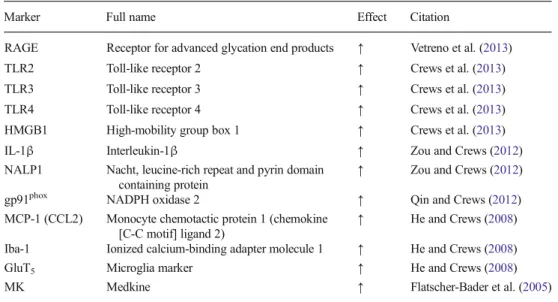
Although this review highlights HMGB1-TLR4 signaling, there are multiple other proinflammatory genes that are in-creased, and we have found many in the post-mortem human alcoholic brain (see Table1). Initial studies found increased markers of microglia and the proinflammatory cytokine MCP1 (CCL2) post-mortem alcoholic ventral tegmental area, amygdala, nucleus accumbens, and hippocampus (He and Crews2008). However, populations of activated microglia in other brain regions remain to be determined in the human post-mortem alcoholic brain. In studies focused on the PFC, we found that post-mortem alcoholic brain has increased levels of HMGB1 as well as TLR2, TLR3, and TLR4 recep-tors (Crews et al.2013) and RAGE (Vetreno et al. 2013). Similarly, NADPH oxidase is increased in alcoholic PFC, the brain region most insulted in alcoholics (Crews and Nixon 2009; Qin and Crews 2012). In other studies focused on hippocampal neurogenesis, we found in-creased IL-1βinflammasome markers in the hippocampus of post-mortem alcoholic brain (Zou and Crews 2012). These studies indicate that multiple neuroimmune genes are upregulated in the human alcoholic brain and likely contribute to neurodegeneration and the neurobiology of alcoholism. These findings further support the role of neuroimmune signaling in human alcoholism and alcohol-ic neurodegeneration.
Fig. 7 Risk of alcoholism and induction of innate immune genes correlate with age of drinking onset in humans.aToll-like receptor 4 (TLR4) and high-mobility group box 1 (HMGB1) expression in the post-mortem human brain is negatively correlated with age of drinking onset adapted from Vetreno et al. (2013).bAn earlier age of drinking onset is predictive of an increased likelihood of developing an alcohol use disorder during an individual’s lifetime. Adapted from Grant (1998)
Table 1 Neuroimmune markers are increased in post-mortem human alcoholic brain
Marker Full name Effect Citation
RAGE Receptor for advanced glycation end products ↑ Vetreno et al. (2013)
TLR2 Toll-like receptor 2 ↑ Crews et al. (2013) TLR3 Toll-like receptor 3 ↑ Crews et al. (2013)
TLR4 Toll-like receptor 4 ↑ Crews et al. (2013) HMGB1 High-mobility group box 1 ↑ Crews et al. (2013)
IL-1β Interleukin-1β ↑ Zou and Crews (2012)
NALP1 Nacht, leucine-rich repeat and pyrin domain containing protein
↑ Zou and Crews (2012)
gp91phox NADPH oxidase 2 ↑ Qin and Crews (2012) MCP-1 (CCL2) Monocyte chemotactic protein 1 (chemokine
[C-C motif] ligand 2)
↑ He and Crews (2008)
Iba-1 Ionized calcium-binding adapter molecule 1 ↑ He and Crews (2008)
GluT5 Microglia marker ↑ He and Crews (2008)
Neuroimmune signaling, hyperexcitability, and neuronal death
Excitotoxicity is associated with alcoholic neurodegeneration and HMGB1-TLR4 signaling. Chronic ethanol treatment of neurons leads to increased sensitivity to excitotoxicity (Chandler et al. 1994). Ethanol potentiates glutamate excitotoxicity in brain slice cultures due to blockade of glial transporters (Zou and Crews2010). However, in neuronal primary cultures, ethanol blocks N-methyl-D-aspartate
(NMDA) excitotoxicity consistent with many studies finding ethanol inhibition of NMDA receptors (Chandler et al.1998). Similar to acute ethanol blocking TLR4 receptor activation when present, ethanol blocks NMDA receptors when present (Chandler et al.1998). Although ethanol can block NMDA responses, glutamate excitotoxicity is increased by ethanol and TNFαin brain slice cultures due in part to glial loss of glutamate uptake (Zou and Crews2005) and perhaps release by ethanol. Further, HMGB1-TLR4 signaling has been shown to activate kinase cascades that lead to phosphorylation of the NR2B subunit of NMDA receptors causing the migration of more NMDA receptors to the synapse that increase synaptic NMDA receptors, neuronal excitability, and excitotoxicity (Balosso et al. 2014; Maroso et al. 2010). Both HMGB1-TLR4 signaling (Balosso et al.2014) and IL1β-IL1R signal-ing (Viviani et al.2003) have been shown to increase NMDA receptor-mediated calcium flux, neuronal excitability, and excitotoxicity through activation of kinase cascades. IL1β -IL1R activation of the Src kinase has been found to increase NMDA calcium flux, excitability, and excitotoxicity. Many studies have found that tyrosine-kinase activation can increase excitability through increases in NR2B-NMDA receptor phosphorylation. Dorit Ron’s group has found that ethanol increases NMDA excitability in hippocampus through kinase activation that alters receptor trafficking leading to increased NR2B-NMDA receptors and increased excitability (Suvarna et al.2005). Another mechanism of chronic ethanol-induced hyperexcitability is neuroimmune inhibition of glial glutamate transporters (Zou and Crews2005). Ethanol releases HMGB1 creating hyperexcitability that disrupts synaptic plasticity and sensitizes to excitotoxicity. HMGB1 is massively released during brain damage activating persistent neuroimmune gene induction (Kim et al.2006). Indeed, Maroso et al. (2010) found increased release of HMGB1 with hippocampal excit-ability that caused seizures leading to persistent increases in HMGB1 and excitability. Ethanol has a modest cumulative effect that, with repeated chronic exposure, increases excit-ability and excitotoxicity due to increased neuroimmune sig-naling (see Fig.4). Thus, the global neurodegeneration with the most severe losses in frontal cortex found in alcoholism is secondary to the persistent and progressive neuroimmune ac-tivation that occurs during alcoholism, which is a chronic relapsing disorder.
Summary
Alcoholism is associated with increased neuroimmune gene expression in the brain. Neuroimmune gene induction appears to occur through two processes, systemic induction of innate immune genes resulting from alcohol-induced increases in gut permeability that result in increased blood cytokines that acti-vate brain neuroimmune genes through multiple mechanisms including transport from blood into the brain. These signals activate neurons and glia through complex signaling that in-cludes amplification through convergence of signaling through NF-κB and AP-1 pathways leading to the induction of proinflammatory cytokines, TLR receptors, RAGE, NADP H oxidase, and other oxidases. Ethanol also induces HMGB1 that contributes to positive loops of amplification of neuroimmune genes through TLR receptors and RAGE. Persistent activation of these pathways leads to a hyperexcit-able state that disrupts neuronal networks contributing to al-coholic psychopathology as well as neurodegeneration. Agents that block ethanol neuroimmune activation may be able to prevent alcoholism and alcoholic neurodegeneration.
Acknowledgments This work was supported by grants from the Na-tional Institutes on Alcohol Abuse and Alcoholism (AA019767, AA11605, and AA007573) and the Neurobiology of Adolescent Drink-ing in Adulthood (NADIA [AAA020023, AA020024, and AA020022]). The authors wish to acknowledge support from The Bowles Center of Alcohol Studies, School of Medicine, The University of North Carolina at Chapel Hill, the National Institute of Health, and the National Institute on Alcoholism and Alcohol Abuse through AA020023, AA020022, AA019767, AA11605, and AA007573.
Conflict of interest The authors have declared that no competing in-terests exist.
Open Access This article is distributed under the terms of the Creative Commons Attribution License which permits any use, distribution, and reproduction in any medium, provided the original author(s) and the source are credited.
References
Alfonso-Loeches S, Guerri C (2011) Molecular and behavioral aspects of the actions of alcohol on the adult and developing brain. Crit Rev Clin Lab Sci 48:19–47
Alfonso-Loeches S, Pascual-Lucas M, Blanco AM, Sanchez-Vera I, Guerri C (2010) Pivotal role of TLR4 receptors in alcohol-induced neuroinflammation and brain damage. J Neurosci Off J Soc Neurosci 30:8285–95
Alfonso-Loeches S, Pascual M, Gomez-Pinedo U, Pascual-Lucas M, Renau-Piqueras J, Guerri C (2012) Toll-like receptor 4 participates in the myelin disruptions associated with chronic alcohol abuse. Glia 60:948–64
Bala S, Marcos M, Gattu A, Catalano D, Szabo G (2014) Acute binge drinking increases serum endotoxin and bacterial DNA levels in healthy individuals. PLoS One 9:e96864
Balosso S, Liu J, Bianchi ME, Vezzani A (2014) Disulfide-containing high mobility group box-1 promotes N-methyl-d-aspartate receptor function and excitotoxicity by activating toll-like receptor 4-dependent signaling in hippocampal neurons. Antioxid Redox Signal 21:1726–40
Banks WA, Erickson MA (2010) The blood–brain barrier and immune function and dysfunction. Neurobiol Dis 37:26–32
Bell RL, Lopez MF, Cui C, Egli M, Johnson KW, Franklin KM, Becker HC (2013) Ibudilast reduces alcohol drinking in multiple animal models of alcohol dependence. Addict Biol
Beynon SB, Walker FR (2012) Microglial activation in the injured and healthy brain: what are we really talking about? Practical and theo-retical issues associated with the measurement of changes in microglial morphology. Neuroscience 225:162–71
Bison S, Crews F (2003) Alcohol withdrawal increases neuropeptide Y immunoreactivity in rat brain. Alcohol Clin Exp Res 27: 1173–83
Blanco AM, Guerri C (2007) Ethanol intake enhances inflammatory me-diators in brain: role of glial cells and TLR4/IL-1RI receptors. Front Biosci: J Virtual Libr 12:2616–30
Blanco AM, Pascual M, Valles SL, Guerri C (2004) Ethanol-induced iNOS and COX-2 expression in cultured astrocytes via NF-kappa B. Neuroreport 15:681–5
Blanco AM, Valles SL, Pascual M, Guerri C (2005) Involvement of TLR4/type I IL-1 receptor signaling in the induction of inflamma-tory mediators and cell death induced by ethanol in cultured astro-cytes. J Immunol 175:6893–9
Blanco AM, Perez-Arago A, Fernandez-Lizarbe S, Guerri C (2008) Ethanol mimics ligand-mediated activation and endocytosis of IL-1RI/TLR4 receptors via lipid rafts caveolae in astroglial cells. J Neurochem 106:625–39
Blednov YA, Bergeson SE, Walker D, Ferreira VM, Kuziel WA, Harris RA (2005) Perturbation of chemokine networks by gene deletion alters the reinforcing actions of ethanol. Behav Brain Res 165:110– 25
Blednov YA, Benavidez JM, Geil C, Perra S, Morikawa H, Harris RA (2011) Activation of inflammatory signaling by lipopolysaccharide produces a prolonged increase of voluntary alcohol intake in mice. Brain Behav Immun 25(Suppl 1):S92–S105
Blednov YA, Ponomarev I, Geil C, Bergeson S, Koob GF, Harris RA (2012) Neuroimmune regulation of alcohol consumption: behavior-al vbehavior-alidation of genes obtained from genomic studies. Addict Biol 17:108–20
Boyadjieva NI, Sarkar DK (2013a) Cyclic adenosine monophosphate and brain-derived neurotrophic factor decreased oxidative stress and ap-optosis in developing hypothalamic neuronal cells: role of microg-lia. Alcohol Clin Exp Res 37:1370–9
Boyadjieva NI, Sarkar DK (2013b) Microglia play a role in ethanol-induced oxidative stress and apoptosis in developing hypothalamic neurons. Alcohol Clin Exp Res 37:252–62
Chandler LJ, Guzman NJ, Sumners C, Crews FT (1994) Magnesium and zinc potentiate ethanol inhibition of N-methyl-D-aspartate-stimulated nitric oxide synthase in cortical neurons. J Pharmacol Exp Ther 271:67–75
Chandler LJ, Harris RA, Crews FT (1998) Ethanol tolerance and synaptic plasticity. Trends Pharmacol Sci 19:491–5
Chavan SS, Huerta PT, Robbiati S, Valdes-Ferrer SI, Ochani M, Dancho M, Frankfurt M, Volpe BT, Tracey KJ, Diamond B (2012) HMGB1 mediates cognitive impairment in sepsis survivors. Mol Med 18: 930–7
Chen CP, Kuhn P, Chaturvedi K, Boyadjieva N, Sarkar DK (2006) Ethanol induces apoptotic death of developing beta-endorphin neu-rons via suppression of cyclic adenosine monophosphate production
and activation of transforming growth factor-beta1-linked apoptotic signaling. Mol Pharmacol 69:706–17
Colton CA (2009) Heterogeneity of microglial activation in the innate immune response in the brain. J NeuroImmune Pharmacol: Off J Soc NeuroImmune Pharmacol 4:399–418
Crews FT (2012) Immune function genes, genetics, and the neurobiology of addiction. Alcohol Res: Curr Rev 34:355–61
Crews FT, Nixon K (2009) Mechanisms of neurodegeneration and regen-eration in alcoholism. Alcohol Alcohol 44:115–27
Crews FT, Vetreno RP (2014) Neuroimmune basis of alcoholic brain damage. Int Rev Neurobiol 118:315–57
Crews FT, Bechara R, Brown LA, Guidot DM, Mandrekar P, Oak S, Qin L, Szabo G, Wheeler M, Zou J (2006) Cytokines and alcohol. Alcohol Clin Exp Res 30:720–30
Crews FT, Zou J, Qin L (2011) Induction of innate immune genes in brain create the neurobiology of addiction. Brain Behav Immun 25(Suppl 1):S4–S12
Crews FT, Qin L, Sheedy D, Vetreno RP, Zou J (2013) High mobility group box 1/Toll-like receptor danger signaling increases brain neuroimmune activation in alcohol dependence. Biol Psychiatry 73:602–12
Dantzer R, O’Connor JC, Freund GG, Johnson RW, Kelley KW (2008) From inflammation to sickness and depression: when the immune system subjugates the brain. Nat Rev Neurosci 9:46–56
Fernandez-Lizarbe S, Pascual M, Guerri C (2009) Critical role of TLR4 response in the activation of microglia induced by ethanol. J Immunol 183:4733–44
Fernandez-Lizarbe S, Montesinos J, Guerri C (2013) Ethanol induces TLR4/TLR2 association, triggering an inflammatory response in microglial cells. J Neurochem 126:261–73
Ferrari D, Chiozzi P, Falzoni S, Dal Susino M, Collo G, Buell G, Di Virgilio F (1997) ATP-mediated cytotoxicity in microglial cells. Neuropharmacology 36:1295–301
Ferrier L, Berard F, Debrauwer L, Chabo C, Langella P, Bueno L, Fioramonti J (2006) Impairment of the intestinal barrier by ethanol involves enteric microflora and mast cell activation in rodents. Am J Pathol 168:1148–54
Flatscher-Bader T, van der Brug M, Hwang JW, Gochee PA, Matsumoto I, Niwa S, Wilce PA (2005) Alcohol-responsive genes in the frontal cortex and nucleus accumbens of human alcoholics. J Neurochem 93:359–70
Gage FH (2002) Neurogenesis in the adult brain. J Neurosci 22:612–3 Gao B, Seki E, Brenner DA, Friedman S, Cohen JI, Nagy L, Szabo G,
Zakhari S (2011) Innate immunity in alcoholic liver disease. Am J Physiol Gastrointest Liver Physiol 300:G516–25
Ge X, Antoine DJ, Lu Y, Arriazu E, Leung TM, Klepper AL, Branch AD, Fiel MI, Nieto N (2014) High mobility group box-1 (HMGB1) participates in the pathogenesis of alcoholic liver disease (ALD). J Biol Chem 289:22672–91
Ghosh S, Hayden MS (2008) New regulators of NF-kappaB in inflam-mation. Nat Rev Immunol 8:837–48
Ginhoux F, Greter M, Leboeuf M, Nandi S, See P, Gokhan S, Mehler MF, Conway SJ, Ng LG, Stanley ER, Samokhvalov IM, Merad M (2010) Fate mapping analysis reveals that adult microglia derive from primitive macrophages. Science 330:841–5
Glass CK, Saijo K, Winner B, Marchetto MC, Gage FH (2010) Mechanisms underlying inflammation in neurodegeneration. Cell 140:918–34
Graeber MB (2010) Changing face of microglia. Science 330:783–8 Grant BF (1998) The impact of a family history of alcoholism on the
relationship between age at onset of alcohol use and DSM-IV alco-hol dependence: results from the National Longitudinal Alcoalco-hol Epidemiologic Survey. Alcohol Health Res World 22:144–7 Guadagno J, Xu X, Karajgikar M, Brown A, Cregan SP (2013)
cells via transcriptional activation of the Bcl-2 family member Puma. Cell Death Dis 4:e538
Guerri C, Pascual M (2010) Mechanisms involved in the neurotoxic, cognitive, and neurobehavioral effects of alcohol consumption dur-ing adolescence. Alcohol 44:15–26
Guerri C, Montoliu C, Renau-Piqueras J (1994) Involvement of free radical mechanism in the toxic effects of alcohol: implications for fetal alcohol syndrome. Adv Exp Med Biol 366:291–305 Harrison NA, Brydon L, Walker C, Gray MA, Steptoe A, Critchley HD
(2009a) Inflammation causes mood changes through alterations in subgenual cingulate activity and mesolimbic connectivity. Biol Psychiatry 66:407–14
Harrison NA, Brydon L, Walker C, Gray MA, Steptoe A, Dolan RJ, Critchley HD (2009b) Neural origins of human sickness in intero-ceptive responses to inflammation. Biol Psychiatry 66:415–22 He J, Crews FT (2008) Increased MCP-1 and microglia in various regions
of the human alcoholic brain. Exp Neurol 210:349–58
Hide I, Tanaka M, Inoue A, Nakajima K, Kohsaka S, Inoue K, Nakata Y (2000) Extracellular ATP triggers tumor necrosis factor-alpha re-lease from rat microglia. J Neurochem 75:965–72
Hoffmann A, Natoli G, Ghosh G (2006) Transcriptional regulation via the NF-kappaB signaling module. Oncogene 25:6706–16
Huh GS, Boulanger LM, Du H, Riquelme PA, Brotz TM, Shatz CJ (2000) Functional requirement for class I MHC in CNS development and plasticity. Science 290:2155–9
Huttunen HJ, Rauvala H (2004) Amphoterin as an extracellular regulator of cell motility: from discovery to disease. J Intern Med 255:351–66 Kettenmann H, Kirchhoff F, Verkhratsky A (2013) Microglia: new roles
for the synaptic stripper. Neuron 77:10–8
Kim JB, Sig Choi J, Yu YM, Nam K, Piao CS, Kim SW, Lee MH, Han PL, Park JS, Lee JK (2006) HMGB1, a novel cytokine-like mediator linking acute neuronal death and delayed neuroinflammation in the postischemic brain. J Neurosci Off J Soc Neurosci 26:6413–21 Kreutzberg GW (1996) Microglia: a sensor for pathological events in the
CNS. Trends Neurosci 19:312–8
Kuhn P, Sarkar DK (2008) Ethanol induces apoptotic death of beta-endorphin neurons in the rat hypothalamus by a TGF-beta 1-depen-dent mechanism. Alcohol Clin Exp Res 32:706–14
Leclercq S, Cani PD, Neyrinck AM, Starkel P, Jamar F, Mikolajczak M, Delzenne NM, de Timary P (2012) Role of intestinal permeability and inflammation in the biological and behavioral control of alcohol-dependent subjects. Brain Behav Immun 26:911–8 Li Q, Verma IM (2002) NF-kappaB regulation in the immune system. Nat
Rev Immunol 2:725–34
Lister MF, Sharkey J, Sawatzky DA, Hodgkiss JP, Davidson DJ, Rossi AG, Finlayson K (2007) The role of the purinergic P2X7 receptor in inflammation. J Inflamm 4:5
Liu Y, Qin L, Wilson B, Wu X, Qian L, Granholm AC, Crews FT, Hong JS (2008) Endotoxin induces a delayed loss of TH-IR neurons in substantia nigra and motor behavioral deficits. Neurotoxicology 29: 864–70
Liu J, Yang AR, Kelly T, Puche A, Esoga C, June HL Jr, Elnabawi A, Merchenthaler I, Sieghart W, June HL Sr, Aurelian L (2011) Binge alcohol drinking is associated with GABAA alpha2-regulated Toll-like receptor 4 (TLR4) expression in the central amygdala. Proc Natl Acad Sci U S A 108:4465–70
Lonze BE, Ginty DD (2002) Function and regulation of CREB family transcription factors in the nervous system. Neuron 35:605–23 Marcos M, Pastor I, Gonzalez-Sarmiento R, Laso FJ (2008)
Interleukin-10 gene polymorphism is associated with alcoholism but not with alcoholic liver disease. Alcohol Alcohol 43:523–8
Maroso M, Balosso S, Ravizza T, Liu J, Aronica E, Iyer AM, Rossetti C, Molteni M, Casalgrandi M, Manfredi AA, Bianchi ME, Vezzani A (2010) Toll-like receptor 4 and high-mobility group box-1 are in-volved in ictogenesis and can be targeted to reduce seizures. Nat Med 16:413–9
Maroso M, Balosso S, Ravizza T, Liu J, Bianchi ME, Vezzani A (2011) Interleukin-1 type 1 receptor/Toll-like receptor signalling in epilep-sy: the importance of IL-1beta and high-mobility group box 1. J Intern Med 270:319–26
Marshall SA, McClain JA, Kelso ML, Hopkins DM, Pauly JR, Nixon K (2013) Microglial activation is not equivalent to neuroinflammation in alcohol-induced neurodegeneration: the importance of microglia phenotype. Neurobiol Dis 54:239–51
Mayfield J, Ferguson L, Harris RA (2013) Neuroimmune signaling: a key component of alcohol abuse. Curr. Opin. Neurobiol
Montoliu C, Sancho-Tello M, Azorin I, Burgal M, Valles S, Renau-Piqueras J, Guerri C (1995) Ethanol increases cytochrome P4502E1 and induces oxidative stress in astrocytes. J Neurochem 65:2561–70
Mulligan MK, Ponomarev I, Hitzemann RJ, Belknap JK, Tabakoff B, Harris RA, Crabbe JC, Blednov YA, Grahame NJ, Phillips TJ, Finn DA, Hoffman PL, Iyer VR, Koob GF, Bergeson SE (2006) Toward understanding the genetics of alcohol drinking through tran-scriptome meta-analysis. Proc Natl Acad Sci U S A 103:6368–73 Okun E, Griffioen KJ, Lathia JD, Tang SC, Mattson MP, Arumugam TV
(2009) Toll-like receptors in neurodegeneration. Brain Res Rev 59: 278–92
Okvist A, Johansson S, Kuzmin A, Bazov I, Merino-Martinez R, Ponomarev I, Mayfield RD, Harris RA, Sheedy D, Garrick T, Harper C, Hurd YL, Terenius L, Ekstrom TJ, Bakalkin G, Yakovleva T (2007) Neuroadaptations in human chronic alcoholics: dysregulation of the NF-kappaB system. PLoS One 2:e930 Osterholm MT (2005) Preparing for the next pandemic. N Engl J Med
352:1839–42
Osterndorff-Kahanek E, Ponomarev I, Blednov YA, Harris RA (2013) Gene expression in brain and liver produced by three different reg-imens of alcohol consumption in mice: comparison with immune activation. PLoS One 8:e59870
Pandey SC, Zhang D, Mittal N, Nayyar D (1999) Potential role of the gene transcription factor cyclic AMP-responsive element binding protein in ethanol withdrawal-related anxiety. J Pharmacol Exp Ther 288:866–78
Pandey SC, Roy A, Mittal N (2001) Effects of chronic ethanol intake and its withdrawal on the expression and phosphorylation of the creb gene transcription factor in rat cortex. J Pharmacol Exp Ther 296: 857–68
Parvathenani LK, Tertyshnikova S, Greco CR, Roberts SB, Robertson B, Posmantur R (2003) P2X7 mediates superoxide production in pri-mary microglia and is up-regulated in a transgenic mouse model of Alzheimer’s disease. J Biol Chem 278:13309–17
Pascual M, Boix J, Felipo V, Guerri C (2009) Repeated alcohol adminis-tration during adolescence causes changes in the mesolimbic dopa-minergic and glutamatergic systems and promotes alcohol intake in the adult rat. J Neurochem 108:920–31
Pascual M, Balino P, Alfonso-Loeches S, Aragon CM, Guerri C (2011a) Impact of TLR4 on behavioral and cognitive dysfunctions associat-ed with alcohol-inducassociat-ed neuroinflammatory damage. Brain Behav Immun 25(Suppl 1):S80–91
Pascual M, Fernandez-Lizarbe S, Guerri C (2011b) Role of TLR4 in ethanol effects on innate and adaptive immune responses in perito-neal macrophages. Immunol Cell Biol 89:716–27
Pastor IJ, Laso FJ, Romero A, Gonzalez-Sarmiento R (2005) Interleukin-1 gene cluster polymorphisms and alcoholism in Spanish men. Alcohol Alcohol 40:181–6
Perkins ND (2007) Integrating cell-signalling pathways with NF-kappaB and IKK function. Nat Rev Mol Cell Biol 8:49–62
Pruett SB, Zheng Q, Fan R, Matthews K, Schwab C (2004) Ethanol suppresses cytokine responses induced through Toll-like receptors as well as innate resistance to Escherichia coli in a mouse model for binge drinking. Alcohol 33:147–55
Qin L, Crews FT (2012) NADPH oxidase and reactive oxygen species contribute to alcohol-induced microglial activation and neurodegen-eration. J Neuroinflammation 9:5
Qin L, Crews FT (2014) Focal thalamic degeneration from ethanol and thiamine deficiency is associated with neuroimmune gene induction, microglial activation, and lack of monocarboxylic acid transporters. Alcohol Clin Exp Res 38:657–71
Qin L, Wu X, Block ML, Liu Y, Breese GR, Hong JS, Knapp DJ, Crews FT (2007) Systemic LPS causes chronic neuroinflammation and progressive neurodegeneration. Glia 55:453–62
Qin L, He J, Hanes RN, Pluzarev O, Hong JS, Crews FT (2008) Increased systemic and brain cytokine production and neuroinflammation by endotoxin following ethanol treatment. J Neuroinflammation 5:10 Qin L, Liu Y, Hong JS, Crews FT (2013) NADPH oxidase and aging
drive microglial activation, oxidative stress, and dopaminergic neu-rodegeneration following systemic LPS administration. Glia 61: 855–68
Raivich G, Bohatschek M, Kloss CU, Werner A, Jones LL, Kreutzberg GW (1999) Neuroglial activation repertoire in the injured brain: graded response, molecular mechanisms and cues to physiological function. Brain Res Brain Res Rev 30:77–105
Raouf R, Chabot-Dore AJ, Ase AR, Blais D, Seguela P (2007) Differential regulation of microglial P2X4 and P2X7 ATP receptors following LPS-induced activation. Neuropharmacology 53:496– 504
Schwarz JM, Bilbo SD (2013) Adolescent morphine exposure affects long-term microglial function and later-life relapse liability in a model of addiction. J Neurosci Off J Soc Neurosci 33:961–71 Sims GP, Rowe DC, Rietdijk ST, Herbst R, Coyle AJ (2010) HMGB1
and RAGE in inflammation and cancer. Annu Rev Immunol 28: 367–88
Sugama S, Takenouchi T, Fujita M, Conti B, Hashimoto M (2009) Differential microglial activation between acute stress and lipopoly-saccharide treatment. J Neuroimmunol 207:24–31
Sullivan EV, Rosenbloom MJ, Lim KO, Pfefferbaum A (2000a) Longitudinal changes in cognition, gait, and balance in abstinent and relapsed alcoholic men: relationships to changes in brain struc-ture. Neuropsychology 14:178–88
Sullivan EV, Rosenbloom MJ, Pfefferbaum A (2000b) Pattern of motor and cognitive deficits in detoxified alcoholic men. Alcohol Clin Exp Res 24:611–21
Suvarna N, Borgland SL, Wang J, Phamluong K, Auberson YP, Bonci A, Ron D (2005) Ethanol alters trafficking and functional N-methyl-D-aspartate receptor NR2 subunit ratio via H-Ras. J Biol Chem 280: 31450–9
Szabo G, Mandrekar P (2009) A recent perspective on alcohol, immunity, and host defense. Alcohol Clin Exp Res 33:220–32
Szabo G, Mandrekar P, Catalano D (1995) Inhibition of superantigen-induced T cell proliferation and monocyte IL-1 beta, TNF-alpha, and IL-6 pro-duction by acute ethanol treatment. J Leukoc Biol 58:342–50 Takeuchi H (2010) Neurotoxicity by microglia: mechanisms and
poten-tial therapeutic strategy. Clin Exp Neuroimmunol 1:12–21 Tang D, Billiar TR, Lotze MT (2012) A Janus tale of two active high
mobility group box 1 (HMGB1) redox states. Mol Med 18:1360–2 Valles SL, Blanco AM, Pascual M, Guerri C (2004) Chronic ethanol treatment enhances inflammatory mediators and cell death in the brain and in astrocytes. Brain Pathol 14:365–71
Venereau E, Casalgrandi M, Schiraldi M, Antoine DJ, Cattaneo A, De Marchis F, Liu J, Antonelli A, Preti A, Raeli L, Shams SS, Yang H, Varani L, Andersson U, Tracey KJ, Bachi A, Uguccioni M, Bianchi ME (2012) Mutually exclusive redox forms of HMGB1 promote cell recruitment or proinflammatory cytokine release. J Exp Med 209:1519–28
Vetreno RP, Crews FT (2012) Adolescent binge drinking increases ex-pression of the danger signal receptor agonist HMGB1 and toll-like receptors in the adult prefrontal cortex. Neuroscience 226:475–88 Vetreno RP, Crews FT (2014) Current hypotheses on the mechanisms of
alcoholism. Handb Clin Neurol 125:477–97
Vetreno RP, Qin L, Crews FT (2013) Increased receptor for advanced glycation end product expression in the human alcoholic prefrontal cortex is linked to adolescent drinking. Neurobiol Dis 59:52–62 Vetreno RP, Broadwater M, Liu W, Spear LP, Crews FT (2014)
Adolescent, but not adult, binge ethanol exposure leads to persistent global reductions of choline acetyltransferase expressing neurons in brain. PLoS One 9:e113421
Viviani B, Bartesaghi S, Gardoni F, Vezzani A, Behrens MM, Bartfai T, Binaglia M, Corsini E, Di Luca M, Galli CL, Marinovich M (2003) Interleukin-1beta enhances NMDA receptor-mediated intracellular calcium increase through activation of the Src family of kinases. J Neurosci Off J Soc Neurosci 23:8692–700
Wahamaa H, Schierbeck H, Hreggvidsdottir HS, Palmblad K, Aveberger AC, Andersson U, Harris HE (2011) High mobility group box pro-tein 1 in complex with lipopolysaccharide or IL-1 promotes an in-creased inflammatory phenotype in synovial fibroblasts. Arthritis Res Ther 13:R136
Wang H, Yang H, Czura CJ, Sama AE, Tracey KJ (2001) HMGB1 as a late mediator of lethal systemic inflammation. Am J Respir Crit Care Med 164:1768–73
Watkins LR, Maier SF, Goehler LE (1995) Cytokine-to-brain communi-cation: a review & analysis of alternative mechanisms. Life Sci 57: 1011–26
Whitman BA, Knapp DJ, Werner DF, Crews FT, Breese GR (2013) The cytokine mRNA increase induced by withdrawal from chronic eth-anol in the sterile environment of brain is mediated by CRF and HMGB1 release. Alcohol Clin Exp Res 37:2086–97
Zhao YN, Wang F, Fan YX, Ping GF, Yang JY, Wu CF (2013) Activated microglia are implicated in cognitive deficits, neuronal death, and successful recovery following intermittent ethanol exposure. Behav Brain Res 236:270–82
Zou JY, Crews FT (2005) TNF alpha potentiates glutamate neurotoxicity by inhibiting glutamate uptake in organotypic brain slice cultures: neuroprotection by NF kappa B inhibition. Brain Res 1034:11–24 Zou J, Crews F (2006) CREB and NF-kappaB transcription factors
reg-ulate sensitivity to excitotoxic and oxidative stress induced neuronal cell death. Cell Mol Neurobiol 26:385–405
Zou J, Crews F (2010) Induction of innate immune gene expression cascades in brain slice cultures by ethanol: key role of NF-kappaB and proinflammatory cytokines. Alcohol Clin Exp Res 34:777–89 Zou J, Crews FT (2012) Inflammasome-IL-1beta signaling mediates
eth-anol inhibition of hippocampal neurogenesis. Front Neurosci 6:77 Zou JY, Crews FT (2014) Release of neuronal HMGB1 by ethanol
through decreased HDAC activity activates brain neuroimmune sig-naling. PLoS One 9:e87915