STRUCTURAL AND BIOCHEMICAL ANALYSIS OF DYNEIN LIGHT CHAIN-MEDIATED HOMODIMERIZATION OF CYTOSKELETAL AND NUCLEAR PORE
PROTEINS
Erin MacKenzie Romes
A dissertation submitted to the faculty of the University of North Carolina at Chapel Hill in partial fulfillment of the requirements for the degree of Doctor of Philosophy in the Department of Biochemistry and Biophysics.
Chapel Hill 2012
ABSTRACT
ERIN M. ROMES: Structural and biochemical analysis of dynein light chain-mediated homodimerization of cytoskeletal and nuclear pore proteins
(Under the direction of Dr. Kevin C. Slep)
The dynein light chain, Lc8/Dyn2, is a ubiquitous protein that acts as a scaffold, binding to many different target proteins in various cellular contexts. Here we describe S. cerevisiae Dyn2’s biophysical and structural interaction with the dynein intermediate chain,
Pac11 at the dynein complex, and also Dyn2’s interaction with a nuclear pore protein, Nup159 in the cytoplasmic fibrils. We also demonstrate the structural and binding similarities between the Drosophila homolog, Lc8 binding to a centriole duplication protein, Ana2 and Dyn2’s interaction with Pac11 or Nup159. We obtained the first high-resolution crystal structure of Dyn2 bound to Nup159 peptides and subsequent structures of a homodimer of Dyn2 bound to two identical peptides of Pac11, and a homodimer of Lc8 bound to two identical Ana2 peptides. We also characterized the thermodynamic binding profiles of Dyn2/Lc8 interacting with Pac11, Ana2, or Nup159 peptide binding sites and discovered that both Dyn2 and Lc8 are capable of two modes of binding peptides, endothermically or exothermically with KDs in the range of 0.5 to 20 µM.
ACKNOWLEDGEMENTS
First of all, I would like to thank my advisor, Dr. Kevin Slep for mentoring me through this huge stage of growth in my career. He helped me learn to find vision in a project and not to get too discouraged when that project was not proceeding according to that vision. I would also like to thank my committee members: Dr. Richard Cheney, Dr. Nikolay Dokholyan, Dr. Hengming Ke, Dr. Matt Redinbo, and Dr. Steve Rogers for guiding me through the essential experiments that resulted in published manuscripts. Finally, thank you to Michael Miley in the Crystallography Core at UNC for the technical assistance and advice. I would like to thank the Slep lab members for technical assistance and well-mannered frivolity. I would like to thank Lauren Slevin for mentoring me through the new field of centrioles, technical support, and for her assistance with editing this dissertation. I would especially like to thank Jaime Campbell for helping me establish the Slep lab and for hours of moral support over the years.
Lastly, I would like to thank my family; none of this would be possible without them. I would like to apologize for hours of lost conversation and quality time due to other lab priorities. I am so fortunate to have Dennis, Linda, and Jonathan Toth to cheer me on from Michigan. The person who deserves most of the credit is my husband, Drew. My life would not be the same without his shoulder to cry on, a dinner to celebrate the small victories, and his unconditional support throughout all of it.
TABLE OF CONTENTS
CHAPTER 1 : INTRODUCTION ... 1
The benefit of a ubiquitous scaffolding protein ... 1
Lc8/Dyn2 is ubiquitously expressed, and a promiscuous protein... 3
Lc8/DYNLL/Dyn2 interactions that are independent of the dynein complex illuminate a role as a dimerization machine ... 7
Motors of the cytoskeleton... 9
The structure of processive kinesin ... 12
Cytoplasmic dynein structure ... 13
Function and regulation of the dynein holoenzyme ... 18
Lc8/Ctp interacts with Ana2 on centrioles... 20
Dyn2 interacts with Nup159 on the cytoplasmic side of the nuclear pore ... 27
Research Objectives ... 29
References ... 32
CHAPTER 2 : THE STRUCTURE OF A YEAST DYNEIN DYN2-PAC11 COMPLEX AND EFFECT ON SINGLE MOLECULE DYNEIN MOTOR ACTIVITY ... 40
Preface... 40
Summary ... 40
Experimental Procedures ... 41
References ... 60
CHAPTER 3 : THE STRUCTURE AND BINDING MODE OF THE DYNEIN LIGHT CHAIN, LC8, WITH AN ESSENTIAL CENTRIOLE DUPLICATION FACTOR, ANA2 ... 62
Preface... 62
Summary ... 62
Experimental Procedures ... 63
Results ... 66
Discussion ... 71
References ... 73
CHAPTER 4 : THE STRUCTURE OF A YEAST DYN2-NUP159 COMPLEX AND THE MOLECULAR BASIS FOR THE DYNEIN LIGHT CHAIN – NUCLEAR PORE INTERACTION ... 75
Preface... 75
Summary ... 75
Experimental Procedures ... 76
Results ... 80
Discussion ... 94
References ... 97
CHAPTER 5 : DISCUSSION AND FUTURE WORK ... 100
Discussion ... 100
Future Work ... 108
LIST OF TABLES
LIST OF FIGURES
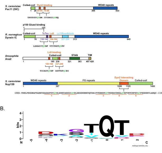
Figure 1-1. Dyn2/Lc8 binds to proteins with coiled-coil domains and a QT motif. ... 4 Figure 1-2. Structure of the Drosophila Lc8 in complex with the rat dynein IC shows
an Lc8 dimer bound to two IC β-strands by two parallel β-sheets. ... 6 Figure 1-3. Cartoon of the dynein holoenzyme shows how all of the canonical
dynein associating factors interact at the N-terminus of the DHC. ... 15 Figure 1-4. Centriole duplication involves seven essential proteins in higher
eukaryotes. ... 22 Figure 1-5. A cartoon diagram of the Nuclear Pore Complex illustrates how exported
mRNA interacts with the cytoplasmic fibrils. ... 27 Figure 2-1. Dynein intermediate chains contain conserved dynein light chain binding
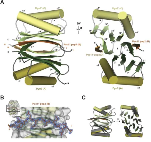
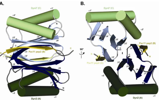
motifs near the amino terminal coiled-coil domain. ... 46 Figure 2-2. The structure of Dyn2 in complex with Pac11 pep2 shows a Dyn2 dimer
bound to two Pac11 pep2s that form the end β-strands on two antiparallel
β-sheets formed within the core of the Dyn2 dimer. ... 48
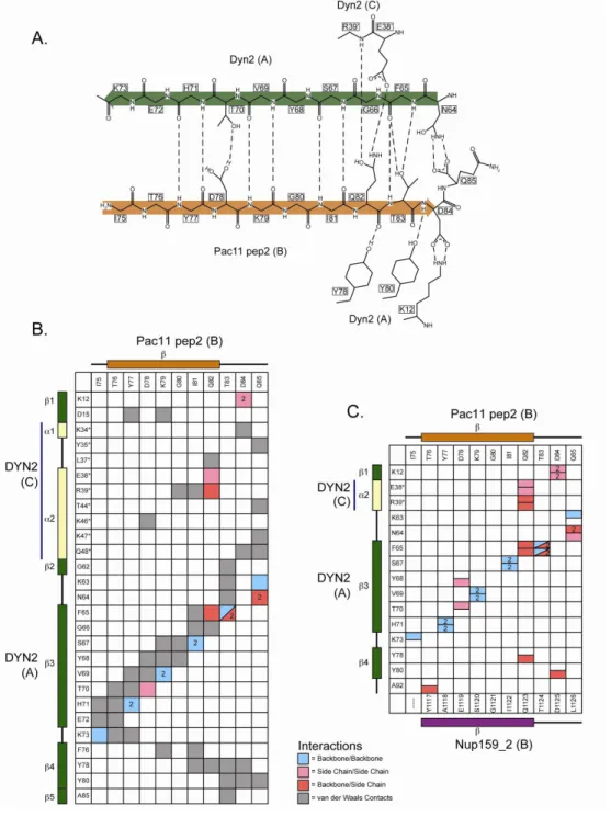
Figure 2-3. The crystal structure of Dyn2 in complex with Pac11 pep2 shows an extensive hydrogen bonding network that involves backbone/backbone
β-strand interactions as well as involvement by the residue side chains. ... 51
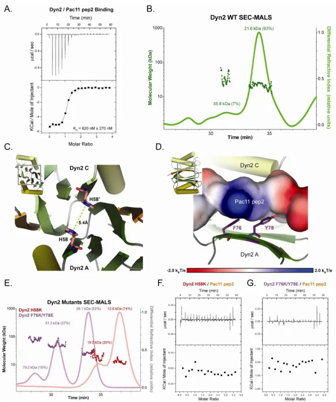
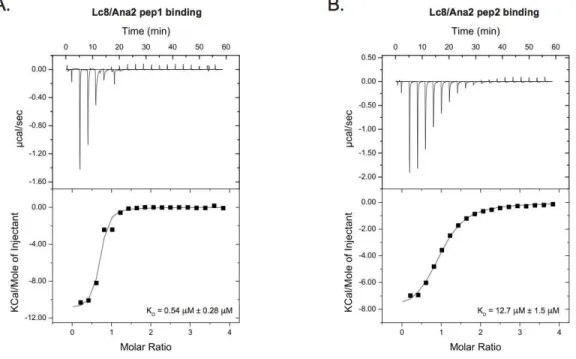
Figure 2-4. Dyn2’s ability to dimerize and bind Pac11 pep2 is abrogated through key mutations. ... 53 Figure 3-1. Lc8 interacts with two Ana2 peptides with an exothermic binding mode
but exhibits different affinities... 67 Figure 3-2. The crystal structure of the Lc8-Ana2 pep1 is a 2:2 complex formed by
an Lc8 homodimer binding two Ana2 pep1. ... 70 Figure 4-1. S. cerevisiae Dyn2 is a conserved dynein light chain involved in diverse
macromolecular complexes including the nuclear pore complex and the cytoplasmic dynein motor complex. ... 81 Figure 4-2. Structure of the Dyn2-Nup159 pep2 complex shows a quaternary
complex composed of a Dyn2 dimer, bound to two Nup159 pep2
peptides through parallel, composite -sheets. ... 84 Figure 4-3. Dyn2 homodimerizes via an extensive network of van der Waals
Figure 4-4. Dyn2 binds substrates through a highly conserved, positively charged
composite groove formed by Dyn2 dimerization. ... 88 Figure 4-5. The Dyn2:Nup159 pep2 interaction is mediated by an extensive
interaction network that recognizes ten contiguous Nup159 residues,
dually conferring specificity and substrate plasticity. ... 90 Figure 4-6. The Dyn2 interaction with Nup159 pep2 and pep4 occur in a 1:1
stoichiometry, and exhibit similar affinities but differ in their thermal
binding modes. ... 91 Figure 4-7. Crystallographic contacts array Dyn2 dimers linearly in an arrangement
that affords polarized binding to arrayed Dyn2 binding motifs. ... 93
LIST OF SUPPLEMENTAL FIGURES
Supplemental Figure 2-1. Pac11 pep2 does not bind to Dyn2 H58K or Dyn2
F76K/Y78E as measured by ITC. ... 61 Supplemental Figure 3-1. Ana2 peptide ITC controls were necessary to determine
the peptide contribution to the overall heat of dilution in each injection. ... 74 Supplemental Figure 4-1. The dynein light chain is highly plastic with regard to
LIST OF ABBREVIATIONS
AAA+ Family= ATPases Associated with various cellular Activities Family ATP = Adenosine Triphosphate
DID = Dynein light chain Interacting Domain FG = Phenylalanine-Glycine
GST = Glutathione S-Transferase HC = dynein heavy chain
IC = dynein intermediate chain
ITC = Isothermal Titration Calorimetry KD = knock down
LB = Luria Broth LC = dynein light chain
LIC = dynein light intermediate chain LIS1 = lissencephaly 1
MTBD = microtubule binding domain NPC = nuclear pore complex
PDB = protein data bank
RMSD = Root Mean Square Deviation
CHAPTER 1 INTRODUCTION
The benefit of a ubiquitous scaffolding protein
In many cases separate proteins with the same or similar function evolved to act in specialized, and often highly regulated functions, such as separate kinases evolving to differentially distinguish protein targets for phosphorylation (Manning, 2002). There is a benefit to having one ubiquitous protein that can accomplish the same function in multiple cellular locations. Ubiquitous proteins do not use as much genetic space as having two separate proteins with the same function, and do not require as much regulatory support for proteins that are not subject to rigorous regulation scrutiny. All of these assumptions are contingent on the ubiquitous protein binding with the same mode in different cellular contexts or having some flexibility in binding.
In the case of scaffolding proteins, their function is to optimize the effectiveness of another protein by providing the opportunity to form a stable complex of two or more proteins so that binding avidity is increased. Additionally, scaffolding proteins often do not have a significant catalytic or biochemical function, but are often the subject of regulation due to their allosteric control of a functional complex (Good, 2011).
α-helical domains that come together to form a coiled-coil. Coiled-coils require a specific
heptad pattern in the amino acid sequence to stabilize the intra-, and inter-helix interactions (Zhou, 1992; Marsden, 2010). Some proteins contain the amino acid sequence pattern for a coiled-coil, but the coils are not long enough for specificity in an exclusive dimerization partner (Marsden, 2010). In cases such as this a scaffolding protein may provide enough specificity or stability for the coiled-coil to satisfactorily dimerize into a fully functional
complex. Short coils have also been shown to convert from an α-helical to a β-strand form; a
transition that is dependent on certain sequence cues, temperature, and peptide concentration
(Aposolovic, 2010; Kammerer, 2006). This transition to β-strands may not be intentional (as in the case of amyloid formation in neurodegenerative diseases), but may provide flexibility as a gain-of-function interaction site for protein-mediated dimerization.
Coiled-coil domains are sufficient for dimerization in some cases but specific patterns
and longer amino acid chains are required to accomplish a dimer than a β-strand-β-strand
interaction between a β-sheet and target protein β-strand (Su, 1994; Khakshoor, 2010). Su et
al. determined that at least three heptad repeats (21 residues) are necessary for forming a
stable two-stranded α-helical coiled-coil, and five heptad repeats (35 residues) are optimal for stabilizing length (Su, 1994). Some coiled-coils successfully form obligate dimers that only function properly when they are homodimerized as is the case for cytoskeleton motor proteins like cytoplasmic dynein and kinesins that are not processive as monomers (Peckham, 2011).
platform for recruitment that might also allow for a point of regulation. Lc8/Dyn2’s mediation may afford less dedicated peptide sequence for a target binding protein that would otherwise rely on a coiled-coil for dimerization, and it may provide greater specificity for forming a homodimer than a coiled-coil of the same length.
The objective of this thesis is to investigate structural and biochemical properties of Dyn2/Lc8 across diverse systems: the dynein motor, centrioles, and at the nuclear pore. These assays will be used to distill the common function of dimerization, as well as to highlight differences between Dyn2/Lc8’s interactions in these systems. The remainder of the introduction will present the three systems: 1) the microtubule cytoskeleton and its motor proteins, specifically dynein, 2) centriole structure and duplication, and 3) the structure and functional regulation of the nuclear pore complex.
Lc8/Dyn2 is ubiquitously expressed, and a promiscuous protein
Sequence conservation of 45 Dyn2/Lc8 natural binding sites includes 41 from Rapali, 2011 and the Pac11 and Ana2 binding sites. Sequence homology is colored according to the following amino acid code; RHK (blue), DE (red), FYW (green), AVILM (cyan), STNQ (black), and CGP (purple).
Lc8/DYNLL/Dyn2 is also a promiscuous protein, involved in a diversity of protein interactions, in a number of different cellular contexts. The binding motifs for the light chain orthologs have been widely debated due to a number of sequence and binding mode exceptions (Rapali, 2011; Benison, 2007; Radnai, 2010). In S. cerevisiae, Dyn2 interacts with the dynein intermediate chain, Pac11, through tandem canonical 10-12 residue stretches, each containing a conserved QT motif (Fig. 1-1A) (Stuchell-Brereton, 2011). Many authors recognize the Lc8 binding motif has a high probability of a glutamine followed by a threonine/valine (Fig. 1-1B). There are, however a number of binding interactions where Lc8 does not utilize this motif, such as p21 activated kinase 1 (Pak1) that contains a divergent serine then proline at the QT positions in the binding cleft (Lightcap, 2008). As of 2011, Rapali et al. reported 41 naturally occurring Lc8 binding motifs and most of the candidates also contained a separate dimerization domain, such as a coiled-coil (Rapali, 2011).
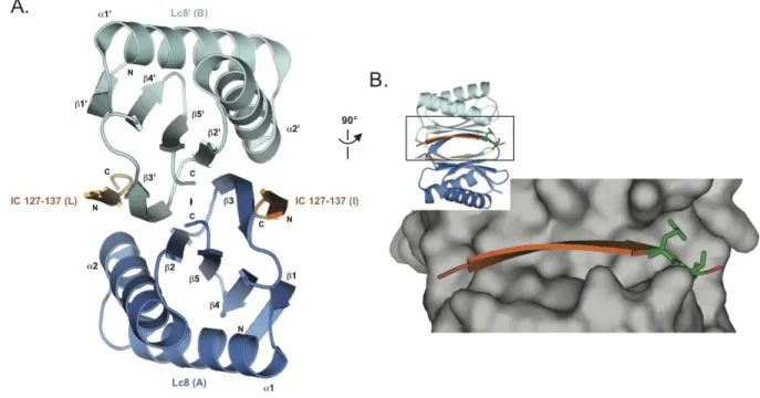
Figure 1-2. Structure of the Drosophila Lc8 in complex with the rat dynein IC shows an Lc8 dimer bound to two IC ββββ-strands by two parallel ββββ-sheets.
A. A cartoon diagram of the Lc8-IC crystal structure shows a quaternary structure of Lc8 dimerized in blue (chain A) and cyan (chain B) with two IC β-strands (residues 127-137) in dark (chain I) or light orange (chain L). There is a two-fold non-crystallographic rotational symmetry operator indicated in the z-axis between the two parallel β-sheets composed of five anti-parallel β-strands arranged β1-β4-β5-β2-β3’. Two α-helices from each monomer are arranged on top and bottom of the β-sheet sandwich that is peripherally bound to the IC through β-strand-β-strand interactions. B. The Lc8 dimer is turned 90° from A. and displayed in surface (gray) to show the zoomed in Lc8 binding pocket. IC (dark orange) binding residues show the wide binding pocket with few steric restrictions. The conserved Q135 and T136 are shown in green sticks to highlight their location near the end of the binding pocket. Figure made with pdb 2PG1 (Williams, 2007).
allows a protein to be more flexible in the selected binding partners, and therefore more promiscuous.
mediate dimerization of target proteins. Previous work has shown that Lc8 is phosphorylated on Ser88 by p21-activated kinase (Pak1), although the specific kinase utilized is hotly debated (Song, 2008; Benison, 2009). Song et al. used an S88E phosphomimetic mutation to show that phosphorylation at this specific Ser88 dissociates the Lc8 dimer and abolishes Lc8’s ability to bind Bim (Song, 2008).
Lc8 is composed of five β-strands that form an anti-parallel β-sheet with two α
-helices on one face (Fig. 1-2A). Lc8 homodimerizes and forms two parallel β-sheets that are
sandwiched by the outer α-helices. One β-strand from each monomer completes the other
monomer’s sheet and forms an anti-parallel β-strand interaction with the peptide that binds
each monomer of Lc8 (Williams, 2007; Benison, 2007; Fan, 2001; Lightcap, 2008). It is therefore remarkable that Lc8 binds many different targets with different thermodynamic binding modes through the same binding cleft (Fig. 1-2B).
While studies to date have biophysically characterized the Lc8/DYNLL, dynein light chains from Drosophila, rat, and human, molecular and biophysical details of the S. cerevisiae Dyn2 have remained outstanding. S. cerevisiae is a leading model system for
biophysical, biochemical and genetic investigations of the nuclear pore complex and the cytoplasmic dynein motor complex.
Lc8/DYNLL/Dyn2 interactions that are independent of the dynein complex illuminate a role as a dimerization machine
protein (Nup159) (Stelter, 2007). There are even a few species of algae and flowering plants, such as Arabidopsis, that do not contain the dynein motor, but maintain the Lc8 homolog of the dynein light chain (Wickstead, 2007). Since only about 30% of Lc8 is tightly associated with cytoplasmic dynein (King, 1996) and some species do not contain other dynein complex proteins (Wickstead, 2007) it is probable Lc8 acts in a greater capacity than just as a scaffolding protein for the dynein motor complex.
Evidence suggests that although other dynein associating factors can act independent of the dynein motor, such as another dynein light chain, TcTex, which remodels actin in neurite outgrowth (Chuang, 2005), many of their characterized functions indirectly affect the dynein motor. A number of proteins that have been shown to interact with Lc8/DYNLL are assumed to also interact with the dynein complex such as Bim (Puthalakath, 1999), Bmf (Day, 2004), and Gephyrin (Fuhrmann, 2002). We challenge these presumed interactions due to the lack of experiments exclusively showing the importance of the dynein motor in these interactions, and a few experiments showing the converse; that the dynein motor domain is not always included in associating factor functions.
Although lc8∆/dyn2∆ does not have a strong phenotype, the deletion phenotypes of
the proteins with which Lc8/Dyn2 interacts at the dynein motor complex have more severe deletion phenotypes (Pac11/DIC, STIL/Ana2, Nup159) (Stuchell-Brereton, 2011; Arquint, 2012; Gorsch, 1995). pac11∆ in S. cerevisiae shows an inability to move the nucleus into the
accumulation of poly(A)+ RNA in the nucleus (Gorsch, 1995). These strong phenotypes in a variety of cellular processes draw attention to the importance of having functional binding interactions with Dyn2/Lc8 to ensure optimal processing in the aforementioned contexts.
Given the diverse set of dynein light chain binding partners, both at the dynein complex and independent of the complex, it has been postulated that the dynein light chain functionally serves as a dimerization machine. This role correlates with the structures of higher Dyn2 orthologs that show dynein light chains complexed 2:2 with a variety of target peptides (Lightcap, 2008; Fan, 2001; Williams, 2007; Benison, 2008; Wang, 2003). In this study we aim to further our structural and biophysical understanding of Dyn2/Lc8 to derive a model for Dyn2’s role as a dimerization machine.
Motors of the cytoskeleton
direction of microtubules (with the exception of kinesin 14 family members) with many different types of kinesins thus delineated because they perform different tasks. The cytoplasmic dynein complex is a 1.2 MDa multifunctional motor protein that processes toward the minus end of microtubules with great importance in cell division, intracellular organelle transport and organization, and delivery of cargos across great distances within a cell (Nyarko, 2004). It is believed that dynein can accomplish such a great variety of tasks through the presentation of different scaffolding and regulatory proteins, called associating factors. Kinesin, however, has several different proteins to accomplish the various plus end directed functions that a single cytoplasmic dynein can accomplish with its associating factors. All three families of cytoskeletal motor proteins utilize ATP hydrolysis to generate force for their various functions, but myosin (such as the 119 kDa Dictyostelium Myosin-1B) and kinesin (such as the approximately 400 kDa Kinesin-1 complex) are much smaller and simpler than the comparative behemoth, dynein (1.2 MDa cytoplasmic complex) (Mooseker, 1995; Kull, 2000; Nyarko, 2004). While much is known about kinesin and myosin family members, far less is known about structure and regulation of the dynein motor and dynein regulators.
In vitro experiments of cytoplasmic dynein and kinesin have aided in our
The structure of processive kinesin
The kinesin motor is comprised of a heavy chain that homodimerizes to form a functional motor, which can process toward the plus end of microtubules with invariant 8 nm steps. The heavy chain contains an approximately 30 kDa globular motor domain that contains the highly conserved microtubule-binding site and catalytic site. Kinesins contain a coiled-coil, or stalk, for dimerization, and a tail that binds to the light chain scaffolding protein (Sack, 1999). The kinesin family can be divided into groups based on the organization of these three domains within the peptide, although the structural folds within the catalytic head domains are invariant (Endow, 2010). The tail has been less well characterized in kinesin, however, the kinesin light chain bound to the tail mediate binding to vesicles and organelles.
Cytoplasmic dynein structure
Cytoplasmic dynein in metazoans is composed of a catalytic homodimer of heavy chains (HC), and a number of non-catalytic subunits; two dynein intermediate chains (IC), three light intermediate chains (LIC), and three light chains (LC) (Fig. 1-3B). In fungi, such as S. cerevisiae, there are only three reported non-catalytic subunits that are canonical dynein components: the intermediate chain, Pac11, a light intermediate chain, Dyn3, and a light chain, Dyn2. Dynein also binds to other non-canonical associating factors such as the dynactin complex, lissencephaly 1 (LIS1)/Pac1, and the RZZ complex (Rod, Zwilch, Zw10) to regulate processivity and contribute to function. The absence of the dynactin or LIS1 components by depletion phenotypically copies the loss of dynein function (Kardon, 2009), therefore these associating factors provide an essential component to dynein in vivo.
Structure of the dynein heavy chain
discovered about the dynein motor structure and possible mechanism, there have not been significant studies on the role that the dimerization domain of the HC plays in coordinating dynein associating factors.
The motor domain of dynein is composed of six ATP binding domains (although only four of six are actually capable of binding ATP) that are concatenated in a single polypeptide, thus making dynein a unique member of the ATPases Associated with various cellular Activities (AAA+) family. Recently, the crystallographic structure of the motor domain of the cytoplasmic dynein heavy chain in Dictyostelium was solved to 2.8 Å (Kon, 2012) and in S. cerevisiae to 3.3 Å resolution (Schmidt, 2012). Both of these structures are high enough
Figure 1-3. Cartoon of the dynein holoenzyme shows how all of the canonical dynein associating factors interact at the N-terminus of the DHC.
ATP hydrolysis through the ring and into the MTBD (Carter, 2008; Carter, 2011; Kon, 2011; Kon, 2012; Schmidt, 2012).
Structure of the canonical cytoplasmic dynein associating factors
Although a full length structure of a dynein intermediate chain has not been published, the domain structure and binding sites for other dynein associating factors have been determined using sequence analysis and biochemical assays (Stuchell-Brereton, 2011; Benison, 2007; Makokha, 2002; Williams, 2007; Hall, 2009). The very N-terminal portion of the IC contains a short, predicted coiled-coil (Makokha, 2002) and also contains the p150, dynactin subunit, binding site (Vaughan, 1995). The IC N-terminal domain follows an inherently disordered domain that gains structure upon binding to Lc8 (Nyarko, 2004), and contains binding sites for the other two LCs. The two light chain-binding sites for human
TcTex and Lc8 were determined by x-ray crystallography to form a β-strand-β-strand interaction with the backbone of IC (Williams, 2007; Benison, 2007). The third LC that binds
to the central domain of the IC, Lc7/Roadblock, interacts with two α-helices from the IC to
form the binding site (Susalka, 2002; Hall, 2010). The final C-terminal domain of the IC
contains seven WD-repeat motifs (Susalka, 2002;) predicted to form a β-propeller (Garcia-Higuera, 1996), which then binds to the dynein heavy chain (Ma, 1999).
The LICs of dynein are isoforms of a single gene that undergoes differential phosphorylation and alternative splicing to form two cytoplasmic proteins (Hughes, 1995). Sequence analysis has shown that there appears to be no significant relationship between the
LICs and the LCs, nor are there regions of predicted α-helical, or β-sheet secondary structure
also shares some homology with the ABC transporter family (Hughes, 1995). Although there is not very much known about the LIC’s function, its sequence homology with the ABC transporter family indicates LIC may bind and hydrolyze ATP (Mische, 2008). Mutations in the putative ATP-binding residues of C. elegans LIC (dli-1) have shown complete rescue of function, indicating that ATP hydrolysis is not necessary for LIC’s function (Yoder, 2001).
In addition to Lc8/Dyn2 there are two more dynein light chains, Lc7/Roadblock and TcTex have similar secondary structural elements to Dyn2/Lc8, but Lc7’s secondary structure is a different fold, and they all have little sequence homology with each other. The light chains are thus named because they are less than 10 kDa on average, and are all non-catalytic subunits. The Lc7/Roadblock structure in humans (Ilangovan, 2005), mouse (Song,
2005), and Drosophila (Hall, 2010) were all shown to contain five β-strands that
homodimerize to form one contiguous β-sheet with each monomer contributing two
peripheral α-helices on either side of the β-sheet. The IC binds Lc7 with two amphipathic
helices laid across the β-sheet and forms interactions throughout the molecule (Hall, 2010).
The final category of dynein LC, TcTex, shares 1.6 Å RMSD strucutural homology with LC8, but is very divergent in sequence with almost no homology (Williams, 2005). TcTex binds non-overlapping sequences on target proteins in the same β-strand-β-strand interaction
mode as Lc8 where each monomer binds a peptide along the midline β-sheet. TcTex, like
Lc8, contains five β-strands and two α-helices that are arranged in the same fashion as Lc8
except that the central β-strands and α-helices are much longer (Williams, 2005; Williams, 2007). Although the three categories of dynein LC are structurally different and bind targets
Function and regulation of the dynein holoenzyme Dynein’s role in vivo
Cytoplasmic dynein has a variety of roles that are instrumental in cell movement, division, and intracellular transport of cargos. One of dynein’s primary roles in metazoans is in organelle transport such as movement of the Golgi apparatus (Corthesy-Theulaz, 1992; Holzbaur, 1994), lysosomes, and endosomes (Lin, 1992). For many of the larger organelles there are several dynein motors attached at any one time, which serves the role of overcoming the stall force for a single dynein motor, and to keep the organelle processively moving along the microtubule track (Bryantseva, 2012). The dynein complex has also been implicated in transport of vesicles, viruses, and packaged mRNA through LC mediation (Holzbaur, 1994; Vallee, 2004). It is not yet clear how dynein is specifically targeted to transport these particular cargos.
the absence of Cin8) so the Kar9 and Cin8 pathways have some overlap with dynein and complete other functions typically associated with dynein in metazoans (Geiser, 1997). Intermolecular regulation of the dynein motor domain
The dynein motor domain is internally regulated by AAA2-4 as well as by external adaptors that are crucial in adjusting the motor to its cellular function (Kardon, 2009). LIS1 is the only known associating protein to bind directly to the dynein motor domain. LIS1 binds directly between AAA3 and AAA4 to regulate communication between the catalytic motor and the microtubule-binding stalk, which prolongs dynein’s interaction with the MT and thereby increases processivity (Huang, 2012; McKenney, 2010). It appears as though this LIS1 binding interaction is most important during high bearing loads like transport of nuclei, movement of kinetochores, and during high tension in the cytoskeleton (McKenney, 2010). NudE is also important in the high load interactions and to recruit LIS1 and dynein to kinetochores (Stehman, 2007). NudE, however, interacts directly with the IC and LIC to abrogate dynein force production (McKenney, 2010).
Dynactin is a 1 MDa complex that has a role in modulating nearly every function of dynein’s behavior. In vitro experiments have shown that dynactin acts as a clutch for increasing dynein processivity (Kardon, 2009 PNAS). There are two microtubule-binding domains in the p150 subunits of dynactin that were thought to tether dynein to the microtubule to enhance processivity (Waterman-Storer, 1995). Mutant and deletion constructs of dynactin show that these microtubule-binding domains are dispensable and therefore not sufficient for increasing processivity (Kim, 2007).
Interactions through the p150 subunit as well as the ARP1 scaffolding subunit both mediate interactions between dynein and cargo (Kardon, 2009). The ARP1 subunit directly interacts
with a protein on the cytosolic surface of the Golgi, βIII spectrin, in the process of
dynein-dynactin mediated vesicle trafficking from the endoplasmic reticulum (Holleran, 2001). The p150 subunit not only binds to ARP1 and the dynein IC, but also to a number of microtubule plus end binding proteins such as EB1 and CLIP170 to target dynein to stably congress at microtubule plus ends (Hayashi, 2005; Hayashi, 2007). Dynein’s binding interactions with non-canonical proteins not only have the ability to modulate dynein’s behavior, but more importantly they alter dynein’s functionality in attachment and delivery of specific cargos during certain times.
Through biochemical and structural studies we are gaining more insight as to how the dynein motor domain is organized and functions to promote motility in vitro, but there is still a great deal to learn about how this molecular machine selects and attaches to cargos for its in vivo functions. One hypothesis for dynein targeting cargos in fungi is that dynein is
targeted to the plus ends of microtubules through the LIS1/NudE interaction or through the dynactin interaction to probe the cytoplasm for cargos as microtubules cycle through dynamic instability (Kardon, 2009). The mechanism for dynein targeting cargos in metazoans is less clear.
Lc8/Ctp interacts with Ana2 on centrioles
aberrations that amount to disease states such as primary microcephaly, male sterility, and cancer (Kitagawa, 2011; Nigg, 2009). The process and regulation of centriole duplication has recently become a popular topic because it is poorly understood and has possible clinical implications in drug therapies.
Centriole structure
The structure of a centriole consists of a cylinder decorated by microtubule triplets with nine-fold radial symmetry; mature centrioles contain distal appendages that help assemble components to build the centrosome, the structure responsible for nucleating the bipolar mitotic spindle. The centrosome consists of a pair of centrioles: a mature (“mother” centriole), and a procentriole tethered by interconnecting fibers. A cloud of protein matrix, called pericentriolar material surrounds the centriole pair (Azimzadeh, 2011; Bornens, 2012).
During centriole duplication, the nucleating procentriole forms perpendicular to the original centriole at the proximal end. The first structure for nucleating a daughter centriole is a central hub that forms like a wheel spoke with nine-fold symmetry accompanied by microtubule triplets around the periphery (Azimzadeh, 2012; Brito, 2012; Nigg, 2011). The central cartwheel, at the proximal end of a procentriole, remains within the daughter to form the scaffold for the remainder of the barrel structure (Brito, 2012; Kitagawa, 2011). The microtubule triplets on the daughter centriole are then elongated around a central lumen to a specified length that is dependent on cell type and species (Schmidt, 2009; Tang, 2009). Centriole biogenesis
Figure 1-4. Centriole duplication involves seven essential proteins in higher
eukaryotes.
In the end of G1/early S phase of the cell cycle the duplication licensing protein, Plk4/Sak/Zyg1 is recruited by Cep152/Asl to begin formation of the cartwheel with Sas4/CPAP (brown). Sas6 and Cep135/Bld10 accumulate at formation of the cartwheel, and Sas-5/STIL/Ana2 interacts with Sas6 to initiate a single daughter centriole. Sas4/CPAP also acts to stabilize newly formed centriole microtubules in the elongation phase of duplication. In the final phase of centriole duplication Cep192/Spd2 stabilizes mitotic microtubules to nucleate the spindle.
each of which contains opportunities for regulation: initiation/licensing, elongation, and capping/termination. The initial step of priming the centriole replication machinery is the onset of the licensing phase, which will commence assembling the daughter centriole cartwheel during the following S-phase of the cell. There are only a few proteins that are essential for centriole duplication including Cep152/Asl, Plk4/Sak/Zyg1, STIL/Sas-5/Ana2, Sas6, Cep135/Bld10, CPAP/Sas4, and Cep192/Spd2 (Vulprecht, 2012; Kitagawa, 2009). When any one of these seven proteins is deficient then centrioles duplicate irregularly or not at all. Although the cascade of proteins involved in the first steps appears to be hierarchical, the specific order of events and players is still poorly understood. The first known protein involved in the initial cascade is Cep152/Asl to recruit Plk4/SAK to centrioles and to act as a scaffold for CPAP/Sas4 to assemble or stabilize centriolar microtubules during the elongation phase (Carvalho, 2010; Stevens, 2010). Sas6 forms the initial cartwheel during initiation, and Sas-5/STIL1/Ana2 is a required Sas6 binding partner that is also required for controlling the number of procentrioles. The coiled-coil and C-terminal half of Ana2 is sufficient for binding to the N-terminal portion of Sas6 (Stevens, 2010).
When centrosome duplication is disturbed there is an increased incidence of genomic instability due to compromised segregation of chromosomes. Therefore, the number of procentrioles generated on a mother centriole and the number of rounds of centriole duplication are crucial to fidelity of balanced centrosome number (Nigg, 2011).
The second phase of assembly is the elongation of the daughter centriole from the cartwheel assembly, which occurs during S to G2 phase of the cell cycle. During this phase,
unknown intrinsic property of the elongation machinery to make the daughter centriole a homogeneous length for the designated cell type and species, although post-translational modifications are suspected (Brito, 2012). The elongation machinery is also capable of distinguishing between daughter and mother centrioles with proteins like centrobin, so that
only daughter centrioles elongate (Marthiens, 2012). Centrobin’s recruitment to α-tubulin core components is dependent on Sas6, so centrobin is capable of distinguishing between centriole types due to the formation of the inner cartwheel (Gudi, 2011). In the absence of centrobin, 58% of cells show daughter centrioles with stunted growth (Gudi, 2011). Therefore, centrobin acts in the elongation/stabilization phase that is directly dependent on the proper assembly of Sas6 during initiation.
The final phase of centriole duplication is capping and disengagement of the newly matured centriole. A daughter centriole acquires distal and subdistal appendages that designate it as a fully matured centriole that is capable of replicating in future rounds of mitosis. During this final phase Cep192 targets AurA to mitotic centrosomes to assist in AurA oligomerization so that it can drive spindle microtubule assembly (Joukov, 2010). The role STIL/Sas-5/Ana2 plays in centriole licensing and elongation
400 amino acid length arthropods (Arquint, 2012). Near the C-terminus there is an area of approximately 90 amino acids called the STAN motif (for STil/Ana2) that shows 31% identity between Drosophila Ana2 and zebrafish STIL (Stevens, 2010). The most divergent member of the STAN motif is in the C. elegans Sas-5 sharing only 12% identity with the zebrafish STIL. At the very C-terminus there is a second motif that shows high sequence similarity, called TIM (Truncated In Microcephaly) (Arquint, 2012). This area of sequence was named because truncation of the very C-terminus of human STIL results in fewer asymmetrically divided progenitor neurons due to abnormal centriole function, thus resulting in microcephaly (Kumar, 2009).
Dynein Lc8 from Drosophila interacts with Ana2 to assist in mitotic spindle orientation
In addition to centriole duplication, Ana2 forms a complex with a number of astral microtubule binding proteins to orient the mitotic spindle for asymmetric division of Drosophila neuroblasts (Wang, 2011). Ana2 mutant neuroblast spindle poles were
disengaged from centrosomes, which resulted in disorganized spindles and misoriented assemblies (Wang, 2011). Severe spindle misorientation in asymmetrically dividing cells like neuroblast progenitors can result in hyperproliferation and tumorigenesis (Caussinus, 2005).
The dynein light chain from Drosophila, Lc8 was shown to interact with the central domain of Ana2 which contains a coiled-coil; co-localizing to the distal ends of centrioles (Wang, 2011). Lc8 mutants on their own were also shown to have a similar, although less severe, misoriented spindle phenotype as the Ana2 mutants (Wang, 2011).
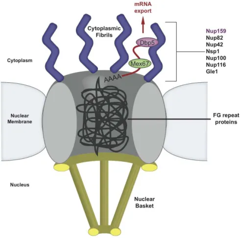
Figure 1-5. A cartoon diagram of the Nuclear Pore Complex illustrates how exported mRNA interacts with the cytoplasmic fibrils.
The nuclear pore is composed of a ring of transmembrane proteins with a nuclear basket structure and cytoplasmic fibrils that have both been identified as structurally and functionally important in nuclear pore transport. The central core of the pore contains a web of proteins with FG repeats, which can move aside to permit translocation. Dbp5 interacts with the mRNA export machinery and the cytoplasmic fibril as the final step in nuclear pore exit.
Dyn2 interacts with Nup159 on the cytoplasmic side of the nuclear pore
mRNA export into the cytoplasm and gated transport of specific proteins into and out of the nucleus.
The proteins that make up this highly coordinated and complex structure form an eight-fold symmetrical pore from a limited number of structural folds (Maul, 1971). The
types of domains in the nuclear pore proteins, or nucleoporins (Nups), are primarily α
-solenoids, β-propellers, Phe-Gly (FG) rich repeats, coiled-coil domains, and transmembrane
Nup159 is a prime component of the Nup82 complex and plays a directed role in coordinating nucleoporins involved in mRNA export rather than protein trafficking between the cytoplasm and nucleus (Del Priore, 1997). Nup159 has an extended multi-component architecture that facilitates its roles in mRNA export, as well as filament localization in the NPC structure (Del Priore, 1997; Weirich, 2004; Gorsch, 1995; Schmitt, 1999). Nup159’s N-terminal domain constitutes a seven-bladed β-propeller that extends into the cytoplasm and mediates Dbp5 binding (Miller, 2004). Deletion of Nup159’s N-terminal domain results in a temperature-sensitive phenotype, lethal at 37°C and hallmarked by Dbp5 mislocalization and constitutive mRNA export defects at 23°C (Del Priore, 1997; Gorsch, 1995). Nup159’s central 700 amino acids form an FG-rich repeat domain. C-terminal to the FG-rich repeats is a 100 amino acid region termed the dynein light chain interacting domain (DID) that uses a pentameric array of dynein light chain binding motifs to bind the yeast dynein light chain Dyn2 (Stelter, 2007). C-terminal to the DID, Nup159 contains a predicted helical region (Kraemer, 1995) that is essential for Nup159’s stability and localization on the NPC, and has recently been shown to form a heterotrimeric structure with Nup82 and Nup116 (Del Priore, 1997; Gorsch, 1995; Yoshida, 2011). Higher-order oligomerization of the Nup82 complex requires both the Nup159 DID region as well as Dyn2 (Stelter, 2007). The functional role of a dynein light chain at the nuclear pore is independent of its role in the cytoplasmic dynein microtubule motor complex (Stelter, 2007).
Research Objectives
We set out to characterize the S. cerevisiae dynein light chain, Dyn2 interacting in the protein complex for which it was named, the dynein complex. It was previously established that Dyn2 interacts directly with the intermediate chain, Pac11 (also S. cerevisiae) but the Dyn2 binding sites on Pac11 were not structurally or biophysically characterized. We solved the co-complex of Dyn2 binding to a peptide of one of the two reputed Pac11 binding sites as well as biophysically characterized the binding affinity of Dyn2 for Pac11 at this site. We hypothesize that Dyn2-mediated dimerization of Pac11 modulates Pac11’s function in attaching to the dynein heavy chain or directly modulating the processivity of the dynein complex. We designed two mutations in Dyn2 to determine whether Dyn2-mediated dimerization of Pac11 is necessary for affecting Pac11’s ability to bind the dynein heavy chain. One double point mutant knocks out Dyn2’s ability to bind Pac11 but maintains a Dyn2 homodimer, and a single point mutation that ablates Dyn2’s ability to homodimerize and also the ability to bind Pac11. Our collaborators will assess the processivity of the dynein complex with in vitro tracking assays utilizing these Dyn2 mutants in future work.
Lc8 in complex with the non-canonical Ana2 peptide-binding site, and we will continue to improve the refinement of the crystals for solving this structure. Lastly, we aim to determine whether Ana2’s interaction with Lc8 is necessary for dimerization, and whether dimerization of Ana2 is required for Ana2 to function properly in centriole duplication and in orienting the mitotic spindle. These in vivo experiments will be part of our future experimentation to determine the importance of the Lc8-Ana2 complex.
For studying how the dynein light chain interacts at the nuclear pore I chose to focus on the S. cerevisiae LC homolog, Dyn2. The budding yeast is a highly utilized model organism for studying dynein, and Dyn2 had not been structurally characterized. This body of work describes the first structure of the S. cerevisiae Dyn2, bound to a Nup159 peptide where only biochemical interactions have been shown with Dyn2. We show that Nup159 is the first known example of a single macromolecule that is capable of binding to Lc8/Dyn2 with one endothermic profile and another site with exothermic favorability. We hypothesized that even though there was only 50% sequence identity between S. cerevisiae Dyn2 and Drosophila Lc8 there would be a similar structural organization and mechanism for binding
peptides.
References
1. Alber, F., Dokudovskaya, S., Veenhoff, L. M., Zhang, W., Kipper, J., Devos, D., Suprapto, A., Karni-Schmidt, O., Williams, R., Chait, B. T., Sali A., and Rout, M. P. (2007) Nature 450, 695-701
2. Allen, N. P. C., Patel, S. S., Huang, L., Chalkley, R. J., Burlingame, A., Lutzmann, M., Hurt, E. C., and Rexach, M. (2002) Mol. Cell. Proteomics 1, 930-946
3. Aposolovic, B., Danial, M., Klok, H. A. (2010) Chem. Soc. Rev. 39, 3541-3575
4. Arquint, C., Sonnen, K. F., Steirhof, Y.-D., and Nigg, E. A. (2011) J. Cell Sci. 125, 1342-1352
5. Arquint, C., Sonnen, K. F., Stierhof, Y.-D., and Nigg, E. A. (2012) J. Cell Sci. 125, 1342-1352
6. Azimzadeh, J., and Marshall, W. F. (2011) Curr. Biol. 20, R816-R825 7. Azimzadeh, J., and Bornens, M. (2012) J. Cell Sci. 120, 2139-2142
8. Bader, J. R., and Vaughan, K. T. (2010) Semin. Cell Dev. Biol. 21, 269-275
9. Bailer, S. M., Balduf, C., Katahira, J., Podtelejnikov, A., Rollenhagen, C., Mann, M., Pante, N., and Hurt, E. C. (2000) J. Biol. Chem. 275, 23540-23548
10. Benison, G., Karplus, P. A., and Barbar, E. (2007) J. Mol. Biol. 371, 457-468
11. Benison, G., Chiodo, M., Karplus, P. A., and Barbar, E. (2009) Biochemistry 48, 11381-11389
12. Bornens, M. (2012) Science 335, 422-426
13. Brito, D. A., Gouveia, S. M., and Bettencourt-Dias, M. (2012) Curr. Opin. Cell Biol. 24, 4-13
14. Bryantseva, S. A. and Zhapparova, O. N. (2012) Cell Biol. Int. 36, 1-6
15. Busson, S., Dujardin, D., Moreau, A., Dompierre, J., and De Mey, J. R. (1998) Curr. Biol. 8, 541-544
16. Carter, A. P., Garbarino, J. E., Wilson-Kubalek, E. M., Shipley, W. E., Cho, C., Milligan, R. A., Vale, R. D., and Gibbons, I. R. (2008) Science 322, 1691-1695
19. Carvalho-Santos, Z., Machado, P., Branco, P., Tavares-Cadete, F., Rodrigues-Martins, A., Pereira-Leal, J. B., and Bettencourt-Dias, M. (2010) J. Cell Sci. 123, 1414-1426 20. Caussinus, E., and Gonzalez, C. (2005) Nat. Genet. 37, 1125-1129
21. Chaudhury, A., Rao, Y. M., and Goyal, R. K. (2008) Am. J. Physiol. Gastrointest. Liver Physiol. 295, G442-G451
22. Cho, C., Reck-Peterson, S. L., and Vale, R. D. (2008) J. Biol. Chem. 283, 25839-25845 23. Chuang, J. Z., Yeh, T. Y., Bollati, F., Conde, C., Canavosio, F., Caceres, A., and Sung,
C. H. (2005) Dev. Cell 9, 75-86
24. Corthesy-Theulaz, I., Pauloin, A., and Pfeffer, S. R. (1992) J. Cell Biol. 118, 1333-1345 25. Cyr, J. L., Pfister, K. K., Bloom, G. S., Slaughter, C. A., and Brady, S. T. (1991) PNAS
88, 10144-10148
26. Day, C. L., Puthalakath, H., Skea, G., Strasser, A., Barsukov, I., Lian, L.Y., Huang, D. C. S., Hinds, M. G. (2004) Biochem. J. 377, 597-605
27. Del Priore, V., Heath, C. V., Snay, C. A., MacMillan, A., Gorsch, L. C., Dagher, S., and Cole, C. N. (1997) J. Cell Sci. 110, 2987-2999
28. Delattre, M. Leidel, S., Wani, K., Baumer, K., Bamat, J., Schnabel, H., Feichtinger, R., Schnabel, R., and Gonczy, P. (2004) Nat. Cell Biol. 6, 656-664
29. Eftink, M. R., Anusiem, A. C., and Biltonen, R. L. (1983) Biochemistry 22, 3884-3896 30. Endow, S. A., Kull, F. J., and Liu, H. (2010) J. Cell Sci. 123, 3420-3424
31. Eshel, D., Urrestarazu, L. A., Vissers, S., Jauniaux, J. C., van Vliet-Reedijk, J. C., Planta, R. J., and Gibbons, I. R. (1993) PNAS 90, 11172-11176
32. Espindola, F.S., Suter, D. M., Partata, L. B. E., Cao, T., Wolenski, J. S., Cheney, R. E., King, S. M., and Mooseker, M. S. (2000) Cell Motil. Cytoskeleton 47, 269-281
33. Fahrenkrog, B., Hurt, E. C., Aebi, U., and Pante, N. (1998) J. Cell Biol. 143, 577-588 34. Fan, J., Zhang, Q., Tochio, H., Li, M., and Zhang, M. (2001) J. Mol. Biol. 306, 97-108 35. Fuhrmann, J. C., Kins, S., Rostaing, P., El Far, O., Kirsch, J., Sheng, M., Triller, A.,
Betz, H., and Kneussel, M. (2002) J. Neurosci. 22, 5393-5402
36. Galy, V., Gadal, O., Fromont-Racine, M., Romano, A., Jacquir, A., and Nehrbass, U. (2004) Cell 116, 63-73
38. Geiser, J. R., Schott, E. J., Kingsbury, T. J., Cole, N. B., Totis, L. J., Bhattacharyya, G., He, L., and Hoyt, M. A. (1997) Mol. Biol. Cell 8, 1035-1050
39. Gennerich, A., Carter, A. P., Reck-Peterson, S. L., and Vale, R. D. (2007) Cell 131, 952-965
40. Gerace, L. (1988) Ann. Rev. Cell Biol. 4, 335-374
41. Good, M. C., Zalatan, J. G., and Lim, W. A. (2011) Science 332, 680-686 42. Gorsch, L. C., Dockendorff, T. C., Cole, C. N. (1995) J. Cell Biol. 129, 939-955
43. Gross, S. P., Welte, M. A., Block, S. M., and Wieschaus, E. F. (2002) J. Cell Biol. 156, 715-724
44. Gudi, R., Zou, C., Li, J., and Gao, Q. (2011) J. Cell Biol. 193, 711-725
45. Habura, A., Tikhonenko, I., Chisholm, R. L., and Koonce, M. P. (1999) J. Biol. Chem. 274, 15447-15453
46. Hall, J., Karplus, P. A., and Barbar, E. (2009) J. Biol. Chem. 284, 33115-33121
47. Hall, J. Son, Y., Karplus, P. A., and Barbar, E. (2010) J. Biol. Chem. 285, 22566-22575 48. Hayashi, I., Wilde, A., Mal, T. K., and Ikura, M. (2005) Mol. Cell 19, 449-460
49. Hayashi, I., Plevin, M. J., and Ikura, M. (2007) Nat. Struct. Mol. Biol. 14, 980-981 50. Hendricks, A. G., Perison, E., Ross, J. L., Schroeder III, H. W., Tokito, M., and
Holzbaur, E. L. F. (2010) Curr. Biol. 20, 697-702
51. Hodge, C. A., Tran, E. J., Noble, K. N., Alcazar-Roman, A. R., Ben-Yishay, R., Scarcelli, J. J., Folkmann, A. W., Shav-Tal, Y., Wente, S. R., Cole, C. N. (2011) Genes Dev. 25, 1052-1064
52. Holleran, E. A., Ligon, L. A., Tokito, M., Stankewich, M. C., Morrow, J. C., and Holzbaur, E. L. F. (2001) J. Biol. Chem. 276, 36598-36605
53. Holzbaur, E. L. F., and Vallee, R. B. (1994) Annu. Rev. Cell Biol. 10, 339-372
54. Huang, J., Roberts, A. J., Leschziner, A. E., and Reck-Peterson, S. L. (2012) Cell 150, 975-986
55. Hughes, S. M., Vaughan, K. T., Herskovits, J. S., and Vallee, R. B. (1995) J. Cell Sci. 108, 17-24
57. Joukov, V., De Nicolo, A., Rodriguez, A., Walter, J. C., and Livingston, D. M. (2010) PNAS 107, 21022-21027
58. Kahana, J. A., Schlenstedt, G., Evanchuk, D. M., Geiser, J. R., Hoyt, M. A., and Silver, P. A. (1998) Mol. Biol. Cell 9, 1741-1756
59. Kammerer, R. A. and Steinmetz, M. O. (2006) J. Struct. Biol. 155, 146-153 60. Kardon, J. R., and Vale, R. D. (2009) Nat. Rev. Mol. Cell Biol. 10, 854-865 61. Kardon, J. R., Reck-Peterson, S. L., and Vale, R. D. (2009) PNAS 106, 5669-5674 62. Khakshoor, O., Lin, A. J., Korman, T. P., Sawaya, M. R., Tsai, S. C., Eisenberg, D.,
and Nowick, J. S. (2010) J. Am. Chem. Soc. 132, 11622-11628
63. Kim, H., Ling, S. C., Rogers, G. C., Kural, C., Selvin, P. R., Rogers, S. L., and Gelfand, V. I. (2007) J. Cell Biol. 176, 641-651
64. King, S. M., Barbarese, E., Dillman III, J. F., Patel-King, R. S., Carson, J. H., and Pfister, K. K. (1996) J. Biol. Chem. 271, 19358-19366
65. Kitagawa, D., Busso, C., Fluckiger, I., and Gonczy, P. (2009) Dev. Cell 17, 900-907 66. Kitagawa, D., Vakonakis, I., Olieric, N., Hillbert, M., Keller, D., Olieric, V., Bortfeld,
M., Erat, M. C., Fluckiger, I., Gonczy, P., and Steinmetz, M. O. (2011) Cell 144, 364-375
67. Kon, T., Nishiura, M., Ohkura, R., Toyoshima, Y. Y., and Sutoh, K. (2004) Biochemistry 43, 11266-11274
68. Kon, T., Sutoh, K. and Kurisu, G. (2011) Nat. Struct. Mol. Biol. 18, 638-642
69. Kon, T., Oyama, T., Shimo-Kon, R., Imamula, K., Shima, T., Sutoh, K., and Kurisu, G. (2012) Nature 484, 345-350
70. Kraemer, D. M., Strambio-de-Castillia, C., Blobel, G., and Rout, M. P. (1995) J. Biol. Chem. 270, 19017-19020
71. Kull, F. J. (2000) Essays Biochem. 35, 61-73
72. Kumar, A., Girimaji, S. C., Duvvari, M. R., and Blanton, S. H. (2009) Am. J. Hum. Gen. 84, 286-290
73. Leidel, C., Longoria, R. A., Gutierrez, F. M., and Shubeita, G. T. (2012) Biophys. J. 103, 492-500
75. Lin, S. X., and Collins, C. A. (1992) J. Cell Sci. 101, 125-137 76. Lumry, R. and Rajender, S. (1970) Biopolymers 9, 1125-1227
77. Ma, S., Trivinos-Lagos, L., Graf, R., and Chisholm, R. L. (1999) J. Cell Biol. 147, 1261-1274
78. Makokha, M., Hare, M., Li, M., Hays, T., and Barbar, E. (2002) Biochemistry 41, 4302-4311
79. Manning, G., Plowman, G. D., Hunter, T., and Sudarsanam, S. (2002) Trends Biochem. Sci. 27, 514-520
80. Marsden, H. R. and Kros, A. (2010) Angew. Chem. Int. Ed. 49, 2988-3005 81. Marthiens, V., Piel, M., and Basto, R. (2012) J. Cell Sci. 125, 3281-3292 82. Maul, G. G. (1971) J. Cell Biol. 51, 558-563
83. McCauley, S. D., Gilchrist, M., and Befus, A. D. (2007) Life Sci. 80, 959-964
84. McKenney, R. J., Vershinin, M., Kunwar, A., Vallee, R. B., and Gross, S. P. (2010) Cell 141, 304-314
85. Mehta, A. (2001) J. Cell Sci. 114, 1981-1998
86. Miller, A. L., Suntharalingam, M., Johnson, S. L., Audhya, A., Emr, S. D., and Wente, S. R. (2004) J. Biol. Chem. 279, 51022-51032
87. Mische, S., He, Y., Ma, L., Li, M., Serr, M., and Hays, T. S. (2008) Mol. Biol. Cell 19, 4918-4929
88. Moore, J. K., Li, J., and Cooper, J. A. (2008) Traffic 9, 510-527
89. Mooseker, M. S., and Cheney, R. E. (1995) Annu. Rev. Cell Dev. Biol. 11, 633-675 90. Nigg, E. A., and Raff, J. W. (2009) Cell 139, 663-678
91. Nigg, E. A., and Stearns, T. (2011) Nat. Cell Biol. 13, 1154-1160
92. Nyarko, A., Hare, M., Hays, T. S., and Barbar, E. (2004) Biochem. 43, 15595-15603 93. Nyarko, A., Hall, J., Hall, A., Hare, M., Kremerskothen, J., and Barbar, E. (2011)
Biophys. Chem. 159, 41-47
94. Peckham, M. (2011) Biochem. Soc. Trans. 39, 1142-1148
96. Radnai, L., Rapali, P., Hodi, Z., Suveges, D., Monar, T., Kiss, B., Becsi, B., Erdodi, F., Buday, L., Kardos, J., Kovacs, M., and Nyitray, L. (2010) J. Biol. Chem. 285, 38649-38657
97. Rapali, P., Radnai, L., Suveges, D., Harmat, V., Tolgyesi, F., Walgren, W. Y., Katona, G., Nyitray, L., and Pal, G. (2011) PLoS One 6, e18818
98. Ribbeck, K., and Gorlich, D. (2001) EMBO J. 20, 1320-1330
99. Rodrigues-Martins, A., Riparbelli, M., Callaini, G., Glover, D. M., and Bettencourt-Dias, M. (2007) Science 316, 1046-1050
100. Rout, M. P., Aitchison, J. D., Suprapto, A., Hjertaas, K., Zhao, Y., and Chait, B. T. (2000) J. Cell Biol. 148, 635-651
101. Sack, S., Kull, F. J., and Mandelkow, E. (1999) Eur. J. Biochem. 262, 1-11
102. Schmidt, H., Gleave, E. S., and Carter, A. P. (2012) Nat. Struct. Mol. Biol. 19, 492-497 103. Schmidt, T. I., Kleylein-Sohn, J., Westendorf, J., Le Clech, M., Lavoie, S. B., Stierhof,
Y. D., and Nigg, E. A. (2009) Curr. Biol. 19, 1005-1011
104. Schmitt, C., von Kobbe, C., Bachi, A., Pante, N., Rodrigues, J. P., Boscheron, C., Rigaut, G., Wilm, M., Seraphin, B., Carmo-Fonseca, M., and Izaurralde, E. (1999) EMBO J. 18, 4332-4347
105. Schwartz, T. U. (2005) Curr. Opin. Struct. Biol. 15, 221-226
106. Song, C., Wen, W., Rayala, S. K., Chen, M., Ma, J., Zhang, M., and Kumar, R. (2008) J. Biol. Chem. 283, 4004-4013
107. Song, J., Tyler, R. C., Lee, M. S., Tyler, E. M., and Markley, J. L. (2005) J. Mol. Biol. 354, 1043-1051
108. Stehman, S. A., Chen, Y., McKenney, R. J., and Vallee, R. B. (2007) J. Cell Biol. 178, 583-594
109. Stelter, P., Kunze, R., Flemming, D., Hoepfner, D., Diepholz, M., Philippsen, P., Boettcher, B., and Hurt, E. C. (2007) Nat. Cell Biol. 9, 788-796
110. Stevens, N. R., Dobbelaere, J., Brunk, K., Franz, A., and Raff, J. W. (2010) J. Cell Biol. 188, 313-323
111. Stuchell-Brereton, M. D., Siglin, A., Li, J., Moore, J. K., Ahmed, S., Williams, J. C., and Cooper, J. A. (2011) Mol. Biol. Cell 15, 2690-2701
113. Susalka, S. J., Nikulina, K., Salata, M. W., Vaughan, P. S., King, S. M., Vaughan, K. T., and Pfister, K. K. (2002) J. Biol. Chem. 277, 32939-32946
114. Tang, C. J., Fu, R. H., Wu, K. S., Hsu, W. B., and Tang, T. K. (2009) Nat. Cell Biol. 11, 825-831
115. Tcheperegine, S. E., Marelli, M., and Wozniak, R. W. (1999) J. Biol. Chem. 274, 5252-5258
116. Tynan, S. H., Gee, M. A., and Vallee, R. B. (2000) J. Biol. Chem. 275, 32769-32774 117. Vale, R. D. (2003) J. Cell Biol. 163, 445-450
118. Vallee, R. B., Williams, J. C., Varma, D., and Barnhart, L. E. (2004) J. Neurobiol. 58, 189-200
119. Vaughan, K. T., and Vallee, R. B. (1995) J. Cell Biol. 131, 1507-1516
120. Vulprecht, J., David, A., Tibelius, A., Castiel, A., Konotop, G., Liu, F., Bestvater, F., Raab, M. S., Zentgraf, H., Izraeli, S., and Kramer, A. (2012) J. Cell Sci. 125, 1353-1362
121. Wang, C., Li, S., Januschke, J., Rossi, F., Izumi, Y., Garcia-Alvarez, G., Gwee, S. S. L., Soon, S. B., Sidhu, H. K., Yu, F., Matsuzaki, F., Gonzalez, C., and Wang, H. (2011) Dev. Cell 21, 520-533
122. Wang, W., Lo, K. W., Kan, H., Fan, J., and Zhang, M. (2003) J. Biol. Chem. 278, 41491-41499
123. Waterman-Storer, C. M., Karki, S., and Holzbaur, E. L. (1995) PNAS 92, 1634-1638 124. Watson, P., Forster, R., Palmer, K. J., Pepperkok, R., and Stephens, D. J. (2005) Nat.
Cell Biol. 7, 48-55
125. Weirich, C. S., Erzberger, J. P., Berger, J. M., and Weis, K. (2004) Mol. Cell 16, 749-760
126. Wickstead, B., and Gull, K. (2007) Traffic 8, 1708-1721
127. Williams, J. C., Xie, H., and Hendrickson, W. A. (2005) J. Biol. Chem. 280, 21981-21986
128. Williams, J. C., Roulhac, P. L., Roy, A. G., Vallee, R. B., Fitzgerald, M. C., and Hendrickson, W. A. (2007) Proc. Natl. Acad. Sci. 104, 10028-10033
129. Yoder, J. H., and Han, M. (2001) Mol. Biol. Cell 12, 2921-2933
131. Zhou, N. E., Zhu, B.-Y., Kay, C. M., and Hodges, R. S. (1992) Biopolymers 32, 419-426
CHAPTER 2
THE STRUCTURE OF A YEAST DYNEIN DYN2-PAC11 COMPLEX AND EFFECT ON SINGLE MOLECULE DYNEIN MOTOR ACTIVITY
Preface
This work is a manuscript in preparation. Lu Rao (Laboratory of Arne Gennerich at Albert Einstein College of Medicine) performed the expression and preparation of rhodamine labeled Dyn2 and Pac11 as well as the single molecule motility assays, which are not presented here but will be part of the final manuscript. Ashutosh Tripathy assisted me with the size exclusion chromatography-multi-angle light scattering experiments as well as the isothermal microtitration calorimetry. I performed the remaining experiments and protein preparation. My advisor, Kevin Slep, and I designed the project and experiments I performed. Kevin Slep, Arne Gennerich, Lu Rao, and I wrote and edited the final manuscript. Romes, EM, Rao, L, Tripathy, A, Slep, K, and Gennerich, A. (manuscript in progress).
Summary
in both of these capacities and binds directly to a dynein light chain, Dyn2 to assist and/or stabilize dimerization of Pac11 at two binding sites (Stuchell-Brereton, 2011). We determined the binding affinity of Dyn2 for the Pac11 second binding site (pep2) to have a KD of 620 nM, which is the tightest Dyn2 binding interaction characterized to date. We also
present the 1.90 Å resolution crystal structure of full length Dyn2 in complex with a peptide from the second of two Pac11 binding sites (pep2). Based on this crystal structure we designed a double point mutant (F76K/Y78E) that ablates Dyn2’s ability to bind to the Pac11 pep2, but maintains Dyn2’s ability to dimerize. We additionally confirmed that a single point mutant discussed in previous work on the Drosophila homolog, Lc8 (H55K), ablates Dyn2 dimerization and consequently also the ability of Dyn2 to bind to the Pac11 pep2. These point mutation tools will likely prove useful in dissecting whether Dyn2’s role in dimerization is necessary for the function of Pac11 interacting with the dynein heavy chain or with cargo. Future experiments will aim to determine the role that Dyn2 plays in modifying dynein processivity and localization, as well as whether Dyn2’s function as a dimerization machine affects Pac11’s ability to modulate dynein processivity.
Experimental Procedures
Cloning, Expression, and Purification of Full Length Dyn2 from S. cerevisiae - Full length Dyn2 was cloned from S. cerevisiae S288c as described (Romes, 2012). Briefly, Dyn2 was cloned into pGEX-6P-2, (GE Healthcare) yielding an N-terminal, cleavable GST tag. GST-Dyn2 was expressed in BL21 DE3 (pLysS) and induced using 0.1 mM
eluted and incubated with PreScission protease (GE Healthcare) to cleave the GST tag. A final purification step was performed using an ion exchange SP Sepharose Fast Flow column (GE Healthcare). The Dyn2 peak was collected and exchanged into 50 mM NaCl, 25 mM Hepes, pH 6.8, and 0.1% β-mercaptoethanol. The final, purified Dyn2 protein was concentrated to 5 mg/mL, snap frozen in liquid nitrogen and stored at -80°C. The final Dyn2 protein contained an N-terminal GPLGS cloning artifact.
Cloning, Expression and Purification of Dyn2 mutants - Dyn2 H58K and F76K/Y78E
mutagenesis was done with the Quikchange method (Strategene) with the following complimentary oligonucleotides for the H58K mutant: 5’-GGATGTCAAATACGGCAATACCTGGAAAGTGATTGTCGGAAAGAACTTTGGG-3’ (where
the underlined portion codes for the mutated codon) and 5’-CCCAAAGTTCTTTCCGACAATCACTTTCCAGGTATTGCCGTATTTGACATCC-3’, and the
complimentary oligonucleotides for F76K/Y78E:
GTGACACACGAAAAGGGCCATAAAGTTGAATTCTATATCGGTCCACTGGCG-3’ and
5’-CGCCAGTGGACCGATATAGAATTCAACTTTATGGCCCTTTTCGTGTGTCAC-3’. Both the
single and double mutants were confirmed by sequencing.The expression and purification of Dyn2 H58K and Dyn2 F76K/Y78E follow the same protocols and buffers as for WT Dyn2.
Crystallization – Final concentrations of 0.5 mM Dyn2 and 0.6 mM Pac11 pep2 were
incubated together to form a complex in 50 mM NaCl, 25 mM Hepes, pH 6.8, and 0.1% β-mercaptoethanol for 30 minutes at ambient temperature. Crystals were obtained by the hanging drop protocol using 2 µL of the Dyn2-Pac11 pep2 mixture and 1 µL of the 1 mL well solution: 0.4 M sodium phosphate monobasic, 0.1 M 1,6-hexanediol, and 25% polyethylene glycol 3350. Crystals grew at 20°C into full-sized thin, individual platesin two weeks. Crystals were transferred to cryoprotection of well solution supplemented with 20% ethylene glycol and flash frozen in liquid nitrogen.
Data Collection, Structure Determination, and Refinement – Diffraction data were collected on Dyn2-Pac11 pep2 crystals at the Advanced Photon Source SER-CAT beamline 22-ID with 1° oscillations over 180° from a single crystal. Data were indexed, integrated and scaled using HKL2000 (Otwinowski, 1997). The structure was determined using the AutoMR molecular replacement program (PHENIX crystallographic suite (Adams, 2010)) and a modified 4DS1 (Romes, 2012) coordinate file in which a monomeric, apo Dyn2 search model was used. The model was built using AutoBuild (PHENIX) (Adams, 2010) and refined iteratively through manual builds in Coot (Emsley, 2010) followed by refinement runs using phenix.refine (PHENIX) (Adams, 2010). Refinement statistics were monitored using a Free R, calculated using 7.8% of the data, randomly excluded from refinement (Brunger, 1992).
Isothermal Microtitration Calorimetry – Pac11 pep2 was exchanged into buffer B using G-25 Sephadex Quick Spin Columns (Roche) to remove additional salts. WT Dyn2, Dyn2 H58K, and Dyn2 F76K/Y78E were individually exchanged into buffer B as before: 50
ITC at 26°C on a Microcal AutoITC200 (GE Healthcare). 19 x 2 µL injections of 1.0 mM
Pac11 pep2 were automatically injected into 200 µL of 50 µM WT Dyn2. The data were analyzed with the Origin 7.0 software package (OriginLab) and the resulting isotherm was fit to a one-site binding model. The experiment was performed in triplicate and the mean KD
value with standard deviation is shown in Fig. 2-4A.
Dyn2 H58K and Dyn2 F76K/Y78E experiments were performed with Pac11 pep2 solubilized in buffer B (but not desalted) and the same concentrations as for WT Dyn2 and did not show binding (Fig. 2-4 F,G). 19 x 2 µL injections of 3.0 mM Pac11 pep2 were
automatically injected into 200 µL of 300 µM Dyn2 H58K (not desalted) or 300 µM Dyn2
F76K/Y78E (not desalted). There was significant heat contributed by the Pac11 pep2 in the control experiment of 19 x 2 µL injections of 3.0 mM Pac11 pep2 that were automatically
injected into 200 µL of buffer B. Because each injection produced a significant heat of
dilution, the control experiment was subtracted from the raw 3.0 mM Pac11 pep2 into 300 µM Dyn2 H58K or 300 µM F76K/Y78E, and no binding was observed for either experiment
(Fig. S2-1 C-E).
Pac11 pep2 was exchanged into buffer B using G-25 Sephadex Quick Spin Columns (Roche) to remove additional salts and the resulting concentration was used for further Dyn2 mutant experiments. 19 x 2 µL injections of 0.5 mM Pac11 pep2 were automatically injected
into 200 µL of 50 µM Dyn2 H58K or 50 µM Dyn2 F76K/Y78E and no binding was
observed (Fig. S2-1 A,B).
Size Exclusion Chromatography and Multi-Angle Light Scattering – Dyn2 H58K and
Dyn2 F76K/Y78E were exchanged into buffer B: 50 mM NaCl, 25 mM Hepes, pH 6.8, and
concentrated to 6.0 mg/mL, Dyn2 H58K was concentrated to 10.0 mg/mL, and Dyn2 F76K/Y78E was concentrated to 6.0 mg/mL and all three proteins were monitored for
precipitation during concentration. 50 µL injections of Dyn2 WT, H58K or F76K/Y78E were
individually injected onto a Superdex 200 10/300 GL size exclusion column in buffer B with 0.2 g/L sodium azide and then passed consecutively through a Wyatt DAWN HELEOS II light scattering instrument and a Wyatt Optilab rEX refractometer. The light scattering and refractive index data were used to calculate the weight-averaged molar mass (MW) and the
mass fraction in each peak using the Wyatt Astra V software program (Wyatt Technology Corp.) (Wyatt, 1993).
Protein Data Bank Accession Number – Coordinates for the Dyn2-Pac11 pep2 complex have been deposited in the Research Collaboratory for Structural Bioinformatics PDB under accession code 4HT6.
Results
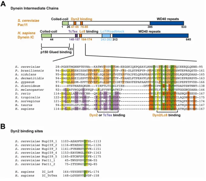
The dynein intermediate chain contains two conserved binding motifs for two Dyn2
homodimers in lower eukaryotes, or for one Dyn2/Lc8 homodimer and one TcTex
homodimer in higher eukaryotes. In a sequence alignment of ICs from 12 species of
Figure 2-1. Dynein intermediate chains contain conserved dynein light chain binding motifs near the amino terminal coiled-coil domain.