CELLULAR & MOLECULAR BIOLOGY LETTERS http://www.cmbl.org.pl
Received: 03 August 2011 Volume 17 (2012) pp 258-273 Final form accepted: 15 February 2012 DOI:10.2478/s11658-012-0008-5 Published online: 25 February 2012 © 2012 by the University of Wrocław, Poland
* Author for correspondence. e-mail: rraffani@hunter.cuny.edu
Abbreviations used: Dex – dexamethasone; CCK-8 – cholecystokinin octapeptide; TGN – Trans Golgi network; ZG – zymogen granules
Research article
Rab3D REGULATES AMYLASE LEVELS, NOT AGONIST-INDUCED AMYLASE RELEASE, IN AR42J CELLS
SAIMA LIMI1, GEORGE OJAKIAN2 and ROBERT RAFFANIELLO1*
1Hunter College, School of Health Professions, Medical Laboratory Sciences
Program, Box 617, 425 East 25th Street, New York, NY 10010, USA,
2SUNY-Downstate Medical Center, Department of Cell Biology, Brooklyn,
New York, USA
Abstract: Rab3D is a low molecular weight GTP-binding protein that associates
with secretory granules in exocrine cells. AR42J cells are derived from rat pancreatic exocrine tumor cells and develop an acinar cell-like phenotype when treated with dexamethasone (Dex). In the present study, we examined the role of Rab3D in Dex-treated AR42J cells. Rab3D expression and localization were analyzed by subcellular fractionation and immunoblotting. The role of Rab3D was examined by overexpressing myc-labeled wild-type-Rab3D and a constitutively active form of Rab3D (Rab3D-Q81L) in AR42J cells. We found that Rab3D is predominantly membrane-associated in AR42J cells and co-localizes with zymogen granules (ZG). Following CCK-8-induced exocytosis, amylase-positive ZGs appeared to move towards the periphery of the cell and co-localization between Rab3D and amylase was less complete when compared to basal conditions. Overexpression of WT, but not mutant Rab3D, resulted in an increase in cellular amylase levels. Overexpression of mutant and WT Rab3D did not affect granule morphology, CCK-8-induced secretion, long-term (48 hr) basal amylase release or granule density. We conclude that Rab3D is not involved in agonist-induced exocytosis in AR42J cells. Instead, Rab3D may regulate amylase content in these cells.
Key words: Amylase, AR42J cells, Exocrine, Rab3D, Secretion, Zymogen
INTRODUCTION
During zymogen granule biosynthesis in exocrine cells, digestive enzymes are synthesized and transported through the secretory apparatus to the trans-Golgi (TGN) network in acinar cells. Condensing vacuoles or immature granules bud off the TGN and mature into zymogen granules (ZG). This maturation involves concentration of ZG contents as well as removal of cargo and membrane proteins that are not intended to be associated with ZGs. The mature ZGs are localized near the apical plasma membrane until the cell receives signals for exocytosis to occur.
Rab proteins are small Ras-like GTPases required in the regulation of vesicle formation, transport, budding, tethering and fusion in eukaryotic cells [1, 2]. More than 70 mammalian isoforms have been identified and, like other small GTPases, Rab proteins cycle between GTP- and GDP-bound conformations. Geranylgeranylation of Rab proteins is essential for their function and allows them to associate with lipid membranes. Rab accessory proteins are involved in regulating the localization and guanine nucleotide binding state of Rab proteins and include Rab-GTPase-activating protein, Rab-guanine nucleotide exchange factor, Rab escort protein (REP) and Rab GDP-dissociation inhibitor (rab-GDI) [3-9]. The guanine nucleotide state of the Rab protein affects its conformation and association with effector molecules. Rab proteins and their effectors play an important role in maintaining membrane specificity during vesicular trafficking. Several Rab proteins have been localized to the ZG of exocrine cells, including Rab3D, Rab11, Rab27B and Rab8 [10-12]. In pancreatic and parotid acinar cells, as well as gastric chief cells, Rab3D is localized to the ZG membrane, suggesting that this protein is involved in regulated exocytosis in exocrine cells [10, 13-16]. However, more recent studies have revealed that Rab3D is present in Golgi compartments in Brunner’s gland acinar cells as well as osteoclasts, raising questions about its role in the late events involved in regulated exocytosis [17, 18]. The point at which Rab3D associates with ZGs in exocrine cells as they form has not been determined. Moreover, work from this laboratory and others have suggested that Rab3D may associate with subpopulations of mature ZG [19, 20].
The AR42J cell line is derived from rat pancreatic exocrine tumor cells and develops an acinar cell-like phenotype when treated with dexamethasone (Dex). Dex treatment results in an increase in amylase levels, secretory granule volume and the number of CCK-8 receptors in these cells [26, 27]. CCK-8 induces amylase release from AR42J cells and, therefore, this cell line represents a convenient model for studying the molecular events involved in regulated secretion in exocrine cells. Rab3D, as well as the other three Rab3 isoforms (A, B and C), are all expressed in AR42J cells [23, 28]. Moreover, Rab3D is upregulated in response to Dex treatment, suggesting a role for this protein in AR42J cells as they develop a more acinar cell-like phenotype [28-30]. The aim of the present study was to examine the role of Rab3D in AR42J cells. We examined the effects of overexpressing wild-type Rab3D and a mutant form of Rab3D (Rab3D-Q81L) that does not hydrolyze GTP on acinar cell function using Dex-treated AR42J cells. More specifically, we examined the effects of overexpression of Rab3Dwt and Rab3D-Q81L on AR42J cell morphology, granule density, amylase levels, and basal and agonist-induced amylase secretion.
MATERIALS AND METHODS
Materials
Antisera which recognize Rab3D were raised in rabbit or chicken using recombinant Rab3D as an antigen and were affinity purified using a recombinant Rab3D column. These antibodies were previously characterized and found to be specific for Rab3D (31). Rab1B and Rab5 antibodies were obtained from Zymed (Carlsbad, CA). Bovine serum albumin (BSA), protein G-agarose and dexamethasone were from Sigma-Aldrich (St. Louis, MO). Optiprep and Alexa-fluor conjugated antibodies were from Invitrogen (Carlsbad, CA). Protease inhibitor mix was from Calbiochem (San Diego, CA). CCK-8 was from American Peptide (Sunnyvale, CA). Plasmids coding for myc-Rab3D wild-type (wt) and myc-Rab3DQ81L were obtained from Dr. R. Regazzi (University of Lausanne, Switzerland).
Cell culture and transfection
AR42J cells were obtained from ATCC (Manassas, VA) and cultured in Dulbecco’s modified Eagle’s medium (DMEM) with 10% fetal bovine serum (FBS), 1 mM pyruvate, 1x penstrep and 2 mM glutamine. Cells were cultured in
a humidified incubator at 37oC in an atmosphere of 95% air and 5% CO
2. For
Dex treatment, the cells were seeded onto 6-well plates, T-75 or T-25 flasks. The following day, complete medium containing 50 nM Dex was added to the cells. The cells were harvested or assayed 2 days after the addition of Dex.
(Invitrogen, CA) containing plasmid DNA/GeneExpresso complexes (1:4 ratio)
and incubated for 20 min at 37oC. The cells were then resuspended in complete
medium and added to flasks, 6-well plates or Nunc chamber slides. The cells were treated with Dex the following day. Following two days of Dex treatment, the transfected cells were used for the experiments described below. All experiments were performed at least three times.
Immunocytochemistry
Cells were grown on Nunc chamber slides and treated with Dex as described above. The cells were fixed with 4% paraformaldehyde for 30 min and washed in phosphate buffered saline (PBS). After incubation in PBS containing 3% BSA, 1% goat serum and 0.2% Triton X-100 (PBS-BSA/GS) for 1 hr to block non-specific binding, the cells were incubated with either chicken antibody against Rab3D (1:100 dilution in PBS-BSA/GS) or mouse antibody against the
myc-tag (1:200) overnight at 4oC. The cells were then incubated with rabbit
antibodies against amylase for 2 hrs, followed by a mixture of Alexa fluor-488-conjugated goat-rabbit IgG and Alexa fluor-568-fluor-488-conjugated goat anti-chicken IgG or anti-mouse secondary antibodies. Fluorescent signals were visualized using a BioRad RadiancePlus confocal microscope. Images were obtained, merged and assembled into figures using Adobe Photoshop software.
Preparation of cell lysates and subcellular fractions
AR42J cells were scraped into PBS, washed and pelleted. Cell pellets were resuspended in lysing solution (20 mM Tris-HCL [pH 7.0], 150 mM NaCl, 1x protease inhibitor mix). To obtain cytosolic and membrane fractions, the cells were freeze-thawed 3x and centrifuged at 120,000 x g for 40 min. The supernatant represented the cytosolic fraction. The pellet was resuspended in an equal volume of lysing solution and represented the membrane fraction.
Density gradient centrifugation
AR42J cells were transfected as described, seeded in T-75 flasks and treated with Dex. After 48 hrs, transfected cells from one or two T-75 flasks were harvested and gently homogenized using a Tissue Tearor (Glen Mills Inc., NJ). A post-nuclear supernatant (PNS) fraction was obtained by centrifugation at 500 x g for 3 min. The PNS (200 µl) was loaded on top of a 10-30% continuous iodixanol gradient (2 ml). The gradient was prepared in 0.25 M sucrose, 10 mM Tris-HCL (pH 7.4) and 1x protease inhibitor mix. Following centrifugation (2 hrs-137,000 x g) in a Beckman TL-100 ultracentrifuge, 150 µl fractions were collected from the top of the gradient. Fractions were assayed for amylase activity.
Immunoblotting of subcellular fractions
incubated with 1% BSA/1% goat serum (GS) in TBST (50 mM Tris pH 7.5, 0.15 M NaCl, 0.05% Tween-20) for > 2 hours to block non-specific binding, followed by incubation with TBST + BSA containing the primary antibody. Blots were washed 4 x 10 min with TBST + BSA and incubated with horseradish peroxidase-conjugated anti-mouse, anti-chicken or anti-rabbit IgG. Following extensive washing with TBST, bands were visualized by chemiluminescence on a GeneGnome Bioimaging system (Syngene, MD). Band intensity was quantitated using Genetools Analysis software (Syngene, MD).
Amylase and protein assays
Protein assays were performed using the BioRad protein assay reagent (BioRad, Hercules, CA) according to the manufacturer’s instructions. For amylase release
assays, the cells were seeded in 6-well plates (2-3 x 10⁵ cells/well). The day of
the assay, the cells were washed with amylase assay buffer (AAB-98 mM NaCl,
4.8 mM KCl, 2 mM CaCl2,1.2 mM MgSO4, 0.1% BSA, 0.01% soybean trypsin
inhibitor, 25 mM HEPES pH 7.5) and incubated with AAB containing no
additions or CCK-8 (1 nM) for 40 min at 37oC. The AAB was removed from the
wells and replaced with AAB + 1% TX-100. Amylase levels in the media and cell extracts were determined using the Phadebas amylase assay (Pharmacia, Stockholm, Sweden). Amylase release is expressed as percent total release into the AAB during the 40 min incubation or amylase release per µg protein. Cellular amylase levels are expressed as amylase units per mg protein.
RESULTS
Expression and subcellular localization of Rab3D in AR42J cells
Treatment with Dex has been shown to induce the differentiation of AR42J cells into acinar-like cells and de novo formation of electron-opaque secretory granules, which contain the major pancreatic zymogens [26, 27]. We and others have previously demonstrated that Rab3D is expressed in AR42J cells and that Rab3D levels increase upon treatment with Dex [23, 28, 30], suggesting that Rab3D plays a role in regulated exocytosis in these cells. It should also be noted that the other three Rab3 isoforms (Rab3A, Rab3B and Rab3C) are expressed in this cell line [23, 28]. Interestingly, the four Rab3 isoforms do not co-localize with one another [23], suggesting that they may have distinct roles in these cells. Treatment of AR42J cells with CCK-8 results in a > 2-fold increase in amylase release over basal release. However, the effect of secretagogues, such as CCK-8, on the subcellular localization of Rab3D in AR42J cells has not been examined. We examined the effects of CCK-8 on Rab3D localization by immunoblotting membrane and cytosolic fractions from AR42J cells treated with 1 nM CCK-8 or no additions for 40 min.
Fig. 1. Localization of Rab3D in AR42J cells. A – AR42J cells were cultured with Dex (50 nM) for 2 days. The cells were incubated with CCK-8 (1 nM) or no additions (NA) for 40 min, then harvested. Cytosol (C) and membrane (M) fractions were prepared and immunoblotted for Rab3D. MW markers are on left (kD). B – AR42J cells were cultured with Dex (50 nM) for 2 days. The cells were treated with CCK-8 (1 nM) or no additions for 40 min, then washed with PBS and fixed with 4% paraformaldehyde in PBS. The cells were immunostained for Rab3D (red) and amylase (green) as described. Scale bars, 10 µm. C – High magnification of areas enclosed in squares in 1B.
be complete. However, in CCK-8-treated cells it appeared that Rab3D/amylase co-localization was not as complete when compared with untreated cells. In many CCK-8-treated cells Rab3D positive staining was more evident in the central portion of the cell in the merged images indicative of the formation of Rab3D–positive/amylase-negative structures following stimulation (Fig. 1B and 1C). Our findings indicate that Rab3D is associated with secretory granule membranes in AR42J cells, and that Rab3D may associate with other membrane organelles following agonist-induced secretion.
Expression of myc-Rab3Dwt and myc-Rab3DQ81L in AR42J cells
To examine the role of Rab3D in AR42J cells, we transfected the cells with plasmids coding for myc-Rab3Dwt and myc-Rab3DQ81L. The Rab3DQ81L mutant is constitutively active and does not cycle between GDP- and GTP-bound forms of the protein. In an attempt to increase transfection efficiency, AR42J cell pellets were incubated directly with the DNA/GeneExpresso complexes for 20 min prior to plating, as opposed to conventional methods in which cells that have already been plated are treated with DNA/transfection reagent complexes. When transfection of plated cells was compared with transfection of cell pellets, transfection efficiencies increased from < 10% to approx. 35%, respectively. Hence, transfection of AR42J cell pellets results in greater transfection efficiency. Therefore, this method was employed for the remainder of the study.
Fig. 2. Overexpression of myc-Rab3D wt and Q81L in AR42J cells. A – AR42J cells were mock-transfected or transfected with myc-Rab3Dwt (WT) or myc-Rab3DQ81L (Q81L) plasmids, then cultured in the presence of Dex for 2 days. Cytosol (C) and membrane (M) fractions were prepared and immunoblotted for Rab3D. The myc-tagged recombinant Rab3D proteins migrated just above endogenous Rab3D. B – Cytosol (C) and membrane (M) fractions from mock-transfected AR42J cells or AR42J cells overexpressing myc-Rab3Dwt (WT) or myc-Rab3DQ81L (Q81L) were immunoblotted with antibodies against Rab5 and Rab1B. Overexpression of neither myc-Rab3Dwt nor myc-Rab3DQ81L altered the subcellular localization of Rab5 or Rab1B. MW markers are on left (kD).
Rab3D can be distinguished by their different molecular weight (MW) on immunoblots, the recombinant forms being larger because of the myc-tag. As shown in Fig. 2A, recombinant Rab3D proteins (wt and Q81L) were predominantly cytosolic, whereas endogenous Rab3D was predominantly membrane-associated. Recombinant myc-Rab3D proteins were also present in the membrane fraction. The amount of membrane-associated Rab3D (wt and Q81L) is increased approx. 3-fold in transfected cells. The cytosolic localization of the majority of recombinant Rab3D proteins is probably due to the limited capacity of the cell to isoprenylate Rab3D. This process involves several Rab accessory proteins including REP, geranylgeranyl transferase II and perhaps Rab-GDI. Other Rab3D-specific proteins may also be involved in lipid modification and membrane delivery of Rab3D in acinar cells. To ensure that overexpression of recombinant Rab3D does not affect the localization of other Rab proteins by interacting with Rab accessory proteins such as REP and/or GDI, we examined the subcellular distribution of Rab5 and Rab1B in cells overexpressing myc-Rab3Dwt and myc-Rab3DQ81L. As shown in Fig. 2B, overexpression of recombinant Rab3D did not alter the subcellular distribution of Rab5 or Rab1B.
The effect of overexpression of myc-Rab3Dwt and myc-Rab3DQ81L on granule morphology was examined next. AR42J cells were grown on chamber slides, transfected as described and treated with Dex. After 48 hrs, the cells were fixed and immunostained with antibodies specific for the myc-epitope and amylase. Mock-transfected cells were devoid of staining with the myc-antibody (not shown). Cells expressing myc-Rab3Dwt or myc-Rab3DQ81L and adjacent non-transfected cells were observed and compared in the same field (Fig. 3). Both myc-Rab3Dwt and myc-Rab3DQ81L co-localized with amylase on the secretory granules in transfected cells. Moreover, amylase staining in cells transfected with myc-Rab3Dwt or myc-Rab3DQ81L was similar when compared with non-transfected cells in the same field. Upon examination of numerous fields in which transfected and non-transfected cells were present, we determined that overexpression of neither myc-Rab3Dwt nor myc-Rab3DQ81L affects the distribution or appearance of amylase-containing granules in AR42J cells (Fig. 3).
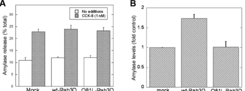
Effect of overexpression of myc-Rab3Dwt or myc-Rab3DQ81L on CCK-8-induced amylase release and amylase levels in AR42J cells
Fig. 3. Overexpression of myc-Rab3D wt and Q81L in AR42J cells does not affect amylase localization. AR42J cells transfected with myc-Rab3Dwt (top panels) or myc-Rab3DQ81L (lower panels) plasmids were cultured in the presence of Dex for 2 days, washed with PBS and fixed with 4% paraformaldehyde in PBS. The cells were immunostained for the myc antigen and amylase as described in Methods. Amylase staining is shown on the left and immunostaining of the myc antigen on the right. Comparison of amylase staining in positive (white arrowheads) and negative cells revealed that overexpression of neither myc-Rab3Dwt nor myc-Rab3DQ81L affects the staining or distribution of amylase in AR42J cells. Scale bars, 10 um.
Next, we measured total amylase levels in mock-transfected AR42J cells and AR42J cells overexpressing myc-Rab3Dwt or myc-Rab3DQ81L. Results were expressed as amylase units per mg protein and normalized to amylase levels in mock-transfected cells. As shown in Fig. 4B, overexpression of myc-Rab3Dwt resulted in a 1.73-fold increase in cellular amylase levels when compared with mock-transfected cells, whereas overexpression of myc-Rab3DQ81L did not affect amylase levels. These findings suggest that Rab3D is involved in regulating the amount of amylase stored within the cell.
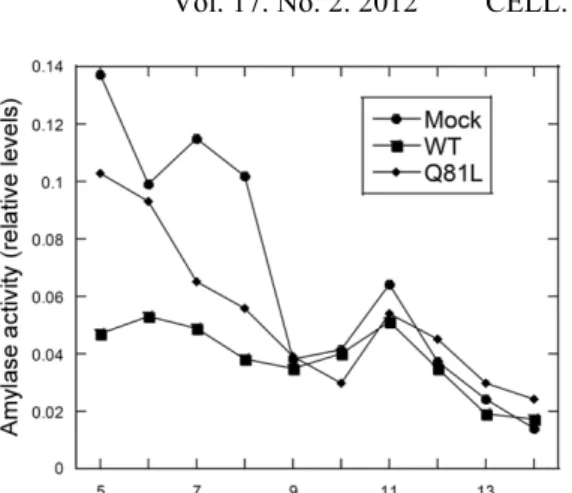
Overexpression of myc-Rab3Dwt or myc-Rab3DQ81L does not affect long-term basal amylase release or granule density
Fig. 5. Density gradient centrifugation of mock-transfected and AR42J cells transfected with myc-Rab3Dwt or myc-Rab3DQ81L. Mock-transfected AR42J cells and AR42J cells transfected with myc-Rab3Dwt or myc-Rab3DQ81L plasmids were treated with Dex for 2 days and harvested into lysing buffer. A post-nuclear supernatant (PNS) was prepared and applied to a 10-30% continuous iodixanol gradient as described in Methods. Fractions (150 ul) were collected from the top of the gradient. Amylase levels in the fractions were determined and the data were normalized to 1, which represents the maximum level of amylase activity. A peak in amylase activity was observed in F11 (arrow) in mock-transfected AR42J cells as well as AR42J cells mock-transfected with Rab3Dwt or myc-Rab3DQ81L plasmids, indicating that overexpression of myc-Rab3D WT or Q81L does not affect granule density. Similar results were observed in three experiments.
DISCUSSION
Although many different approaches have been employed in examining the role of Rab3D in exocrine cell function, its role remains unclear. In the present study, we examined the role of Rab3D in Dex-treated AR42J cells, a cell line derived from rat pancreatic cancer cells that resembles pancreatic acinar cells when treated with Dex [26, 27]. Immunoblotting of subcellular fractions revealed that approximately 80% of cellular Rab3D is membrane-associated in AR42J cells, a value similar to that observed in parotid acinar cells [16]. We examined the effect of CCK-8-stimulated amylase release on the subcellular localization of Rab3D in AR42J cells and found that the level of membrane-associated Rab3D did not change following CCK-8 treatment. In a previous study we observed an increase in membrane-associated Rab3D following agonist-induced amylase release in isolated parotid acini [16]. However, in those studies agonist-induced amylase release from isolated parotid acini was increased >4-fold over basal levels, whereas in the present study CCK-8-induced amylase release was increased approximately 2-fold over basal levels. Hence, detectable redistribution of Rab3D may require a relatively robust secretory response.
[23, 28], the effect of secretory agonists on cell morphology has not been examined. We found that in response to treatment with CCK-8, the Rab3D-positive amylase-containing granules appear to move toward the periphery of the cell. Moreover, Rab3D-positive, amylase-negative structures were observed in the cell interior following stimulation with CCK-8. These Rab3D-positive structures may represent granule membrane that has been retrieved following regulated exocytosis. We did not observe Rab3D on the plasma membrane under any conditions in AR42J cells, suggesting that Rab3D-positive membrane components are removed from the granule membrane prior to fusion with the cell membrane. This is consistent with an earlier study in which a Rab3-like protein became associated with the Golgi, not the plasma membrane, following agonist-induced secretion in pancreatic acinar cells [32]. Moreover, we found an increase in the amount of Rab3D associated with low-density membrane fractions in isolated parotid acini following agonist-induced amylase release [20]. Taken together, these findings suggest that Rab3D may be recycled from the secretory granules to the Golgi prior to granule fusion with the cell membrane. Exactly how this redistribution occurs remains to be determined. To further elucidate the role of Rab3D in acinar cell function, we overexpressed wild-type and a constitutively active form of Rab3D in AR42J cells. We employed a modified transfection method using GeneExpresso transfection reagent in which the cells were transfected by incubation with the DNA/reagent complexes prior to plating. This method improved the transfection efficiency from <10% to >35%. Although immunoblotting of subcellular fractions revealed that the majority of recombinant Rab3D was localized in the cytosol, membrane-associated Rab3D levels were increased >3-fold over mock transfected cells. Overexpression of myc Rab3D (mutant or WT) did not affect the localization of endogenous Rab3D.
With respect to functional studies, we found that overexpression of Rab3D (WT or mutant) did not affect agonist-induced amylase release from AR42J cells. This is consistent with what has been previously observed with this cell line [23]. However, others have demonstrated that overexpression of Rab3D does affect regulated exocytosis in exocrine cells using other approaches and cell systems. For example, overexpression of Rab3D in transgenic mice resulted in an increase in agonist-induced amylase release from pancreatic acini [33]. In contrast to these findings, overexpression of WT and the constitutively active mutant Rab3D in pancreatic acini using adenoviral vectors did not affect agonist-induce amylase release, whereas overexpression of a dominant-negative mutant (deficient in guanine nucleotide binding) inhibited amylase release [34]. Although different effects of mutant and WT Rab3D proteins were observed, these studies suggest a role for Rab3D in exocytosis.
and inactive forms of Rab3D may be required for these effects to be seen. Immunocytochemistry revealed that overexpression of Rab3D did not alter granule morphology or localization in AR42J cells. Moreover, density gradient centrifugation studies showed that granule density was unchanged in cells overexpressing mutant or WT Rab3D. We proposed that the increase in amylase levels in cells overexpressing myc-Rab3Dwt may be due to a subtle change in basal amylase release. However, long-term basal amylase release was not altered by overexpression of mutant or WT Rab3D. Hence, Rab3D most likely modulates amylase content in AR42J cells by regulating granule biosynthesis. Several studies indicate a role for Rab3D in granule biogenesis as opposed to late fusion events. In a Rab3D-deficient mouse model, agonist-induced amylase release was not altered in parotid or pancreas [25]. Interestingly, granule size was doubled in these mice, suggesting a role for Rab3D in granule formation [25]. With respect to studies using Rab3D-deficient mice, one may argue that other Rab proteins expressed in the acinar cell, specifically Rab27B, may overlap in function with Rab3D, and, therefore, offset the loss of Rab3D in Rab3D-deficient mice. This issue remains to be resolved. However, another more recent study has demonstrated that the transcription factor MIST1 induces the formation of large granules in gastric chief cells by inducing expression of Rab3D and Rab26 [35]. Similarly, overexpression of mutant Rab3D interfered with the formation of Weibel-Palade bodies in platelets [36]. All of these studies support a role for Rab3D in regulating granule formation and, perhaps, granule content. Although we did not measure granule size in the present study, the increase in amylase levels we observed in AR42J cells overexpressing myc-Rab3Dwt may be a result of an increase in granule size and/or number.
Others have demonstrated that Rab3D is associated with subpopulations of secretory granules [20, 37] and is actually shed from granule membranes prior to
exocytosis [19]. Valentijn et al. [19] found that pancreatic granules engaging in
exocytosis were coated with actin while Rab3D was released from the granule membrane. Whether or not Rab3D is involved in regulating actin binding to granules remains to be elucidated. More recently, Rab3D has been localized to the early Golgi compartments in Brunner’s gland acinar cells and intestinal goblet cells [18], suggesting a role for Rab3D much earlier in the secretory pathway than previously considered. These findings, along with those in the present study, further support the hypothesis that Rab3D may not be involved in the actual fusion process in exocrine cells.
To conclude, we examined the expression and function of Rab3D in AR42J cells using what we believe is a more efficient transfection procedure. Although Rab3D is associated with amylase-positive secretory granules in these cells, our results indicate that Rab3D is not involved in agonist-induced exocytosis. Rather, Rab3D may regulate amylase content in these cells.
REFERENCES
1. Jordens, I., Marsman, M., Kuijl, C. and Neefjes, J. Rab proteins, connecting
transport and vesicle fusion. Traffic 6 (2005) 1070-1077.
2. Grosshans, B.L., Ortiz, D. and Novick, P. Rabs and their effectors:
achieving specificity in membrane traffic. Proc. Natl. Acad. Sci. USA. 103
(2006) 11821-11827.
3. Burton, J. and De Camilli, P. A novel mammalian guanine nucleotide
exchange factor (GEF) specific for rab proteins. Adv. Second Messenger
Phosphoprotein Res. 29 (1994) 109-119.
4. Takai, Y., Sasaki, T. and Matozaki, T. Small GTP-binding proteins. Physiol.
Rev. 81 (2001) 153-208.
5. Collins, R.N. “Getting it on"-GDI displacement and small GTPase
membrane recruitment. Mol. Cell 12 (2003) 1064-1066.
6. Wu, S.K., Zeng, K., Wilson, I.A. and Balch, W.E. Structural insights into
the function of the rab-GDI superfamily. TIBS 21 (1996) 472-476.
7. Alory, C. and Balch, W.E. Molecular evolution of the
Rab-escort-protein/guanine-nucleotide-dissociation-inhibitor superfamily. Mol. Biol.
Cell 14 (2003) 3857-3867.
8. Pfeffer, S.R. Rab GDP dissociation inhibitor: Putting rab GTPases in the
right place. J. Biol. Chem. 270 (1995) 17057-17059.
9. Wu, Y.W., Tan, K.T., Waldmann, H., Goody, R.S. and Alexandrov, K.
Interaction analysis of prenylated Rab GTPase with Rab escort protein and
GDP dissociation inhibitor explains the need for both regulators. Proc. Natl.
Acad. Sci. USA 104 (2007) 12294-12299.
10.Ohnishi, H., Ernst, S.A., Wys, N., McNiven, M. and Williams, J.A. Rab3D
localizes to zymogen granules in rat pancreatic acini and other exocrine
glands. Am. J. Physiol. 271 (1996) G531-G538.
11.Chen, X., Walker, A.K., Strahler, J.R., Simon, E.S., Tomanicek-Volk, S.L.,
Nelson, B.B., Hurley, M.C., Ernst, S.A., Williams, J.A. and Andrews P.C. Organellar proteomics: analysis of pancreatic zymogen granule membranes.
Mol. Cell Proteomics 5 (2006) 306-312.
12.Chen, X., Li, C., Izumi, T., Andrews, P.C. and Williams, J.A. Rab27b
localizes to zymogen granules and regulates pancreatic acinar exocytosis.
Biochem. Biophys. Res. Commun. 323 (2004) 1157-1162.
13.Valentijn, J.A., Gumkowski, M.D. and Jamieson, J.D. The expression
pattern of rab3D in the developing rat exocrine pancreas coincides with the
acquisition of regulated exocytosis. Eur. J. Cell Biol. 71 (1996) 129-136.
14.Raffaniello, R.D., Lin, J., Wang, F. and Raufman, J-P. Expression of rab3D
in dispersed chief cells from guinea pig stomach. Biochem. Biophys. Acta
1311 (1996) 111-116.
15.Tang, L.H., Gumkowski, F.D., Sengupta, D., Modlin, I.M. and Jamieson, J.D.
Rab3D protein is a specific marker for zymogen granules in gastric chief
16.Raffaniello, R.D., Lin, J., Schwimmer, R. and Ojakian, G.K. Expression and
localization of Rab3D in rat parotid gland. Biochem. Biophys. Acta 1450
(1999) 352-363.
17.Pavlos, N.J., Xu, J., Riedel, D., Yeoh, J.S., Teitelbaum, S.L., Papadimitriou,
J.M., Jahn, R., Ross, F.P. and Zheng, M.H. Rab3D regulates a novel vesicular trafficking pathway that is required for osteoclastic bone
resorption. Mol. Cell Biol. 25 (2005) 5253-5269.
18.Valentijn, J.A., van Weeren, L., Ultee, A. and Koster, A.J. Novel
localization of Rab3D in rat intestinal goblet cells and Brunner's gland
acinar cells suggests a role in early Golgi trafficking. Am. J. Physiol.
Gastrointest. Liver Physiol. 293 (2007) G165-177.
19.Valentijn, J.A., Valentijn, K., Pastore, L.M. and Jamieson, J.D. Actin
coating of secretory granules during regulated exocytosis correlates with the
release of Rab3D. Proc. Natl. Acad. Sci. USA 97 (2000) 1091-1095.
20.Chan, D., Lin, J. and Raffaniello, R.D. Expression and localization of rab
escort protein isoforms in parotid acinar cells from rat. J. Cell Physiol. 185
(2000) 339-347.
21.Williams, J.A., Chen, X. and Sabbatini, M.E. Small G proteins as key
regulators of pancreatic digestive enzyme secretion. Am. J. Physiol.
Endocrinol. Metab. 296 (2009) E405-414.
22.Gevrey, J.C., Laurent, S., Saurin, J.C., Némoz-Gaillard, E., Regazzi, R.,
Chevrier, A.M., Chayvialle, J.A. and Abello, J. Rab3a controls exocytosis in
cholecystokinin-secreting cells. Febs. Lett. 503 (2001) 19-24.
23.Piiper, A., Leser, J., Lutz, M.P., Beil, M. and Zeuzem, S. Subcellular
distribution and function of Rab3A-D in pancreatic acinar AR42J cells.
Biochem. Biophys. Res. Commun. 287 (2001) 746-751.
24.Nguyen, D., Jones, A., Ojakian, G.K. and Raffaniello, R.D. Rab3D
redistribution and function in rat parotid acini. J. Cell Physiol. 197 (2003)
400-408.
25.Riedel, D., Antonin, W., Fernandez-Chacon, R., Alvarez de Toledo, G., Jo, T.,
Geppert, M., Valentijn, J.A., Valentijn, K., Jamieson, J.D., Südhof, T.C. and Jahn, R. Rab3D is not required for exocrine exocytosis but for the
maintenance of normally sized secretory granules. Mol. Cell Biol. 22 (2002)
6487-6497.
26.Logsdon, C.D., Moessner, J., Williams, J.A. and Goldfine, I.D.
Glucocorticoids increase amylase mRNA levels, secretory organelles, and
secretion in pancreatic acinar AR42J cells. J. Cell Biol. 100 (1985) 1200-1208.
27.Logsdon, C.D. Glucocorticoids increase cholecystokinin receptors and
amylase secretion in pancreatic acinar AR42J cells. J. Biol. Chem. 261
(1986) 2096-2101.
28.Klengel, R., Piiper, A., Pittelkow, S. and Zeuzem, S. Differential expression
of Rab3 isoforms during differentiation of pancreatic acinar cell line AR42J.
29.Qiu, X., Valentijn, J.A. and Jamieson, J.D. Carboxyl-methylation of Rab3D
in the rat pancreatic acinar tumor cell line AR42J. Biochem. Biophys. Res.
Commun. 285 (2001) 708-714.
30.Raffaniello, R.D., Fedorova, D., Ip, D. and Rafiq, S. Hsp90 co-localizes with
rab-GDI-1 and regulates agonist-induced amylase release in AR42J cells.
Cell. Physiol. Biochem. 24 (2009) 369-378.
31.Raffaniello, R.D. and Raufman, J-P. Cytosolic RAB3D is associated with
RAB escort protein (REP), not RAB-GDP dissociation inhibitor (GDI), in
dispersed chief cells from guinea pig stomach. J. Cell. Biochem. 72 (1999)
540-548.
32.Jena, B.P., Gumlowski, F.D., Konieczko, E.M., VonMollard, G.F., Jahn, R.
and Jamieson, J.D. Redistribution of a rab3-like GTP-binding protein from secretory granules to the golgi complex in pancreatic acinar cells during
regulated exocytosis. J. Cell Biol. 124 (1994) 43-53.
33.Ohnishi, H., Samuelson, L.C., Yule, D.I., Ernst, S.A. and Williams, J.A.
Overexpression of Rab3D enhances regulated amylase secretion from
pancreatic acini of transgenic mice. J. Clin. Invest. 100 (1997) 3044-3052.
34.Chen, X., Edwards, J.A.S., Logsdon, D., Ernst, S.A. and Williams, J.A.
Dominant-negative Rab3D inhibits amylase release from mouse pancreatic
acini. J. Biol. Chem. 277 (2002) 18002-18009.
35.Tian, X., Jin, R.U., Bredemeyer, A.J., Oates, E.J., Błazewska, K.M.,
McKenna, C.E. and Mills, J.C. RAB26 and RAB3D are direct transcriptional targets of MIST1 that regulate exocrine granule maturation.
Mol. Cell Biol. 30 (2010) 1269-1284.
36.Knop, M., Aareskjold, E., Bode, G. and Gerke, V. Rab3D and annexin A2
play a role in regulated secretion of vWF, but not tPA, from endothelial
cells. EMBO J. 23 (2004) 2982-2992.
37.van Weeren, L., de Graaff, A.M., Jamieson, J.D., Battenburg, J.J. and
Valentijn, J.A. Rab3D and actin reveal distinct lamellar body subpopulations
in alveolar epithelial type II cells. Am. J. Respir. Cell Mol. Biol. 30 (2004)