CELLULAR & MOLECULAR BIOLOGY LETTERS http://www.cmbl.org.pl
Received: 05 April 2007 Volume 13 (2008) pp 230-239 Revised form accepted: 17 September 2007 DOI:10.2478/s11658-007-0049-3 Published online: 16 November 2007 © 2007 by the University of Wrocław, Poland
*
Author for correspondence: Department of Cardiology, 1st Chair of Cardiology and Cardiac Surgery, Medical University in Łódź, Poland. Sterlinga 1/3; 91-425 Łódź, Poland, e-mail: m.banach@termedia.pl, tel/fax: +48 42 636 44 71Abbreviations used: catalase – CAT; CHD – coronary heart disease; GSH-Px – glutathione peroxidase; ROS – reactive oxygen species; SOD-1 – superoxide dismutase
Research article
CARVEDILOL MODIFIES ANTIOXIDANT STATUS OF PATIENTS WITH STABLE ANGINA
JAN KOWALSKI1 , MACIEJ BANACH2*, MARCIN BARYLSKI1, ROBERT IRZMANSKI1 and LUCJAN PAWLICKI1
1Department of Internal Medicine and Cardiological Rehabilitation, Medical University of Łódź, Poland, 2Department of Cardiology, 1st Chair of Cardiology
and Cardiac Surgery, Medical University of Łódź, Poland
Key words: Stable angina, Carvedilol, Superoxide dismutase, Peroxidase, Catalase, Plasma antioxidative activity
INTRODUCTION
There is growing evidence on the role of lower systemic antioxidant defense in the development of ischemic changes in patients with stable angina. Activated neutrophils are recognized as a major source of the free radicals that are responsible for lipid peroxidation, the inhibition of prostacyclin synthesis, and disturbance in the production of nitric oxide, the fundamental biological function of which is relaxation of the smooth muscles and inhibition of platelet aggregation [1].
Beta-blockers are one of the basic groups of drugs used in coronary heart disease (CHD). A particular place in this group belongs to carvedilol, a beta-blocker with vasodilating and antioxidative activity. It is thought that its antioxidative activity is connected with the cardioprotective effect of the drug [1-3]. Moreover, unlike other beta-blockers, carvedilol is supposed to modify the lipid profile of the plasma beneficially [4, 5].
Although the benefits of carvedilol are well documented, its antioxidative activity in patients with stable angina is still not well understood. Therefore, we conducted our study in order to estimate the effect of carvedilol on erythrocyte superoxide dismutase (SOD-1), glutathione peroxidase (GSH-Px) and catalase (CAT) activities and on plasma antioxidative activity in patients with stable angina.
MATERIALS AND METHODS
Study population
Patients who smoked, had been diagnosed with diabetes mellitus, liver or kidney failure, or systemic diseases, or were taking steroids and/or antioxidants (vitamins C, A, E, angiotensin-converting enzyme inhibitors, statins, angiotensin receptor blockers) were excluded from the study. The rationale for these exclusion criteria was that these conditions could influence the antioxidant status of the patients and yield incorrect results. After applying these exclusion criteria to 75 patients, a group of 30 patients with diagnosed stable angina were qualified for investigations.
acetylsalicylic acid administered in a daily dose of 75 mg and temporarily short-acting nitrates 2 months prior to and during carvedilol therapy.
The control group consisted of 12 clinically healthy subjects (8 men and 4 women), aged 39-49 years (mean 45.7 ± 3.7 years). They were healthy volunteers, without coronary heart disease. These subjects did not smoke cigarettes and did not take any drugs, including antioxidative vitamins, and they were qualified for the study at the same time as the patients.
There were no significant differences in terms of the presence of risk factors for atherosclerosis between the group of healthy subjects and the patients with stable angina (i.e. genetic predispositions, elevated total cholesterol in the serum, bad nutritional habits, obesity, high body weight, low physical activity). Detailed patient characteristics are shown in Tab. 1.
Tab. 1. Patients’ detailed characteristics.
Study Group Control Group
Number of Pts. (n) 30 12
Sex M/F (n [%]) 20 [66.7%]/10 [33.3%] 8 [66.7%]/4 [33.3%] Age - range [yrs]
- mean [yrs]
37-49 45.6 ± 4.2*
39-49 45.7 ± 3.7
BMI (kg/m²) 29.2 ± 3.7* 28.7 ± 4.1
TC (mg/dl) 212.24 ± 30.98* 207.45 ± 3.67
HDL (mg/dl) 44.59 ± 11.82* 43.07 ± 12.58
LDL (mg/dl) 137.14 ± 27.05* 131.09 ± 3.15
Acetylsalicylic acid in a daily dose
of 75 mg no drugs
Other drugs during
the study Short-acting nitrates 2 months prior
to and during carvedilol therapy. no drugs * P > 0.05, not significant
Blood samples (5 ml) were collected from the cubital vein in the early morning, about 12 h after the previous meal. They were collected immediately before drug administration, and 4, 8 and 12 weeks after its administration to the patients, and once from the control group. All the determinations were performed within 2 h of blood collection from the examined subject, according to the applied method. The laboratory staff who performed the determinations were blind to the group the sample came from.
Antioxidative enzymes
Erythrocyte SOD-1 (EC 1.15.1.1) activity was determined according to the method of Misra and Fridovich [6]. Erythrocyte GSH-Px (EC 1.11.1.9) activity was measured according to the method of Little et al. using coumene hydroperoxide as a substrate [7]. Erythrocyte catalase (EC 1.1.11.6) activity was measured according to Beers et al. [8]. The antioxidative activity of the blood plasma was determined via the method of Bartosz et al. [9], based on the reduction of the preformed cation radical of 2,2,azinobis (3-ethylbenzothiazo-line-6-sulphonic acid) by the blood plasma.
Statistical analysis
The data was analyzed using Student’s t-test for independent groups. The comparisons between each measurement were performed with the t-test for paired data. Values are expressed as a mean ± SD. The probability value p < 0.05 was considered significant.
RESULTS
Erythrocyte superoxide dismutase activity
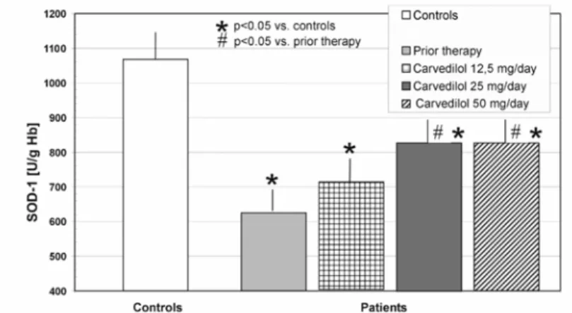
The SOD-1 activity in the erythrocytes was markedly lower (p < 0.05) in patients with stable angina qualified to carvedilol therapy (627.0 ± 60.5 U/gHb) than in healthy subjects (1068.0 ± 78.2 U/gHb) (Fig. 1). The activity increased to 712.5 ± 75.0 U/gHb after 4 weeks of carvedilol therapy (12.5 mg/24 h) and to 830.0 ± 70.5 U/gHb after the next 4 weeks (25 mg/24 h) (Fig. 1), and did not change after a further 4 weeks of treatment with carvedilol (50 mg/24 h), when it was 837.0 ± 75.05 U/gHb (Fig. 1). The increase in SOD-1 activity in erythrocytes was observed during carvedilol therapy, but its value remained significantly lower (p < 0.05) than in healthy subjects.
Comparing the results of SOD-1 activity in erythrocytes in the groups of patients with stable angina qualified to carvedilol therapy, we did not observe significant changes between the groups. However, markedly different results were obtained for the comparison of patients before carvedilol therapy and patients who received carvedilol in doses of 25 and 50 mg/24 h (p < 0.05) (Fig. 1).
Erythrocyte glutathione peroxidase activity
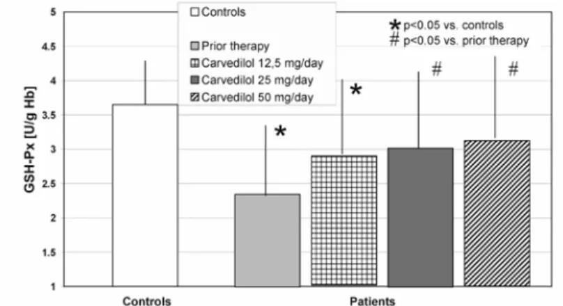
The GSH-Px activity in the erythrocytes was significantly lower (p < 0.05) in patients with stable angina qualified to carvedilol therapy than in healthy subjects (2.36 ± 1.02 U/gHb vs. 3.65 ± 0.64 U/gHb) (Fig. 2). The activity increased to 2.90 ± 1.10 U/gHb after 4 weeksof carvedilol therapy (12.5 mg/24 h) but was still significantly lower (p < 0.05) than in the healthy subjects (Fig. 2).
Fig. 2. The effect of carvedilol on glutathione peroxidase (GSH-Px) activity in erythrocytes. Values are expressed as means ± SD (# means significant changes between pts. without and with carvedilol therapy).
After another 4 weeks of carvedilol therapy (25 mg/24 h), the GSH-Px activity in erythrocytes increased to 3.05 ± 1.10 U/gHb and did not differ significantly from that observed in the healthy subjects (Fig. 2). Similarly results were obtained after a further 4 weeks of the drug therapy (50 mg/24 h): erythrocyte GSH-Px activity increased to 3.15 ± 1.28 U/gHb and did not differ from that observed in the healthy subjects (Fig. 2). Comparing the results of GSH-Px activity in erythrocytes in the groups of patients with stable angina qualified to carvedilol therapy, we did not observe significant changes between the groups, but markedly different results were obtained during the comparison of patients before carvedilol therapy and patients who had carvedilol administered in doses of 25 and 50 mg/24 h (p < 0.05) (Fig. 2).
Erythrocyte catalase activity
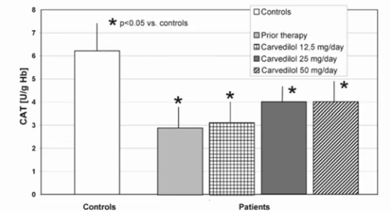
after the next 4 weeks (12.5 mg/24 h) and to 4.01 ± 0.85 U/gHb after a further 4 weeks of the carvedilol therapy (25 mg/24 h) (Fig. 3). The CAT activity in the erythrocytes did not change after the next 4 weeks of the treatment (50 mg/24 h) (4.5 ± 0.90 U/gHb) (Fig. 3). An increase in CAT activity in the erythrocytes was observed during carvedilol therapy, but it was still lower (p < 0.05) than in the healthy subjects. Comparing the results of the CAT erythrocyte activity in the groups of patients with stable angina qualified to carvedilol therapy, we did not observe significant changes between the groups (Fig. 3).
Fig. 3. The effect of carvedilol on catalase(CAT) activity in erythrocytes. Values are expressed as means ± SD.
Plasma antioxidative activity
Plasma antioxidative activity was significantly lower (p < 0.05) in patients with stable angina qualified to carvedilol therapy than in healthy subjects (327.65 ± 70.1 vs. 544.2 ± 50.12 μmol Trolox equivalents/litre of plasma) (Fig. 4). After four weeks of carvedilol therapy (12.5 mg/24 h), an increase (p < 0.05; 349.24 ± 76.20 μmol Trolox equivalents/litre of plasma) in plasma antioxidative activity was found compared to the initial value (Fig. 4). An increase (p < 0.05) in plasma antioxidative activity (388.8 ± 81.52 μmol Trolox equivalents/litre of plasma) was also observed after the next 4 weeks of carvedilol therapy (25 mg/24 h) (also as compared to the initial value) (Fig. 4). The activity did not change after a further 4 weeks of carvedilol therapy (50 mg/24 h) (391.74 ± 82.8 μmol Trolox equivalents/litre of plasma) (Fig. 4). The plasma antioxidative activity increased during carvedilol therapy, but was still markedly lower (p < 0.05) than in healthy subjects.
Fig. 4. The effect of carvedilol on plasma antioxidative activity. Values are expressed as means ± SD (# means significant changes between pts. without and with carvedilol therapy).
DISCUSSION
In our study, we observed an increase in the activities of antioxidative enzymes (SOD-1, GSH-Px and CAT) in the erythrocytes during carvedilol administration to patients with stable angina pectoris. Furthermore, during carvedilol administration, an increase in plasma antioxidative activity was observed.
Red blood cells are particularly exposed to free radical action as they carry molecular oxygen which are a potential source of ROS (reactive oxygen species). To prevent a peroxidation reaction, red blood cells are equipped with various enzymatic and non-enzymatic defense mechanisms, which become exhausted under conditions of oxidative stress.
In our study, the baseline values of SOD-1, GSH-Px and CAT activity were significantly lower in patients with stable angina pectoris as compared with those for the group of healthy subjects. During carvedilol therapy, an increase in SOD-1 and CAT activities was found in the erythrocytes, but their activity remained markedly lower than in the healthy subjects. Also, over the whole course of the therapy, an increase in GSH-Px activity was observed in the red blood cells. In experimental and clinical studies, decreased activity of antioxidative enzymes was indicated in CHD and other diseases [10-15].
Increased production of free radicals induces adaptative reactions in the organism. Initially, an increase in enzymes synthesis is observed. Then there is a decrease in their activity, which can indicate exhaustion of the antioxidative defense mechanisms.
Based on published reports, it may be concluded that the intensity of antioxidative enzyme biosynthesis depends on the intensification of ROS formation [18, 19]. In the case of long-lasting and intensive exposure to ROS, a decrease in the activity of these enzymes comes about. The decrease in their activity indicates deep disturbances in the oxidation-reduction balance in the blood of patients with stable angina. The observed increase in SOD-1, GSH-Px and CAT activity in the erythrocytes of patients treated with carvedilol may indicate its antioxidative action.
In our studies, we found that patients with stable angina have decreased plasma antioxidative activity. During carvedilol administration, an increase in plasma antioxidative activity was observed, but throughout the whole course of therapy, it remained significantly lower than that seen in healthy subjects. The demonstrated carvedilol antioxidative activity speaks for an additional mechanism of its action. Most probably, it is associated with the fact that carvedilol, by causing a decrease in neutrophil O2 generation, indirectly affects lower consumption of anti-oxidative enzymes and thus, their activity increases. Furthermore, there is an increased possibility of their regeneration owing to the substrate decrease (O2). The observed changes may also result from carvedilol having a protective effect on endothelium oxidative and nitrosative stress.
In physiochemical, biochemical and cellular assays, carvedilol and several of its metabolites inhibited lipid peroxidation, scavenged ROS, inhibited the formation of ROS and prevented the depletion of endogenous antioxidants such as vitamin E and glutathione [20-24]. Since the elements of the non-enzymatic component of the system protecting against the effects of the excessive activity of ROS may, as the exposure proceeds, become exhausted during the adaptative mechanism, their functions are gradually adopted by antioxidative enzymes. Their activity may temporarily increase, and a decrease in their activity evidences deep disturbances of oxidation-reduction balance. Extensive disturbances in the antioxidative defense mechanism observed in angina patients are the result of atherosclerosis developing for many years [25-27].
REFERENCES
1. Gilbert, E.M., Abraham, W.T. and Olsen, S. Comparative hemodynamic, left ventricular functional, and antiadrenergic effects of chronic treatment with metoprolol versus carvedilol in the failing heart. Circulation 94 (1996) 2817-2825.
2. Banach, M., Goch, A., Misztal, M., Rysz, J., Barylski, M., Jaszewski, R. and Goch, J.H. Low output syndrome following aortic valve replacement. Predictors and prognosis. Arch. Med. Sci. 3 (2007) 117-122.
3. Ruffolo, R.R. Jr. and Feurwstein, G.Z. Pharmacology of carvedilol: rationale for use in hypertension, coronary artery disease, and congestive heart failure. Cardiovasc. Drugs Ther. 11 (1997) 247-256.
4. Heusch, F., Skyschally, A., Leineweber, K., Haude, M., Erbel, R. and Heusch, G. The interaction of coronary microembolization and ischemic preconditioning: A third window of cardioprotection through TNF-alpha. Arch. Med. Sci. 3 (2007) 83-92.
5. Hauf-Zachariou, U. A double-blind comparison of the effects of carvedilol and captopril on the lipid profile in patients with mild to moderate hypertension and dyslipidemia. Eur. J. Clin. Pharmacol. 45 (1993) 95-100.
6. Misra, H.P. and Fridovich, I. The role of superoxide anion in the autooxidation of epinephrine and simple assay for superoxide dismutase. J. Biol. Chem. 247 (1972) 3170-3175.
7. Little, C. and O’ Brien, P.J. An intracellular GSH-peroxidase with a lipid peroxide substrate. Bioch. Biophys. Res. Comm. 31 (1968) 145-150.
8. Beers, R. and Sizer, J.W. Spectrofotometric method for measuring the breakdown of hydrogen peroxide by catalase. J. Biol. Chem. 195 (1952) 133-140. 9. Bartosz, G., Janaszewska, A. and Ertel, D. Spectrophotometric determination of peroxyl radical-trapping capacity with ABAP and ABTS. Curr. Topics. Biophys. 22 (1998) 11-13.
10. Landmesser, U., Merten, R., Spiekermann, S., Büttner, K., Drexler, H. and Hornig, B. Vascular extracellular superoxide dismutase activity in patients with coronary artery disease: relation to endothelium-dependent vasodilation. Circulation 101 (2000) 2264-2270.
11. Janssen, M., Yandermeer, P. and Dejong, J.W. Antioxidant defences in rat, pig, guinea pig and human hearts - comparison with xanthine oxidoreductase activity. Cardiovasc. Res. 27 (1993) 2052-2057.
12. Hapyn, E., Czerwonka-Szaflarska, M. and Drewa, G. Enzymatic efficiency of erythrocyte antioxidant barrier and lipid peroxidation in children from families with high risk of early atherosclerosis. Med. Sci. Monit. 6 (2000) 112-116. 13. Rogowicz, A., Litwinowicz, M., Pilacinski, S., Zozulinska, D. and
Wierusz-Wysocka, B. Does early insulin treatment decrease the risk of microangiopathy in non-obese adults with diabetes. Arch. Med. Sci. 3 (2007) 129-135.
15. Rysz, J., Blaszczak, R., Banach, M., Kedziora-Kornatowska, K., Kornatowski, T., Tanski, W. and Kedziora, J. Evaluation of chosen parameters of antioxidative system in patients with type 2 diabetes in different periods of metabolic compensation. Arch. Immunol. Ther. Exp. 55 (2007) in press. 16. Kowalski, J., Barylski, M., Banach, M., Grycewicz, J., Irzmanski, R. and
Pawlicki, L. Neutrophil superoxide anion generation during atorvastatin and fluvastatin therapy used in coronary heart disease primary prevention. J. Cardiovasc. Pharmacol. 48 (2006) 143-147.
17. Jayakumari, N., Ambikakumari, V. and Balakrishnan, K.G. Antioxidant status in relation to free radical production during stable and unstable anginal syndromes. Atherosclerosis 94 (1992) 183-190.
18. Cai, H. and Harrison, D.G. Endothelial dysfunction in cardiovascular diseases: the role of oxidant stress. Circ. Res. 87 (2000) 840-840.
19. Harrison, D.G. Cellular and molecular mechanisms of endothelial dysfunction. J. Clin. Invest. 100 (1998) 2153-2157.
20. Santos, D.J and Moreno, A.J. Inhibition of heart mitochondrial lipid peroxidation by non-toxic concentrations of carvediolol and its analog BM-910228. Biochem. Pharmacol. 61 (2001) 155-164.
21. Banach, M., Okonski, P., Kosmider, A., Irzmanski, R., Rysz, J., Olszewski, R., Zwolinski, R. and Zasłonka, J. Aortic valve replacement in patients with heart failure. Pol. Merkur. Lekarski 20 (2006) 642-645.
22. Feuerstein, G.Z. and Ruffolo, R.R. Carvedilol, a novel vasodilating beta-blocker with the potential for cardiovascular organ protection. Eur. Heart J. 17 (1996) 24-29.
23. Kowalski, J., Błaszczyk, J., Petecka, E., Irzmański, R., Kowalczyk, E., Kowalska, E., Cegliński, T. and Pawlicki, L. Neutrophils superoxide anion generation during carvedilol therapy in patients with stable angina. Int. J. Cardiol. 102 (2005) 397-402.
24. Tadolini, B. and Franconi, F. Carvedilol inhibition of lipid peroxidation. A new antioxidative mechanism. Free. Radic. Res. 5 (1998) 377-387.
25. Banach, M., Drozdz, J., Okonski, P. and Rysz, J. Immunological aspects of the statins’ function in patients with heart failure. A raport from the Annual Conference of ESC – Heart Failure 2005. Cell. Mol. Immunol. 2 (2005) 433-437.
26. Irzmanski, R., Piechota, M., Barylski, M., Banach, M., Gławęda, B., Kowalski, J., Cierniewski, C., Kośmider, M. and Pawlicki, L. Dynamics of changes of the BNP concentration in patients with stable angina pectoris qualified for PTCA. Dependence on the selected morphological and haemodynamic parameters. Arch. Med. Sci. 2 (2006) 15-19.