University of Zurich
Zurich Open Repository and Archive
Winterthurerstr. 190 CH-8057 Zurich http://www.zora.uzh.ch
Year: 2010
Application of label-free absolute quantitative proteomics in
human gingival crevicular fluid by LC/MS E (gingival
exudatome)
Bostanci, N; Heywood, W; Mills, K; Parkar, M; Nibali, L; Donos, N
Bostanci, N; Heywood, W; Mills, K; Parkar, M; Nibali, L; Donos, N (2010). Application of label-free absolute quantitative proteomics in human gingival crevicular fluid by LC/MS E (gingival exudatome). Journal of Proteome Research, 9(5):2191-2199.
Postprint available at: http://www.zora.uzh.ch
Posted at the Zurich Open Repository and Archive, University of Zurich. http://www.zora.uzh.ch
Originally published at:
Journal of Proteome Research 2010, 9(5):2191-2199.
Bostanci, N; Heywood, W; Mills, K; Parkar, M; Nibali, L; Donos, N (2010). Application of label-free absolute quantitative proteomics in human gingival crevicular fluid by LC/MS E (gingival exudatome). Journal of Proteome Research, 9(5):2191-2199.
Postprint available at: http://www.zora.uzh.ch
Posted at the Zurich Open Repository and Archive, University of Zurich. http://www.zora.uzh.ch
Application of label-free absolute quantitative proteomics in
human gingival crevicular fluid by LC/MS E (gingival
exudatome)
Abstract
Periodontal disease is perhaps the most common infectious disease in humans. Gingival crevicular fluid (GCF) is a local inflammatory exudate of the periodontal tissues. Its composition greatly varies between health and periodontal disease. GCF collection is rapid and noninvasive, but previous approaches aiming to analyze its composition have mainly involved single protein biomarkers. The aim of this study was to perform analysis of the GCF exudatome from healthy and periodontally diseased sites by
LC/MS(E), a label-free mass spectrometry method that enables simultaneous protein identification and absolute quantification in biological fluids. In total, 154 proteins of human, bacterial, and viral origin were identified in the 40 GCF samples obtained from the 10 subjects (five healthy and five generalized aggressive periodontitis). The proportion of bacterial, viral, and yeast protein was increased in disease, compared to health. The presence of host defense-related proteins, such as Cystatin-B and defensins, was confirmed to be present only in health. Among the newly identified GCF proteins were L-plastin detected only in disease (15.6 +/- 12.1 fmol) and Annexin-1 detected in 5-fold higher levels in health. Nevertheless, pro-inflammatory cytokines or periodontal pathogen proteins were rarely detected. Conclusively, the LC/MS(E) technology may facilitate characterization of GCF proteome in periodontal health and disease, thus conferring prognostic and diagnostic value. Larger cohort studies are required to characterize the complete GCF proteome in health and disease.
Application of label free-absolute quantitative proteomics in human gingival crevicular fluid by LC/MSE (Gingival exudatome)
N. Bostanci †§*, W. Heywood‡, K. Mills‡, M. Parkar#, L. Nibali†, N. Donos†*
†Periodontology Unit, Division of Clinical Research, UCL Eastman Dental Institute, London, UK
§ Institute of Oral Biology, Center for Dental and Oral Medicine and
Cranio-Maxillofacial Surgery, Faculty of Medicine, University of Zürich, Zürich, Switzerland
# Division of Biomaterials and Tissue Engineering, UCL Eastman Dental Institute,
London, UK
‡ Biochemistry Group, Clinical and Molecular Genetics Unit, UCL Institute of Child
Health, London, UK
*Corresponding Authors:
Prof. Nikolaos Donos. Unit of Periodontology, Department of Clinical Research, UCL Eastman Dental Institute, 256 Grays Inn Road, London WC1X 8LD.
E-mail: [email protected]. Tel: +44 20 7915 1075; Fax: +44 20 7915 1137 Dr. Nagihan Bostanci. Institute of Oral Biology, Center for Dental and Oral Medicine and Cranio-Maxillofacial Surgery, Faculty of Medicine, University of Zürich, Plattenstrasse 11, 8032, Zürich, Switzerland
E-mail: [email protected]. Tel: +41 44 634 57 53 Fax: +41 44 634 43 10
Key words: Gingival crevicular fluid, periodontal disease, aggressive periodontitis, proteomics, biomarkers, diagnosis, LC/MSE, qTOF MS
Abstract
Periodontal disease is perhaps the most common infectious disease in humans. Gingival crevicular fluid (GCF) is a local inflammatory exudate of the periodontal tissues. Its composition greatly varies between health and periodontal disease. GCF collection is rapid and non-invasive, but previous approaches aiming to analyse its composition have mainly involved single protein biomarkers. The aim of this study was to perform analysis of the GCF exudatome from healthy and periodontally diseased sites by LC/MSE, a label-free mass spectrometry method that enables
simultaneous protein identification and absolute quantification in biological fluids. In total, 154 proteins of human, bacterial and viral origin were identified in the 40 GCF samples obtained from the 10 subjects (five healthy and five generalized aggressive periodontitis). The proportion of bacterial, viral and yeast protein was increased in disease, compared to health. The presence of host defence-related proteins, such as Cystatin-B and defensins was confirmed to be present only in health. Among the newly identified GCF proteins were L- plastin detected only in disease (15.6 ± 12.1 fmol) and Annexin-1 detected in 5-fold higher levels in health. Nevertheless, pro-inflammatory cytokines or periodontal pathogen proteins were rarely detected. Conclusively, the LC/MSE technology may facilitate characterisation of GCF
proteome in periodontal health and disease, thus conferring prognostic and diagnostic value. Larger cohort studies are required to characterise the complete GCF proteome in health and disease.
Introduction
Periodontal disease is perhaps the most common infectious disease in humans caused by bacteria present in the oral cavity, which attach on the teeth and cause inflammation to the surrounding (periodontal) tissues. Longitudinal epidemiological studies show that whilst 90% of the population will develop some signs of the early disease namely gingivitis, only 7-13% appear to be at high risk of developing severe destructive periodontitis (Brown and Loe, 1993). Two main forms of destructive periodontal disease are currently recognized, chronic and aggressive periodontitis. Aggressive periodontitis is often characterized by a rapid and severe periodontal destruction in mainly younger individuals and a tendency for cases to aggregate in families (Albandar and Rams, 2002). In its severe forms, periodontitis may lead to loosening and drifting of the teeth and ultimately tooth loss, resulting in a significant impact on masticatory function and quality of life. Moreover, recent evidence has indicated that periodontal disease could contribute to the development of life-threating conditions namely cardiovascular disease (CVD) or stroke (Danesh et al., 1997). The growing global burden of periodontitis creates a need to identify biomarkers for this disease, establish novel diagnostic tests, or develop new therapies directed at the attenuation of the action of these markers.
Gingival crevicular fluid (GCF) is a serum transudate or an inflammatory exudate that results from the interaction between the bacterial biofilm adherent to the tooth surfaces and the cells of the periodontal tissues (Alfano, 1974). GCF plays a special part in maintaining the structure of junctional epithelium and in the antimicrobial defence of periodontium and acts as a medium for the transport of bacterial products into and host derived products out of the periodontal environment (Ebersole, 2003;
Krasse, 1996). The collection of GCF is a non-invasive, safe procedure, and perhaps the most applicable aid for analysis for development of diagnostic markers(Lamster et al., 1988), since it is closely approximated to the tissues where periodontal disease begins and consequently opens to the whole body (Champagne et al., 2003). Because GCF composition reflects the nature and amplitude of the host response to the microbial plaque challenge, determination of GCF constituent levels may help identify subjects undergoing a transition from health to disease (Bostanci et al., 2007). The circulating GCF proteome holds great promise as a reservoir of information that will be applicable for diagnosis of periodontal disease.
The identification of the composition of GCF as opposed to that of serum or saliva has been more challenging due to its small sample volume. Initial protein analysis of GCF has been elucidated using techniques such as crossed immunoelectrophoresis, sodium dodecyl sulfate polyacrylamide gel electrophoresis (SDS-PAGE), two-dimensional gel electrophoresis coupled with mass spectrometry (MS) and ELISA (Estreicher et al., 1996; Golub and Kleinberg, 1976; Pisano et al., 2005). Using these approaches, more than 60 individual components of GCF have been assessed as candidate markers of disease progression (Lamster and Ahlo, 2007; Ozmeric, 2004; Uitto, 2003). Specifically, plasma proteins, bacterial and host enzymes, various inflammatory mediators have been among the myriad components studied. Although these strategies have provided some insights, it is acknowledged that studying individually these markers is of limited value. To date, however, there is a lack of information regarding proteomic approaches and sample processing in order to analyse the expression of various proteins present in GCF. We anticipate that integrated analysis of the proteome of GCF is required to dissect fully the molecular
mechanisms responsible for periodontal diseases and consequently linked systemic diseases such as CVD and diabetes mellitus (DM).
New developments in proteomic analysis technology could allow for successful analysis of the full complement of proteins in GCF. Recent reports employing label-free quantitative liquid chromatography/tandem mass spectrometry (2D-LC-MS/MS) have demonstrated that protein profiles of oral samples such as saliva may be useful for diagnosis of DM(Rao et al., 2009) . More recently, a new variant of label-free quantification known as LC/MSE was introduced for quadrupole time-of-flight
(Q-Tof) mass spectrometers. For this method, alternating scans of low collision energy and elevated collision energy during LC/ MS analysis are used to obtain both protein quantification and protein identification data in a single sample (Silva et al., 2006; Silva et al., 2005). This technology offers unique advantages including reduced sample consumption, improved detection sensitivity and enhanced data quality for proteomic studies. The aim of this study was to perform a comprehensive analysis of the GCF exudatome from healthy and periodontally diseased sites by LC/MSE. The
results of the present study suggest that the current technology, which requires only a small volume of fluid, is an appropriate platform for analysis of GCF proteome and allowed us to uncover a number of proteins that were not previously known to be present in GCF.
Research Design and Methods
Subjects. From a case-control study, 10 subjects were chosen for GCF proteome analysis. The study had been reviewed and approved by the Joint UCL/UCLH Committees on the Ethics of Human Research (REF:05/Q0502/84). Subjects with
chronic systemic or oral infections, pregnant or lactating, receiving systemic medications meeting, or current smokers were excluded. The selection of the subjects was made according to the criteria proposed by the 1999 International World Workshop for a Classification of Periodontal Disease and Conditions(Armitage, 1999). Patients fulfilling the inclusion criteria were provided with an written informed consent. 40 GCF samples were obtained from healthy subjects (n=5) and patients with generalized aggressive periodontitis (G-AgP) (n=5). Clinical periodontal parameters including probing pocket depth (PPD), clinical recession level (REC), full-mouth bleeding scores (FMBS) and plaque scores (FMPS) were assessed by two calibrated examiners at six sites/tooth with a UNC-15 periodontal probe with a force of 0.3N. Presence or absence of bleeding on probing (BOP) and plaque accumulation was recorded in a dichotomous way. If bleeding occurred within 15 sec after retrieval of the probe, the site is recorded as BOP-positive (Muhlemann and Son, 1971). Presence of visible plaque after running the probe along the gingival margin was recorded as positive. The mean FMPS and FMPS are given as percentage (Table 1). The healthy group consisted of 3 females and 2 males ranging in age from 27 to 38 years with a mean age of 32.4 ± 3.9 years. They exhibited PPD < 3 mm, and no radiographic evidence of alveolar bone loss. The G-AgP group consisted of 3 females and 2 males between the ages of 25 and 44 years (mean of 34.6 ± 6.9 years) with a generalized pattern of severe periodontal tissue destruction and loss of periodontal support inconsistent with age and plaque levels (Armitage, 2004). They exhibited clinical attachment loss ≥ 5 mm on eight or more teeth; at least three of those were other than central incisors or first molars. Statistical analysis for clinical and demographic data included normality testing using the Kolmogorov-Smirnov test and Student t tests or Mann-Whitney tests as appropriate. A P value of less than 0.05 was considered
statistically significant. All data analyses were performed using the SPSS 12.0 software.
Sample Collection and Processing. The GCF samples were collected from the four deepest periodontal pockets (one site per quadrant) in G-AgP patients. In the healthy group, the GCF samples were collected from four standard sites (one first molars and one central incisors in each jaw) exhibiting PPD up to 3 mm without bleeding on probing or radiographic evidence of bone loss. The selected sites were isolated from salivary contamination. A sterile Periopaper strip (ProFlow Inc. Amityville, NY) was then gently inserted into the periodontal pocket and left in place for 30 seconds (Lamster, 1997). Mechanical irritation avoided and the strips contaminated with blood were discarded.
The GCF sample volume was measured with a calibrated Periotron 8000 (Periotron 8000, Proflow, Inc., Amityville, NY, USA) and then the readings were converted to an actual volume (µl) by reference to the standard curve. Four samples from each subject were pooled in an Eppendorf tube prior to storing at -80°C prior to analysis. On the day of the analysis, the GCF samples were re-eluted in 100µl PBS, then centrifuged at 13000 x g for 15 min.
MSe Label Free Quantitation. 10 µl of 100mM Tris, pH 7.8, containing 6M urea was added to 10µl of eluted GCF sample protein and was left shaking at room temperature for 1hr. Disulphide bridges were reduced by the addition of 3 µl of 100mM Tris-HCL, pH 7.8 containing 5M DTE and incubation at room temp for 60 min. Free thiol groups were carboamidomethylated followed by incubation with 6 µl
of 100mM Tris-HCL, pH 7.8 containing 5M iodoacetamide. The solution was then diluted with 155 µl H2O, vortexed and 2 µg of sequence grade trypsin (Sigma
Aldrich, UK) added to the solution. Samples were incubated overnight at 37 0C in a
water bath. After incubation, digested peptides were extracted using self made C8 resin columns. Peptides in aqueous solution were loaded onto the columns washed with 0.1% TFA and eluted in 50% ACN 0.1%TFA. Eluted peptides were then dried down using a Jouan R10.22 Speeedvac. Dried GCF peptides were then re-suspended in 3% ACN with 50fmol of MassPrepTM yeast enolase digestion standard (Waters,
Milford, USA) prior to LC/MSE analysis. Each sample was analyzed in two
independent experimental runs. The LC/MSE analysis was performed using 10 µl of
the final tryptic digest. A standard yeast enolase digest was run before and after the samples to monitor sensitivity.
GCF proteins were identified and quantitated by direct analysis of the reaction mixture described above. All analyses were performed using a nanoAcquity HPLC and QTOF Premier mass spectrometer (Waters Corporation, Manchester, UK). Peptides were trapped and desalted prior to reverse phase separation using a Symmetry C18 5µm, 5mm X 300µm pre-column. Peptides were then separated prior to mass spectral analysis using a 15cm X 75 µm C18 reverse phase analytical column. Peptides were loaded onto the pre-column at a flow rate of 4µl / min in 0.1% formic acid for a total time of 4 mins. Peptides were eluted off the pre-column and separated on the analytical column using a gradient of 3-40% acetonitrile [0.1% formic acid] over a period of 90 min and at a flow rate of 300 nl/min. The column was washed and re-generated at 300 nl/min for 10 min using a 99% acetonitrile [0.1%] rinse. After all non-polar and non peptide material was removed the column was re-equilibrated at
the initial starting conditions for 20 min. All column temp were maintained at 35 0C.
Mass accuracy was maintained during the run using a lock spray of the peptide [glu1]-fibrinopeptide B delivered through the auxiliary pump of the nanoAcquity at a concentration of 300 fmol/l and at a flow rate of 300 nl/min.
Peptides were analysed in positive ion mode using a Q-Tof Premier mass spectrometer (Waters Corp., Manchester, UK) and was operated in v-mode with a typical resolving power of 10,000 FWHM. Prior to analyses, the TOF analyser was calibrated using [glu1]-fibrinopeptide B fragments obtained using collision energy of 25 eV and over the mass range 50-2000 m/z. Post calibration of data files were corrected using the doubly charged precursor ion of [glu1]-fibrinopeptide B (785.8426 m/z) with a sampling frequency of 30 sec. Accurate mass LC-MS data were colleted in a data independent and alternating, low and high collision energy mode. Each low/high acquisition was 1.5s with 0.1 sec interscan delay. Low energy data collections were performed at constant collision energy of 4 eV, high collision energy acquistions were performed using a 15-40eV ramp over a 1.5 sec time period and a complete low/high energy acquisition achieved every 3.2 sec.
Data Processing and Database Searching.
ProteinLynx GlobalServer version 2.3.5 was used to process all data acquired. Protein identifications were obtained by searching UniProt/Swiss-Prot databases to which the sequence of yeast enolase was added manually. Protein identification from the low/high collision spectra for each sample was processed using a hierarchical approach where more than three fragment ions per peptide, seven fragment ions per protein and more than two peptides per protein had to be matched. Since
single-peptide protein identifications are more likely to represent false-positive data points, all proteins with greater than two peptides identified with high confidence were considered for protein quantification. Carboamidomethylation was set as a fixed modification and oxidation was set as variable modification. The average intensity value of the top three ionizing peptides to yeast enolase was used to convert the average intensity of the top three ionizing peptides for proteins to the corresponding absolute quantity of protein loaded on column. The lowest detectable concentration was 0.1 fmol. Quantitative data in fmol was analysed in Microsoft Excel.
Enzyme-linked immunosorbent assay (ELISA)
To validate the LC/MSE findings, neutrophil defensin (HNP) was selected as marker.
HNP levels were measured in the same GCF samples by a commercially available ELISA kit, according to the manufacturer’s instructions (HyCult Biotechnology, Uden, The Netherlands). The absorbance was measured at 450 nm with a wavelength correction set at 570nm, using a microplate reader (Tecan Infinite 200, Tecan Group Ltd, Switzerland). A standard curve was generated using a four parameter logistic (4-PL) curve-fit (Microsoft Office Excel). The values of the samples were assigned according to the generated standard curve. The sensitivity of the assay was 0.41 pg/ml.
Results and Discussion
Identification and Quantification of GCF Proteins.
In the present study we used label-free quantitative LC/MSE analysis approach to
characterise multiple proteins simultaneously in the GCF samples collected from healthy subjects and patients with a generalized severe form of periodontal disease.
There are several reports in the literature that have investigated single potential biomarkers by employing immunoassays using this biological material (Uitto, 2003). To our knowledge, this is the first report to make use of the label-free LC/MSE
approach for full identification and quantification of proteins in the GCF, unveiling a large number of proteins that were not previously known to be present in GCF.
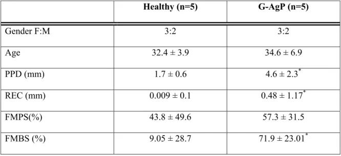
The demographic and clinical data are shown in Table 1 and Table 2. There were no significant differences between the groups in terms of gender and age (Table 1). Gender-mixing in a small sample size would have been a limitation if we sought to verify disease markers in such a small sample. Since the aim of the study was to evaluate if this particular method is an appropriate platform for detection of proteins in gingival crevicular fluid, a mixed gender sample in both groups is potentially more suitable in this context.
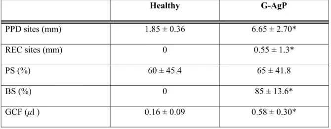
When full mouth clinical parameters of the studied subjects were compared, the G-AgP group had significantly higher mean PPD, REC and FMBS (p<0.05) (Table 1). G-AgP groups had also significantly higher mean PPD, REC and BS scores of sampling sites than the healthy group (P<0.05) (Table 2). In contrast, no significant differences were detected between the two groups regarding the plaque levels (P>0.05).
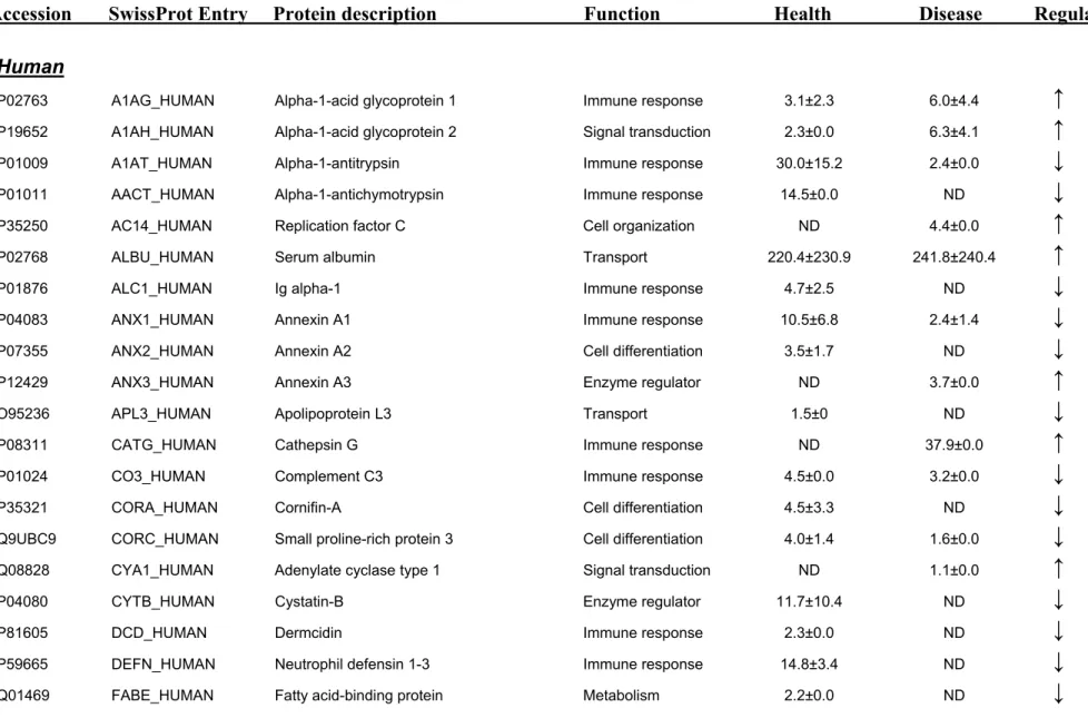
A total of 154 proteins with at least two unique peptide hits, validated from two different runs, were identified and quantified in the two subject groups (Table 3). When the identified proteins are categorized based on their origin, the disease group exhibited an increase in the proportion of bacterial, yeast, and viral protein, at the expense of human-derived proteins (Figure 1 A-B). This is rationale, as the
microorganism load is expected to be higher in disease. The pie chart in Figure 2 A-B also shows the functional classification of the GCF human-origin proteome based on their annotations in the SwissProt database. The annotation of the biological process revealed no quantitative changes in the proportions of human proteins involved in immune response, transportation, metabolism, signal transduction and enzyme regulation. However, the percentage of proteins involved in cell differentiation was decreased in disease, while that of cell organization was increased. The plausible explanation is that in the disease status, the cells undergo morphological alteration in response to the microorganism challenge, while the cell differentiation mechanisms are diminished, denoting a impeded tissue formation. Although the percentage of immune response regulated proteins was similar in health and disease, qualitative differences exist, as elaborated further.
Greater number of proteins were identified in diseased samples (n=115) than healthy ones (n=88). Interestingly, a few markers were found exclusively in either group, whereas a greater number of them were found present in both groups, albeit at different relative levels. The putative roles of these proteins in relation to periodontal health/disease are discussed below. There was also great variability of GCF analytes between the subjects, even within the same group. The mean +/- SD value derives from five individual samples in each group. The fact that some SDs are 0.0 is due to the protein was detected only in one single sample in that group. This suggests that the protein composition of GCF is rather individual specific, and pooling of the samples from the same clinical presentation based on diagnosis may result in loss of important information. Since the sample size was small with the large SD, it was not possible to perform further statistical analysis.
Among the large number of proteins that were found inboth diseased and healthy subjects, the most abundant proteins were human albumin, followed by immunoglobulins and various keratins. Previously, around 70 individual components of GCF have been assessed as potential markers for the disease (Lamster and Ahlo, 2007). These have been mostly proteolytic enzymes and pro-inflammatory cytokines. However, in the present study we rarely identified cytokines. This could potentially be due to their low concentrations or low molecular weight being masked by the presence of albumin or immunoglobulins (Jacobs et al., 2005). While many studies employing proteomic technologies apply techniques to deplete serumand plasma of high-abundance proteins prior to analysis, recent studies have shown that these practices may concomitantly remove potentially important diagnosticinformation. For instance, human albumin is a protein vehicle within the blood stream that binds to hormones, cytokines,and lipoproteins (Dea et al., 2002). In the present study, albumin concentration was the highest among all detected proteins which was at the level of 220.4 ± 230.9fmol in health and 241.8 ± 240.4 fmol in periodontitis. On the contrary to cytokines, we did identify a number of enzymes associated with polymorphonuclear neutrophils (PMNs), such as matrix metalloproteinase-8 (MMP-8), cathepsin G and myeloperoxidase, all of which were previously identified and proposed as candidate diagnostic markers for periodontal disease (Sorsa et al., 1999; Tervahartiala et al., 1996; Yamalik et al., 2000). Interestingly, MMP-8 and cathepsin G were not detected in GCF samples from healthy sites, but were present in periodontitis. MMPs are a family of zinc-dependent endopeptidases that are expressed in many inflammatory conditions including periodontitis and contribute to connective tissue breakdown. The fact that some other MMPs were not detected by this method,
despite that they have been detected by conventional protein detection assays suggests that the current method has its limitations. In the case of relatively low abundant proteins, such as MMPs and potentially cytokines, a drop of peptide signal is still a limiting factor for detection. Selective enrichment or depletion of the highly abundant GCF proteins prior to mass spectrometric analysis may help overcome this problem (Freije et al., 2008). Although the current approach was quite successful, clearly there is more room for improvement identifications of whole GCF proteins.
Myeloperoxidase was detected only in one out of five healthy samples, but in all periodontitis samples, which was on average 2-fold greater. Other proteins in the diseased group were most notably heat shock protein 70 kDa (HSP70). HSP70 has been shown to be an activator of the innate immune system (Kiang and Tsokos, 1998) and its release can be induced by periodontal pathogens (Saba et al., 2007).
Among the proteins not previously identified in GCF, the present data indicates that L-plastin (Plastin-2), also known as lymphocyte cytosolic protein 1, is of special interest. This protein was not detected in any of the five healthy samples, whereas it was present in 4 out of 5 diseased samples, with a mean concentration of 15.6 ± 12.1 fmol. L-plastin belongs to the actin-binding protein family, which is found in cells of the hematopoetic origin such as leukocytes. A role for L-plastin in the regulation of leukocyte adhesion and PMN signal transduction has been suggested (Jones et al., 1998). However, there is no known literature suggesting a role for L-plastin in periodontal disease. Higher concentrations of this protein in GCF may facilitate recruitment of PMNs at sites of inflammation. This may be relevant in view of the
potential constitutive PMN hyper-reactivity in periodontitis patients (Matthews et al., 2007).
In contrast, cystatin B and neutrophil defensins were not detected in any of the periodontitis group samples. The average concentrations in health were 11.7 ± 10.4 fmol for cystatin B, and 14.8 ± 3.4 fmol for neutrophil defensin. A commercially available ELISA for human neutrophil defensin was also employed to validate the findings from the proteomic analysis. Accordingly, human neutrophil defensin levels were under detection limit in the periodontal disease samples, whereas the mean concentration of the healthy samples was 125.13 ± 129.04 ng/ml. These findings are consistent with the LC/MSE data, as well as previously published data (Lundy et al.,
2005). The fact that neutrophil defensin, which has an antimicrobial properties was absent in the diseased samples supports the idea that the progression of periodontal disease could be linked to a decrease in the defensin barrier (Lundy et al., 2005). To this extent, defensins are suggested to have an important protective role for the host immune response to infection by periodontal pathogens (Brogden et al., 2003). Cystatin B, a neutral cysteine proteinase inhibitor, belonging to the cystatin family that minimizes proteolysis-associated tissue damage in inflammation (Chen et al., 1998; Dickinson, 2002). Proteins belong to the cystatin family such cystatin A, but not cystatin B, C or S, have previously been detected in GCF by immunoblotting or ELISA (Blankenvoorde et al., 1997). Cystatin B in particular could have been derived from serum, or from local cellularsecretions such as monocytes, epithelial cells and PMNs (Jarvinen et al., 1987).
Members of the calcium binding protein family including Calgranulin B, S100-P and Annexins were also differentially present in GCF. The S100 family proteins are thought to modulate biological activity via calcium binding and they expressed by macrophages and by epithelial cells in acutely inflamed tissues (Marenholz et al., 2004). Annexins are a group of Ca2+-binding proteins that are associated with
inflammatory and defense responses (Perretti and D'Acquisto, 2009). In the present study, annexin-1 appears to be more associated with a healthy status, since it was more frequently detected in health (four out of five samples in health vs two out of five in disease) and at 5-fold higher levels compared to disease. Likewise, Annexin-2 was not detected in any of the disease samples, but was found in two out of five healthy samples, at a mean concentration of 3.5 ± 1.7 fmol. Annexin-1 has long been suggested to be a critical endogenous negative regulator of pro-inflammatory mediators including IL-1, IL-6andcyclooxygenase-2 (Parente and Solito, 2004). In several disease models, such as experimental animal models of endotoxemia or arthritis, absence of Annexin-1 has been associated with increased levels of cytokines and exacerbation of acute inflammation (Damazo et al., 2005; Yang et al., 1999). Therefore, it is tempting to postulate that the higher levels of annexins in GCF may be associated with a healthy periodontal status, or that reduced levels or absence may haveapotential to explain many aspects of inflammatory status in periodontal disease. These proteins, which were mainly found in health, may have an important protective role and their function in periodontal disease needs to be addressed in a larger cohort of patient samples.
Other proteins of the immune system, such as α1-antitrypsin, α1-1-antichymotrypsin,
detected in two out of five healthy samples (30.0 ± 15.2 fmol), but was found in only one disease sample (2.4 ± 0.0 fmol). Presence of α-1-antitrypsin in GCF has been previously reported (Cox et al., 2006). Deficiencies in α-1-antitrypsin are known to predispose individuals to destructive inflammatory disease, including periodontitis (Peterson and Marsh, 1979; Yang et al., 2000). The balance between proteases and protease inhibitors is critical for maintenance of healthy tissue, and imbalances may lead to tissue destruction. The α-1-acid glycoprotein is one of the acute phase proteins, which is increased in response bacterial infection. In addition, its serum concentrations remain stable under physiological conditions (Fournier et al., 2000; Hochepied et al., 2003).A comparison of the healthy samples versus the diseased ones showed that α-1-acid glycoprotein 1 levels were up-regulated by 2-fold in periodontitis. Higher levels of this molecule have been previously reported in the sera of subjects with periodontitis (Glurich et al., 2002).
Confirming previous reports (Eley and Cox, 2003; Mooney and Kinane, 1997; Reinhardt et al., 1989; Wilton et al., 1993), GCF was enriched with immunoglobulins including four IgG subclass proteins and IgA. IgG concentrations were lower in GCF from patients with periodontitis than healthy ones. IgG1 protein was detected in both health and disease and was at highest concentration in healthy samples (86.2 ± 61.5 fmol). IgG3 was not present in any of the disease samples, whereas its mean concentration in health was 11.6 ± 0.0 fmol. IgA was present only in health (in 2 samples out of 5) with a mean concentration of 4.7 ± 2.5 fmol. Immunoglobulins arrive at the periodontal lesion site from both systemic and local tissue sources. The relative abundance of immunoglobulins in GCF may play a crucial role in the local defence mechanisms.
Notably, twenty-five different keratins were identified and quantified in GCF. However, this is not surprising since a great complexity of cytokeratins have been shown to be expressed in the gingiva or periodontal pockets (Mackenzie and Gao, 1993; Mackenzie et al., 1991; Ouhayoun et al., 1985). Although it should not be excluded that keratins are usual contaminants from the air, skin or hair, in the present study this was controlled for by simultaneous digestion of all samples. The present results indicated different and distinctive patterns of keratin protein expression in healthy and diseased GCF. For instance, cytokeratins 12 and 23 were only present in GCF from healthy sites, whereas cytokeratin 1 was consistently found in all samples. Other cytokeratins were detected at variable levels, but clearly their concentrations were lower in periodontal disease compared to health. This may be expected since the normal turnover rate of healthy sulcular epithelium is known to be one of the most rapid of all the epithelial tissues (Rowat and Squier, 1986). Therefore, it would be expected that healthy GCF contains large number of cytokeratins resulting from this vigorous turnover. In periodontal disease, where the periodontal pocket epithelium is ulcerated, the presence of cytokeratins in GCF may be attributed to passive release due to epithelial cell detachment or lysis (Ouhayoun et al., 1990).
There were also proteins from bacteria, viruses and yeast present in the GCF, since protein identification searches were not only limited to human protein sequence database. Evidently, greater proportions of these organisms are present in the GCF samples from periodontal disease than healthy ones. The higher presence of viral proteins, such as herpes virus protein 2, in the diseased samples corroborates previous reports that support the involvement of viral infection in the pathogenesis of
periodontal disease (Slots, 2007). Interestingly, no proteins from putative periodontal pathogens were detected in any of the diseased samples, with one exception, the methylmalonyl-CoA mutase of Porphyromonas gingivalis, which is one of the major pathogens implicated in periodontitis (Curtis et al., 2001). Although previous studies have shown that P. gingivalis proteolytic enzymes can be detectable in GCF (Aas et al., 2007), the present approach may not be optimal for the detection of periodontal pathogens. Notably, E. coli proteins were detected in GCF of both healthy and periodontally diseased subjects. Since the oral cavity is not a natural habitat for E. coli, the presence of its proteins in GCF detected in the present study may reflect their transfer from non-oral sites via the serum route.
Conclusions
The findings of the present study demonstrate that label-free quantitative LC/MSE
based mass spectrometry technology is an appropriate platform for the analysis of GCF proteome. The protein content of GCF could potentially be used as a diagnostic aid in periodontal disease, since the characterisation of the GCF proteome will greatly increase the clinical importance of this biological exudate. The comprehensive analysis of GCF by the suggested approach may provide better understanding of the pathological processes involvedin periodontal disease, and facilitate the development of accurate diagnostic and prognostic markers. Due to the interindividual variations of protein biomarkers in GCF, larger cohort studies are required to characterise the complete GCF proteome in health and disease, before its diagnostic and prognostic value reaches optimal full potential.
Abbreviations:
CVD, cardiovascular disease; DM, diabetes mellitus; DTT, dithiothreitol; ELISA, enzyme-linked immunosorbent assay; G-AgP: generalized aggressive periodontitis; GCF, gingival crevicular fluid; GO, gene ontology; IL, interleukin; LC, liquid chromatography; LC-MS/MS, liquid chromatography/tandem mass spectrometry; PD, periodontal disease; PPD, probing pocket depth; REC, recession level; SDS-PAGE, sodium dodecyl sulfate polyacrylamide gel electrophoresis; Q-Tof, quadrupole time-of-flight.
Acknowledgement
This study was partially supported by the research account of the Periodontology Unit and UCLH/UCL Comprehensive Biomedicine Centre. Dr. NB was employed as Walport Clinical Academic Lecturer at Eastman Dental Institute, funded by the NIHR.
References
Aas, J. A., Barbuto, S. M., Alpagot, T., Olsen, I., Dewhirst, F. E., and Paster, B. J. (2007). Subgingival plaque microbiota in HIV positive patients. J Clin Periodontol 34, 189-195.
Albandar, J. M., and Rams, T. E. (2002). Risk factors for periodontitis in children and young persons. Periodontol 2000 29, 207-222.
Alfano, M. C. (1974). The origin of gingival fluid. J Theor Biol 47, 127-136.
Armitage, G. C. (1999). Development of a classification system for periodontal diseases and conditions. Ann Periodontol 4, 1-6.
Armitage, G. C. (2004). Periodontal diagnoses and classification of periodontal diseases. Periodontol 2000 34, 9-21.
Blankenvoorde, M. F., Henskens, Y. M., van der Weijden, G. A., van den Keijbus, P. A., Veerman, E. C., and Nieuw Amerongen, A. V. (1997). Cystatin A in gingival crevicular fluid of periodontal patients. J Periodontal Res 32, 583-588.
Bostanci, N., Ilgenli, T., Emingil, G., Afacan, B., Han, B., Toz, H., Atilla, G., Hughes, F. J., and Belibasakis, G. N. (2007). Gingival crevicular fluid levels of RANKL and OPG in periodontal diseases: implications of their relative ratio. J Clin Periodontol 34, 370-376.
Brogden, K. A., Heidari, M., Sacco, R. E., Palmquist, D., Guthmiller, J. M., Johnson, G. K., Jia, H. P., Tack, B. F., and McCray, P. B. (2003). Defensin-induced adaptive immunity in mice and its potential in preventing periodontal disease. Oral Microbiol Immunol 18, 95-99.
Brown, L. J., and Loe, H. (1993). Prevalence, extent, severity and progression of periodontal disease. Periodontol 2000 2, 57-71.
Champagne, C. M., Buchanan, W., Reddy, M. S., Preisser, J. S., Beck, J. D., and Offenbacher, S. (2003). Potential for gingival crevice fluid measures as predictors of risk for periodontal diseases. Periodontol 2000 31, 167-180.
Chen, H. Y., Cox, S. W., and Eley, B. M. (1998). Cathepsin B, alpha2-macroglobulin and cystatin levels in gingival crevicular fluid from chronic periodontitis patients. J Clin Periodontol 25, 34-41.
Cox, S. W., Rodriguez-Gonzalez, E. M., Booth, V., and Eley, B. M. (2006). Secretory leukocyte protease inhibitor and its potential interactions with elastase and cathepsin B in gingival crevicular fluid and saliva from patients with chronic periodontitis. J Periodontal Res 41, 477-485.
Curtis, M. A., Aduse-Opoku, J., and Rangarajan, M. (2001). Cysteine proteases of Porphyromonas gingivalis. Crit Rev Oral Biol Med 12, 192-216.
Damazo, A. S., Yona, S., D'Acquisto, F., Flower, R. J., Oliani, S. M., and Perretti, M. (2005). Critical protective role for annexin 1 gene expression in the endotoxemic murine microcirculation. Am J Pathol 166, 1607-1617.
Danesh, J., Collins, R., and Peto, R. (1997). Chronic infections and coronary heart disease: is there a link? Lancet 350, 430-436.
Dea, M. K., Hamilton-Wessler, M., Ader, M., Moore, D., Schaffer, L., Loftager, M., Volund, A., and Bergman, R. N. (2002). Albumin binding of acylated insulin (NN304) does not deter action to stimulate glucose uptake. Diabetes 51, 762-769. Dickinson, D. P. (2002). Cysteine peptidases of mammals: their biological roles and potential effects in the oral cavity and other tissues in health and disease. Crit Rev Oral Biol Med 13, 238-275.
Ebersole, J. L. (2003). Humoral immune responses in gingival crevice fluid: local and systemic implications. Periodontol 2000 31, 135-166.
Eley, B. M., and Cox, S. W. (2003). Proteolytic and hydrolytic enzymes from putative periodontal pathogens: characterization, molecular genetics, effects on host defenses and tissues and detection in gingival crevice fluid. Periodontol 2000 31, 105-124. Estreicher, A., Broggiato, A., Duroux, P., Andersen, E., and Cimasoni, G. (1996). Low molecular-weight proteins in human gingival crevicular fluid. Arch Oral Biol 41, 733-738.
Fournier, T., Medjoubi, N. N., and Porquet, D. (2000). Alpha-1-acid glycoprotein. Biochim Biophys Acta 1482, 157-171.
Freije, R., Klein, T., Ooms, B., Kauffman, H. F., and Bischoff, R. (2008). An integrated high-performance liquid chromatography-mass spectrometry system for the activity-dependent analysis of matrix metalloproteases. J Chromatogr A 1189, 417-425.
Glurich, I., Grossi, S., Albini, B., Ho, A., Shah, R., Zeid, M., Baumann, H., Genco, R. J., and De Nardin, E. (2002). Systemic inflammation in cardiovascular and periodontal disease: comparative study. Clin Diagn Lab Immunol 9, 425-432.
Golub, L. M., and Kleinberg, I. (1976). Gingival crevicular fluid: a new diagnostic aid in managing the periodontal patient. Oral Sci Rev, 49-61.
Hochepied, T., Berger, F. G., Baumann, H., and Libert, C. (2003). Alpha(1)-acid glycoprotein: an acute phase protein with inflammatory and immunomodulating properties. Cytokine Growth Factor Rev 14, 25-34.
Jacobs, J. M., Adkins, J. N., Qian, W. J., Liu, T., Shen, Y., Camp, D. G., 2nd, and Smith, R. D. (2005). Utilizing human blood plasma for proteomic biomarker discovery. J Proteome Res 4, 1073-1085.
Jarvinen, M., Rinne, A., and Hopsu-Havu, V. K. (1987). Human cystatins in normal and diseased tissues--a review. Acta Histochem 82, 5-18.
Jones, S. L., Wang, J., Turck, C. W., and Brown, E. J. (1998). A role for the actin-bundling protein L-plastin in the regulation of leukocyte integrin function. Proc Natl Acad Sci U S A 95, 9331-9336.
Kiang, J. G., and Tsokos, G. C. (1998). Heat shock protein 70 kDa: molecular biology, biochemistry, and physiology. Pharmacol Ther 80, 183-201.
Krasse, B. (1996). Serendipity or luck: stumbling on gingival crevicular fluid. J Dent Res 75, 1627-1630.
Lamster, I. B. (1997). Evaluation of components of gingival crevicular fluid as diagnostic tests. Ann Periodontol 2, 123-137.
Lamster, I. B., and Ahlo, J. K. (2007). Analysis of gingival crevicular fluid as applied to the diagnosis of oral and systemic diseases. Ann N Y Acad Sci 1098, 216-229. Lamster, I. B., Oshrain, R. L., Fiorello, L. A., Celenti, R. S., and Gordon, J. M. (1988). A comparison of 4 methods of data presentation for lysosomal enzyme activity in gingival crevicular fluid. J Clin Periodontol 15, 347-352.
Lundy, F. T., Orr, D. F., Shaw, C., Lamey, P. J., and Linden, G. J. (2005). Detection of individual human neutrophil alpha-defensins (human neutrophil peptides 1, 2 and 3) in unfractionated gingival crevicular fluid--a MALDI-MS approach. Mol Immunol
42, 575-579.
Mackenzie, I. C., and Gao, Z. (1993). Patterns of cytokeratin expression in the epithelia of inflamed human gingiva and periodontal pockets. J Periodontal Res 28, 49-59.
Mackenzie, I. C., Rittman, G., Gao, Z., Leigh, I., and Lane, E. B. (1991). Patterns of cytokeratin expression in human gingival epithelia. J Periodontal Res 26, 468-478.
Marenholz, I., Heizmann, C. W., and Fritz, G. (2004). S100 proteins in mouse and man: from evolution to function and pathology (including an update of the nomenclature). Biochem Biophys Res Commun 322, 1111-1122.
Matthews, J. B., Wright, H. J., Roberts, A., Ling-Mountford, N., Cooper, P. R., and Chapple, I. L. (2007). Neutrophil hyper-responsiveness in periodontitis. J Dent Res
86, 718-722.
Mooney, J., and Kinane, D. F. (1997). Levels of specific immunoglobulin G to Porphyromonas gingivalis in gingival crevicular fluid are related to site disease status. Oral Microbiol Immunol 12, 112-116.
Muhlemann, H. R., and Son, S. (1971). Gingival sulcus bleeding--a leading symptom in initial gingivitis. Helv Odontol Acta 15, 107-113.
Ouhayoun, J. P., Goffaux, J. C., Sawaf, M. H., Shabana, A. H., Collin, C., and Forest, N. (1990). Changes in cytokeratin expression in gingiva during inflammation. J Periodontal Res 25, 283-292.
Ouhayoun, J. P., Gosselin, F., Forest, N., Winter, S., and Franke, W. W. (1985). Cytokeratin patterns of human oral epithelia: differences in cytokeratin synthesis in gingival epithelium and the adjacent alveolar mucosa. Differentiation 30, 123-129. Ozmeric, N. (2004). Advances in periodontal disease markers. Clin Chim Acta 343, 1-16.
Parente, L., and Solito, E. (2004). Annexin 1: more than an anti-phospholipase protein. Inflamm Res 53, 125-132.
Perretti, M., and D'Acquisto, F. (2009). Annexin A1 and glucocorticoids as effectors of the resolution of inflammation. Nat Rev Immunol 9, 62-70.
Peterson, R. J., and Marsh, C. L. (1979). The relationship of alpha1-antitrypsin to inflammatory periodontal disease. J Periodontol 50, 31-35.
Pisano, E., Cabras, T., Montaldo, C., Piras, V., Inzitari, R., Olmi, C., Castagnola, M., and Messana, I. (2005). Peptides of human gingival crevicular fluid determined by HPLC-ESI-MS. Eur J Oral Sci 113, 462-468.
Rao, P. V., Reddy, A. P., Lu, X., Dasari, S., Krishnaprasad, A., Biggs, E., Roberts, C. T., and Nagalla, S. R. (2009). Proteomic identification of salivary biomarkers of type-2 diabetes. J Proteome Res 8, 239-245.
Reinhardt, R. A., McDonald, T. L., Bolton, R. W., DuBois, L. M., and Kaldahl, W. B. (1989). IgG subclasses in gingival crevicular fluid from active versus stable periodontal sites. J Periodontol 60, 44-50.
Rowat, J. S., and Squier, C. A. (1986). Rates of epithelial cell proliferation in the oral mucosa and skin of the tamarin monkey (Saguinus fuscicollis). J Dent Res 65, 1326-1331.
Saba, J. A., McComb, M. E., Potts, D. L., Costello, C. E., and Amar, S. (2007). Proteomic mapping of stimulus-specific signaling pathways involved in THP-1 cells exposed to Porphyromonas gingivalis or its purified components. J Proteome Res 6, 2211-2221.
Silva, J. C., Denny, R., Dorschel, C., Gorenstein, M. V., Li, G. Z., Richardson, K., Wall, D., and Geromanos, S. J. (2006). Simultaneous qualitative and quantitative analysis of the Escherichia coli proteome: a sweet tale. Mol Cell Proteomics 5, 589-607.
Silva, J. C., Denny, R., Dorschel, C. A., Gorenstein, M., Kass, I. J., Li, G. Z., McKenna, T., Nold, M. J., Richardson, K., Young, P., and Geromanos, S. (2005). Quantitative proteomic analysis by accurate mass retention time pairs. Anal Chem 77, 2187-2200.
Slots, J. (2007). Herpesviral-bacterial synergy in the pathogenesis of human periodontitis. Curr Opin Infect Dis 20, 278-283.
Sorsa, T., Mantyla, P., Ronka, H., Kallio, P., Kallis, G. B., Lundqvist, C., Kinane, D. F., Salo, T., Golub, L. M., Teronen, O., and Tikanoja, S. (1999). Scientific basis of a matrix metalloproteinase-8 specific chair-side test for monitoring periodontal and peri-implant health and disease. Ann N Y Acad Sci 878, 130-140.
Tervahartiala, T., Konttinen, Y. T., T, I. N., Hayrinen-Immonen, R., Ding, Y., and Sorsa, T. (1996). Cathepsin G in gingival tissue and crevicular fluid in adult periodontitis. J Clin Periodontol 23, 68-75.
Uitto, V. J. (2003). Gingival crevice fluid--an introduction. Periodontol 2000 31, 9-11. Wilton, J. M., Bampton, J. L., Hurst, T. J., Caves, J., and Powell, J. R. (1993). Interleukin-1 beta and IgG subclass concentrations in gingival crevicular fluid from patients with adult periodontitis. Arch Oral Biol 38, 55-60.
Yamalik, N., Caglayan, F., Kilinc, K., Kilinc, A., and Tumer, C. (2000). The importance of data presentation regarding gingival crevicular fluid myeloperoxidase and elastase-like activity in periodontal disease and health status. J Periodontol 71, 460-467.
Yang, P., Tremaine, W. J., Meyer, R. L., and Prakash, U. B. (2000). Alpha1-antitrypsin deficiency and inflammatory bowel diseases. Mayo Clin Proc 75, 450-455. Yang, Y., Hutchinson, P., and Morand, E. F. (1999). Inhibitory effect of annexin I on synovial inflammation in rat adjuvant arthritis. Arthritis Rheum 42, 1538-1544.
Figure Legends
Figure 1
The pie-charts show the distribution of the proportion of gingival crevicular fluid (GCF) proteins according to their source of origin, in health (A) and disease (B). The proportions are expressed as percentage (%) of the total protein in each group. Four soureces of origin were identified, namely human, bacetrial, yeast and viral. An increase in the proportion of bacterial, viral and yeast origin proteins was detected in disease, compared to health.
Figure 2
The pie-charts show the distribution of the proportion of gingival crevicular fluid (GCF) proteins according to their biological function, in health (A) and disease (B). The proportions are expressed as percentage (%) of the total protein in each group. The sorting of protein function was based on the UniProt/Swiss-Prot database annotations. A functional classification was assigned to each unique protein, and the total number and percentage of proteins in each class were determined.
Figure 1
A)
Health 80% 12% 4% 4% Human Bacterial Yeast ViralB)
Disease 68% 16% 11% 5% Human Bacterial Yeast ViralFigure 2
A)
Health 11% 3% 9% 27% 2% 5% 43% Transport Signal transduction Metabolism Immune response Enzyme regulator Cell organization Cell differentiationB)
Disease 12% 11% 8% 26% 3% 11% 29% Transport Signal transduction Metabolism Immune response Enzyme regulator Cell organization Cell differentiationTable 1: Demographic and full mouth clinical parameters of the studied subjects (five healthy and five patients with generalized aggressive periodontitis (G-AgP) (mean ± SD) Healthy (n=5) G-AgP (n=5) Gender F:M 3:2 3:2 Age 32.4 ± 3.9 34.6 ± 6.9 PPD (mm) 1.7 ± 0.6 4.6 ± 2.3* REC (mm) 0.009 ± 0.1 0.48 ± 1.17* FMPS(%) 43.8 ± 49.6 57.3 ± 31.5 FMBS (%) 9.05 ± 28.7 71.9 ± 23.01*
To determine the clinical periodontal status, all subjects had a clinical periodontal examination including the measurement of probing pocket depth (PPD), clinical recession level (REC), full-mouth bleeding (FMBS) and full-mouth plaque scores (FMPS) at six sites around each tooth with a manual probe as detailed in Materials and Methods. The asterisk (*) indicates significant difference from the healthy group (Mann–Whitney U-test, p<0.05).
Table 2: Clinical parameters of the sampling areas in the study groups (mean ± SD) Healthy G-AgP PPD sites (mm) 1.85 ± 0.36 6.65 ± 2.70* REC sites (mm) 0 0.55 ± 1.3* PS (%) 60 ± 45.4 65 ± 41.8 BS (%) 0 85 ± 13.6* GCF (µl ) 0.16 ± 0.09 0.58 ± 0.30*
The gingival crevicular fluid (GCF) samples were collected from the four deepest periodontal pockets (one site per quadrant) in G-AgP patients. In the healthy group, the GCF samples were collected from four standard sites (one first molars and one central incisors in each jaw) exhibiting PPD up to 3 mm without bleeding on probing and radiographic evidence of alveolar bone loss. The GCF sample volume was measured with a calibrated Periotron 8000. PPD, probing pocket depth; REC, clinical recession level; BS, bleeding score; PS, plaque score. The asterisk (*) indicates significant difference from the healthy group (Mann–Whitney U-test, p<0.05).
Table 3
Gingival crevicular fluid Proteins Identified Using LC/MSE
GCF proteins were identified and quantitated in healthy subjects (n=5) and patients with generalized aggressive periodontitis (n=5) by LC/MSE. Proteins having at least
two peptide identifications found in human GCF are listed by alphabetical order with their UniProt/Swiss-Prot entry name, accession number and description. Proteins are grouped according to their origin. The absolute quantity of each protein is expressed as fmol. The arrows indicate the relative up-regulation or down-regulation of any given protein in disease, compared to health. The mean and +/- SD value is derived from five individual samples in each group.
Table 3. Gingival crevicular fluid Proteins Identified Using LC/MSE
Accession SwissProt Entry Protein description Function Health Disease Regulation Human
P02763 A1AG_HUMAN Alpha-1-acid glycoprotein 1 Immune response 3.1±2.3 6.0±4.4
↑
P19652 A1AH_HUMAN Alpha-1-acid glycoprotein 2 Signal transduction 2.3±0.0 6.3±4.1↑
P01009 A1AT_HUMAN Alpha-1-antitrypsin Immune response 30.0±15.2 2.4±0.0
↓
P01011 AACT_HUMAN Alpha-1-antichymotrypsin Immune response 14.5±0.0 ND
↓
P35250 AC14_HUMAN Replication factor C Cell organization ND 4.4±0.0
↑
P02768 ALBU_HUMAN Serum albumin Transport 220.4±230.9 241.8±240.4
↑
P01876 ALC1_HUMAN Ig alpha-1 Immune response 4.7±2.5 ND
↓
P04083 ANX1_HUMAN Annexin A1 Immune response 10.5±6.8 2.4±1.4
↓
P07355 ANX2_HUMAN Annexin A2 Cell differentiation 3.5±1.7 ND
↓
P12429 ANX3_HUMAN Annexin A3 Enzyme regulator ND 3.7±0.0
↑
O95236 APL3_HUMAN Apolipoprotein L3 Transport 1.5±0 ND
↓
P08311 CATG_HUMAN Cathepsin G Immune response ND 37.9±0.0
↑
P01024 CO3_HUMAN Complement C3 Immune response 4.5±0.0 3.2±0.0
↓
P35321 CORA_HUMAN Cornifin-A Cell differentiation 4.5±3.3 ND
↓
Q9UBC9 CORC_HUMAN Small proline-rich protein 3 Cell differentiation 4.0±1.4 1.6±0.0
↓
Q08828 CYA1_HUMAN Adenylate cyclase type 1 Signal transduction ND 1.1±0.0↑
P04080 CYTB_HUMAN Cystatin-B Enzyme regulator 11.7±10.4 ND
↓
Q7Z6J6 FRMD5_HUMAN FERM domain protein 5 Signal transduction ND 0.5±0.0
↑
P04406 G3P2_HUMAN GAPDH 3 Metabolism 8.3±6.5 ND
↓
P01857 GC1_HUMAN Ig gamma-1 chain C Immune response 86.2±61.5 53.8±45.2
↓
P01859 GC2_HUMAN Ig gamma-2 chain C Immune response 19.9±10.6 25.2±15.3↑
P01860 GC3_HUMAN Ig gamma-3 chain C Immune response 11.6±0.0 ND
↓
P01861 GC4_HUMAN Ig gamma-4 chain C Immune response 26.4±17.9 20.8±10.9
↓
P52565 GDIR_HUMAN Rho GDP-dissociation inhibitor 1 Signal transduction ND 4.6±3.7↑
P14136 GFAP_HUMAN Glial fibrillary acidic protein Signal transduction ND 4.7±0.0↑
P62807 H2BA_HUMAN Histone H2B type 1 Immune response 3.3±0.0 71.2±0.0
↑
P57053 H2BS_HUMAN Histone H2B type F-S Immune response ND 71.2±0.0
↑
P62805 H4_HUMAN Histone H4 Cell organization ND 1.2±0.0
↑
P69905 HBA_HUMAN Hemoglobin subunit alpha Transport 0.3±0.0 59.3±60.5
↑
P68871 HBB_HUMAN Hemoglobin subunit beta Transport ND 22.5±0.0
↑
P02042 HBD_HUMAN Hemoglobin subunit delta Transport ND 158.2±196.7
↑
P00738 HPT2_HUMAN Haptoglobin Metabolism 10.9±9.1 12.0±4.0
↑
P11142 HSP7C_HUMAN Heat shock 71 kDa protein Immune response ND 3.1±0.0
↑
P54652 HSP72_HUMAN Heat shock-related 70 kDa Immune response ND 1.2±0.0↑
P01777 HV3P_HUMAN Ig heavy chain V-III region Immune response ND 2.8±0.0↑
P35527 K1CI_HUMAN Keratin, type I cytoskeletal 9 Cell differentiation 10.0±9.2 6.6±0.0↓
P13645 K1CJ_HUMAN Keratin, type I cytoskeletal 10 Cell differentiation 22.2±13.6 8.7±0.0↓
Q99456 K1CL_HUMAN Keratin, type I cytoskeletal 12 Cell differentiation 26.6±9.4 ND↓
P13646 K1CM_HUMAN Keratin, type I cytoskeletal 13 Cell differentiation 32.2±23.0 10.4±7.1↓
P02533 K1CN_HUMAN Keratin, type I cytoskeletal 14 Cell differentiation 16.5±17.5 7.5±3.5↓
P19012 K1CO_HUMAN Keratin, type I cytoskeletal 15 Cell differentiation 21.7±16.6 8.2±4.6↓
P30654 K1CP_HUMAN Keratin, type I cytoskeletal 16 Cell differentiation 27.7±16.0 10.3±0.0↓
P35900 K1CT_HUMAN Keratin, type I cytoskeletal 20 Cell differentiation 13.7±6.3 1.4±0.0
↓
Q9C075 K1CW_HUMAN Keratin, type I cytoskeletal 23 Cell differentiation 3.9±0.0 ND↓
P35908 K22E_HUMAN Keratin, type II cytoskeletal 2 Cell differentiation 73.3±97.6 10.4±5.8↓
Q01546 K22O_HUMAN Keratin, type II cytoskeletal 2 oral Cell differentiation 11.0±10.4 4.6±0.6↓
P04264 K2C1_HUMAN Keratin, type II cytoskeletal 1 Cell differentiation 60.1±51.8 15.8±9.6↓
P12035 K2C3_HUMAN Keratin, type II cytoskeletal 3 Cell differentiation 9.1±7.3 ND↓
P19013 K2C4_HUMAN Keratin, type II cytoskeletal 4 Cell differentiation 8.4±7.5 1.2±0.0↓
P13647 K2C5_HUMAN Keratin, type II cytoskeletal 5 Cell differentiation 85.6±114.4 7.5±1.7↓
P08729 K2C7_HUMAN Keratin, type II cytoskeletal 7 Cell differentiation 9.2±0.0 ND↓
P05787 K2C8_HUMAN Keratin, type II cytoskeletal 8 Cell differentiation 8.7±7.6 4.5±0.0↓
P02538 K2CA_HUMAN Keratin, type II cytoskeletal 6A Cell differentiation 66.0±61.4 11.5±2.7↓
P04259 K2CB_HUMAN Keratin, type II cytoskeletal 6B Cell differentiation 168.5±0.0 12.4±3.8↓
P48668 K2CC_HUMAN Keratin, type II cytoskeletal 6C Cell differentiation 203.9±262.4 11.5±2.7↓
P48667 K2CD_HUMAN Keratin, type II cytoskeletal 6D Cell differentiation 119.6±178.2 6.9±8.0↓
P48668 K2CE_HUMAN Keratin, type II cytoskeletal 6E Cell differentiation 151.2±195.9 10.5±2.9↓
P48669 K2CF_HUMAN Keratin, type II cytoskeletal 6F Cell differentiation 62.7±55.4 17.8±0.0↓
P01834 KAC_HUMAN Ig kappa chain C Immune response 15.5±13.5 6.8±2.6
↓
P78368 KC12_HUMAN Casein kinase I isoform gamma-2 Signal transduction ND 0.5±0.0
↑
Q9C0H6 KHL4_HUMAN Kelch-like protein 4 Cell organization 1.8±0.0 ND
↓
P14618 KPY2_HUMAN Cytosolic thyroid hormone protein Metabolism 0.6±0.0 ND
↓
P30613 KPYR_HUMAN Pyruvate kinase 1 Metabolism ND 1.4±0.0
↑
P04206 KV3G_HUMAN Rheumatoid factor Immune response ND 2.7±0.3
↑
P01842 LAC_HUMAN Ig lambda chain C Immune response 12.8±18.4 4.6±1.1
↓
P00695 LYC_HUMAN Lysozyme C Metabolism 12.1±14.7 3.0±1.4
↓
P22712 MPB1_HUMAN C myc promoter binding protein Cell differentiation 1.5±0.0 ND
↓
P35579 MYH9_HUMAN Myosin-9 Immune response ND 4.2±0.0
↑
P80188 NGAL_HUMAN Neutrophil gelatinase-lipocalin Transport ND 18.9±11.2
↑
P11678 PERE_HUMAN Eosinophil peroxidase Transport 1.2±0.0 ND
↓
P05164 PERM_HUMAN Myeloperoxidase Immune response 3.1±0.0 6.8±4.5
↑
P13796 PLSL_HUMAN Lymphocyte cytosolic protein 1 Transport ND 15.6±12.1
↑
P13797 PLST_HUMAN Plastin-3 Cell organization ND 9.8±7.9
↑
P07737 PRO1_HUMAN Profilin-1 Cell organization 9.4±0.0 13.9±0.0
↑
Q596K9 Q596K9_HUMAN Migration-inducing protein 12 Cell organization ND 24.5±0.0
↑
Q6ICQ8 Q6ICQ8_HUMAN ARHG protein Signal transduction ND 2.1±0.0
↑
Q6NSB4 Q6NSB4_HUMAN HP protein Enzyme regulator ND 5.4±0.0
↑
Q01201 RELB_HUMAN Transcription factor RelB Cell organization 55.8±0.0 ND
↓
P02753 RETB_HUMAN Retinol-binding protein 4 Transport 3.3±0.0 ND
↓
Q13671 RIN1_HUMAN Ras interaction/interference protein 1 Signal transduction ND 0.2±0.0
↑
P06702 S109_HUMAN Calgranulin B Immune response 10.3±0.3 11.0±0.2
↑
P25815 S10E_HUMAN Protein S100-P Transport ND 1.3±0.0
↑
O15056 SYJ2_HUMAN Synaptojanin-2 Signal transduction 0.6±0.0 ND
↓
P17542 TAL1_HUMAN T-cell acute lymphocytic leukemia 1 Cell differentiation ND 0.9±0.0
↑
P10599 THIO_HUMAN Thioredoxin Metabolism ND 2.5±0.0
↑
P29401 TKT_HUMAN Transketolase Metabolism 0.8±0.0 ND
↓
Q03169 TNAP2_HUMAN TNFalpha-induced protein 2 Immune response 0.6±0.0 ND
↓
P02787 TRFE_HUMAN Serotransferrin Transport 16.8±12.06 6.4±4.7
↓
P02788 TRFL_HUMAN Lactotransferrin Transport ND 2.9±0.0
↑
P62328 TYB4_HUMAN Thymosin beta-4 Cell organization ND 1.1±0.0
↑
P49427 UBC3_HUMAN Ubiquitin-conjugating enzyme E2 R1 Cell organization ND 1.4±0.0
↑
Bacterial
P57448 AMPA_BUCAI Cytosol aminopeptidase Bacterial ND 1.0±0.0
↑
Q9ZL64 ACO2_HELPJ H. pylori aconitate hydratase Bacterial ND 0.4±0.0
↑
P0A1B5 AROF_SALTY S.typhimurium DAHP synthetase Bacterial ND 1.6±0.0
↑
P0A1Y2 ASRC_SALTY S. typhimurium sulfite reductase Bacterial ND 2.6±0.0
↑
P12999 BIOC_ECOLI E. coli biotin synthesis protein Bacterial 0.9±0.0 ND
↓
P0A518 CH61_MYCTU M.tuberculosis chaperonin Bacterial 17.9±0.0 ND
↓
Q02929 CPS_CLOTM Putative sensory transducer Bacterial ND 2.1±0.0
↑
P0A9F3 CYSB_ECOLI E. coli cysB Bacterial ND 0.7±0.0
↑
P31680 DJLA_ECOLI E. coli DnaJ-like protein Bacterial 2.9±0.0 ND
↓
P66004 DLDH_MYCTU M. tuberculosis dihydrolipoyl DH Bacterial 20.8±0.0 ND
↓
Q9PHE3 DNAA_XYLFA Xylella fastidiosa dnaA Bacterial ND 2.9±0.0
↑
O25242 DP3B_HELPY H. pylori DNA polymerase Bacterial ND 8.8±0.0
↑
Q9ZML6 DXR_HELPJ H. pylori reductoisomerase Bacterial ND 15.7±0.0
↑
P06176 FLIC_SALCH S. choleraesuis flagellin Bacterial ND 2.2±0.0
↑
Q05755 GLTB_AZOBR A. brasilense glutamate synthase Bacterial 13.9±0.0 ND
↓
P07018 MCP4_ECOLI E. coli MCP-IV Bacterial ND 2.4±0.0
↑
Q57714 PORB_METJA Pyruvate synthase porB Bacterial 46.6±0.0 6.2±0.0
↓
Q59677 MUTB_PORGI P. gingivalis Methylmalonyl-CoA Bacterial ND 0.6±0.0
↑
P11278 PELP_YERPS Yersinia pseudotuberculosis pelY Bacterial 12.7±0.0 ND
↓
P16682 PHND_ECOLI E. coli phosphonates-binding protein Bacterial 17.7±0.0 ND
↓
O07497 SECA_BORBU Protein translocase Bacterial ND 1.7±0.0
↑
Q9Z7P1 SYH_CHLPN C. pneumoniae h-tRNA synthetase Bacterial ND 1.0±0.0
↑
P16966 TTR_PSESY P. syringae acetyltransferase Bacterial 1.9±0.0 ND
↓
P54390 YPIB_BACSU B. subtilis protein ypiB Bacterial 6.2±0.0 ND
↓
Yeast
O94049 ACSA_CANAL C.albicans acetyl coenzyme A Yeast ND 0.2±0.0
↑
O93803 CET1_CANAL C. albicans mRNA-capping Yeast ND 1.9±0.0
↑
P87131 CYSK_SCHPO S. pombe cysteine synthase Yeast ND 1.4±0.0
↑
P14020 DPM1_YEAST S. cerevisiae DPM synthase Yeast ND 3.2±0.0
↑
Q02207 FOX2_YEAST S. cerevisiae β-oxidation protein Yeast 0.1±0.0 ND
↓
P22147 KEM1_YEAST S. cerevisiae exoribonuclease 1 Yeast 9.9±0.0 ND
↓
P40957 MAD1_YEAST S.cerevisia MAD1 Yeast ND 2.0±0.0
↑
P33755 NPL4_YEAST S. cerevisiae NPL4 Yeast ND 10.7±0.0
↑
P46673 NU85_YEAST S. cerevisiae NUP85 Yeast ND 2.1±0.0
↑
P07922 PYRF_KLULA C. sphaerica OMP decarboxylase Yeast 6.1±0.0 21.3±8.9
↑
P33332 SEC3_YEAST Protein PSL1 Yeast ND 0.5±0.0
↑
P26637 SYLC_YEAST S. cerevisiae l-tRNA synthetase Yeast ND 3.9±0.0
↑
P34252 YKK8_YEAST SLD2 Yeast ND 1.3±0.0
↑
P36113 YKZ7_YEAST RING finger protein Yeast ND 3.5±0.0
↑
Viral
P00544 FGR_FSVGR Sarcoma virus tyrosine- kinase Viral 4.2±0.0 11.3±9.2
↑
P36848 FIBP_ADE31 Human adenovirus fiber protein Viral 10.2±0.0 ND
↓
P13121 RPC1_BPP1 Enterobacteria phage C1 Virus ND 0.4±0.0
↑
P69202 RPC2_BPP22 Enterobacteria phage P22 C2 Virus ND 2.1±0.0
↑
P26109 RRP3_IARUD Polymerase basic protein 2 Virus ND 0.81±0.0
↑
Q08099 US02_HSVBS Herpes virus Protein US2 Virus ND 4.4±0.0
↑
P21044 VC13_VACCC Vaccinia virus Protein C13 Virus 1.9±0.0 ND
↓
Table of Contents Synopsis
GCF is a local inflammatory exudate of the diseased periodontal tissues that can be used to study potential disease markers. As opposed to previous approaches studying single biomarkers, the present study used label-free LC/MSE and identified 154 proteins of human, bacterial and viral origin, in health and periodontitis. This technology may facilitate characterisation of GCF proteome in periodontal health and disease, aiding the development of accurate diagnostic and prognostic markers.
Gingival exudatome
analysis by LC/MS
EData Processing and Database Searching
Protein origin in health and disease
Clinical
Radiographic GCF
Periodontal disease
Health 80% 12% 4% 4% Human Bacterial Yeast Viral Disease 68% 16% 11% 5% Human Bacterial Yeast Viral