Targeting the Epigenetic Lesion in MLL-Rearranged Leukemia
(Article begins on next page)
The Harvard community has made this article openly available.
Please share
how this access benefits you. Your story matters.
Citation
No citation.
Accessed
February 19, 2015 11:44:51 AM EST
Citable Link
http://nrs.harvard.edu/urn-3:HUL.InstRepos:10436226
Terms of Use
This article was downloaded from Harvard University's DASH
repository, and is made available under the terms and conditions
applicable to Other Posted Material, as set forth at
http://nrs.harvard.edu/urn-3:HUL.InstRepos:dash.current.terms-of-use#LAA
T
ARGETING THE
E
PIGENETIC
L
ESION IN
MLL-
REARRANGED
L
EUKEMIA
A dissertation presented
by
Liying Michelle Chen
to
the Department of Molecular, Cellular and Biology
in partial fulfillment of the requirements
for the degree of
Doctor of Philosophy
in the subject of
Biochemistry
Harvard University
Cambridge, Massachusetts
December 2012
© 2012
by
Liying Chen
iii
Dissertation Advisor: Professor Scott A. Armstrong Liying Chen
Targeting the Epigenetic Lesion in MLL-rearranged Leukemia
Abstract
It has become increasingly apparent that the misregulation of histone modification actively contributes to cancer. The histone H3 lysine 79 (H3K79) methyltransferase Dot1l has been implicated in the development of leukemias bearing translocations of the Mixed Lineage Leukemia (MLL) gene. We studied the global epigenetic profile for H3K79 dimethylation and found abnormal H3K79 dimethylation profiles exist not only in leukemias driven by MLL-fusion proteins with nuclear partners like AF9, but also in leukemia with MLL-fusions containing cytoplasmic partners like AF6. Genetic inactivation of
Dot1l led to downregulation of fusion target genes and impaired both in vitro bone marrow
transformation and in vivo leukemia development by MLL-AF10, CALM-AF10 as well as MLL-AF6,
suggesting that aberrant H3K79 methylation by DOT1L sustains fusion-target gene expression in MLL
rearranged leukemias and CALM-AF10 rearranged leukemias. Pharmacological inhibition of DOT1L
selectively killed MLL-AF10 and MLL-AF6 transformed cells but not Hox9/Meis1 transformed cells, pointing to DOT1L as a potential therapeutic target in MLL-rearranged leukemia.
We further characterized the DOT1L complex under physiological conditions from human leukemia cells and identified AF10 as a key DOT1L complex component. Given the importance of H3K79 methylation in MLL-rearranged leukemia, we sought to study the role of DOT1L complex component AF10 in H3K79 methylation and MLL leukemia. We generated conditional knockout mice in which the Dot1l-interacting octapeptide-motif leucine-zipper (OM-LZ) domain of Af10 was flanked by LoxP sites. Cre induced deletion of Af10OM-LZ is predicted to abrogate the Af10-Dot1l interaction. Our histone mass spectrometry
iv
data demonstrated that deletion of the endogenous Af10OM-LZ domain abrogated global H3K79
dimethylation but retained H3K79 monomethylation. Interestingly, bone marrow transformation by MLL-AF6 and MLL-AF9 is abrogated by induced deletion of endogenous Af10OM-LZ, while bone marrow transformation by MLL-AF10 and CALM-AF10 is not affected by deletion of endogenous Af10OM-LZ, confirming the importance of Af10-Dot1l interaction in MLL- or CALM fusion-leukemias. Moreover, we showed Af10OM-LZ deletion prolonged survival of MLL-AF9 leukemia in vivo and led to chromotin compaction and downregulation of MLL fusion targets in MLL-AF9 leukemia. Therefore our results demonstrate a role for Af10 in the conversion of H3K79 monomethylation to dimethylation and reveal the AF10-DOT1L interaction as an attractive therapeutic target in MLL-rearranged leukemias.
v
Table of Contents
Title Page………..………...…... i
Copyright Notice………...…. ii
Abstract ... iii
Table of Contents ... v
Acknowledgements ... viii
List of Figures and Tables... x
List of Abbreviations ... xii
Chapter 1 Introduction ... 1
Characteristics of MLL-rearranged Leukemia ... 2
Normal Hematopoiesis, Leukemogenesis, and Leukemia Cell of Origin ... 6
Epigenetics and Cancer Therapy ... 9
Chapter 2 Characterization of the Dot1l Complex in Leukemia ... 12
Introduction ... 13
Results ... 14
Discussion ... 21
Material and Methods ... 24
Chapter 3 MLL-AF10 and CALM-AF10 Require Dot1l in Leukemia Initiation and Maintanence
... 30
Introduction ... 30
Results ... 33
Discussion ... 50
Materials and Methods ... 52
Chapter 4 Leukemic Transformation by the MLL-AF6 Fusion Requires the H3K79
Methyltransferase Dot1l ... 57
Introduction ... 57
Results ... 59
Discussion ... 70
Material and Methods ... 72
Chapter 5 The Interaction between Dot1l and Af10 Is Required for H3K79 Dimethylation and
MLL Leukemogenesis ... 76
Introduction ... 76
vi
Discussion ... 87
Materials and Methods ... 91
Chapter 6 Conclusions ... 96
Dedicated to
My Family
Tianqing Chen & Zhen Ye
Qiguo Zhang & Quanying Zhang
Haifei Zhang, Eric Zhang, Alan Zhang, Alex Zhang
&
viii
Acknowledgements
I’d like to thank many people who are very special to me. It is their help and support that has
made my PhD journey possible.
First of all, I am heartily thankful to my dissertation advisor, Scott A. Armstrong, whose
encouragement, guidance and support from the initial to the final stage of my PhD training had
made my Harvard experience a truly enjoyable one. Often I went into a meeting with Scott,
feeling inadequate about my study, only to come out feeling very motivated and enthusiastic to
approach the answer. During my most difficult times in the graduate school, I had doubted my
ability to pursue a scientific career with my family responsibilities, but his encouragement and
mentorship keep inspiring me and pushing me forward, in a very positive way. His supports
enabled me to develop fully as a scientist and as a person. It has been my great honor to be his
graduate student.
Special thanks are also in order for my dissertation advisory committee, Stuart Schreiber of
Broad Institute, Raymond Erikson of Harvard MCB, and Leonard Zon of Harvard Medical
School, and former committee member Richard Losick and Nicole Francis of Harvard MCB for
their critical advice and encouragement over the past five years.
I also want to express my dearest gratitude to my colleagues and friends in the Armstrong lab, in
particular Aniruddha Deshpande, Kathrin Bernt, Amit Sinha, Nan Zhu, Andrei Krivtsov, Deepti
Banka, Stuart Dias, Maurizio Fazio, Joerg Farber, David Chen, Zhaohui Feng, Demetrios
Kalaitzidis, Cherry Ng, Matthew Stubbs, Jennie Krasker, and Jenny Chang. They are some of the
most intelligent, hard-working and fun people I have met. It has been a pleasure working
alongside such an outstanding group of people.
I am especially indebted to Aniruddha Deshpande, an exceptional postdoctoral fellow in the
Armstrong lab who has introduced me leukemia mouse model and guided me patiently over the
last three years. I have been privileged to work with Aniruddha on the studies presented in
Chapter 3-5, and I cannot give him enough credit for training me scientifically. He has been very
successful as an independent investigator, and I have no doubt that he will lead a terrific group
very soon.
We had the good fortune of collaborating closely with excellent scientists in other laboratories
and core facilities. The large scale DOT1L complex studies in Chapter 2 were done in
collaboration with Andrew Woo and Alan Cantor in Children’s Hospital Boston. The complex
component identification and histone modification studies were done with the help from Ross
Tomaino in Harvard Medical School Taplin Biological Mass Spectrometry Facility. Renee Rubio
ix
in Dana Farber CCCB facility has helped us with the next-generation DNA sequencing. And
Ronald Mathieu in Flow Cytometry Facility has guided us on cell analysis and sorting. I am
deeply appreciative of their scientific insight and openness that led to the fruitfulness of these
joint endeavors.
I must thank some special people on the other side of this planet as well. I owe a lot to my
teachers and friends at my alma mater, Peking University, especially Long-Chuan Yu, my
mentor for my first research grant Chun-Tsung Fellowship. I also owe much to Baoping Wang,
my supervisor for my first job in Beijing Novo Nordisk R&D, and my undergraduate thesis
mentor Xing Wang Deng of National Institute of Biological Sciences in China. They encouraged
me to pursue my interest in science. I would not be here without them, and I am extremely lucky
to have their continuous support.
I would also like to take this opportunity to acknowledge all my classmates as well as our
program coordinator Michael Lawrence at the Department of Molecular and Cellular Biology at
Harvard. They are all remarkable individuals. They graciously helped me from day one and gave
me the sense of community in my 5.5-year experience of studying abroad.
Last but not least, I thank my family, who has provided me with unconditional love and support
over the years and to whom I dedicate this work. My husband Haifei Zhang and I started
graduate school together at Harvard in 2007. We have shared our happiness and sorrows,
supported and encouraged each other on our way towards our dreams. We have had three
adorable kids, Eric Zhang, Alan Zhang and Alex Zhang. And our dear parents are always ready
to help us when we need. I could not have asked for a more supportive and loving family and we
are very happy to celebrate the end of this long chapter.
October 2012
Cambridge, MA
x
List of Figures and Tables
Chapter 1
Figure 1-1 Distribution of major MLL fusion partner genes in de novo childhood and adult
leukaemias... 4
Figure 1-2 MLL rearrangement in HSC and early progenitor cells leads to clinical leukemia. ... 7
Figure 1-3 H3K79me2 is enriched in MLL target loci
HOXA6-11
in
MLL-AF9
rearranged
human leukemia cell line MOLM and
MLL-AF9
rearranged human primary leukemia. ... 10
Chapter 2
Figure 2-1 Tandem affinity purification of DOT1L-containing multiprotein complex. ... 16
Table 2-1 Identification of DOT1L associated proteins in SEMK2 and REH cells ... 17
Figure 2-2 Characterization of DOT1L complex in leukemia cells. ... 18
Figure 2-3 Suppression of AF10 and AF17 have different phenotypes in leukemia. ... 20
Chapter 3
Figure 3-1 Cre-mediated deletion of
Dot1l
leads to loss of H3K79me2 in MLL-AF10 and
CALM-AF10 immortalized murine bone marrow cells ... 34
Figure 3-2 Loss of Dot1l leads to decreased colony forming potential and increased
differentiation of MLL-AF10 or CALM-AF10 transformed cells. ... 36
Figure 3-3 EPZ004777 selectively inhibits proliferation of MLL-AF10 and CALM-AF10
transformed murine bone marrow cells ... 39
Figure 3-4 EPZ004777 causes cell cycle arrest and apoptosis in MLL-AF10 and CALM-AF10
transformed bone marrow cells ... 41
Figure 3-5 EPZ004777 decreased the colony forming potential and induced differentiation in
MLL-AF10 and CALM-AF10 transformed bone marrow cells
in vitro
and EPZ004777
pretreatment diminished spleen-colony forming potential
in vivo
... 43
Figure 3-6 Dot1l is required for initiation and maintenance of MLL-AF10-driven leukemia
in
vivo
... 47
Figure 3-7
Dot1l
deleted CALM-AF10 leukemic cells failed to repopulate in secondary
recipients. ... 49
Chapter 4
Figure 4-1 H3K79 methylation in MLL-AF6 transformed cells. ... 61
Figure 4-2 Dot1l deletion impairs the transforming capacity of MLL-AF6 transformed bone
marrow cells. ... 63
Figure 4-3 Selective inhibition of MLL-AF6 transformed cells by EPZ004777 ... 66
xi
Figure 4-5 Selective inhibition of the ML2 cell line by EPZ004777. ... 69
Chapter 5
Figure 5-1 Generation of Af10
f/fmice and abrogation of H3K79me2 in
Hoxa9/Meis1-transformed Af10
-/-mouse bone marrow cells. ... 79
Figure 5-2 Loss of AF10 decreased colony forming potential of MLL-AF6 and MLL-AF9
transformed bone marrow cells, similar to loss of Dot1l. ... 81
Figure 5-3
AF10
deletion prolonged survival of secondary MLL-AF9 leukemia
in vivo
... 82
Figure 5-4 AF10 is dispensable for bone marrow transformation by MLL-AF10 as well as
CALM-AF10... 84
Figure 5-5 H3K79me2 ChIP-qPCR from in vitro transformed cells on day 9 after transduction
with Cre. ... 86
Figure 5-6 Loss of AF10 decreased the chromatin accessibility of MLL-target genes specifically.
... 87
xii
List of Abbreviations
AEP, AF4 family/ENL family/pTEFb complex; AF1P, ALL1 (MLL) fused gene from chromosome 1p; AF4, ALL1 (MLL) fused gene from chromosome 4; AF6, ALL1 (MLL) fused gene from chromosome 6; AF9, ALL1 (MLL) fused gene from chromosome 9; AF10, ALL1 (MLL) fused gene from chromosome 10; AF17, ALL1 (MLL) fused gene from chromosome 17; ALL, acute lymphoblastic leukemia;
AML, acute myeloid leukemia;
BCR-ABL, breakpoint cluster region gene fused with abelson gene; CALM, clathrin assembly lymphoid myeloid leukemia protein; CBP, CREB-binding protein;
CDK, cyclin dependent kinase; ChIP, chromatin immunoprecipitation; CLP, common lymphoid progenitor; CMP, common myeloid progenitor;
DOT1L, disruptor of telomeric silencing 1-like; ELL, eleven-nineteen lysine-rich leukemia; ENL, eleven nineteen leukemia;
GMP, granulocyte monocyte progenitor; HAT, histone acetyl transferase;
HDAC, histone deacetylase; HMT, histone methyl transferase; HD, homeodomain;
HOX, homeobox;
HSC, long term-hematopoietic stem cell;
L-GMP, leukemia-granulocyte monocyte progenitor; MEIS1, myeloid ecotropic integration site 1;
MEP, megakaryocyte erythrocyte progenitor; MLL, mixed lineage leukemia;
xiii
MOZ,
monocytic leukemia zinc finger protein;
MPP, multipotent progenitor;
NPM, Nucleophosmin;
PAFc, polymerase-associated factor complex; PHD, plant homeodomain;
SET, Su(var), Enhancer of zeste, Trithorax; TIF2, transcription intermediary factor 2; RNAP II, RNA polymerase II (Pol II).
1
Chapter 1
2
Characteristics of MLL-rearranged Leukemia
Chromosomal translocations which encode fusion proteins are frequently associated with human leukemias. Current advances in biology, biochemistry and pharmacology have raised the prospects of highly targeted leukemia therapeutics that maximize efficacy and minimize systemic toxicity. One outstanding example is targeting the fusion protein BCR-ABL kinase by imatinib in chronic myeloid leukemia (1). In our lab, we focus on studying leukemia bearing translocation involving the Mixed
Lineage Leukemia (MLL) gene on chromosome band 11q23. MLL-rearranged leukemia is a subset of
leukemia with distinct biological and clinical characteristics (2). Despite the advances in treatments in other leukemias, leukemias bearing MLL rearrangement are still associated with a very poor prognosis.
Translocations involving the MLL gene are present in about 10% of all human leukemias, but over 70% of infant leukemias (3, 4). Since its first identification in 1991, MLL gene has been identified by molecular studies to be recurrently fused with a diverse array of partner genes in acute lymphoblastic leukemia (ALL), acute myeloid leukemia (AML), biphenotypic or acute mixed-lineage leukemias, and secondary leukemia after chemotherapy with Topoisomerase II inhibitors (5). Whereas children with acute leukemia that does not harbor a MLL translocation have an overall survival of >80%, infants diagnosed with MLL-rearranged leukemia fail to benefit from intensive chemotherapy, radiation therapy or allogeneic stem cell transplantation, and have an overall survival of 30-50% (6, 7). Thus new therapeutic approaches are clearly needed for patients with MLL-rearranged hematopoietic malignancies.
Although the oncogenic property of MLL fusion proteins has been proven by several retroviral models (8, 9) and MLL fusion knock-in mouse models (10-12), the underlying mechanisms of leukemogenesis is yet unknown. Wildtype MLL is a mammalian homolog of the Drosophila Trithorax protein, and it has multiple functional domains including the N terminal AT hoods that bind to DNA and the C terminal Suv39, Enhancer of Zeste, Trithorax (SET) domain that methylates histone H3 lysine 4 (13). In mammals,
3
MLL maintains the expression pattern of Homeobox (HOX) genes during development, and is essential for hematopoiesis (14). Mll knockout mice show the expression of stage-specific Hox genes is initiated but not maintained in the absence of Mll. As a result of MLL translocations, the N terminus of MLL fuses with the C terminus of more than 60 different partner proteins, among which AF4, AF9, ENL, AF10, AF6, ELL, AF1P, AF17 and SEPT6 are the most common (5). There are three main functional categories for MLL fusion partners. One category of fusion partners normally coexist in a P-TEFb containing transcriptional elongation complex or a histone methyltransferase Dot1l-containing complex (DotCom) (15) in the nucleus. Many MLL fusion partners, such as AF10, ENL, ELL, AF9, AF4 and AF5q31, have been identified in either DotCom or transcriptional elongation complexes called AF4, ENL, P-TEFb Complex (AEP) (16) or Super Elongation Complex (SEC) (17). It is thought that the MLL fusion protein recruits DOT1L or AEP/SEC to promote RNA polymerase II-mediated transcription of MLL target genes. The second category of fusion partners are cytoplasmic proteins important in various functions such as cellular adhesion, endocytosis, cytoskeleton organization, and signal transduction. This category includes coiled-coil domain-containing proteins such as AF6, AF1q, GAS7, EEN, and the SEPTIN family. The common feature of these cytoplasmic proteins is their ability to self-associate via the coiled-coil domains (18). It is noteworthy that these oligomerization domains are necessary and sufficient for MLL fusion mediated transformation and even MLL artificial homodimer can immortalize hematopoietic cells (18). The third category of MLL fusion partners includes transcriptional factors, such as forkhead family members AFX (19) and chromatin remodeling factors, such as histone acetyltransferase CBP and p300 (20, 21). These proteins are important for the recruitment of RNA polymerase II and the initiation of MLL target gene transcription in their respective fusion events.
4
Figure 1-1 Distribution of major MLL fusion partner genes in de novo childhood and adult leukaemias.
Adapted from review (2). AF4 is predominantly found in ALL and AF9 and AF10 are predominantly found in AML.
Despite the remarkably diverse MLL fusion partners, all the MLL-rearranged leukemias share a similar and distinct leukemogenic gene expression program, with aberrant activation of MLL target genes including HOXA cluster genes and MEIS1 (2, 22). HOX genes are transcription factors that participate in
the development of multiple organs and hematopoiesis. HOXA cluster genes are normally highly
expressed only in early hematopoietic progenitors (23). Wildtype MLL can maintain the expression of HOXA cluster genes in early hematopoietic progenitors but also allow the down-regulation during normal development. However MLL fusion partners constitutively drive the expression of HOX genes. Strikingly, when ectopically expressed together, HOXA9 and MEIS1 can transform hematopoietic cells in vitro and induce leukemia in retrovial mouse models that recapitulate human MLL-rearranged leukemia (24). How a heterogeneous group of partner proteins leads to similar leukemia features in MLL-rearranged leukemia is still unclear. It is believed that MLL fusion event disrupt the normal function and gain new functions from wild-type MLL and its fusion partners. This is supported by functional studies of the minimal fusion that can transform hematopoietic cells, and clinical observation that all the MLL fusion proteins retained the open reading frame of the fusion partners, both implying the importance of fusion partner in the leukemogenesis. It is easy to understand that MLL fusion partners that reside in the nucleus recruit Pol II to the promoter regions or release Pol II from stalling at the transcriptional start sites. But it is less clear
5
why MLL fusion with cytoplasmic fusion partners that do not have transcriptional activation function and MLL N-terminal artificial homodimer can transform hematopoietic cells. Another hypothesis is that the homodimerization of MLL creates new domains with which the transcriptional elongation complexes or transcriptional coactivators interact. It was shown that MLL-AF6 does not recruit AEP/SEC or DOT1L complex, so this hypothesis still waits to be tested with more detailed biochemical studies.
Recently, we and others have demonstrated another unique characteristic of MLL-rearranged leukemia- the genome-wide epigenetic lesion formed by histone H3 lysine 79 (H3K79) methyltransferase Disruptor of telomeric silencing 1-like (DOT1L) (12, 16, 25). MLL-rearranged leukemia is a perfect example of how genetic corruption and epigenetic corruption cooperate together to initiate and maintain a disease. As discussed above, wildtype MLL is an epigenetic modifier with N-terminal DNA binding domains and C-terminal H3K4 methyltransferase domain called SET domain. Methylation of H3K4 is usually associated with positive regulation of gene expression. Interestingly, MLL always loses its SET domain after rearrangement with fusion partners, and the subtle change in H3K4 methylation profile in MLL-rearranged leukemia would not be able to explain the global deregulation of gene expression in MLL-rearranged leukemia. Through chromatin immunoprecipitation coupled with sequencing (ChIP-seq), our lab studied changes of different epigenetic marks comparing leukemia stem cells and the common progenitors from which they arose, and found aberrantly high levels of H3K79 dimethylation across the gene bodies of MLL target genes only exist in leukemia stem cells but not in the common progenitors (12). H3K79 dimethylation is ubiquitously coupled with positive regulation of gene expression in normal cells but it is remarkably more enriched in MLL target genes than in other highly expressed genes in MLL-rearranged leukemias (25). DOT1L is the sole enzyme responsible for methylation at H3K79. Interestingly, several fusion partners of MLL, for example, AF9, ENL, AF4, and AF10, are shown to bind to DOT1L either directly or indirectly (26-29). However, DOTl is not a part of AEP/SEC in MLL fusion leukemias, and recent results indicate that AF9 or ENL cannot simultaneously bind P-TEFb and DOT1L. Moreover, MLL fusion partners that normally reside in the cytoplasm cannot directly recruit DOT1L to
6
MLL targets. Therefore, how DOT1L is recruited to MLL target genes in MLL-rearranged leukemia is still a mystery.
Normal Hematopoiesis, Leukemogenesis, and Leukemia Cell of Origin
Hematopoiesis is a hierarchy where hematopoietic stem cells (HSC) possess significant self-renewal potential and their progeny cells with a restricted cell fate, have lost this potential. This developmental process is well-controlled by spacial and temporal expression of developmentally important genes, that are turned on and off by transcription factors and perhaps maintained by epigenetic mechanisms. Definitive hematopoiesis starts with HSC, which have both the ability for unlimited self-renewal and the ability to differentiate into multipotent progenitors (MPPs). MPPs have limited self-renewal ability and can differentiate into common lymphoid progenitors (CLPs) and common myeloid progenitors (CMPs). CLP can ultimately differentiate into B and T cells and CMP can differentiate into granulocyte-macrophage progenitors (GMP), which give rise to various granulocytes and granulocyte-macrophages, and megakaryocyte-erythrocyte progenitors (MEP), which give rise to platelets and red blood cells.
If in the journey of hematopoiesis, MLL rearrangement happens in hematopoietic stem cells or early hematopoietic progenitors, the mutant cells will have unlimited self-renewal potential, expand without differentiation, crowd out normal bone marrow cells and infiltrate into other organs, leading to clinical
leukemia. MLL rearrangement breaks MLL in the breakpoint cluster region (BCR) region encompassing
exons 5 through 11, where several topoisomerase II (topo II) cleavage sites, DNase I hypersensitive sites and scaffold attachment regions coexist and the free energy level for unwinding double strand DNA is very low (30). There is also a strong non-homologous end joining (NHEJ) repair signature including
deletion, inversion and duplication in virtually all MLL rearrangement, suggesting MLL-rearrangement is
caused by accidental cleavage of chromosomes and subsequent mistakes in NHEJ repair in hematopoietic cells (30).
7
Figure 1-2 MLL rearrangement in HSC and early progenitor cells leads to clinical leukemia.
Adapted from review(2).
Mouse models provide an extremely useful tool for understanding the mechanisms of MLL-fusion-mediated leukemogenesis and for preclinical development of rational therapeutic strategies. Mll-AF9
knock-in mouse models that constitutively express Mll-AF9 under wildtype MLL promoter, and Mll-Af9
conditional knock-in mouse models that have Mll-Af9 interchromosomal translocation in hematopoietic cells both have a propensity of developing AML (10, 11). Conditional Mll-Enl knock-in mouse models
have rapid onset of AML, whereas conditional Mll-CBP knock-in model results in myelomonocytic
hyperplasia that can progress to a myeloproliferative or myelodysplastic disorder only after additional chemical or radiation-induced mutagenesis (20, 31). These knock-in experiments demonstrated the
8
leukemogenic function of Mll fusions and also demonstrated that the N-terminal Mll knock-in alone is not leukemogenic in mice. As MLL-ENL and MLL-AF9 translocations are most frequently found in acute
leukemias and MLL-CBP translocation is most frequently found in myelodysplastic syndrome or
therapeutics-induced acute leukemias, these mouse models appear to truly recapitulate the disease found in humans. Since MLL-AF4 is the most frequent translocation found in ALL patients, it would be useful to develop an Mll-Af4 mouse model that faithfully recapitulates human MLL-AF4 leukemia. However,
initial mouse models of constitutive Mll-AF4 knock-in only resulted in mixed lymphoid/myeloid
hyperplasia and mature B-cell neoplasms in mice (32). Conditional expression of Mll-AF4 by
interchromosomal recombination in lymphoid-specific cells produced mature B-cell lymphomas but failed to cause any ALL (32). Nevertheless, a more recently developed conditional Mll-AF4 knock-in mouse model that specifically knock-in Mll-AF4 in hematopoietic stem cell and progenitors, developed ALL or AML with a median latency around 100 days, and the mouse Mll-AF4 leukemia cells share very similar global gene expression patterns and H3K79 methylation profiles with human MLL-AF4 leukemia
cells (12). This Mll-AF4 mouse model demonstrated that Mll-AF4 leukemia have a distinct gene
expression pattern and a distinct H3K79me2 profile from MLL-wildtype leukemia cells. Interestingly, although MLL-fusion proteins are also expressed in other tissue like brain and kidney in the constitutive MLL-fusion knock-in mouse models, only hematopoietic related malignancies develop, suggesting that MLL-fusion proteins are only cancerous for the hematopoietic system.
Besides knock-in mouse models, mouse models based on retroviral gene transfer and bone marrow transplantation have also been a useful approach for understanding MLL fusion functions in leukemia development. In retroviral models, normal bone marrow cells are harvested from mice, transduced with a retrovirus carrying the MLL fusion gene and the cells are then analyzed in vitro or injected into syngenic mice to assess leukemia development in vivo. Using this approach, it has been shown that MLL-ENL and MLL-AF9 can induce acute leukemia from mouse bone marrow progenitors as well as human cord blood
9
progenitors (33, 34). Retroviral models also demonstrated that MLL-GAS7, MLL-AF9, and MOZ-TIF2, but not BCR-ABL can transform committed progenitors that have limited self-renewal potential as well as hematopoietic stem cells that have unlimited self-renewal potential (8, 35). When reprogrammed by MLL fusion genes, the committed progenitors can maintain the global identity of the progenitor from which they arose while activating a self-renewal associated program (8). This finding defines progression from normal progenitor to cancer stem cell and suggests that targeting a self-renewal program expressed in an abnormal context may be possible.
Epigenetics and Cancer Therapy
In eukaryotic cells, DNA is wrapped around a histone octamer comprised of one (H3/H4)2 heterotetramer
and two H2A/H2B dimers to form the nucleosome, the fundamental building block of chromatin. Genetic information is stored in the genome sequence and it serves as a bank of information for all the cells in a multicellular eukaryotic organism. During normal development or disease progression, cells change their identities, express various sets of genes and function differently. Unlike genetics, epigenetics refers to all the heritable changes of cellular status that are not related to the change of DNA sequence. Epigenetics involves DNA methylation, histone modification and chromatin remodeling and is crucial for the establishment and maintenance of gene expression patterns within any given cell type of an organism.
The histone molecules are susceptible to a variety of covalent modifications including acetylation, phosphorylation, sumoylation, ubiquitination, and methylation among others. Studies in the past decade have shown that covalent histone modifications influence chromatin structure and/or function directly or indirectly through the recruitment of effector proteins to specific chromatin regions (36-38), thus playing positive or negative roles in gene expression. Generally, histone methylation on H3K4, H3K36 and
10
H3K79 and histone lysine acetylation in H3 tails correlates with gene transcriptional activation, while histone methylation on H3K9, H3K27, and H4K20 correlates with gene transcriptional repression (38).
Interestingly, it has become increasingly apparent that the misregulation of histone modification, which is caused by the deregulation of factors that mediate the modification installation, removal and/or interpretation, actively contributes to human cancer. Recent whole genome sequencing data support the roles of epigenetic modifiers in cancer progression. For example, UTX, JARID1A, EZH2 are frequently found mutated in cancers. In many cases, epigenetic deregulation directly drives cancer. For example, MOZ-TIF2 is a recurrent translocation in human AML. MOZ itself is a histone acetyltransferase. The oncogenic potential of MOZ-TIF2 fusion protein is dependent on the ability of TIF2 to bind another histone acetyltransferase CBP (39). NUP98-NSD1 is another recurrent translocation in human AML. It has been demonstrated that the H3K36 methyltransferase activity of NSD1 is essential for the activation
of Hoxa genes and leukemogenesis in NUP98-NSD1 leukemia (40). As mentioned in the previous
sections, MLL rearrangement abrogates the H3K4 methyltransferase activity of MLL protein, but leads to aberrantly high H3K79 methylation in MLL target genes, possibly by directly or indirectly recruiting DOT1L to the MLL target loci (Figure 1-3).
Figure1-3 H3K79me2 is enriched in MLL target loci HOXA6-11 in MLL-AF9 rearranged human leukemia cell line
11
As a cell state, cancer is influenced by the epigenetic program that determines how chromatin is packaged and what genes are expressed and silenced. We rationalize that targeting epigenetic modifiers may correct the deregulated epigenetic program in cancer, and set the cell state back to normal. The FDA approval of DNA methyltransferase inhibitors (azacytidine and decitabine) for the treatment of myelodysplastic syndrome (41, 42), and the more recent approval of HDAC inhibitors (vorinostat and romidepsin) for use in refractory cutaneous T cell lymphoma (43) have validated epigenetic enzymes as attractive targets for pharmaceutical drug development. A number of inhibitors of histone acetyltransferases (HATs), histone methyltransferases (HMTs) and histone demethylases (HDMs) are also in various stages of preclinical
development (reviewed in (44)). MLL rearranged leukemia is another example of how epigenetic
deregulation contributes to cancer. The involvement of DOT1L enzymatic activity in leukemogenesis driven by MLL fusion proteins raises the possibility of targeting DOT1L for therapeutic intervention. In our project, we sought to answer whether H3K79 methyltransferase DOT1L is required for a subset of MLL rearranged leukemia and whether DOT1L and its complex components can serve as a new therapeutic target for MLL-rearranged leukemia.
My hypothesis is that DOT1L and its associated factors play a fundamental role in MLL-rearranged leukemia, and I tested this hypothesis through three specific approaches.
1. Identify DOT1L complexes in leukemic cells using biochemical approaches and study the function of DOT1L complex components in epigenetic regulation via knock-down experiments.
2. Determine whether Dot1l and aberrant H3K79 methylation is essential for MLL-AF10, CALM-AF10, or MLL-AF6 mediated leukemogenesis. MLL-AF10 represents MLL fusions with nuclear partners, and MLL-AF6 represents MLL fusions with cytoplasmic partners.
3. Assess the therapeutic potential of inhibiting the function of Dot1l complex by knock-out of a key Dot1l complex component in leukemia.
12
Chapter 2
Characterization of the Dot1l Complex in
Leukemia
Addendum
Liying Chen and Scott Armstrong designed the experiments. Liying Chen performed the
experiments. The mass spectrometry analysis was done with the help of Ross Tomaino in Taplin
Mass Spec Core Facility.
13
Introduction
Disruptor of Telomeric Silencing-1 (Dot1) is an evolutionarily conserved histone H3 lysine (K) 79 methyltransferase. It was discovered originally in Saccharomyces cerevisiae as a disruptor of telomeric silencing. In yeast, Dot1 mediated H3K79 methylation is associated with telomere silencing, meiotic checkpoint control, and DNA damage response. In multicellular organisms, Dot1l is essential for embryonic development, hematopoiesis, cardiac function, and the development of leukemia (reviewed in (45)).
Dot1l is a class I SAM-dependent methyltransferase. It catalyzes the mono-, di-, and tri-methylation of H3K79 in a nonprocessive manner (45, 46). Gene inactivation experiments demonstrated that Dot1l is the only H3K79 methyltransferase in yeast, flies, and mice (47-49), and despite many studies, no H3K79 demethylase has yet been reported. Unlike other histone lysine methyltransferase, the enzymatic domain of Dot1l does not belong to the Su(var)3-9, Enhancer of Zeste, and Trithorax (SET) histone lysine methyltransferase family but shares structural similarity with the histone arginine methyltransferase domain (50, 51). In contrast to other histone methyltransferases that modify the histone tails, Dot1l catalyzes the methylation of lysine in the globular domain of histone H3. Interestingly, characterization of DOT1L enzymatic activity revealed that DOT1L requires the nucleosome as its substrate (52). The fact that most histone methyltransferases have activity on structures as simple as H3 tail peptides but DOT1l has minimal activity on recombinant H3 or core histones, suggests that the activity of DOT1L may recognize other features of the nucleosome. In agreement with the substrate preference, the crystal structure of DOT1L suggested a broad interaction surface between DOT1l, DNA, H3/H4 tetramer and H2A/H2B dimer.
DOT1l was brought to the attention of oncologists by Yi Zhang’s paper which revealed the interaction between DOT1l and AF10, a gene recurrently fused with MLL and CALM in AML and T-ALL patients
14
(29, 53). Then it was found that another MLL-fusion partner, ENL, co-purified with DOT1L (28). It was found later by several other biochemical studies that Dot1l directly or indirectly interacts with MLL fusion partner AF4, AF17 and AF9 (26, 27, 54). Indeed, the H3K79 hypermethylation associated with MLL-target genes is remarkable in MLL-AF4 and MLL-AF9 mouse leukemia and human leukemia. Like H3K4 methylation and H3K36 methylation, H3K79 methylation is usually associated with active transcribed region. However, in MLL-rearranged leukemia, H3K79 methylation, but not H3K4 or H3K36 methylation, is much higher at MLL targets than non-MLL targets that have similar gene expression levels, suggesting Dot1l and H3K79 methylation may be involved in driving the leukemia. Using a conditional knock-out mouse model, our lab as well as other labs have recently shown that Dot1l is required for transformation of murine bone marrow cells with MLL-AF9 and MLL-GAS7, but not with E2a-Pbx1(25, 55-57). One of the hypotheses is that Dot1l is attracted to MLL-fusion by the fusion partner. However, recent studies assessing the MLL-fusion complex either in its respective MLL-fusion leukemia, or in 293T overexpression system, failed to find DOT1l co-purification. Furthermore, detailed biochemical studies showed the interaction of DOT1l and ENL, and AEP and ENL, is mutually exclusive (16, 58).
To study the role of the DOT1L complex in the context of leukemia, we purified the DOT1L multiprotein complex under physiological conditions in SEMK2 leukemia cells, which represent MLL-AF4 ALL (the most common MLL rearrangement) and REH leukemia cells, which is an human ALL cell line with wildtype MLL. We aimed to characterize the DOT1L complex in leukemia, to understand the relation between super elongation complex and DOT1L complex.
Results
To understand molecular mechanisms of DOT1L function in MLL-rearranged leukemia, I started with biochemical studies of the DOT1L complex in two human pre-B cell leukemia cell lines— SEMK2,
15
which carries the most common MLL translocation MLL-AF4, and REH, which has germline MLL. In order to obtain best recovery efficiency and purity, I took advantage of a recently described technique for metabolic biotin and FLAG double tagging of protein in mammalian cells (59, 60) (Figure 2-1A). SEMK2 and REH cell lines that stably express the E. coli biotin ligase BirA alone (as a control), or BirA and DOT1L with a FLAG epitope followed by a 23 amino-acid BirA recognition motif have been generated. To minimize spurious association caused by overexpression, cell lines that express recombinant DOT1L at levels similar to the endogenous levels were used for subsequent nuclear protein extraction and complex characterization (Figure 2-1B). Western blot showed that H3K79 is hypermethylated when DOT1L is overexpressed, suggesting that the recombinant DOT1L is functional in vivo (Figure 2-1B). Nuclear extraction and affinity purification of FLAG-Bio-DOT1L was performed. Co-purified proteins were separated by SDS-PAGE, trypsinized and analysed by microcapillary liquid chromatography/tandem mass spectrometry. The peptide sequences were determined by matching protein or translated nucleotide databases with the acquired fragmentation pattern by the software program SEQUEST (ThermoFinnigan). Protein abundance was determined by the presence of unique peptide sequences, and proteins identified in SEM-BirA or REH-BirA cells were considered as background/noise. Together, four independent experiments were done with SEMK2 cell line, and one tandem affinity purification experiment was done with REH cell line. Figure 2-1C compared the DOT1L complex components identified in MLL-AF4 rearranged SEMK2 cells and MLL-wildtype REH cells, and Table 2-1 summarizes the functions of DOT2-1L complex components in SEMK2 cells and REH cells.
16
Figure 2-1Tandem affinity purification of DOT1L-containing multiprotein complex.
(A) Schematic representation of experimental procedure. Recombinant DOT1L containing the N-terminal FLAG and the BirA recognition site was co-expressed with BirA, a biotin ligase in E. coli. Nuclear extraction and tandem affinity purification was followed by mass spectrometry to determine complex components. (B) Western blot analysis of nuclear extracts from SEMK2 cell lines that stably expressing BirA only (left lane) or both BirA and
Flag-Bio
DOT1L (right lane). The staining with anti-DOT1L antibody shows sub-endogenous level of DOT1L overexpression, and the staining with Streptavidin-horseradish peroxidase shows the metabolically labeled recombinant DOT1L. Lower panel showed western blot analysis of whole cell lysates from SEMK2 cell lines that stably expressing BirA only or both BirA and Flag-BioDOT1L, probed with anti-di-methyl-H3K79 antibody. (C) Graph showing the components of DOT1L complex in SEMK2 (yellow circle) and REH (green circle).
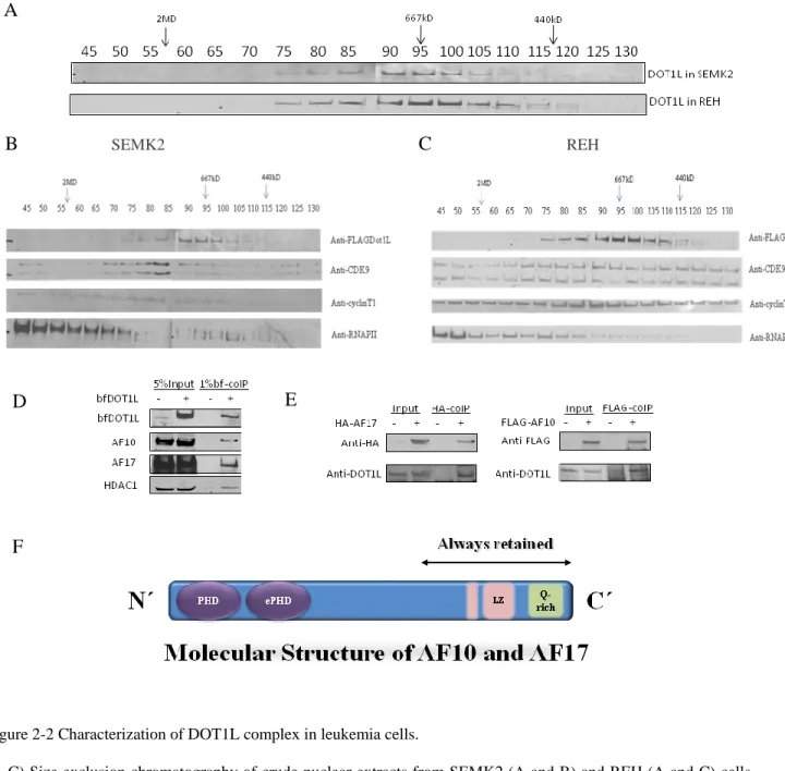
As seen in Figure 2-1C and Table 2-1, mass spectrometry results demonstrated that AF10 and AF17 are two stable components in DOT1L complex in both SEMK2 and REH cells, while other proteins, such as transcription factors, histone modification factors, and scaffold proteins associate with Dot1L in a cell-type dependent manner. The interaction on the list is further verified by co-immunoprecipitation (co-IP) and reciprocal co-IP (see Figure 2-2D).
B
A
17
Table 2-1 Identification of DOT1L associated proteins in SEMK2 and REH cells
Dot1L complex in SEMK2 (4 experiments in total) Dot1L complex in REH (1 experiment in total) Name Average
Peptide Number
Annotation Name Peptide Number
Annotation
Dot1L 63 H3K79 methyltransferase Dot1L 58 H3K79 methyltransferase
AF10 16 MLL fusion partner, putative transcription factor
AF10 17 MLL fusion partner, putative transcription factor
AF17 11 MLL fusion partner, putative transcription factor
AF17 8 MLL fusion partner, putative transcription factor
ILF2 3 NFAT transcription factor TCOF1 7 Treacher Collins-Franceschetti syndrome 1 isoform a
HDAC1 3 Histone deacetylase 1 ARHGEF2 5 Rho guanine nucleotide exchange factor 2
MEF2C 1 Transcription activator in B cells
GTF2I 2 General transcription factor II-I
COE1 (2)*1 Early B-cell factor SMC2 2 Structural maintenance of chromosomes protein 2 CDK9 (1)*2 Transcriptional elongation factor PCM1 2 Pericentriolar material 1 protein NPM1 (5)*3 Nucleophosmin, involved in ribosome biogenesis, histone assembly, cell proliferation
NPM1 1 Nucleophosmin, involved in ribosome biogenesis, histone assembly, cell proliferation
CCAR1 1 Cell division cycle and apoptosis regulator 1
AHDC1 1 AT hook containing protein that may interact with MLL, ENL, AF9, AF10
Note: I performed one experiment for REH cells with tandem affinity purification and four experiments for SEMK2 cells, once with single anti-FLAG purification, twice with single strepavidin purification, and once with tandem affinity purification. All the listed proteins in SEMK2 cells have repeatedly appeared in SEM-BirA-Dot1L samples but not in SEM-BirA control except NPM1, CDK9, and COE1(see*).
*1: COE1 was identified once with 2 unique peptides in the tandem affinity purification in SEMK2 cells. *2: CDK9 was identified once with 1 unique peptide in Streptavidin affinity purification in SEMK2 cells.
*3: NPM is known to be an abundant and sticky protein in the nucleus. It was detected in both SEM-BirA-Dot1L sample and SEM-BirA control in single step purifications, but it was identified only in SEM-BirA-Dot1L and REH-BirA-DOT1L samples (5 peptides and 1 peptide detected respectively) but not in SEM-BirA or REH-BirA controls using tandem affinity purification.
18
Figure 2-2 Characterization of DOT1L complex in leukemia cells.
(A-C) Size exclusion chromatography of crude nuclear extracts from SEMK2 (A and B) and REH (A and C) cells showed that DOT1L complexes elute at the size around 670kD (fractions 90-95) while P-TEFb elute at a broader range (enriched infractions 65-95) . Western blot was performed with antibodies against Dot1L, CDK9, cyclinT1 and RNAPII CTD, respectively. (D) The interaction between DOT1L and AF10/AF17/HDAC1 was confirmed by immunoprecipitation. (E) Reciprocal co-IP showed AF10 and AF17 interact with DOT1L in vivo. (F) The molecular structure of AF10 and AF17.
A
B
SEMK2C
REHD
E
19
To further characterize DOT1L complex, I performed gel filtration on nuclear extracts from SEMK2 and REH cells. As shown in Figure 2-2, the DOT1L complex was eluted as a sharp peak about 670kDa in both SEMK2 cells and REH cells. In contrast, RNA polymerase II (RNAPII), CDK9 and cyclin T1 were all eluted as very broad peaks, consistent with the diversity of RNAPII and P-TEFb containing complexes. In our study, RNAPII and P-TEFb subunits were mainly co-eluted at sizes above 1MDa, in agreement with recent papers that showed the Pol II elongation complex AEP or SEC at the size of about 1.5MDa (17).
There are several important points raised by this study. First, though SEMK2 cells have MLL-AF4 translocation, neither MLL or AF4 is found to interact with DOT1L, suggesting that DOT1L is not recruited to MLL target loci through direct association with MLL-AF4 fusion protein or that the association is weak or transcient. Second, except CDK9, none of the DOT1L complex components identified in leukemic cells appears in AEP or SEC, and the DOT1L complex was eluted at different sizes than AEP or SEC, suggesting that the DOT1L complex and AEP or SEC are two distinct complexes. Third, proteins that only associate with DOT1L in SEMK2 or REH cells may have biological relevance. For example, it has been shown that transcription activator MEF2C is highly expressed in MLL-rearranged AML and its up-regulation may be responsible for the aggressive nature of MLL-MLL-rearranged AML (61). DOT1L may cooperate with MEF2C to promote homing and invasiveness of MLL-rearranged leukemic cells.
As shown in Figure 2-2, the two most stable DOT1L interacting partners are AF10 and AF17. AF10 and AF17 were both identified as MLL fusion partners in acute myeloid leukemias. They are highly homologous proteins with similar N-terminal plant homeodomain (PHD) fingers, and C-terminal Leucine Zipper (LZ) domain. What are their functions in DOT1L complex? Why does nature evolve two similar proteins? We hypothesized that the two proteins serve different functions in vivo. To characterize the
20
function of AF10 and AF17, I started with shRNA-mediated knock-down experiments in human acute leukemia cell lines. Five shRNAs against human AF10 and five shRNAs against human AF17 were tested, among which shAF10.1 and shAF17.7 showed consistent knock-down in leukemia cell lines (Figure 2-3A and 2-3B) and no toxicity in 293T cells.
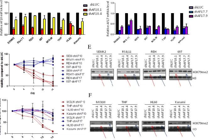
Figure 2-3 Suppression of AF10 and AF17 have different phenotypes in leukemia.
(A-B). Reverse transcription and q-PCR showing effective knock-down of AF10 (A) and AF17 (B) 4 days after transduction with lentivirus carrying shRNA construct. AF10 and AF17 expression levels were normalized to GAPDH and expressed relative to control cells which were transduced with lentivirus carrying shRNA against Luciferase gene (shLUC, set to 100%). (C) Growth of human ALL cell line SEM (MLL-AF4) and RS4;11(MLL-AF4), REH (non-MLL-rearranged), and 693 (non-MLL-rearranged) cells after transduction with shAF10.1 and shAF17.7. Viable cells were counted every 3 to 4 days, and cell numbers after transduction with shLUC were set as 100%. Result is representive of 3 indipendent experiments. (D) Growth of human AML cell line MOLM (MLL-AF9) and THP (MLL-AF9), HL60 (non-MLL-rearranged), and Kasumi (non-MLL-rearranged) cells after transduction with shAF10.1 and shAF17.7. Viable cells were counted every 3 to 4 days, and cell numbers after transduction with shLUC were set as 100%. Result is representive of 3 indipendent experiments. Western blot showing AF17 protein level after shRNA-mediated knock-down. (E-F) Western blot showing H3K79me2 level 9 days after transduction with shRNA (red arrows points to reduced H3K79me2 level) in 4 ALL cell lines (E) and 4 AML cell lines (F).
A
B
C
D
E
21
Knock-down of AF10 in human leukemia cell lines led to a global decrease of H3K79me2 9 days after introduction of shRNA by lentivirus (red arrows, Figure 2-3E and 2-3F), while knock-down of AF17 did not significantly affect H3K79me2 level. In addition, AF10 knock-down affected cell viability of
MLL-rearranged leukemia cell line SEM, RS4;11, MOLM, and THP, while AF17 knock-down does not affect
cell viability (Figure 2-3C and 2-3D). Therefore, AF10 and AF17 may function differently and AF10 is more important regarding H3K79 dimethylation by Dot1l. In Chapter 5, I focus more on the role of AF10 in MLL leukemogenesis.
Discussion
The diversity of MLL fusion partners poses a challenge to developing a unified mechanistic model for
MLL leukemias. However, gene expression signatures of MLL rearranged leukemias are remarkably
similar and can be reliably used to distinguish leukemias with MLL rearrangements from other subtypes. Our lab has shown that MLL rearranged leukemias with nuclear fusion partners (12, 25) and with cytoplasmic fusion partners (see Chapter 4) both have aberrantly high histone methylation at H3K79 in MLL target genes. Thus this epigenetic lesion may be involved in driving the gene expression signatures in MLL leukemias. The development of molecular therapies to specifically target epigenetic modifiers is a promising new field. In this project, I sought to study the requirement of DOT1L complex in several MLL rearranged leukemias with dismal prognosis. To understand the function of DOT1L in leukemia and to suggest new therapeutic strategies, however, requires information about the DOT1L complex in the right context. Therefore we purified DOT1L complex in human acute leukemia cells with the most common
MLL rearrangement, MLL-AF4, or without MLL rearrangement. Our mass spectrometry data showed
AF10 and AF17 are the two most stable DOT1L-interacting proteins in leukemia cells. Moreover, shRNA mediated knock-down of AF10 in human acute leukemia cell lines diminished global H3K79 methylation
22
level and affected the survival of MLL-rearranged leukemia cells, while knock-down of AF17 showed no effect on global H3K79 methylation or in vitro leukemia cell survival.
In our study, we purified the DOT1L complex under physiological conditions from human acute leukemia cells. While we further characterized the functions of DOT1L complex components, Ali Shilatifard group published the first paper describing Dot1l complex, which they called DotCom. In their study, they overexpressed FLAG-tagged Dot1l in 293T cells and performed mass spectrometry after one-step affinity purification. Similar to our study, they also identified AF10 and AF17 in the complex. Besides AF10 and AF17, they found two more MLL fusion partner AF9 and ENL, and a scaffolding protein TRRAP in DotCom. There are some possibilities to explain the discrepancy between the two studies. First, while the human kidney fibroblast cell line 293T is a useful mammalian system to study protein-protein interaction, it is known for overexpression of recombinant proteins at an artificially high level, which may pick up false interactions that do not happen under the physiological protein concentration. Second, human kidney fibroblast cells and human leukemia cells express different sets of proteins. The difference in the cellular proteome may explain the difference of DOT1L/Dot1l interacting network in the two studies. The interaction between DOT1L and MEF2C or ILF2 may have important functions in hematopoietic malignancy, but MEF2C and ILF2 may not be expressed at a detectable level in kidney fibroblasts. It is possible that AF9 and ENL are less abundant in human acute lymphoblastic cells than in 293T cells,
therefore even if they interact with DOT1L in vivo, they will not be identified in our study with human
ALL cell lines.
Our study brought the function of the AF10-DOT1L complex into sharp focus, leading us to study its role in the epigenetic activation of gene expression and involvement in MLL rearranged leukemias. The interaction between AF10 and DOT1L was first discovered in yeast-two-hybrid screen by Yi Zhang’s group. They also demonstrated that the C-terminal octapeptide motif-leucine zipper (OM-LZ) domain of AF10, which is required for association with DOT1L, is the “minimal” fusion domain necessary and
23
sufficient for bone marrow transformation by MLL-AF10 and CALM-AF10. The PHD finger, 50-80 amino acids in length, is a common domain that reads histone modifications. It is found in many human proteins and is involved in chromatin-mediated gene regulation. For example, the PHD finger in ING2 and NURF binds to H3K4me3, and the PHD finger in JARID1C binds to H3K9me3. Many histone modifying enzymes have well characterized chromatin binding domains, DOT1L, however, does not have any known domain to read histone modifications. Although our study with H3 tail peptide binding assay failed to identify the histone modification AF10 may read (data not shown), it is still possible that AF10 completes DOT1L complex function by targeting DOT1L to specific histone modifications.
One straight-forward hypothesis for MLL rearranged leukemia would be that the MLL-fusion partner attracts Dot1l to MLL targets, promotes H3K79 methylation and gene transcription. This hypothesis could explain the mechanisms of MLL-AF10, and possibly MLL-AF9 and MLL-ENL. However, our study of DOT1L complex in MLL-AF4 expressing cells did not find AF4 or MLL by mass spectrometry. The DotCom identified by Ali Shilartifard group also does not include other common MLL fusion partners like AF4, CBP, GAS7 or AF6. Moreover, recently it was revealed that AF4, AFF4, AF9 and ENL, all of which are MLL fusion partners that exist in the nucleus, form a higher-order transcriptional elongation complex called AEP or SEC, with the positive transcription elongation factor (P-TEFb). It was demonstrated that AEP or SEC was hijacked to MLL targets in the context of MLL-fusion leukemia, contributing to leukemogenesis. Consistent with our study, their studies also showed Dot1l is not part of the MLL-fusion-AEP/SEC complex, although H3K79 methylation is aberrantly high at MLL targets. The detailed structure-function study revealed that although ENL binds to Dot1l, the binding of Dot1l and ENL or AEP and ENL is mutual exclusive. (16, 17) Therefore, how DOT1L is recruited to MLL targets in MLL-rearranged leukemias remains elusive and more detailed studies about the functions of DOT1L interacting proteins need to be done.
24
It is intriguing that two homologous proteins AF10 and AF17 serve different functions in vivo. Knock-down of AF10 affects global H3K79 dimethylation level and cellular survival, while AF17 does not. When we were characterizing the function of AF10 with knock-out mouse model, a AF17 knock-out mouse model was reported (62, 63). In agreement with our knock-down study, AF17 knock-out has minimal effect on H3K79 methylation and hematopoiesis, but plays a role in sodium homeostasis and blood pressure control.
Material and Methods
Generation of Dot1l expressing human leukemia cell lines
pEF-BirA-V5-His, MSCV-BirA-V5-His, pEF-FLAG-Bio-puro, and MSCV-FLAG-Bio-puro were gifts from Orkin lab. pEF-FLAG-Bio-DOT1L-puro was generated by amplifying DOT1L cDNA using 5’-ATT GGA TCC TGG GGG AGA AGC TGG AGC TGA GAC TGA AG-3’ (forward primers) and 5’-TAT AGC TAG CGA ATT CTA GTT ACC TCC AAC TGT GCC GCC TGC CAC CCC-3’ (reverse primer), and inserting the cDNA into the BamHI/XbaI sites of pEF-FLAG-Bio-puro vector. MSCV-FLAG-Bio-DOT1L-puro was assembled by ligating the DOT1L cDNA fragment and the MSCV-FLAG-Bio-puro vector at the EcoRI site. Packaging plasmids VSV.G and M57 gag/pol were used with MSCV plasmids to make amphotropic retrovirus carrying BirA and DOT1L. And packaging plasmids pMD2.G and psPAX2 were used with pLKO plasmids to make lentivirus carrying shRNA. Coding region human AF10, AF17 and control shRNA in pLKO.1-lentiviral vectors were kindly synthesized by the Harvard/MIT Broad Institute RNA consortium (http://www.broad.mit.edu/rnai/trc). A shRNA targeting Luciferase served as control.
25
The following human cell lines were obtained from American Type Culture collection: MLL-AF4 rearranged ALL cell line RS4;11 (ATCC CRL-1873),
MLL wildtype ALL cell line REH (ATCC CRL-8286), MLL-AF9 rearranged AML cell line THP-1 (ATCC TIB-202), MLL wildtype AML cell line HL-60 (ATCC CCL-240).
The following cell lines were purchased from the Deutsche Sammlung von Mikroorganismen und
Zellkulturen (DSMZ, Germany):
MLL-AF9 rearranged AML cell line MOLM-14 (DMSZ ACC 554), MLL wildtype AML cell line KASUMI-1 (DMSZ ACC 220).
MLL wildtype ALL cell line 697 cells were provided by Dr. Kimberly Stegmaier (Dana Farber Cancer Institute).
MLL-AF4 rearranged ALL cell line SEMK2-M1 are previously described (Faber et al., 2009).
All the leukemia cell lines were maintained in RPMI-1640 media (Invitrogen) supplemented with heat-inactivated 10% fetal bovine serum, 2 mM L-glutamine and non-essential amino acids and 50 U/ml Penicillin/Streptomycin (all Gibco, Invitrogen, Carlsbad, CA). 293T cells were cultured in DMEM media (Invitrogen) supplemented with heat-inactivated 10% fetal bovine serum, 2 mM L-glutamine and non-essential amino acids and 50 U/ml Penicillin/Streptomycin. All cells were cultured in the appropriate
medium in a humidified incubator at 37°C in 5% CO2.
Lentiviral shRNA experiment
The knock-down efficiency of different shRNAs against AF10 and AF17 was tested by q-PCR and western blotting in 293T and human cell lines. The two most efficient shRNAs among the five were selected for further experiments. Human leukemia cell lines were transduced with pLKO.1 puro lentiviral shRNA against AF10, AF17, or luciferase. Lentiviral particles were produced by cotransfection of 293T cells with pLKO.1 constructs and packaging plasmids pMD2.G and psPAX2. Transfections were carried out with FuGENE 6 (Roche Diagnostics), and virus was harvested 48 and 72 hr after transfection.
26
Polybrene (Sigma-Aldrich, St Louis, MO) was added to a final concentration of 7 g/ml, and cells were
overlaid with viral supernatant. Spinocculation was performed at 2250 rpm for 90 minutes at 37°C. 12 hours after the transduction, media was replaced. 24 hours after transducation, infected cells were selected with 2 μg/ml puromycin. Media was replaced with fresh media with 2 μg/ml puromycin and the number of viable cells was counted with trypan blue stain under the microscope every 3 days.
Generation of human leukemia cell lines stably expressing BirA with and without DOT1L
Amphotropic retroviral supernatants were produced by cotransfection of 293T cells with VSV.G, M57 gag/pol, and the plasmid of interest using FuGENE6 (Roche Molecular Biochemicals, Indianapolis, IN). Virus containing supernatant medium was collected 2-3 days after the transfection. For transduction, 2,000,000 cells/well were plated into 6 well plates in the appropriate culture media. Polybrene (Sigma-Aldrich, St Louis, MO) was added to a final concentration of 7 g/ml, and cells were overlaid with viral supernatant. Spinocculation was performed at 2250 rpm for 90 minutes at 37°C. 12 hours after the transduction, media was replaced with 2 ml of the appropriate fresh culture media. 24 hrs after the
transduction, cells were selected
with 0.5 mg/ml G418 for one week (for establishing BirA
expressing cells first) or
in 0.5 mg/ml G418 and 5 g/ml puromycin (Sigma-Aldrich) for three days (for establishing DOT1L expressing cells based on BirA expressing cells), and maintained in 0.5 mg/ml G418alone (for BirA expressing cells without DOT1L) or 0.5 mg/ml G418 and 2.5 g/ml puromycin (for BirA
expressing cells with DOT1L) throughout the remainder of the observation period.
Stably transfected
cells that express lower level of DOT1L were cloned by limiting dilution.
Large-scale purification of DOT1L complex and proteomic analysis
Nuclear extracts are prepared simultaneously from BirA expressing cells with and without tagged DOT1L, and affinity purification is processed simultaneously. Cell culture were scaled up to 5-10 liter in 850ml roller bottles (BD falcon) and kept in a rolling incubator at 37°C in 5% CO2. Crude nuclear extracts were
27
prepared from ∼1 × 10¹⁰ cells essentially as described previously (60). Briefly, cells were collected by centrifugation at 2400g for 5 mins and washed with ice-cold 1X PBS twice. Cells were rapidly resuspended with ~5X packed cell volume (PCV) of ice-cold Hypotonic Buffer (10 mM HEPES [pH 7.9], 1.5 mM MgCl2, 10 mM KCl, 0.5 mM DTT, 0.2 mM PMSF, 1:100 mammalian protease inhibitor cocktail [Sigma]), and centrifuged at 3000 rpm at 4oC for 5 mins. Cells were then resuspended in 3X PCV of ice-cold hypotonic buffer and incubated on ice for 10 mins to swell cells. And then cells were transfered to glass Dounce Homogenizer and slowly homogenized up and down 12 times with type B pestle. Cells were checked for cell lysis by examinzation of small aliquot in tyypan blue stain under microscope. Homogenate was centrifuged at 4000 rpm at 3300g at 4oC and supernatant was removed. Nuclei was resuspended by first adding ½ packed nuclear volume of ice cold low-salt buffer (20 mM HEPES [pH 7.9], 25% glycerol (v/v), 1.5 mM MgCl2, 0.02 M KCl, 0.2 mM EDTA, 0.5 mM DTT, 0.2 mM PMSF, 1:100 mammalian protease inhibitor cocktail [Sigma]) and then adding ½ packed nuclear volume of ice-cold high-salt buffer (20 mM HEPES [pH 7.9], 25% glycerol [v/v], 1.5 mM MgCl2, 1.5 M KCl, 0.2 mM EDTA, 0.5 mM DTT, 0.2 mM PMSF, 1:100 mammalian protease inhibitor cocktail [Sigma]). Nuclei suspension was incubated on a rotating wheel for 30 mins at 4oC, and centrifuged at 25,000 g for 30 mins at 4oC. The supernatant (nuclear extract) was transferred to fresh tubes and the centrifugation was repeated to remove any insoluble material.
Nuclear extracts were immediately dialyzed against 50 volumes of BC139K buffer (139 mM KCl, 12 mM
NaCl, 0.8 M MgCl2, 20 mM Tris-HCl [pH 7.9], 0.5% NP-40, 0.2 mM EDTA, 20% [v/v] glycerol, 1 mM
dithiothreitol, 0.2 mM phenylmethylsulfonyl fluoride, 0.1% mammalian protease inhibitor cocktail) at 4°C using a 10,000-molecular-weight-cutoff dialysis cassette (Pierce). The dialysate was centrifuged twice at 25,000 × g for 30 min at 4°C. Between 50 and 120 mg of total nuclear protein was then precleared with protein A/G beads (Roche) for 1 h on a rotating wheel at 4°C. The precleared supernatant was incubated with anti-FLAG M2-agarose beads (Sigma) overnight (14 to 16 h) on a rotating wheel. The beads were collected by centrifugation and washed four times with cold BC139K buffer on a rotating
28
wheel at 4°C for 15 min each. Bound material was eluted by incubating beads in BC139K containing 0.1 mg/ml FLAG peptide (Sigma) for 90 min at 4°C on a rotating wheel. Material from four successive elutions was pooled and incubated with streptavidin-agarose beads (Invitrogen) for 14 to 16 h at 4°C on a rotating wheel. The beads were washed as described above, transferred into BC139 K buffer in which NaCl was substituted for KCl, and heated at 95°C to 100°C in Laemmli sodium dodecyl sulfate (SDS) sample buffer for 5 min. The eluted material was concentrated using a YM-10 Centricon (Millipore) device and resolved by SDS-polyacrylamide gel electrophoresis (PAGE) on 10% acrylamide gels running 2.5 cm into the separating gel. Proteins were visualized with colloidal Coomassie blue stain (Invitrogen), and the lanes were divided into three sections as “low Molecular Weight (MW)”, “medium MW”, and “high MW”.
Excised acrylamide gel sections were cut into approximately 1-mm3 pieces. Gel pieces were then subjected to a modified in-gel trypsin digestion procedure (64). Gel pieces were washed and dehydrated with acetonitrile for 10 min, followed by the removal of acetonitrile. Pieces were then completely dried in a Speed-Vac. Rehydration of the gel pieces was done with 50 mM ammonium bicarbonate solution containing 12.5 ng/μl modified sequencing-grade trypsin (Promega) at 4°C. After 45 min, excess trypsin solution was removed and replaced with 50 mM ammonium bicarbonate solution to just cover the gel pieces. Samples were then placed in a 37°C room overnight. Peptides were later extracted by removing the ammonium bicarbonate solution, followed by one wash with a solution containing 50% acetonitrile and 5% acetic acid. The extracts were dried in a Speed-Vac (∼1 h) and then stored at 4°C until analysis. On the day of analysis, the samples were reconstituted in 5 to 10 μl of high-performance liquid chromatography solvent A (2.5% acetonitrile, 0.1% formic acid). A nanoscale reverse-phase high-performance liquid chromatography capillary column was created by packing 5-μm C18 spherical silica beads into a fused silica capillary (100-μm inner diameter by ∼12-cm length) with a flame-drawn tip (65). After equilibrating the column, each sample was loaded onto the column via a Famos autosampler (LC Packings). A gradient was formed, and peptides were eluted with increasing concentrations of solvent B
29
(97.5% acetonitrile, 0.1% formic acid). As each peptide was eluted, they were subjected to electrospray ionization and fed into an LTQ linear ion-trap mass spectrometer (ThermoFinnigan). Eluting peptides were detected, isolated, and fragmented to produce a tandem mass spectrum of specific fragment ions for each peptide. Peptide sequences (and, hence, protein identity) were determined by matching protein or translated nucleotide databases with the acquired fragmentation pattern by the software program SEQUEST (ThermoFinnigan) (66).
Sephacryl S400 gel filtration chromatography
Crude nuclear extracts were prepared from human leukemia cells as described above and dialyzed against
50 volumes of cold BC100 buffer (100 mM KCl, 12 mM NaCl, 0.8 M MgCl2, 20 mM Tris-HCl [pH 7.9],
0.2 mM EDTA, 20% [vol/vol] glycerol, 1 mM dithiothreitol, 0.2 mM phenylmethylsulfonyl fluoride, 0.1% protease inhibitor cock