STATE-OF-THE-ART PAPER
Vitamin D Deficiency
An Important, Common, and Easily
Treatable Cardiovascular Risk Factor?
John H. Lee, MD,* James H. O’Keefe, MD,* David Bell, MD,† Donald D. Hensrud, MD, MPH,‡ Michael F. Holick, MD, PHD§
Kansas City, Missouri; Birmingham, Alabama; Rochester, Minnesota; and Boston, Massachusetts Vitamin D deficiency is a highly prevalent condition, present in approximately 30% to 50% of the general popula-tion. A growing body of data suggests that low 25-hydroxyvitamin D levels may adversely affect cardiovascular health. Vitamin D deficiency activates the renin-angiotensin-aldosterone system and can predispose to hyperten-sion and left ventricular hypertrophy. Additionally, vitamin D deficiency causes an increase in parathyroid hor-mone, which increases insulin resistance and is associated with diabetes, hypertension, inflammation, and in-creased cardiovascular risk. Epidemiologic studies have associated low 25-hydroxyvitamin D levels with coronary risk factors and adverse cardiovascular outcomes. Vitamin D supplementation is simple, safe, and inexpensive. Large randomized controlled trials are needed to firmly establish the relevance of vitamin D status to cardiovas-cular health. In the meanwhile, monitoring serum 25-hydroxyvitamin D levels and correction of vitamin D defi-ciency is indicated for optimization of musculoskeletal and general health. (J Am Coll Cardiol 2008;52: 1949–56) © 2008 by the American College of Cardiology Foundation
Traditionally, vitamin D has been associated primarily with bone health, and it is well understood that vitamin D deficiency leads to rickets in children and osteomalacia and osteoporosis in adults (1). However, it is now known that adequate vitamin D status is important for optimal function of many organs and tissues throughout the body, including the cardiovascular (CV) system (2). Vitamin D receptors (VDRs) are present on a large variety of cell types, including myocytes, cardiomyocytes, pancreatic beta-cells, vascular endothelial cells, neurons, immune cells, and osteoblasts (1). Vitamin D deficiency or insufficiency is prevalent in prac-tically every segment of the U.S. population, including children and young adults (1). This worldwide pandemic remains generally unrecognized and untreated.
Evolving data indicate that vitamin D deficiency is playing an important role in the genesis of coronary risk factors and CV disease. Vitamin D deficiency seems to predispose to hypertension, diabetes and the metabolic syndrome, left ventricular hypertrophy, congestive heart failure, and chronic vascular inflammation (1,2).
Epidemi-ologic studies have also recently linked vitamin D deficiency with increased risk of major adverse CV events (3). A study of male health professionals showed a 2-fold risk of myo-cardial infarction (MI) in subjects who were vitamin D deficient compared with those in the sufficient range (4). Similarly, a recent prospective cohort study measured the vitamin D levels in 3,258 German adults who were under-going elective cardiac catheterization. During a mean follow-up of 7.7 years, individuals in the lowest quartile for baseline serum 25-hydroxyvitamin D [25(OH)D] had a risk-adjusted 2-fold increased risk of death, especially CV death, compared with those in the highest quartile of vitamin D (5).
This review focuses on the relationship between 2 wide-spread problems: vitamin D deficiency and CV disease. The issues addressed will include: 1) the role of vitamin D deficiency in the genesis of coronary risk factors and adverse CV events; 2) how repletion of vitamin D stores may improve CV health and prognosis; and 3) practical and specific recommendations for restoring and maintaining a healthy vitamin D status in CV patients, because no guidelines have been published on this topic yet.
Vitamin D Basics
Vitamin D comes in 2 forms: vitamin D2(ergocalciferol)
and vitamin D3 (cholecalciferol). Vitamin D2, found in
plants, is the product of ultraviolet B (UVB) (290 to 315 mm) irradiation of ergosterol, and can be consumed as From the *Mid America Heart Institute and University of Missouri, Kansas City,
Missouri; †Southside Endocrinology, Birmingham, Alabama; ‡Mayo Clinic, Roch-ester, Minnesota; and §Boston University Medical Center, Boston, Massachusetts. Dr. O’Keefe is an unpaid scientific consultant to CardioTabs. Funds derived from this company are used for marketing and patient education at Cardiovascular Consultants, the group practice with which Dr. O’Keefe is affiliated. Dr. Holick is a speaker and consultant for the National Dairy Council and the UV Foundation.
Manuscript received June 4, 2008; revised manuscript received August 6, 2008, accepted August 13, 2008.
a supplement or in fortified foods (1). Vitamin D3, a
prod-uct of UVB irradiation of 7-dehydrocholesterol, is synthe-sized in the human epidermis or consumed in the form of oily fish, fortified foods, or a supple-ment. Excessive sunlight expo-sure cannot cause vitamin D tox-icity because UVB converts excess vitamin D3to biologically
inert isomers (1); however, exces-sive oral vitamin D intake can cause toxicity at very high doses (6).
Vitamin D is converted in the liver to 25(OH)D, which is the major circulating metabolite of vitamin D. Serum 25(OH)D concentrations, which reflect both vitamin D intake and endogenous production, should be measured to clinically assess vitamin D status (1). In the kidney, 25(OH)D is converted by 1␣-hydroxylase to its active form, 1,25-dihydroxyvitamin D [1,25(OH)2D], which plays a
vital role in maintaining bone and muscle health by regu-lating calcium metabolism. Although 1,25(OH)2D is the
active form of vitamin D, its serum level does not correlate with overall vitamin D status and thus is generally not clinically useful (1).
Vitamin D in the form of 1,25(OH)2D is a hormone,
because it is produced primarily in 1 organ (the kidney) and then circulates throughout the body, where it exerts wide-ranging effects. The VDR is present in most tissues, including endothelium, vascular smooth muscle, and myo-cardium (2). In addition, both vascular smooth muscle and endothelial cells may have the ability to convert 25(OH)D to 1,25(OH)2D (7). Circulatory 1,25(OH)2D crosses the
cell membrane and cytoplasm and reaches the nucleus, where it binds to the VDR. The VDR-bound 1,25(OH)2D
in turn binds to the retinoic acid x-receptor and serves as a nuclear transcription factor, altering gene function and inducing protein synthesis (1). Directly or indirectly, 1,25(OH)2D regulates over 200 genes, including those
involved in renin production in the kidney, insulin produc-tion in the pancreas, release of cytokines from lymphocytes, production of cathelicidin in macrophages, and growth and proliferation of both vascular smooth muscle cells and cardiomyocytes (1).
Definition and Prevalence of Vitamin D Deficiency
Although a consensus regarding the optimal level of serum 25(OH)D has not yet been established, most experts define vitamin D deficiency as a 25(OH)D level of⬍20 ng/ml (50 nmol/l) and vitamin D insufficiency as 21 to 29 ng/ml (Table 1). For all studied end points to date, the optimal concentration of 25(OH)D is at least 30 ng/ml (8).
A rapidly evolving knowledge base indicates that vitamin D deficiency is much more prevalent than previously
rec-ognized and is present in up to 50% of young adults (9) and apparently healthy children (1). The Third National Health and Nutrition Examination Survey (NHANES III) re-ported the prevalence of vitamin D deficiency in the U.S. to be between 25% and 57% of adults (10).
The prevalence of vitamin D deficiency increases in proportion to distance from the equator because of increased atmospheric filtering of UVB radiation caused by the oblique angles of the sun’s rays at higher latitudes. Addition-ally, ethnic groups with darker skin require proportionally more sun exposure to synthesize equivalent amounts of vitamin D compared with people with lighter skin coloration (11).
Modern human cultures produce less vitamin D cutane-ously, in part because of increasingly indoor lifestyles and efforts to minimize sun exposure by using sunscreens and other sun avoidance strategies. Sunscreen with a sun pro-tection factor of 15 blocks approximately 99% of the cutaneous vitamin D production (12). Additionally, obesity is associated with vitamin D deficiency (13), probably because of a decreased bioavailability of vitamin D that is sequestered in the fat of individuals with excess adipose tissue (14). After equivalent exposure to UVB radiation or a bolus dose of vitamin D2, obese individuals showed 50%
lower blood levels of vitamins D3 and D2compared with
nonobese individuals, probably because of sequestering of 25(OH)D in adipose tissue (14). Older age also reduces the capacity for UVB-induced cutaneous synthesis of vitamin D. After equal doses of sunlight exposure, a 70-year-old person produces 75% less vitamin D3 than a 20-year-old
person (15). Other risk factors for vitamin D deficiency are listed in Table 2.
Vitamin D Deficiency and CV Disease
Epidemiological studies report that the rates of coronary heart disease, diabetes, and hypertension, like vitamin D deficiency, increase in proportion to increasing distance from the equator (16). Deficient or insufficient serum 25(OH)D levels have been documented in patients with myocardial infarction (17), stroke (18), heart failure (2), diabetic CV disease (19), and peripheral arterial disease (20). Recently, the relationship between CV risk factors and 25(OH)D levels was explored among the 15,088 subjects from the NHANES III national cohort registry. In this cross-sectional study, 25(OH)D levels were inversely asso-ciated with hypertension, diabetes mellitus, hypertriglycer-idemia, and obesity (21). Other cross-sectional studies have
Vitamin D Status
Table 1 Vitamin D Status
Serum 25-Hydroxyvitamin D (ng/ml) Vitamin D Status
ⱕ10 Severe deficiency 10–20 Deficiency 21–29 Insufficiency ⱖ30 Sufficiency ⬎150 Toxicity Abbreviations and Acronyms 25(OH)Dⴝ 25-hydroxyvitamin D CVⴝcardiovascular MIⴝmyocardial infarction PTHⴝparathyroid hormone UVBⴝultraviolet B VDRⴝvitamin D receptor
confirmed the links between vitamin D deficiency and both hypertension and diabetes (22,23). Additionally, vitamin D deficiency predisposes to insulin resistance, pancreatic beta cell dysfunction (24), and the metabolic syndrome (24,25). One study reported that a daily intake of 800 IU of vitamin D compared with a daily intake of⬍400 IU of vitamin D reduced the risk of type 2 diabetes by one-third (26). A study of 10,366 Finnish children who were given 2,000 IU of vitamin D3 per day throughout the first year of life
experienced a 78% reduced risk of type 1 diabetes over the ensuing 31 years of follow-up (27). Subsequently, this finding has been confirmed by a meta-analysis performed on 5 observational studies by a group in England (28).
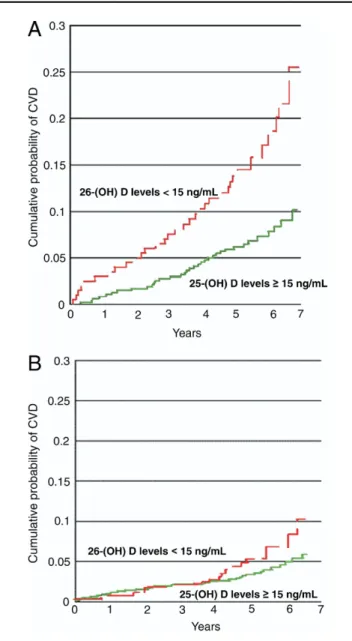
A correlation between vitamin D deficiency and sub-sequent major adverse CV events was found among the 1,739 Framingham Offspring Study participants who were free of CV disease at baseline (3). In this prospective observational study, 25(OH)D levels were measured at baseline and subjects were followed up for a mean of 5.4 years. The rate of a composite CV end point (fatal or nonfatal MI, ischemia, stroke, or heart failure) was 53% to 80% higher in people with low vitamin D levels. The increased CV risk associated with vitamin D deficiency was magnified in the cohort of Framingham offspring with hypertension (Figs. 1and 2).
Vitamin D deficiency predisposes to up-regulation of the renin-angiotensin-aldosterone system and hypertrophy of both the left ventricle and vascular smooth muscle cells (2) (Fig. 3). In vitamin D-deficient animals there is an in-creased incidence of hypertension, left ventricular hypertro-phy, and atherosclerosis (29). Human studies indicate that 1,25(OH)2D inhibits renin synthesis, which may lower
blood pressure (30). Krause et al. (31) showed that increased exposure to UVB radiation in a tanning bed 3 times per week for 3 months led to a 180% increase in 25(OH)D levels and a 6-mm Hg reduction in both systolic and diastolic pressures. A small, randomized, placebo-controlled study of patients with type 2 diabetes and low baseline 25(OH)D levels showed that a single dose of 100,000 IU of vitamin D2reduced systolic blood pressure by a mean of 14
mm Hg and significantly improved endothelial function as measured by forearm blood flow (32). In the NHANES III study, the mean systolic blood pressure was about 3 mm Hg lower in those in the individuals in the highest quintile of serum 25(OH)D levels compared with those in the lowest quintile (22).
Hyperparathyroidism Increases CV Risk
Chronic vitamin D deficiency causes secondary hyperpara-thyroidism, which in turn may mediate many of the detri-mental CV effects of inadequate vitamin D levels. The
Figure 1 Vitamin D Levels Associated With CVD
Vitamin D deficiency increases CV risk. Kaplan-Meier curves show the probabil-ity of major adverse CV events in participants with 25(OH)D levelsⱖ15 ng/ml
(green lines)and 25(OH)D levels⬍15 ng/ml(red lines). The increased CV risk was more apparent in patients with(A)than in patients without(B) hyper-tension. Reprinted, with permission, from Wang et al. (3). 25(OH)D⫽ 25-hy-droxyvitamin D; CV⫽cardiovascular; CVD⫽cardiovascular disease.
Risk Factors for Vitamin D Deficiency
Table 2 Risk Factors for Vitamin D Deficiency
Elderly
Darkly pigmented skin Institutionalized or homebound Increased distance from equator Winter season
Cover-up clothing and/or sunscreen Air pollution Smoking Obesity Malabsorption Renal disease Liver disease
Medications: anticonvulsants, glucocorticoids, antirejection, and human immunodeficiency virus medications
threshold for elevation of parathyroid hormone (PTH) is a 25(OH)D level of⬍30 ng/ml. Further decreases in serum 25(OH)D levels will result in proportionally higher PTH levels to maintain serum and total body calcium (Fig. 4). Vitamin D deficiency reduces intestinal calcium absorption by more than 50% (1). The attendant decrease in serum calcium levels triggers PTH release, which quickly corrects the calcium level by mobilization of calcium from bone, increased renal tubular calcium reabsorption, and increased renal production of 1,25(OH)2D.
The effects of primary hyperparathyroidism on CV out-comes were shown in a study that reported approximately 40% lower relative risks of MI, stroke, and death in patients who had surgical parathyroidectomy compared with obser-vation (33). This link between increased PTH and CV disease was further corroborated by a study of patients with renal failure and secondary hyperparathyroidism [caused by decreased conversion of 25(OH)D to 1,25(OH)2D]. In this
study, patients with a PTH levelⱖ250 pg/ml had a 2-fold risk of CV disease compared with those with PTH levels
⬍250 pg/ml (34). Additionally, a recent observational study found that elevated PTH levels in elderly individuals was associated with a doubling of mortality during follow-up compared with those with normal PTH levels (35).
An increased PTH level is associated with increases in both blood pressure (36) and myocardial contractility, which eventually lead to hypertrophy, apoptosis, and fibrosis of both the left ventricle and vascular medial smooth muscle (2). Vitamin D deficiency and/or increased PTH also predispose to calcification of heart valves, mitral annulus, and myocardium, especially in patients with moderate or severe chronic kidney disease (37).
Chronic kidney disease is associated with markedly in-creased CV risk (38), which may in part be mediated by inadequate vitamin D levels. Vitamin D deficiency is asso-ciated with increased mortality rates in the setting of chronic kidney disease (39), and repleting vitamin D in such patients improves outcomes. Recent observational studies of patients with chronic kidney disease and hyperparathyroid-ism found that the oral administration of 1,25(OH)2D3 Figure 2 Hazard Ratio for CVD by Vitamin D Levels
Multivariable adjusted hazard ratios for major adverse
CV events. Data from Wang et al. (3). Abbreviations as in Figure 1.
Figure 3 Potential Mechanisms for CV Toxicity of Vitamin D Deficiency
(also known as activated vitamin D or calcitriol) was associated with significantly improved survival (40). A placebo-controlled study of 30 pre-dialysis renal failure patients with secondary hyperparathyroidism showed that treatment with 1,25(OH)2D3 improved left ventricular
diastolic function (41). Vitamin D analogs used in patients on dialysis have been shown to improve survival at all doses tested thus far (42).
Low 25(OH)D levels and increased PTH levels increase both inflammation and the risk of adverse CV events (36). Vitamin D deficiency increases systemic inflamma-tion, as documented by elevated levels of C-reactive protein and interleukin-10 (2). Furthermore, administration of 1,25(OH)2D to vitamin D-deficient individuals
down-regulated inflammatory markers (C-reactive protein, and so on) and conferred an antiproliferative effect (43). Extrarenal synthesis of 1,25(OH)2D occurs through cytokine
stimula-tion (7) and is locally important in the paracrine regulastimula-tion of cellular growth, differentiation, and function (44). This may explain why vitamin D deficiency has been associated with type 1 diabetes, cancer, and multiple sclerosis (45). Outcome Studies
A recent meta-analysis of 18 randomized controlled trials comprising 57,000 individuals showed that a vitamin D intake⬎500 IU/day improved all-cause mortality, in part by decreasing CV deaths (46). Moreover, the data regarding both efficacy and CV safety for vitamin D appear to be superior to that for calcium supplements. Indeed, calcium supplementation has recently been implicated as possibly increasing the risk of adverse CV events, particularly in
patients with chronic kidney disease (47,48). Calcium sup-plements acutely increase serum calcium levels, which might accelerate arterial calcification (49). In contrast, serum vitamin D levels are inversely associated with coronary artery calcification (50).
Osteoporosis and atherosclerotic CV disease share many common risk factors, and a pathologic link between these 2 highly prevalent age-related diseases has been suggested (51). A large number of middle-aged to elderly individuals at risk for both CV disease and osteoporosis may benefit from therapies that are likely to improve both conditions, such as an anti-inflammatory diet, daily exercise (especially weight-bearing forms), avoidance of both tobacco and heavy alcohol intake, and possibly vitamin D supplementation. Vitamin D and Myopathy
Myalgias are generally the first manifestation of vitamin D deficiency. Severe vitamin D deficiency with corresponding elevations of PTH were reported in 88% of women who presented with muscle pains and weakness (52). Another study investigated 150 patients with nonspecific musculo-skeletal pains and reported that 25(OH)D levels were insufficient in 93% of individuals and severely deficient in 28% (53). A meta-analysis of 5 randomized clinical trials reported that vitamin D supplementation reduced the risk of falls, most likely from improved muscle function and strength (54). Myalgia, the most common complaint re-ported by patients on statin therapy, may be at least in part caused by underlying vitamin D deficiency. Anecdotally, we have observed that repletion of 25(OH)D levels predictably improves or resolves statin-related myalgias.
Supplementing Vitamin D
Traditionally, up to 95% of the body’s vitamin D require-ment comes from the synthesis in the epidermis on sun exposure, with the remainder ingested from dietary sources (Table 3) (55). The U.S. government’s current recommen-dation for oral vitamin D is 200 IU daily for individuals age
⬍50 years, 400 IU daily for individuals between age 50 and 70 years, and 600 IU for those older than age 70 years. Studies indicate that the average U.S. adult consumes about 230 IU vitamin D per day (56). However, it has been
Figure 4 Relationship of Serum Levels of Parathyroid Hormone and Vitamin D
The inverse relationship between serum 25-hydroxyvitamin D levels and serum parathyroid hormone levels. Reprinted, with permission, from Zittermann et al. (2).
Selected Food Sources of Vitamin D
Table 3 Selected Food Sources of Vitamin D
Food IU per Serving
Cod liver oil, 1 tablespoon 1,360
Wild-caught salmon, 3 oz 600–1,000
Farmed salmon, 3 oz 100–250
Mackerel, cooked, 3 oz 345
Tuna fish, canned in oil, 3 oz 200
Sardines (with bones), canned in oil, drained, 1 oz 250 Milk, nonfat, reduced fat, and whole,
vitamin D-fortified, 1 cup
98
estimated that 1,000 to 2,000 IU is necessary to satisfy the body’s needs for most people (8). Many experts in the field suggest the recommended daily intake of vitamin D be increased to at least 800 to 2,000 IU of vitamin D daily, doses that are difficult to achieve without supplementation, particularly in higher latitudes and in areas of extreme winter climate. A dose of vitamin D3up to 2,000 IU daily
has been deemed by the U.S. Food and Drug Administra-tion’s nutritional guidelines to be generally recognized as safe. A recent review concluded that the safe upper limit for vitamin D consumption is 10,000 IU per day (57); doses
above this increase risk of renal calculi formation, especially in patients with absorptive hypercalcuria and end-stage renal disease patients on dialysis (58).
A study of 340 children ages 10 to 17 years found that increasing the intake of oral vitamin D 10-fold, from the currently recommended dose of 200 to 2,000 IU daily, was required to reach a 25(OH)D level of 30 ng/ml (the lower end of the optimal range) (59). The investigators concluded that doses equivalent to 2,000 IU of vitamin D3daily were
not only safe for adolescents, but also necessary for achiev-ing the desirable vitamin D levels.
The most potent sources of vitamin D are sunlight (about 3,000 IU vitamin D3per 5 to 10 min of mid-day, midyear
exposure of arms and legs for a light-skinned Caucasian) or prescription oral supplements of 50,000 IU capsule of either vitamin D2or D3every 2 weeks (1). Among foods, oily fish
have the highest content of vitamin D3, which ranges from
100 to 1,000 IU per 3.5 oz (1,60), whereas other sources such as milk or orange juice fortified with vitamin D contain up to 100 IU per serving.
As a general rule, every 100 IU vitamin D ingested daily increases the 25(OH)D level by about 1 ng/ml (61,62) (Fig. 5). Over-the-counter dietary supplements of vitamin D2
and D3typically contain 400 to 5,000 IU per capsule. Oral
supplementation with either vitamin D2or D3initially will
increase vitamin D levels equally well (63), although the increases in serum 25(OH)D levels seem to persist longer after a bolus dose of vitamin D3than D2(64).
Treatment of vitamin D-deficient individuals should be initiated with 50,000 IU of vitamin D2or D3weekly for a
period of 8 to 12 weeks. Once the initial repletion phase is complete, maintenance therapy can be continued in 1 of 3 ways: 1) 50,000 IU vitamin D2 or D3 every 2 weeks; 2) Figure 5 Response of Serum
Vitamin D Levels to Supplementation
Effect of dose and duration of vitamin D supplementation on the mean serum 25-hydroxyvitamin D [25(OH)D] concentration achieved. Data from Vieth et al. (62).
Figure 6 Treatment Recommendations for Vitamin D Deficiency
1,000 to 2,000 IU vitamin D3 daily; and 3) sunlight
exposure for 5 to 10 min for Caucasians (longer times required for people with increased skin pigmentation) be-tween the hours of 10AMto 3PM(spring, summer, and fall) (1,61) (Fig. 6).
Acknowledgment
The authors thank Lori J. Wilson for her help in prepara-tion of this article.
Reprint requests and correspondence: Dr. James H. O’Keefe, 4330 Wornall Road, Suite 2000, Kansas City, Missouri 64111. E-mail:jhokeefe@cc-pc.com.
REFERENCES
1. Holick MF. Vitamin D deficiency. N Engl J Med 2007;357:266 – 81. 2. Zittermann A. Vitamin D and disease prevention with special refer-ence to cardiovascular disease. Prog Biophys Mol Biol 2006;92:39 – 48. 3. Wang TJ, Pencina MJ, Booth SL, et al. Vitamin D deficiency and risk
of cardiovascular disease. Circulation 2008;117:503–11.
4. Giovannucci E, Liu Y, Hollis BW, Rimm EB. 25-hydroxyvitamin D and risk of myocardial infarction in men: a prospective study. Arch Intern Med 2008;168:1174 – 80.
5. Dobnig H, Pilz S, Scharnagl H, et al. Independent association of low serum 25-hydroxyvitamin d and 1,25-dihydroxyvitamin D levels with all-cause and cardiovascular mortality. Arch Intern Med 2008;168: 1340 –9.
6. Koutkia P, Chen TC, Holick MF. Vitamin D intoxication associated with an over-the-counter supplement. N Engl J Med 2001;345:66 –7. 7. Zehnder D, Bland R, Chana RS, et al. Synthesis of 1,25-dihydroxyvitamin D(3) by human endothelial cells is regulated by inflammatory cytokines: a novel autocrine determinant of vascular cell adhesion. J Am Soc Nephrol 2002;13:621–9.
8. Bischoff-Ferrari HA, Giovannucci E, Willett WC, Dietrich T, Dawson-Hughes B. Estimation of optimal serum concentrations of 25-hydroxyvitamin D for multiple health outcomes. Am J Clin Nutr 2006;84:18 –28.
9. Tangpricha V, Pearce EN, Chen TC, Holick MF. Vitamin D insufficiency among free-living healthy young adults. Am J Med 2002;112:659 – 62.
10. Looker AC, Dawson-Hughes B, Calvo MS, Gunter EW, Sahyoun NR. Serum 25-hydroxyvitamin D status of adolescents and adults in two seasonal subpopulations from NHANES III. Bone 2002;30: 771–7.
11. Clemens TL, Adams JS, Henderson SL, Holick MF. Increased skin pigment reduces the capacity of skin to synthesise vitamin D3. Lancet 1982;1:74 – 6.
12. Matsuoka LY, Ide L, Wortsman J, MacLaughlin JA, Holick MF. Sunscreens suppress cutaneous vitamin D3 synthesis. J Clin Endocri-nol Metab 1987;64:1165– 8.
13. Wortsman J, Matsuoka LY, Chen TC, Lu Z, Holick MF. Decreased bioavailability of vitamin D in obesity. Am J Clin Nutr 2000;72: 690 –3.
14. Blum M, Dolnikowski G, Seyoum E, et al. Vitamin D(3) in fat tissue. Endocrine 2008;33:90 – 4.
15. Holick MF. Sunlight and vitamin D for bone health and prevention of autoimmune diseases, cancers, and cardiovascular disease. Am J Clin Nutr 2004;80:1678S– 88S.
16. Rostand SG. Ultraviolet light may contribute to geographic and racial blood pressure differences. Hypertension 1997;30:150 – 6.
17. Scragg R, Jackson R, Holdaway IM, Lim T, Beaglehole R. Myocardial infarction is inversely associated with plasma 25-hydroxyvitamin D3 levels: a community-based study. Int J Epidemiol 1990;19:559 – 63. 18. Poole KE, Loveridge N, Barker PJ, et al. Reduced vitamin D in acute
stroke. Stroke 2006;37:243–5.
19. Cigolini M, Iagulli MP, Miconi V, Galiotto M, Lombardi S, Targher G. Serum 25-hydroxyvitamin D3 concentrations and prevalence of cardiovascular disease among type 2 diabetic patients. Diabetes Care 2006;29:722– 4.
20. Melamed ML, Muntner P, Michos ED, et al. Serum 25-hydroxyvitamin D levels and the prevalence of peripheral arterial disease. Results from NHANES 2001 to 2004. Arterioscler Thromb Vasc Biol 2008;28:1179 – 85.
21. Martins D, Wolf M, Pan D, et al. Prevalence of cardiovascular risk factors and the serum levels of 25-hydroxyvitamin D in the United States: data from the Third National Health and Nutrition Examina-tion Survey. Arch Intern Med 2007;167:1159 – 65.
22. Scragg R, Sowers M, Bell C. Serum 25-hydroxyvitamin D, ethnicity, and blood pressure in the Third National Health and Nutrition Examination Survey. Am J Hypertens 2007;20:713–9.
23. Scragg R, Sowers M, Bell C. Serum 25-hydroxyvitamin D, diabetes, and ethnicity in the Third National Health and Nutrition Examina-tion Survey. Diabetes Care 2004;27:2813– 8.
24. Riachy R, Vandewalle B, Moerman E, et al. 1,25-Dihydroxyvitamin D3 protects human pancreatic islets against cytokine-induced apopto-sis via down-regulation of the Fas receptor. Apoptoapopto-sis 2006;11:151–9. 25. Chiu KC, Chu A, Go VL, Saad MF. Hypovitaminosis D is associated with insulin resistance and beta cell dysfunction. Am J Clin Nutr 2004;79:820 –5.
26. Pittas AG, Dawson-Hughes B, Li T, et al. Vitamin D and calcium intake in relation to type 2 diabetes in women. Diabetes Care 2006;29:650 – 6.
27. Hyppönen E, Läärä E, Reunanen A, Järvelin MR, Virtanen SM. Intake of vitamin D and risk of type 1 diabetes: a birth-cohort study. Lancet 2001;358:1500 –3.
28. Zipitis CS, Akobeng AK, Vitamin D supplementation in early childhood and risk of type 1 diabetes: a systematic review and meta-analysis. Arch Dis Child 2008;93:512–7.
29. Simpson RU, Hershey SH, Nibbelink KA. Characterization of heart size and blood pressure in the vitamin D receptor knockout mouse. J Steroid Biochem Mol Biol 2007;103:521– 4.
30. Li YC. Vitamin D regulation of the renin-angiotensin system. J Cell Biochem 2003;88:327–31.
31. Krause R, Buhring M, Hopfenmuller W, Holick MF, Sharma AM. Ultraviolet B and blood pressure. Lancet 1998;352:709 –10. 32. Sugden JA, Davies JI, Witham MD, Morris AD, Struthers AD.
Vitamin D improves endothelial function in patients with type 2 diabetes mellitus and low vitamin D levels. Diabetes Med 2008;25: 320 –5.
33. Vestergaard P, Mosekilde L. Cohort study on effects of parathyroid surgery on multiple outcomes in primary hyperparathyroidism. BMJ 2003;327:530 – 4.
34. Soubassi LP, Chiras TC, Papadakis ED, et al. Incidence and risk factors of coronary heart disease in elderly patients on chronic hemodialysis. Int Urol Nephrol 2006;38:795– 800.
35. Bjorkman MP, Sorva AJ, Tilvis RS. Elevated serum parathyroid hormone predicts impaired survival prognosis in a general aged population. Eur J Endocrinol 2008;158:749 –53.
36. Ogard CG, Engelmann MD, Kistorp C, Nielsen SL, Vestergaard H. Increased plasma N-terminal pro-B-type natriuretic peptide and markers of inflammation related to atherosclerosis in patients with primary hyperparathyroidism. Clin Endocrinol (Oxf) 2005;63:493– 8. 37. Andersson P, Rydberg E, Willenheimer R. Primary
hyperparathyroid-ism and heart disease—a review. Eur Heart J 2004;25:1776 – 87. 38. Sarnak MJ, Levey AS, Schoolwerth AC, et al. Kidney disease as a risk
factor for development of cardiovascular disease: a statement from the American Heart Association Councils on Kidney in Cardiovascular Disease, High Blood Pressure Research, Clinical Cardiology, and Epidemiology and Prevention. Circulation 2003;108:2154 – 69. 39. Wolf M, Shah A, Gutierrez O, et al. Vitamin D levels and early
mortality among incident hemodialysis patients. Kidney Int 2007;72: 1004 –13.
40. Kovesdy CP, Ahmadzadeh S, Anderson JE, Kalantar-Zadeh K. Association of activated vitamin D treatment and mortality in chronic kidney disease. Arch Intern Med 2008;168:397– 403.
41. Singh NP, Sahni V, Garg D, Nair M. Effect of pharmacological suppression of secondary hyperparathyroidism on cardiovascular he-modynamics in predialysis CKD patients: a preliminary observation. Hemodial Int 2007;11:417–23.
42. Lee GH, Benner D, Regidor DL, Kalantar-Zadeh K. Impact of kidney bone disease and its management on survival of patients on dialysis. J Ren Nutr 2007;17:38 – 44.
43. Schleithoff SS, Zittermann A, Tenderich G, Berthold HK, Stehle P, Koerfer R. Vitamin D supplementation improves cytokine profiles in patients with congestive heart failure: a double-blind, randomized, placebo-controlled trial. Am J Clin Nutr 2006;83:754 –9.
44. Mitsuhashi T, Morris RC Jr., Ives HE. 1,25-dihydroxyvitamin D3 modulates growth of vascular smooth muscle cells. J Clin Invest 1991;87:1889 –95.
45. Lips P. Vitamin D physiology. Prog Biophys Mol Biol 2006;92:4 – 8. 46. Autier P, Gandini S. Vitamin D supplementation and total mortality: a meta-analysis of randomized controlled trials. Arch Intern Med 2007;167:1730 –7.
47. Jones G, Winzenberg T. Cardiovascular risks of calcium supplements in women. BMJ 2008;336:226 –7.
48. Bolland MJ, Barber PA, Doughty RN, et al. Vascular events in healthy older women receiving calcium supplementation: randomised con-trolled trial. BMJ 2008;336:262– 6.
49. Asmus HG, Braun J, Krause R, et al. Two year comparison of sevelamer and calcium carbonate effects on cardiovascular calcification and bone density. Nephrol Dial Transplant 2005;20:1653– 61. 50. Watson KE, Abrolat ML, Malone LL, et al. Active serum vitamin D
levels are inversely correlated with coronary calcification. Circulation 1997;96:1755– 60.
51. Shaffer JR, Kammerer CM, Rainwater DL, et al. Decreased bone mineral density is correlated with increased subclinical atherosclerosis in older, but not younger, Mexican American women and men: the San Antonio Family Osteoporosis Study. Calcif Tissue Int 2007;81:430–41.
52. Glerup H, Mikkelsen K, Poulsen L, et al. Commonly recommended daily intake of vitamin D is not sufficient if sunlight exposure is limited. J Intern Med 2000;247:260 – 8.
53. Plotnikoff GA, Quigley JM. Prevalence of severe hypovitaminosis D in patients with persistent, nonspecific musculoskeletal pain. Mayo Clin Proc 2003;78:1463–70.
54. Bischoff-Ferrari HA, Dawson-Hughes B, Willett WC, et al. Effect of vitamin D on falls: a meta-analysis. JAMA 2004;291:1999 – 2006.
55. Dietary Supplement Fact Sheet: Vitamin D. Office of Dietary Sup-plements, NIH Clinical Center, National Institutes of Health. 2007. Available at: http://dietary-supplements.info.nih.gov/factsheets/ vitamind.asp.Accessed July 30, 2008.
56. Moore C, Murphy MM, Keast DR, Holick MF. Vitamin D intake in the United States. J Am Diet Assoc 2004;104:980 –3.
57. Hathcock JN, Shao A, Vieth R, Heaney R. Risk assessment for vitamin D. Am J Clin Nutr 2007;85:6 –18.
58. Daudon M, Jungers P. Drug-induced renal calculi: epidemiology, prevention and management. Drugs 2004;64:245–75.
59. Maalouf J, Nabulsi M, Vieth R, et al. Short-term and long-term safety of weekly high-dose vitamin D3 supplementation in school children. J Clin Endocrinol Metab 2008;93:2693–701.
60. Chen TC, Chimeh F, Lu Z, et al. Factors that influence the cutaneous synthesis and dietary sources of vitamin D. Arch Biochem Biophys 2007;460:213–7.
61. Heaney RP, Davies KM, Chen TC, Holick MF, Barger-Lux MJ. Human serum 25-hydroxycholecalciferol response to extended oral dosing with cholecalciferol. Am J Clin Nutr 2003;77:204 –10. 62. Vieth R. Vitamin D supplementation, 25-hydroxyvitamin D
concen-trations, and safety. Am J Clin Nutr 1999;69:842–56.
63. Holick MF, Biancuzzo RM, Chen TC, et al. Vitamin D2 is as effective as vitamin D3 in maintaining circulating concentrations of 25-hydroxyvitamin D. J Clin Endocrinol Metab 2008;93:677– 81. 64. Armas LA, Hollis BW, Heaney RP. Vitamin D2 is much less effective than vitamin D3 in humans. J Clin Endocrinol Metab 2004;89:5387–91.
Key Words:vitamin Dy25-hydroxyvitamin Dycalciumycoronary disease preventionyhypertensionydiabetes.