ZOOTAXA
ISSN 1175-5326 (print edition)ISSN1175-5334(online edition)
Copyright © 2018 Magnolia Press
Zootaxa 4455 (1): 068–098
http://www.mapress.com/j/zt/
Article
https://doi.org/10.11646/zootaxa.4455.1.3
http://zoobank.org/urn:lsid:zoobank.org:pub:2EA2EE4A-2BA4-438A-A1CB-D9C54F37DC78
Integration of nuclear and mitochondrial gene sequences and morphology
reveals unexpected diversity in the forest cobra (
Naja melanoleuca
)
species complex in Central and West Africa (Serpentes: Elapidae)
WOLFGANG WÜSTER1,11, LAURENT CHIRIO2, JEAN-FRANÇOIS TRAPE3, IVAN INEICH4,
KATE JACKSON5, ELI GREENBAUM6, CESAR BARRON6, CHIFUNDERA KUSAMBA7,
ZOLTÁN T. NAGY8, RICHARD STOREY1, CARA HALL1, CATHARINE E. WÜSTER1,
AXEL BARLOW1,9 & DONALD G. BROADLEY10†
1Molecular Ecology and Fisheries Genetics Laboratory, School of Biological Sciences, Bangor University, Bangor LL57 2UW, United
Kingdom. E-mail: [email protected]
214 Rue des Roses, 06130 Grasse, France. [email protected]
3Laboratoire de Paludologie et Zoologie médicale, Institut de Recherche pour le Développement (IRD), MIVEGEC, B.P. 1386, Dakar,
Sénégal. E-mail: [email protected]
4Institut Systématique Evolution Biodiversité (ISYEB), Muséum national d'Histoire naturelle, CNRS, Sorbonne Universités, EPHE, 57
rue Cuvier, CP 30, 75005 Paris, France. E-mail : [email protected]
5Department of Biology, Whitman College, 345 Boyer Avenue, Walla Walla, WA 99362, USA. E-mail: [email protected]
6Department of Biological Sciences, University of Texas at El Paso, 500 W University Avenue, El Paso, TX 79968, USA.
E-mail: [email protected]
7Laboratoire d’Herpétologie, Département de Biologie, Centre de Recherche en Sciences Naturelles, Lwiro, DR Congo.
E-mail: [email protected]
8Hielscherstr. 25, D-13158 Berlin, Germany. E-mail: [email protected]
9Evolutionary and Adaptive Genomics Group, Institute for Biochemistry and Biology, University of Potsdam, Karl-Liebknecht-Str.
24-25, Haus 29, 14476 Potsdam (Golm), Germany E-mail: [email protected]
10Natural History Museum of Zimbabwe, PO Box 240, Bulawayo, Zimbabwe.
11Corresponding author. E-mail [email protected]
Abstract
Cobras are among the most widely known venomous snakes, and yet their taxonomy remains incompletely understood, particularly in Africa. Here, we use a combination of mitochondrial and nuclear gene sequences and morphological data to diagnose species limits within the African forest cobra, Naja (Boulengerina) melanoleuca. Mitochondrial DNA se-quences reveal deep divergences within this taxon. Congruent patterns of variation in mtDNA, nuclear genes and mor-phology support the recognition of five separate species, confirming the species status of N. subfulva and N. peroescobari, and revealing two previously unnamed West African species, which are described as new: Naja (Boulengerina) guineensis sp. nov. Broadley, Trape, Chirio, Ineich & Wüster, from the Upper Guinea forest of West Africa, and Naja (Boulengerina) savannula sp. nov. Broadley, Trape, Chirio & Wüster, a banded form from the savanna-forest mosaic of the Guinea and Sudanian savannas of West Africa. The discovery of cryptic diversity in this iconic group highlights our limited under-standing of tropical African biodiversity, hindering our ability to conserve it effectively.
Key words: Integrative taxonomy, Africa, Naja melanoleuca, Naja guineensis sp. nov., Naja savannula sp. nov., Elapi-dae, systematics
Introduction
In recent decades, the availability of increasingly practical and affordable molecular methods has revolutionised both our concept of what a species is (De Queiroz, 1998, 2007), and the way species are discovered and delimited. Prior to the 1960s, species were delimited solely on the basis of morphological differences. Since the 1990s, DNA
sequence information has assumed a pivotal role in species delimitation, and the process of species discovery has increasingly focussed on the identification of historical lineages rather than phenotypically distinct or reproductively isolated entities. For many years, mitochondrial DNA (mtDNA) has been the mainstay of the phylogeographic approach to species delimitation (Avise, 2000), due in large part to its rapid rate of evolution, which provides a strong phylogenetic signal even for recent divergences. However, due to the matrilineal, non-recombining mode of inheritance of mtDNA, this marker on its own can be inadequate for the delimitation of species boundaries, due to effects such as male-mediated gene flow crossing boundaries between mtDNA haplotype clades (Thorpe & Richard, 2001; Ogden & Thorpe, 2002; Thorpe et al., 2010), introgressive hybridisation (Babik et al., 2005), and its inability to reveal intergradation on its own.
The inadequacies of mitochondrial DNA on its own have increasingly led to integrative approaches to species delimitation (Padial et al., 2010), whereby multiple approaches are combined to define species limits. For instance, a combination of morphological variation and mtDNA phylogeography has been used successfully to delimit species (e.g., Wüster & Broadley, 2003, 2007; Malhotra et al., 2011a) as well as to question them (Puorto et al., 2001).
More recently, nuclear sequences have become increasingly widely used in phylogeographic analyses and to test species boundaries as part of an integrative approach to taxonomy. Since nuclear loci are not part of a single linkage unit with mtDNA, they provide an independent test of species boundaries, although interpretation and use is complicated by several factors (Hare, 2001). One of the difficulties in using nuclear genes to reconstruct phylogenies at low taxonomic levels is the fact that most nuclear genes have considerably lower rates of evolution than most mitochondrial genes, so that they often carry little phylogenetic signal in analyses around the species level, and their greater effective population size leads to slower lineage sorting and thus greater difficulty in identifying recently isolated lineages. Recent efforts have identified several genes that evolve at faster rates than more traditional nuclear genes (Townsend et al., 2008), making the use of single copy nuclear genes for species delimitation a more realistic possibility.
Understanding species limits is of practical as well as academic importance. Conservation requires the recognition and identification of threatened taxa (May, 1990; Hekkala et al., 2011). Moreover, for medically important taxa such as venomous snakes, a sound taxonomic framework provides the essential underpinnings for research into venom variation and the preparation of effective antivenoms (Fry et al., 2003; Williams et al., 2011) as well as for studies on the causes and correlates of venom variation (Barlow et al., 2009; Casewell et al., 2013, 2014).
Among venomous snakes, cobras (Naja) have been the subject of extensive revision, bringing the number of recognised species from five (Department of the Navy, 1965) to a current total of 29 species (Wüster et al., 2007; Wallach et al., 2009; Ceríaco et al., 2017). Wüster et al. (2007) identified four major lineages within the genus
Naja, the Asian cobras, the African spitting cobras, the Cape/Egyptian cobra group and a mostly Central African group containing the forest cobra Naja melanoleuca as well as the water cobras (Naja annulata and N. christyi) and the burrowing cobra (N. multifasciata). The water and burrowing cobras were previously recognised as distinct genera, Boulengerina and Paranaja, respectively. Wüster et al. (2007) and Wallach et al. (2009) emphasised the need to conserve the nomenclature stability of the genus Naja, a group of iconic and medically important taxa. Wallach et al. (2009) recognised the four major clades identified by Wüster et al. (2007) as four subgenera within
Naja: Naja, Afronaja, Uraeus and Boulengerina. This arrangement recognises the major monophyletic groups contained within Naja while retaining nomenclatural stability. However, Wallach et al. (2014), and subsequently Ceríaco et al. (2016) recognised these subgenera at full generic level without providing any explanation or justification for this act. Since this not only undermines the nomenclatural stability of a long-established genus but also leads to the loss of a tier of phylogenetic information conveyed by the use of the genus-subgenus combination, we retain Naja as a single genus sensu Wallach et al. (2009) and recommend against raising the subgenera to genus level.
The species limits within three of the four subgenera have been extensively revised: Naja now contains 11 Asian species (Wüster & Thorpe, 1991; Wüster, 1996; Slowinski & Wüster, 2000), Afronaja seven African spitting cobras (Wüster et al., 2007), and Uraeus six African non-spitting, open-formation species (Broadley & Wüster, 2004; Trape et al., 2009). In contrast, species limits within the remaining subgenus, the morphologically and ecologically diverse African forest subgenus Boulengerina, have remained largely understudied. Four species have been widely recognised for decades. Three are relatively infrequently encountered and appear to have specialised
life histories: two water cobras, Naja (Boulengerina) annulata and N. (B.) christyi and the so-called burrowing cobra, N. (B.) multifasciata, previously Paranaja multifasciata.
The fourth long-recognised species of the subgenus is the forest cobra, Naja (Boulengerina) melanoleuca Hallowell, 1857, which is widespread and frequently encountered in all forested parts of tropical Africa as well as adjoining forest-savanna mosaic formations. Naja melanoleuca was originally described from Gabon as a variety of Naja haje (Linnaeus), and was not recognised as a full species until late in the 19th century (Matschie, 1893). The
species has since received little taxonomic interest. Geographic variation in pattern and, to a lesser extent, scalation has been noted (e.g., Boulenger, 1896; Laurent, 1955, 1956, 1973; Broadley, 1968; Spawls & Branch, 1995; Spawls et al., 2002, 2018). Laurent (1955) described Naja melanoleuca subfulva from South Kivu Province of Democratic Republic of the Congo (DRC), with paratypes from Rwanda and Burundi. Broadley (1968), in a brief review of the African species of Naja, placed subfulva in the synonymy of N. melanoleuca, noting that specimens from Uganda agreed with melanoleuca in colouration, but with subfulva in ventral counts. Recognition of the name
subfulva has been sporadic (but see Laurent, 1973), but Chirio & Ineich (2006) and Ceríaco et al. (2017) recognised it as a separate species, the latter with supporting molecular evidence. Stucki-Stirn (1979) described N.
m. aurata from the grassfields of western Cameroon, but this subspecies was not recognised by subsequent authors. Finally, Ceríaco et al. (2017) found molecular and morphological evidence to describe the São Tomé population as a new, endemic species, Naja peroescobari.
Preliminary work by one of us (WW) and Ceríaco et al. (2017) revealed deep phylogeographic splits and extensive mtDNA diversity within N. melanoleuca in West and Central Africa, suggestive of the presence of multiple species, which led to the present study. Our aim is to use an integrative taxonomic approach and a combination of nuclear and mitochondrial gene sequences as well as morphology to delimit the species within the
N. melanoleuca complex. We follow the basic approach of Padial et al. (2010): mitochondrial DNA will be used to identify candidate species within the complex, and these will then be further tested with nuclear DNA sequences and morphological data to either confirm or reject their status as full species. Here, we recognise as candidate species sets of populations separated by mitochondrial p-distances of 0.035 or greater, corresponding to those observed between sister species in other Naja (Slowinski & Wüster, 2000; Wüster & Broadley, 2003, 2007; Wüster
et al., 2007; Trape et al., 2009). We then recognise them as full species if they also display at least one of the following: diagnostic character states; clear separation in multivariate morphometric analyses; lack of shared single-copy nuclear haplotypes; evidence of distinctiveness in Bayesian species delimitation that is robust to different priors; and distinctness in multilocus genetic distance.
Materials and methods
Molecular methods. We obtained tissue samples (scale clippings, blood, shed skins, dermal tissue or liver) from 71 individuals, providing broad geographic sampling across the full distribution of the N. melanoleuca complex in Africa. Details of samples and vouchers are given in Appendix 1. Whole genomic DNA was extracted using a Qiagen DNeasy™ Tissue Kit (catalogue no. 69506), following the manufacturer’s instructions, except for blood samples, where PBS buffer was not used and 200µl of blood in Tris-EDTA buffer was added to 20µl proteinase K. Where shed skins were used, the samples were left to lyse overnight. The amount of DNA present in each extraction was then estimated using a NanodropTM spectrophotometer. Fragments of the mitochondrial genes
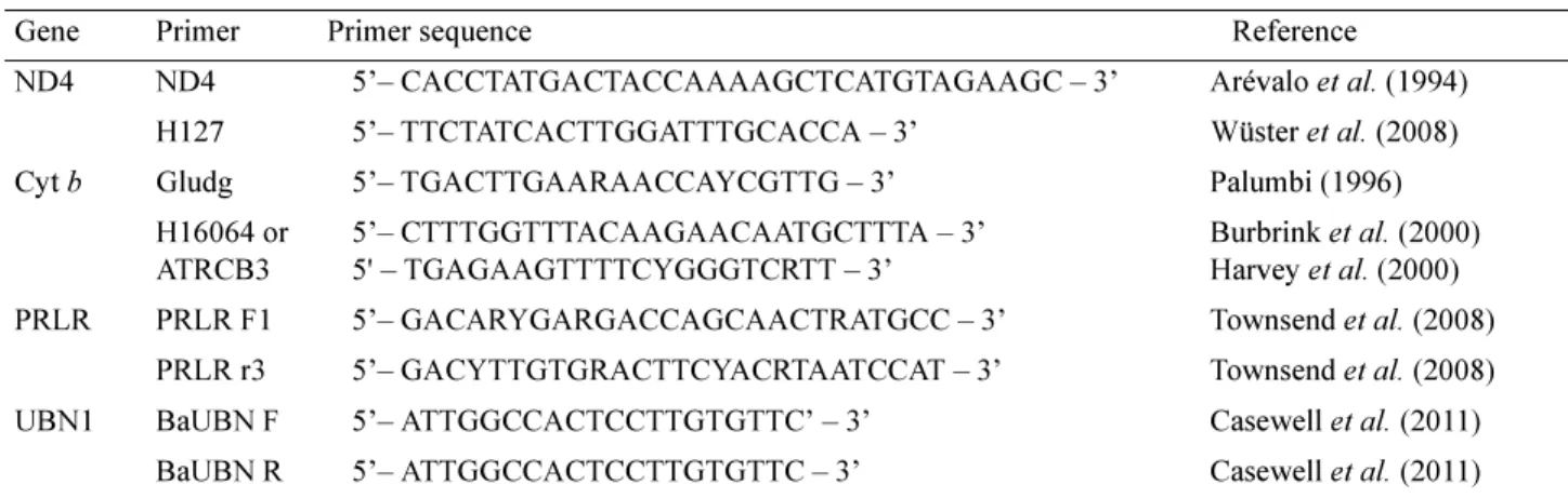
NADH dehydrogenase subunit 4 (ND4) and cytochrome b (cyt b), and two single copy nuclear genes, the prolactin receptor gene (PRLR) and ubinuclein 1 (UBN1) (Townsend et al., 2008), were PCR-amplified and sequenced for all individuals. Primer combinations are given in Table 1.
The PCR reaction mix consisted of 0.3 µl of each primer, 0.8–2.0 µl template DNA and 9.6 µl ABgene® 1.1x ReddyMixTM (1.25 units Thermoprime Plus DNA Polymerase, 75mM Tris-HCL (pH 8.8 at 25oC), 20mM
(NH4)2SO4, 2.0mM MgCl2, 0.01% (v/v) Tween® 20, 0.2mM of each dNTP and a red dye and precipitant for gel electrophoresis (ABgene 2005). 0.8 µl of template DNA was used for ND4 and cyt b, whereas 2.0 µl of template was used for PRLR and UBN1.
The thermocycling regimes involved an initial denaturation at 94°C for 2 minutes (m); 35–40 cycles of: 30 seconds (s) denaturation at 94°C, 30s annealing at 47°C (cyt b), 54°C (ND4) or 45°C, 1m extension at 72°C; and a final extension for 5m at 72°C. All PCR products were cleaned by adding 1µl shrimp alkaline phosphate (SAP) and
0.1µl exonuclease 1 (EXO) to each sample. Samples were then run in a thermal cycler for an initial incubation period of 37°C for 1 hour, 80°C for 15m, followed by a termination period of 4°C for 30s. Direct sequencing was carried out by Macrogen Inc. (dna.macrogen.com) using the same forward primers used in PCRs for mitochondrial fragments and both forward and reverse primers for nuclear loci.
TABLE 1. PCR and sequencing primer sequences and sources.
Sequence chromatograms were proofread and aligned using the software CodonCode Aligner Version 3.5.6 (www.codoncode.com). All sequences were translated to check that no frame shift mutations or unexpected stop codons were present. Heterozygous positions in nuclear sequences were identified by a combination of visual inspection for double peaks and typically low quality Phred scores (Ewing et al., 1998) for the bases surrounding a heterozygous position. Individual allele sequences (haplotypes) were estimated from diploid nuclear loci using the software PHASE v. 2.1.1 (Stephens et al., 2001; Stephens & Scheet, 2005) over 5000 iterations with a burn-in of 500 and a thinning interval of 10, after preparation of the sequence data using SEQPHASE (Flot, 2010). PHASE was run three times to confirm burn-in and convergence across multiple runs.
A mitochondrial DNA phylogeny was generated by maximum likelihood (ML) analysis using the GTR+I+G model identified as optimal in MEGA 6.06 (Tamura et al., 2013) and a neighbour-joining starting tree, and branch support was assessed by means of 1000 bootstrap replicates. Sequences of four species of Naja representing the other subgenera were included to root the tree: N. (Naja) naja, N. (Uraeus) senegalensis, N. (Afronaja) ashei and N.
(Afronaja) nubiae. Deep mitochondrial haplotype clades were then identified as candidate species to be tested in subsequent analyses.
Patterns of haplotype sharing in the two nuclear loci were visualised using the programme Network 4.6.1.0 (Fluxus Technology Ltd. – www.fluxus-engineering.com), and networks drawn in the associated software Network Publisher 1.3.0.0. We obtained an overall measure of nuclear genetic distance between individuals using the program POFAD v.1.03 (Joly & Bruneau, 2006). Allele distance matrices were generated for each nuclear locus under the Kimura two-parameter model (K2P; Kimura, 1980) in PAUP*4.0b10 (Swofford, 2002). A matrix of standardised inter-individual distances across both loci was then calculated in POFAD. This was then input into the software MVSP (Kovach Computing Services, Pentraeth, UK; www.kovcomp.co.uk), and subjected to a principal coordinates analysis to generate a graphical representation of the inter-individual distances.
Bayesian species delimitation was implemented using the reversible-jump Markov Chain Monte Carlo (rjMCMC) method of Yang & Rannala (2010), using the software BPP Version 3 (Rannala & Yang, 2003; Yang & Rannala, 2014), which incorporates species tree uncertainty. This method accommodates the species phylogeny as well as lineage sorting due to ancestral polymorphism while using a nearest-neighbour interchange (NNI) to alter the species tree.
In any Bayesian analysis, the results are likely to be critically dependent on the priors. The key priors for Bayesian species delimitation are the ancestral population size (θ) and root age (τ0). Both can affect the posterior probabilities for models (Yang & Rannala, 2010). Since neither of these values can be inferred for the Naja
melanoleuca group, we instead assessed the effects of different combinations of priors on the robustness of species inferences. We therefore varied values of θ and τ0 to simulate different ancestral population sizes and ages of nodes (Leaché & Fujita, 2010). We considered the four possible combinations of extremes of small or large ancestral
Gene Primer Primer sequence Reference
ND4 ND4 5’– CACCTATGACTACCAAAAGCTCATGTAGAAGC – 3’ Arévalo et al. (1994) H127 5’– TTCTATCACTTGGATTTGCACCA – 3’ Wüster et al. (2008) Cyt b Gludg 5’– TGACTTGAARAACCAYCGTTG – 3’ Palumbi (1996)
H16064 or ATRCB3 5’– CTTTGGTTTACAAGAACAATGCTTTA – 3’ 5' – TGAGAAGTTTTCYGGGTCRTT – 3’ Burbrink et al. (2000) Harvey et al. (2000) PRLR PRLR F1 5’– GACARYGARGACCAGCAACTRATGCC – 3’ Townsend et al. (2008)
PRLR r3 5’– GACYTTGTGRACTTCYACRTAATCCAT – 3’ Townsend et al. (2008) UBN1 BaUBN F 5’– ATTGGCCACTCCTTGTGTTC’ – 3’ Casewell et al. (2011)
population size and shallow or deep divergences. Large and small ancestral population size estimates were modelled as θ ~ G(1,10) and θ ~ G(2,2000), respectively, and deep and shallow divergences as τ0 ~ G(1,10) and τ0 ~ G(2,2000) respectively (Leaché & Fujita, 2010). Priors assuming large ancestral population sizes and shallow divergences are likely to be more conservative in terms of numbers of species estimated. This applies particularly since most taxa of the N. melanoleuca complex occupy large areas and there is no specific reason for assuming a small founder stock (except in the case of Naja peroescobari on São Tomé Island). All BPP3 runs involved a sample size of 100,000, a burnin of 20,000 and a sampling frequency of 2, and were run in duplicate to ensure burnin and consistency of results.
Morphology. Based on mtDNA data showing deeply divergent clades in the complex in Central and West Africa, we examined all available material from west of the Rift Valley and surrounding areas. Further south and east, considerable variation in colour pattern and scalation is not accompanied by deep mitochondrial divergence, but is likely to confound morphological discrimination between distinct lineages in West and Central Africa. We therefore excluded material from southern and parts of eastern Africa from our morphological analyses, pending additional work.
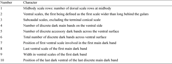
The candidate species identified in this species complex show no diagnostic features in their head scalation, and there is limited interspecific variation in classical scale counts. Dorsal colouration and the patterns of dark crossbars on the venter provide useful diagnostic characters, but these may be confounded by ontogenetic changes. The ventral pattern of the N. melanoleuca complex can be summarised as follows: the anterior venter, throat and chin are predominantly white, with one or several darker crossbands. Following on from this banded anterior ventral region, in most specimens the venter then darkens gradually towards the vent, becoming uniformly dark brown or black more or less rapidly along the posterior half of the venter. We recorded the pattern of ventral banding by noting the ventral scale number of all dark pigmented ventral scales. The ventral bands were divided into main bands and accessory bands. In the N. melanoleuca complex, the main bands are generally well defined, wide (4 or more ventral scales) and in similar positions across most specimens. Additional narrower (usually covering 1–2 ventrals, and not always the entire scale) accessory bands may be present between some of the main bands and under the anterior throat, and the number of these was recorded as well. Finally, we recorded the last ventral of the posteriormost discrete dark main band as a proxy for the posterior extent of well-defined banding along the ventral side. The characters retained for the final analysis are listed in Table 2. The initial separation of taxa was based on the analysis of meristic data for most specimens available in museum collections, collected by DGB over a period of 40 years from personal inspection, literature records, correspondence and relevant data supplied by Barry Hughes.
TABLE 2. Morphological characters used in analyses of the Naja melanoleuca complex
Our final analysis used 605 specimens of the N. melanoleuca complex. Collection acronyms follow Leviton et
al. 1985, with the following additions: E3M= University of Ghana, Legon (B. Hughes); EBM = Estação Biologia Maritima, Inhaca Island, Mozambique; JPT = Zinave National Park, Mozambique (J.P. Tello); KMH = University of Dar es Salaam, Tanzania (K.M. Howell). The specimens examined in the Museu Bocage, Lisbon (MBL) were subsequently destroyed by fire.
Number Character
1 Midbody scale rows: number of dorsal scale rows at midbody
2 Ventral scales, the first being defined as the first scale wider than long behind the gulars 3 Subcaudal scales, excluding the terminal conical scale
4 Number of discrete dark main bands on the ventral side
5 Number of discrete accessory dark bands across the ventral surface 6 Total number of discrete dark bands across ventral surface 7 Position of first ventral scale involved in the first main dark band 8 Last ventral scale of the first main dark band
9 Width in ventral scales of the first dark band
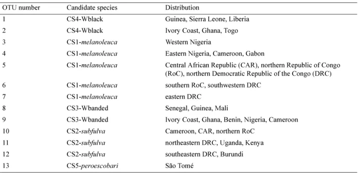
Our principal method of analysis was Canonical Variate Analysis (CVA), which requires the grouping of specimens into operational taxonomic units (OTUs). OTUs were initially defined based on mitochondrial candidate species; these were then further subdivided based on collecting gaps within their distributions. Since the mitochondrial and nuclear DNA data suggested sympatry between different candidate species, the proposed OTUs were initially checked for homogeneity by means of principal components analyses (PCA) run on standardised data, and the assignment of specimens to OTUs corrected based on the results. Once OTUs were defined (Table 3), a CVA was run on characters 1–5, 7, 9 and 10 (since characters 6 and 8 duplicated information contained in the others). The final analysis was run without OTU 13 as the sample size for CS5-peroescobari was too small to be useful for canonical variates analysis.
TABLE 3. OTUs defined for canonical variates analysis of the Naja melanoleuca complex. DRC = Democratic Republic of Congo.
Results
Molecular data. We aligned 716 b.p. of cytochrome b, 660 b.p. of ND4, 510 b.p. of nuclear PRLR and 436 b.p. of nuclear UBN1 sequence. Of the 1376 mitochondrial base pair positions, 449 are variable and 321 informative under the parsimony criterion (314 variable and 259 parsimony informative when outgroup sequences are excluded). Corresponding figures for PRLR and UBN1 (ingroup only) are 20 variable and 14 parsimony-informative and 13 variable and 11 parsimony-parsimony-informative positions, respectively. All sequences have been deposited with GenBank (Accession numbers GQ359493, GQ359497, GQ359575, GQ359579, GQ387080, GQ387109, MH337375–MH337634).
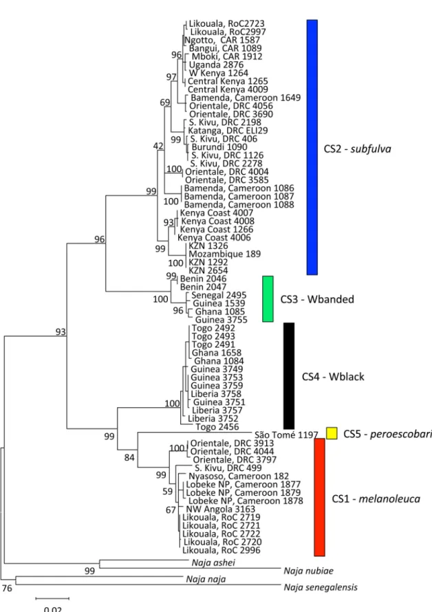
Mitochondrial phylogeny. The phylogenetic tree derived from the mitochondrial data (Fig. 1) reveals five major clades, here considered as candidate species (CS) (Padial et al., 2010), differing by average p-distances of 0.065 to 0.108 across cytochrome b and ND4:
CS1-melanoleuca: A Central African forest form extending from western Cameroon across the Congo Basin to northwestern Angola and Lake Kivu, including topotypical populations of the nominate form N. melanoleuca
melanoleuca from Gabon.
CS2-subfulva: A widespread eastern form extending from western Cameroon to coastal Kenya and south to South Africa (northern KwaZulu-Natal) as well as across the Congo Basin, including topotypical populations of Naja
melanoleuca subfulva.
CS3-Wbanded: A West African form with obvious dorsal banding extending from Senegal to Cameroon in the West African savanna belt.
OTU number Candidate species Distribution
1 CS4-Wblack Guinea, Sierra Leone, Liberia
2 CS4-Wblack Ivory Coast, Ghana, Togo
3 CS1-melanoleuca Western Nigeria
4 CS1-melanoleuca Eastern Nigeria, Cameroon, Gabon
5 CS1-melanoleuca Central African Republic (CAR), northern Republic of Congo (RoC), northern Democratic Republic of the Congo (DRC) 6 CS1-melanoleuca southern RoC, southwestern DRC
7 CS1-melanoleuca eastern DRC
8 CS3-Wbanded Senegal, Guinea, Mali
9 CS3-Wbanded Ivory Coast, Ghana, Benin, Nigeria, Cameroon
10 CS2-subfulva Cameroon, CAR, northern RoC
11 CS2-subfulva northeastern DRC, Uganda, Kenya
12 CS2-subfulva southeastern DRC, Burundi
FIGURE 1. Maximum likelihood mitochondrial phylogeny of the Naja melanoleuca complex. Node support values indicate % bootstrap support; support values for the most distal nodes not shown. Country abbreviations: CAR = Central African Republic, DRC = Democratic Republic of Congo, KZN = KwaZulu-Natal Province, South Africa, RoC = Republic of Congo. Mitochondrial candidate species (CS) are shown in the same colours as in Figures 2–4. For specimen information see Appendix 1.
CS4-Wblack: A frequently melanistic West African forest form extending from Guinea to Togo. CS5-peroescobari, endemic to the island of São Tomé in the Gulf of Guinea.
Mean pairwise mitochondrial sequence divergences among these candidate species are shown in Table 4. TABLE 4. Mean between-group p-distances across 1326 b.p. of mitochondrial cyt b and ND4 sequence.
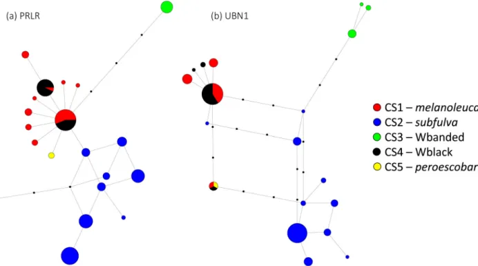
Nuclear loci. Networks of PRLR and UBN1 haplotypes (Fig. 2) congruently show that candidate species
CS2-subfulva and CS3-Wbanded contain exclusive sets of haplotypes, none of which are shared with the other forms in either gene. The remaining three candidate species display sharing of some haplotypes. In both genes, one shared haplotype was found to be widespread across the range of CS1-melanoleuca and CS4-Wblack. In PRLR, the remaining CS4-Wblack specimens contained a near-exclusive haplotype, which was otherwise shared only by a single specimen of the Central African CS1-melanoleuca from Cameroon. The single specimen of N. peroescobari contained a unique, unshared haplotype in PRLR, but shared its single UBN1 haplotype with specimens of the Central African CS1-melanoleuca and the West African CS4-Wblack. CS2 displays considerable variation among its haplotypes. In PRLR, all specimens from the Indian Ocean coast sampled shared a unique, exclusive haplotype, whereas in UBN1, the coastal populations contained three unique and exclusive haplotypes, whereas other specimens shared a haplotype with other specimens from across the range of the species.
FIGURE 2. Haplotype networks for single copy nuclear loci. (a) PRLR; (b) UBN1. Small black circles indicate unsampled haplotypes.
The principal coordinates analysis (PCD) of standardised multilocus distances generated by POFAD (Fig. 3) shows clear separation of CS2-subfulva and CS3-Wbanded from each other and the remaining candidate species.
CS1-melanoleuca CS2-subfulva CS3-Wbanded CS4-Wblack CS1-melanoleuca –
CS2-subfulva 0.09020 –
CS3-Wbanded 0.09210 0.06747 –
CS4-Wblack 0.06458 0.09452 0.09238 –
Reanalysis under exclusion of these highly distinct forms reveals partial separation between CS1-melanoleuca, CS4-Wblack and N. peroescobari, although some specimens of CS1-melanoleuca and CS4-Wblack share identical combinations of haplotypes.
FIGURE 3. Ordination of individual specimens in a Principal Coordinates Analysis of standardised multilocus distances of PRLR and UBN1 scnDNA sequence data. (a) All specimens; (b) Analysis repeated under exclusion of CS2 and CS3.
TABLE 5. Candidate species and their posterior probabilities in BPP v3. Small ancestral population, shallow divergences Large ancestral population, shallow divergences Large ancestral population, deep divergences Small ancestral population, deep divergences Ancestral population size and
divergence age priors
θ = 2 2000 τ = 2 2000 θ = 1 10 τ=2 2000 θ = 1 10 τ = 1 10 θ = 2 2000 τ = 1 10 CS1-melanoleuca > 0.995 > 0.95 > 0.95 > 0.999 CS1-melanoleuca + CS5-peroescobari 0.0005–0.0012 0.043–0.050 0.022–0.044 < 0.0005 CS5-peroescobari > 0.995 0.897–0.898 0.924–0.928 > 0.995 CS4-Wblack + CS5-peroescobari 0.0013–0.0025 0.060–0.0751 0.0281–0.0540 < 0.002 CS3-Wbanded + CS5-peroescobari 0 <0.0002 <0.0002 0 CS4-Wblack >0.995 0.925–0.940 0.946–0.972 > 0.995 CS3-Wbanded 1 >0.9998 >0.9998 1 CS2-subfulva 1 1 1 1
Posterior probability for 4 species
0.002–.003 0.101–0.102 0.072–0.076 0.0005–0.0016 Posterior probability for 5
species
Species delimitation analyses. The Bayesian species delimitation analyses using BPP3 supported the status of all five candidate species as distinct species under most priors of ancestral population size and divergence age (Table 5). The candidate species CS2-subfulva and CS3-Wbanded were consistently recognised as separate species. This applied irrespective of gamma parameters for θ and τ. CS1-melanoleuca (Central African forests) and CS4-Wblack (Upper Guinea forests) were also consistently recovered as separate species. CS5-peroescobari (São Tomé – N. peroescobari) was strongly supported as a separate species under reconstructions assuming small ancestral population sizes, but much more weakly under large ancestral population size scenarios, when dissenting trees were approximately evenly split between placing this population into conspecificity with either CS1-melanoleuca and CS4-Wblack. Nevertheless, both the latter remained supported as separate species irrespective of the position of CS5-peroescobari.
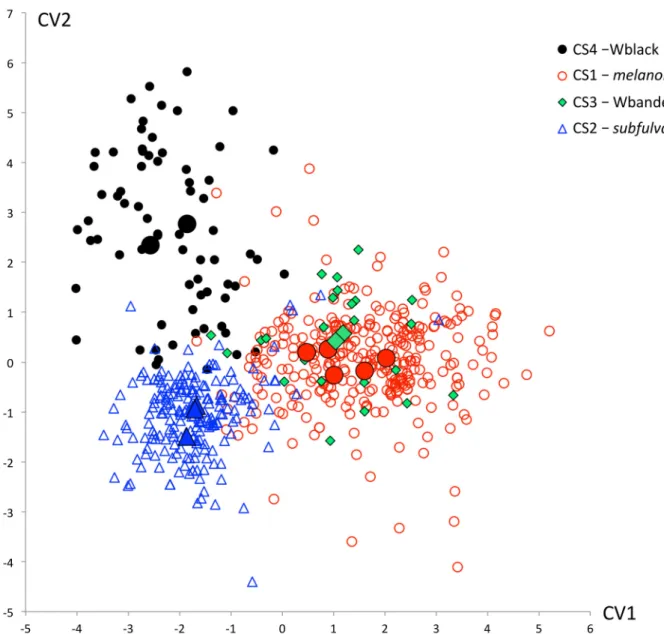
FIGURE 4. Ordination of individual specimens and OTU centroids of four of the mitochondrially defined candidate species of the N. melanoleuca complex along the first two canonical variates. CS5-peroescobari was omitted due to the small available sample size. Canonical variates 1 and 2 account for 57.9 and 22.8% of total variance, respectively. Enlarged symbols indicate OTU centroids.
Morphology. The morphological analyses revealed relatively subtle patterns of distinctness between the candidate species. Fig. 4 shows the ordination of individual specimens and OTU means of the canonical variates analysis of the morphological data of the full data of the five candidate species, and Table 6 shows the canonical variate loadings for the 8 characters retained in the analysis. The specimens representing the OTUs of
whereas specimens of the less well sampled CS3-Wbanded are not separated from CS1-melanoleuca. Additional analyses (not shown) did not improve the resolution of CS3.
TABLE 6. Pooled within-group correlations between discriminating variables and standardized canonical discriminant functions. Character numbers follow Table 2.
Taxonomy
In summary, the combined evidence from mitochondrial and nuclear DNA sequences and morphology support recognition of five mitochondrially defined candidate species as separate species. While not all species criteria support separate species status in all pairwise comparisons between candidate species, each pairwise comparison reveals one or several lines of evidence for the taxa involved. The evidence supporting pairwise species-level distinctness between the mitochondrially defined candidate species is summarised in Table 7.
TABLE 7. Distinguishing datasets and analyses supporting species status for the five mitochondrially defined candidate species considered in this study. Above the diagonal: evidence from nuclear loci; below the diagonal: evidence from morphology.
The mitochondrial DNA analyses revealed deep haplotype clades with geographically cohesive distributions and partial range overlap. The levels of mtDNA sequence divergence between these candidate species equal or exceed those found between sister species of cobra in other subgenera of Naja (Slowinski & Wüster, 2000; Wüster
1st canonical variate 2nd canonical variate
1. Midbody scale rows 0.237 -0.698
2. Ventrals 0.471 0.146
3. Subcaudals 0.261 0.287
4. Number of main ventral bands 0.534 -0.266
5. Number of accessory ventral bands 0.203 0.115
7. First dark ventral of first main band 0.0173 0.603
9. Width of 1st main ventral band -0.161 0.021
10. Last dark ventral of last discrete main band 0.773 -0.097
CS1-N. melanoleuca CS2- N. subfulva CS3-Wbanded N. savannula CS4-Wblack N. guineensis CS5-N. peroescobari CS1-N. melanoleuca – BPP POFAD no haplotype sharing in sympatry BPP POFAD no haplotype sharing BPP (POFAD) BPP (POFAD) CS2-N. subfulva phenotypically distinct – BPP POFAD no haplotype sharing BPP POFAD no haplotype sharing BPP POFAD no haplotype sharing CS3-Wbanded N. savannula
poorly distinct phenotypically distinct – BPP POFAD no haplotype sharing in sympatry BPP POFAD no haplotype sharing CS4-Wblack N. guineensis phenotypically distinct phenotypically distinct phenotypically distinct – BPP CS5-N. peroescobari diagnostic character states diagnostic character states diagnostic character states diagnostic character states –
& Broadley, 2003, 2007; Trape et al., 2009). These results are largely mirrored by the nuclear loci: species delimitation and POFAD analyses of the two loci analysed support the recognition of all five candidate species.
CS2-subfulva and CS3-Wbanded are unambiguously supported as distinct by a lack of haplotype sharing with any of the other candidate species, despite extensive sympatry with CS1-melanoleuca and CS4-Wblack respectively. This is reflected in unambiguous support for species status in BPP analyses and distinctness in POFAD ordinations. CS2-subfulva is additionally largely distinct from the other candidate species in the morphological analyses. We therefore recognise CS2-subfulva and CS3-Wbanded as distinct species.
CS1-melanoleuca and CS4-Wblack display haplotype sharing in both PRLR and UBN1. Such sharing of haplotypes can be the result of ongoing gene flow between the two forms, or the retention of ancestral haplotypes. These can be distinguished in part through the geographic distribution of haplotypes: retention of ancestral haplotypes predicts the distribution of the ancestral haplotype throughout the range of all taxa containing it, whereas gene flow predicts that shared haplotypes should be found primarily near the contact zone between the two taxa in at least one of them. In PRLR, the only extensively shared haplotype is found across the range of the Central African CS1-melanoleuca and the West African CS4-Wblack, from Guinea and Liberia to the Rift Valley, suggesting that this is a retained ancestral haplotype. Another common haplotype is nearly exclusive to CS4-Wblack, and the only specimen of CS1-melanoleuca that shares it originates from southwestern Cameroon, the westernmost population of CS1-melanoleuca included in the molecular study. In UBN1, an extensively shared haplotype is again found across the range of both CS1-melanoleuca and CS4-Wblack, suggesting that it represents a shared ancestral haplotype. Both CS1-melanoleuca and CS4-Wblack contain exclusive private haplotypes not shared with each other. The BPP analysis consistently categorised the Central and West African forest populations as separate species, and the morphological analyses also supported their distinctness. We therefore regard them as distinct species.
CS5-peroescobari: genetically, our single specimen of N. peroescobari is homozygous for a unique haplotype in PRLR, but shares a haplotype with a Ghanaian specimen of CS4-Wblack and two Congolese specimens of
CS1-melanoleuca (N. melanoleuca) in UBN1. In BPP, support for species status of CS5-peroescobari was dependent on ancestral population size and root age, with more conservative parameter values resulting in reduced support for species status. Interestingly, the BPP iterations not recognising CS5-peroescobari as distinct were approximately equally split in associating it with either the CS1-melanoleuca or the CS4-Wblack, while strongly supporting the latter two as separate species. The species was described on the basis of the diagnostic character of separated posterior chin shields (not included in our analyses) and the restriction of white ventrals to the first 22 ventral scales. Our data contradict the latter character: several of our specimens from São Tomé have light ventrals extending further along the venter, to ventrals 47 (BMNH 1906.3.30.80) and even 56 (MBL 1954). Nevertheless, given the data presented in Ceríaco et al. (2017) and here, we recognise Naja (Boulengerina) peroescobari as an island endemic species restricted to São Tomé.
In terms of nomenclature, three of the five candidate species have available names. The Central African forest form (CS1) corresponds to Naja melanoleuca Hallowell, 1857, described from Gabon, and also includes Naja haje var. leucosticta Fischer, 1885. CS2-subfulva contains the holotype of Naja melanoleuca subfulva Laurent, 1955, described from Lwiro, South Kivu Province, Democratic Republic of Congo, and we therefore recognise it as a distinct species, Naja (Boulengerina) subfulva, which also includes the Cameroonian grassfields populations described as Naja melanoleuca aurata by Stucki-Stirn (1979). The name Naja peroescobari Ceríaco et al. 2017 is available for CS5.
In contrast, the two West African candidate species do not appear to have available names (we follow Kaiser et
al., 2013, Measey, 2013, and Kaiser, 2014 in not considering certain names published outside the peer-reviewed literature as part of the permanent scientific record), and we therefore describe them as new.
Naja (Boulengerina) guineensis sp. nov. Broadley, Trape, Chirio, Ineich & Wüster
Naia melanoleuca (not Hallowell) Boulenger, 1896: 376 (part, var. B [c,d], C).
Naja sp. 2 cf. melanoleuca Hallowell, 1857 (blackish dorsum) Trape & Baldé, 2014: 318. Naja sp. 2 cf. melanoleuca (forest form). Trape & Baldé, 2014: 336.
Holotype: MNHN 1921.0485, a male from N’Zébéla, Macenta Prefecture, Nzérékoré region of forested southeastern Guinea (8° 05’N, 9° 05’W), elev. 490 m, Coll. Paul Chabanaud (1876–1959) between 1919–1920 (Chabanaud, 1921: 471) (Fig. 5).
Paratype: BMNH 1960.1.3.72, a male from Njala, Kori, Sierra Leone, Coll. C.T. Pyne.
Diagnosis. Naja guineensis can be distinguished from the partly sympatric N. savannula sp. nov. by lacking extended dorsal banding, often having 17 rather than 19 dorsal scale rows at midbody, a generally lower subcaudal scale count, fewer ventral bands, a lesser posterior extent of the ventral banding, and a strong tendency towards melanism in adults. Specimens with 19 midbody dorsal scale rows can be distinguished from N. melanoleuca through the reduced number of ventral bands, lesser posterior extent of banding and tendency of ontogenetic melanism from N. subfulva in lacking a lighter anterior dorsum and through ontogenetic melanism, and from N.
peroescobari in having the posterior chin shields in contact.
FIGURE 5. Naja (Boulengerina) guineensis sp. nov. Left and top right: holotype, MNHN 1921.0485, dorsal and ventral view and side view of head. Note extensive mottling of throat and anterior ventral side and limited posterior extent of lighter ventral markings. Bottom right: live adult specimen measuring approximately 200 cm total length, from Sekondi-Takoradi, Western Region, Ghana, displaying dark suffusion of throat and anterior venter (not preserved; photo L. Chirio).
Description of holotype. Dimensions: A male specimen with dissected tail base, snout–vent length 1850 mm, tail length 370 mm, total length 2220 mm, ratio total length: tail length 6:1.
Body scalation: 23 scale rows around hood, 17 around midbody, 13 one head length ahead of the vent, all smooth and oblique; 208 ventrals, 62 subcaudals, all divided, anal single.
Head scalation: 7/7 supralabials, 3rd & 4th contact orbit, 6th largest; 8/8 infralabials, first 4 contact anterior chin
shields; anterior pair of chin shields and infralabials 3–4 with an anomaly on both sides, they are warty and covered with circumvolutions; 1/1 preocular, twice as long as high; 3/3 postoculars; 1/1 anterior temporal; posterior temporals 3/3; rostral broader than high and slightly damaged anteriorly, clearly visible from above; 7 temporals and nuchals contacting both parietals.
Pattern: upper side of head and upper temporal region dark brown including parietals and temporals but black behind them, lips and ventral side of head creamy yellowish without dark mottling on chin and anterior throat; each posterior supralabial edge broadly outlined in black from eye level to mouth corner; temporals uniformly brownish. Dorsum uniform brownish-black throughout. Venter: anterior first 32 cm (14% of total length) of venter with
alternating yellow and blackish irregular bands, the posterior yellow bands increasingly suffused with dark mottling. Only ventral 1 is uniform clear yellow, ventrals 2–10, 19, 28–29, 32–35 yellow but always increasingly suffused with dark mottling, remaining ventrals and underside of tail black (after ventral 35).
Variation. Midbody dorsal scale row counts of 17 and 19 are approximately equally common in this species. In large adults, light pattern elements on the head and throat often become heavily suffused with black pigment, leading to a virtually entirely melanistic snake. Some specimens have 1–4 generally faint or poorly defined light bands across the neck, and occasionally an ocellate hood marking.
Largest recorded: 1818+437 = 2255 mm, from Ballassou, Guinea (IRD 4213.G),but larger specimens have been observed. Based on locality, Menzies’ (1966) report of a specimen measuring “eight feet, eight inches” (264 cm) from Bo, Sierra Leone, is likely to refer to this species.
Etymology. The specific epithet guineensis means “from Guinea” and is chosen to reflect the distribution of the species in the Upper Guinea forests of West Africa, part of the West African Forests biodiversity hotspot (Myers et al., 2000).
Suggested common name. Black forest cobra.
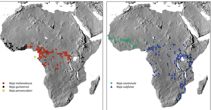
Distribution. The distribution of Naja guineensis appears to be restricted to the Upper Guinea Forests of western Africa, from western Togo to Liberia and Guinea (Trape & Baldé, 2014) (Fig. 6). There is a single record from Contuboel, Guinea Bissau (MBL 535). All other records 10°N or lower.
FIGURE 6. Distribution of the five species of the N. melanoleuca complex. A full list of voucher specimens beyond those included in multivariate analyses and Appendix 2 is available on request from the first author.
Naja (Boulengerina) savannula sp. nov. Broadley, Trape, Chirio & Wüster
Naia melanoleuca (not Hallowell) Boulenger, 1896: 376 (part, var. D). Naja “banded form” Hughes, 2013: 128.
Naja sp. 1 cf. melanoleuca Hallowell, 1857 (yellow banded dorsum) Trape & Baldé, 2014:318. Naja sp. 1 cf. melanoleuca (banded savanna form) Trape & Baldé, 2014: 336.
Holotype: MNHN 2018.0002, an adult male collected by Laurent Chirio on 31/03/2007 in Niénié, W Biosphere Reserve, Benin, 11.05920 °N, 2.20488 °E, elevation 272 m (Fig. 7).
Paratype: BMNH 1975.657, a male specimen collected on the grounds of Amadou Bello University Hospital, Zaria, Nigeria, by D.A. Warrell.
Diagnosis: Distinguishable from other species of the N. melanoleuca complex by the presence of 3 to 8 semidivided yellowish or whitish bands on the anterior dorsal forebody, becoming uniform black caudad; venter yellow with 2 to 8 black bands. Generally higher mean subcaudal scale counts than the other species. Genetically diagnosable through possession of unique mitochondrial haplotypes (cytochrome b: GenBank MH337597–602; ND4: MH337403–408) and unique PRLR and UBN1 haplotypes (PRLR: MH337501–504; UBN1: MH337532– 535).
Description of holotype. Dimensions: A male specimen, snout–vent length 1815 mm, tail length 376 mm, total length 2191 mm, ratio total length: tail length 5.8.
Body scalation: 25 scale rows around hood, 19 around midbody, 15 one head length ahead of the vent, all smooth and oblique. Dorsal scales smooth, oblique. Vertebral row not enlarged. 213 ventrals (and one incomplete ventral before the anal), 69 subcaudals, all divided, anal single.
Head scalation: 7/7 supralabials, 3rd & 4th contact orbit, 6th largest; 8/8 infralabials, first 4 contact anterior chin
shields; posterior chin shields in narrow contact at their anterior end, separated by median scale along most of their length; 1/1 preocular, twice as long as high; 3/3 postoculars; 1/1 anterior temporal; 3/3 posterior temporals; rostral broader than high, clearly visible from above; 7 temporals and nuchals contacting the parietals.
Pattern: upper side of head brown, lips and ventral side of the head pale creamy yellowish with each labial scale laterally bordered with black. Neck brown, dorsum becoming gradually black towards rear. Two very distinct pale dorsal crossbands at the level of ventrals (V) 24 to 28, two other at level of V 40–45, three at level of V 58–64, three less distinct at level of V 77–83, and three slightly marked at level of V 98–104. Ventrals 1 to 11, 13–19, 23– 31, 38–47 and 57–66 pale creamy yellowish, V 20–22 and 32–37 black; V 12 and 48–56 yellowish spotted with black spots. From ventral 67 to the vent, ventrals spotted with black gradually become completely black. Subcaudals black.
Variation: Dorsal scale rows on neck 19–25, at midbody 19, before vent 12–15; ventrals 211–233, subcaudals 63–77 (Table 8). Dorsal semi-divided yellow bands 3–8; ventral principal black bands 2–8 (Fig. 7).
FIGURE 7. Naja (Boulengerina) savannula sp. nov. Top row and bottom left: holotype, MNHN 2018.0002. Bottom right: live specimen from Kindia, Guinea, showing conspicuous, broad dorsal bands and ventral banding, including narrow accessory bands (not vouchered). Photos J.-F. Trape.
Etymology: The name is derived from the contraction of its savanna habitat and annulated colour pattern and was coined by Barry Hughes in an unpublished 1968 manuscript. We have retained this name at the request of our colleague Barry Hughes.
Largest recorded: 1825+405 = 2230 mm, from Medina Djikoye, Senegal (IRD 6155.S).
Suggested common name: West African banded cobra.
Distribution: Senegal and Gambia east to northern Cameroon (Fig. 6). Naja savannula appears to be restricted to gallery forest areas in Africa in Guinean Forest/Savanna Mosaic, extending northwards into West Sudanian Savanna (Chirio, 2003, 2013; Monasterio et al., 2016). Our records are mostly from latitudes 10–14°N, except in the Dahomey Gap in eastern Ghana and Benin, where the species approaches the Gulf of Guinea Coast. The eastern extent of the range is poorly understood. A specimen from Margui Wandala district, northern Cameroon (approx. 10.5°N 13.6°E; MNHN 1962.0022) appears to be assignable to this species, and one of us (JFT) recently collected a specimen from Mboura, 20 km SW Baïbokoum, Logone Oriental Province, Chad (7.598°N, 15.596°E; IRD 2281.N), and there is a recent record from near Niamey, Niger (LC, unpublished data). It seems likely that the species has a wider distribution in northern Cameroon, extreme southern Chad and possibly even extreme northwestern Central African Republic (CAR).
Naja (Boulengerina) melanoleuca Hallowell, 1857
Naia haie var. melanoleuca Hallowell, 1857, Proc. Acad. Nat. Sci. Philadelphia: 61. Type locality: Gabon, syntypes ANSP
6875–76, 6878–79.
Naja haje var. leucosticta Fischer, 1885, Jahr. Hamburg. Wiss. Anst. 2: 115, pl. v, fig. 11. Type locality: Cameroon and Ogooué
River, Gabon, syntypes ZMH 4280, 7048, 7299–7302.
Naia melanoleuca; Boulenger, 1896: 376 (part, vars. A [a,b] & B [a,b]) Naja melanoleuca melanoleuca; Laurent, 1956: 290, pl. xxvi, fig. 2. Naja (Boulengerina) melanoleuca; Wallach et al., 2009.
Boulengerina melanoleuca; Wallach et al., 2014: 122.
Aspidelaps bocagei Sauvage, 1884: 204 (type locality: Gabon and Majumba; holotype MNHN 1884.0015) has been listed as a synonym of N. melanoleuca (e.g., Broadley, 1983; Wallach et al., 2014; Ceríaco et al., 2017), but is in fact a synonym of Naja annulata, as is evident from Sauvage’s description, which notes approximately 20 dark double bands along the entire body length, and 21 mid-dorsal scale rows (see also Schmidt, 1923).
Diagnosis: Dorsum black, often with 1–3 semidivided yellow crossbands on the neck, the first may be an ocellus; venter yellow with 4 to 6 black bands in the first 100 ventrals, thereafter uniform black.
Variation: Dorsal scale rows on neck 19–27, at midbody 19 (very rarely 17 or 21); ventrals 209–230; subcaudals 59–74 (Table 8). Supralabials 7, the third and fourth entering the orbit; infralabials 8, the first four in contact with the anterior sublinguals, no cuneate; preocular 1; postoculars 3 (very rarely 2 or 4); temporals 1+2 or 1+3; nuchals bordering temporals 5–9, usually 7.
FIGURE 8. Naja (Boulengerina) melanoleuca. Adult specimens from Yaoundé, Cameroon (left—photo J.-F. Trape) and Tsibilé, Gabon (right—photo L. Chirio). Note the diffuse but distinct hood mark that is often present in this species, and the combination of broad main bands and narrow accessory bands on the ventral side.
TAB LE 8 . V ariation in m eristic c haract ers in th e speci es o f th e N . m el ano le uc a co m ple x. Na ja guineensis N =71 Na ja mel an oleuca N = 307 Na ja s avan nu la N =24 Na ja s ub fu lva (ex cl. eas te rn and so ut he rn A fr ic an pops.) N = 202 Na ja per oes co ba ri N = 8 Mean ± 1SD Ran ge Mean ± 1S D Range Mean ± 1 SD Range Mean ± 1 S D Range Mean ± 1SD R an ge Mid bod y d or sal scal e r ow s 17 .0 ± 1. 0 17 –1 9 19 .0 ± 0 .3 17–21 19 .0 ± 0 19 19 .0 ± 0. 2 18–21 19 .0 ± 0 .0 19 V en tral s 21 1.8 ± 4 .2 203–221 21 7. 8 ± 4 .4 20 6– 23 2 216.9 ± 4. 2 211–226 210.0 ± 4. 8 19 7– 22 8 209.4 ± 3. 4 204–215 Su bc audals 65 .1 ± 2 .3 60 –7 0 65 .5 ± 2 .7 57–74 70 .0 ± 1 .6 67 –7 3 62 .8 ± 2 .8 57–72 66 .2 ± 4 .6 57 –7 0 Fir st d ark ventral o f f ir st m ain ba nd 18 .0 ± 2 .3 10 –2 3 16 .3 ± 1. 7 11 –2 1 16 .4 ± 2 .2 11 –2 1 14 .2 ± 1 .7 10–22 18 .2 ± 3 .4 14 –2 2 L as t d ar k ve ntr al of f irs t m ain ba nd 22 .2 ± 2 .3 16 .2 6 19 .9 ± 1. 9 15 –2 5 19 .4 ± 2 .4 14 –2 5 18 .2 ± 2 .1 13–26 22 .63 ± 6 .4 15 –2 9 W idt h of f irs t m ai n ba nd ( in v en tral s cales ) 5. 2 ± 1 .4 2–9 4. 5 ± 1. 0 2– 9 4. 0 ± 0 .8 3– 5 5. 0 ± 1 .0 2– 10 5. 4 ± 3. 1 2–9 N um be r of m ai n ba nd s 1. 5 ± 0. 7 1–4 3. 7 ± 0 .9 1–7 3. 6 ± 1. 3 1–7 2. 5 ± 0. 8 1–5 1. 7 ± 0 .7 1– 3 N um be r of a cc es so ry ba nd s 0. 8 ± 1. 2 0–5 1. 4 ± 1 .7 0–9 1. 4 ± 1 .9 0– 6 0. 3 ± 0 .79 0–4 0. 7 ± 0 .9 0– 2 T otal n um ber o f d iscre te d ark ba nd s 2. 3 ± 1. 4 1– 6 5. 1 ± 1 .7 2–12 5. 0 ± 2. 5 2–12 2. 8 ± 1. 1 1– 8 2. 4 ± 1 .2 1– 5 L as t ve ntral o f last d iscret e d ark ba nd 28 .5 ± 9 .2 17 –5 2 68 .6 ± 15 .4 24 –1 14 59 .4 ± 1 9. 1 19 –1 03 36 .8 ± 1 0. 0 13–82 27 .6 ± 1 5. 4 15 –5 5
Colouration: Head brown, the supralabials barred black and yellow, chin yellow. Black above, sometimes a yellow monocellate marking on the hood, or 1–3 small yellow blotches, the dorsal scales may be tipped with white in juveniles (Fig. 8). Yellow or white below with 4 to 6 black bands on the first 100 ventrals, usually uniform black thereafter.
Largest recorded: 2250+420 = 2670 mm, from Moniya, Ibadan, Nigeria (Butler, 1982: 110).
Suggested common name: Central African forest cobra.
Distribution: Centered on the Congo Basin, west to southwestern Nigeria and possibly southern Benin, south to northern Angola, not extending east of the Albertine Rift Valley, where it is replaced by N. subfulva. The western range limits are poorly understood. Populations from southeastern Nigeria are clearly assignable to this form. A few specimens from Lamta, southern Benin (IRD 12.B, IRD 54.B, IRD 60.B), and Ghana (MNHN 1983.0663–64; no further locality information) also appear to be assignable to N. melanoleuca.
Naja (Boulengerina) subfulva Laurent, 1955
Naia melanoleuca (not Hallowell); Boulenger, 1896: 376 (part, vars A [c] & E).
Naja melanoleuca subfulva Laurent, 1955, Revue Zool.– Bot. Afr. 51: 132. Type locality: Lwiro, 1850 m, Kabare, Kivu, Belgian Congo [= Democratic Republic of Congo], holotype MRAC 17514.
Naja melanoleuca aurata Stucki-Stirn, 1979, Snake Report 721: 617–620. Type locality: Bamenda, Cameroon. No type designated.
Naja subfulva; Chirio & Ineich, 2006: 54.
Naja melanoleuca subfulva; Chirio & LeBreton, 2007: 584.
Naja (Boulengerina) melanoleuca subfulva; Broadley & Blaylock, 2013: 139. Naja (Boulengerina) melanoleuca; Conradie et al., 2016: 28.
Diagnosis. Midbody scale rows 19, except along coastal regions of East Africa (Kenya, Tanzania), where most specimens have 17 rows. Pattern highly variable. Adults of most populations distinguishable in having a brown forebody, often with spots, generally becoming darker or blackish posteriorly. Labial pattern may be attenuated in many adults. Venter with several black, dark brown or greyish crossbands on the first 50 ventrals, gradually becoming uniform black caudad in some populations, but often remaining entirely light, often with extensive darker spotting or speckling. Where present, the light forebody and/or light posterior venter are diagnostic for this species. Generally fewer ventral bands and ventral scales than N. melanoleuca or N. savannula and fewer subcaudals than N. savannula (Table 8). Genetically diagnosable through possession of unique mitochondrial haplotypes (cyt b: GenBank MH337603–633; ND4: MH337409–439) and unique PRLR and UBN1 haplotypes (PRLR: MH337441–471; UBN1: MH337531, MH337536–562, MH337564–566).
Variation. Dorsal scale rows on neck 19–27, at midbody 19 (very rarely 17 or 21); ventrals 196–226; anal entire; subcaudals 55–71 (Table 8). Supralabials 7 (very rarely 5 or 6), the third and fourth entering orbit; infralabials 8 (rarely 7), the first four (rarely three) in contact with the anterior sublinguals, no cuneate (very rarely one); preocular 1; postoculars 3; temporal 1+2 or 1+3; temporal bordering parietals 5–9, usually 7.
Colouration. Head brown, the supralabials usually barred black and yellow, chin yellow. Dorsum uniform black, or with faint white crossbars or white-tipped dorsal scales, resulting in a stippling effect, in all juveniles. Adults from the periphery of Lake Victoria and parts of the Congo Basin (Fig. 9) have a uniformly black dorsum as adults, whereas in most of the range, adults become brown anteriorly. There may be 2 or 3 faint yellow divided crossbars on the neck. Ventral banding often faint in adults, with grey or pale brown ventral bands that may be very inconspicuous, or lacking altogether. Narrow accessory ventral bands usually absent.
Largest recorded. 2016+380 = 2396 mm (tail truncated) [JPT 1856 – Zinave, Mozambique], but NMZB-UM 17594 from Inyangani Tea Estates, Zimbabwe, had a total length of 2690 mm (only head and neck preserved).
Suggested common name. Brown forest cobra. This suggestion reflects the fact that in most populations, with the exception of the surroundings of Lake Victoria and some locations in the Congo Basin, the anterior body of adults is brown rather than black. We note that the distribution of this species includes many non-forested habitats, including savannas and open wetlands (Spawls et al., 2018). However, retention of the word “forest” in the common name conveys the phylogenetic information that this species is part of the Naja melanoleuca complex.
Distribution. Forest/savanna mosaic, encircling the Congo Basin, from the grassfields of western Cameroon northward, reaching 13.45°N at Bol, Chad, on the northern shore of Lake Chad (IRD 2802.N), east through the
CAR and South Sudan to western Ethiopia, south through Uganda, western Kenya, eastern DRC, Rwanda, Burundi and western Tanzania to northern Malawi, west through Zambia and Katanga to Angola and the Lower Congo region (Fig. 6). The distribution extends disjunctly east and south to the East African coast from Kenya to northern KwaZulu-Natal (South Africa), including inland locations in western Zimbabwe. The precise distribution of this taxon in the Congo region remains to be ascertained: we have here provided molecular evidence of multiple specimens of this species from the evergreen forest zone of northern Republic of Congo (Likouala Forest – Jackson
et al., 2007) and the northeastern DRC (northwestern Orientale Province along the Congo River). Some of these specimens and populations are difficult to differentiate from sympatric N. melanoleuca, necessitating the use of DNA barcoding approaches (Hebert et al., 2003; Clause et al., 2016) to ensure reliable identification. Two specimens from southeastern Nigeria with low ventral scale counts and lacking ventral banding or displaying a single faded band may also be assignable to this species (Degema, Rivers State: BMNH 1902.11.10.9, and “Oil River”: BMNH 88.8.29.20), suggesting a wider distribution in eastern Nigeria.
Comment. This species displays considerable geographic variation in pattern and scalation, as well as indications of phylogeographic structure. The present study focused on the relationships among the five mitochondrially defined candidate species, and did not encompass the full range of morphological variation in N.
subfulva. It is possible that future work may identify additional cryptic diversity within this wide-ranging and variable species.
FIGURE 9. Naja (Boulengerina) subfulva. Variation in colour and pattern. Top left: specimen from Kakamega, western Kenya, illustrating the typically deep black and white specimens with strong facial markings from the periphery of Lake Victoria. Bottom left: specimen from Chuka, Mount Kenya, Kenya, illustrating an extreme of the brown forebody and reduced facial pigmentation typical of the species in much of its range. Photos W. Wüster, courtesy Royjan Taylor / Bio-Ken snake farm live collection, Watamu, Kenya. Right: specimen from Bamenda, Cameroon, representing the form described by Stucki-Stirn (1979) as Naja melanoleuca aurata. Note the indistinct ventral bands and the lack of accessory ventral bands, as is typical of this species. Photo J.-F. Trape.
Naja (Boulengerina) peroescobari Ceríaco et al., 2017
Diagnosis. In the original description, Naja peroescobari was diagnosed from the other members of the N.
dorsum, and the separation of the posterior chin shields. Our sample does not fully support the diagnostic value of these characters: at least one specimen (BMNH 1906.3.30.80) has the posterior chin shields in contact, and discrete dark bands separated by lighter bands (although often suffused with dark brown pigment) extend as far back as ventrals 45 and 55, respectively, in BMNH 1906.3.30.80 and MBL 1954. Naja peroescobari is distinct from N.
subfulva in never having a brown forebody or a light posterior venter and in lacking dark speckling or spotting on the forebody. It displays greatly reduced ventral banding compared to N. melanoleuca and N. savannula, and, unlike N. guineensis, never has 17 midbody dorsal scale rows.
The species is also diagnosable through unique mitochondrial haplotypes (Ceríaco et al., 2017; cyt b: GenBank MH337634; ND4: MH337440) and a unique PRLR haplotype (MH337499)
Variation. See Table 8 and Ceríaco et al. (2017) for variation in scale counts and ventral banding in this species.
Distribution. Restricted to the volcanic island of São Tomé in the Gulf of Guinea, where it seems to inhabit primarily the forested parts of the centre and south of the island, whereas it seems to be missing from the more open northeast (Ceríaco et al., 2017).
Discussion
The results of this study illustrate the level to which we still remain ignorant of the species diversity of even some of the most iconic organisms alive today. Forest cobras are common in captive collections of venomous snakes in much of the world, and they are one of the most common groups of snakes in many parts of their distribution. Nevertheless, little effort had been made until now to investigate the status of the various populations of the complex.
Some of this confusion is due to subtle patterns of morphological variation in the complex. Broadley (1968) noted the inconsistency between scalation and pattern when assessing the status of Laurent’s N. m. subfulva when it came to Ugandan and western Kenyan specimens. Moreover, while the distinction between species may be reasonably obvious in some places, it may not be so in others: Laurent (1955), Stucki-Stirn (1979) and Chirio & Ineich (2006) had little difficulty in separating N. melanoleuca and N. subfulva in eastern DRC, Cameroon and CAR, whereas one of us (Jackson et al., 2007) collected both species from sites approximately 80 km apart in the Likouala Forest of the northern Republic of the Congo, and remained unaware of the heterogeneity of the samples prior to the genetic results: the specimens involved are morphologically virtually indistinguishable, with the local
N. subfulva (from Ganganya Brousse) displaying a pattern virtually identical to that usually associated with N.
melanoleuca as found by Jackson et al. (2007) at their second site, Impongui.
Morphologically virtually indistinguishable cryptic species are common in some groups (see Bickford et al., 2007, for a review), but have not been reported commonly among larger snakes: even though species delimitation in many groups has required molecular and multivariate morphometric approaches, the recognised species, once delimited, were normally diagnosable without undue difficulty (e.g., Slowinski & Wüster, 2000; Broadley & Wüster, 2004; Wüster & Broadley, 2003, 2007; Trape et al., 2009; but see Malhotra et al., 2011b; Mrinalini et al., 2015). The N. melanoleuca group may be unusual in this regard. We acknowledge, however, that our morphological data were limited in terms of numbers of usable characters, and the use of multiple sources in the compilation of the data may have introduced unintended imprecision into the data presented here, as well as making the assignment of some specimens to species difficult. Rigorous and thorough studies of the external and internal morphology of this complex may yet reveal reliable diagnostic character states for the different species.
The intraspecific variability, lack of clear morphological differentiation and occurrence in macrogeographic sympatry (with, more than likely, parapatry on a smaller geographical scale—Laurent, 1955; Stucki-Stirn, 1979) of several species of the complex complicates the diagnosis of species, the identification of some specimens, and the establishment of the distribution of the species of the complex. A combined morphological and molecular barcoding approach would be helpful to confirm the morphological diagnosis and distribution of the species of this complex, especially around the Dahomey Gap and adjoining parts of Nigeria, Cameroon and southern Chad, where all four mainland species approach each other. In particular, while all southern Togolese material has been assignable to N. guineensis, a series of specimens from Lamta, Benin, appears to be assignable to N. melanoleuca. Further east, the distributions of N. melanoleuca, N. savannula and N. subfulva appear to approach each other or
overlap in northern Cameroon. Specimen IRD 2281.N, from Mboura, Chad (see above) was assignable to N.
savannula, whereas another from Moundou, 100 km to the NE (8.57°N, 16.07°E; IRD 2600.N), was assignable to
N. subfulva. The distribution and diagnosis of N. melanoleuca and N. subfulva in the Congo Basin also remain insufficiently resolved. Because Central Africa is one of the most poorly sampled regions in Africa by herpetologists for a variety of reasons (Tolley et al., 2016; Greenbaum, 2017), extensive fieldwork efforts will be required to fill in gaps in sampling and improve understanding of the geographic distribution of these cobras.
The taxonomic division of widespread species raises the possibility that some of the newly recognised species may be much more threatened than previously suspected (e.g., Melzer et al., 2017; Shaney et al., 2017): Naja
melanoleuca in its original sense has never been assessed for its IUCN Red List status, but its wide distribution and common occurrence in many areas suggests that it is unlikely to be highly threatened as a whole. This may not be true of some of the newly recognised species with much more restricted distributions. As indicated by the common name, all the species of this complex are primarily snakes of wooded or forested habitats, although N. savannula and N. subfulva also occupy gallery forests and wetlands in savanna habitats (Trape & Mané, 2006; Marais & Jubber, 2010). Although, like most cobras, forest cobras adapt well to moderate anthropogenic changes (Menzies, 1966), there is evidence that they are prone to being replaced by N. nigricollis in deforested habitats in at least some parts of their range (Luiselli & Angelici, 2002), suggesting that they are likely to be vulnerable to the effects of forest loss, and their exploitation for bushmeat has also been documented (Eniang et al., 2006). This would be a particular concern for N. guineensis, which is restricted to the Upper Guinean Forest sector of the West African Forests Global Biodiversity Hotspot (Myers et al., 2000). Equally, N. peroescobari is restricted to the island of São Tomé, a WWF-designated priority ecoregion (Jones, 1994; WWF & IUCN, 1994), and the species is potentially vulnerable due to its isolated insular range as well as persecution out of fear, or for traditional Chinese medicine (Ceríaco et al., 2017).
The hitherto unsuspected diversity of the N. melanoleuca complex revealed in this study also suggests that a reassessment of venom composition and its variation in the complex is required, with a particular view towards testing the efficacy of the available antivenoms against the different venoms of the complex. This group has been largely neglected in the recent toxinological literature, but a recently published proteomic study (Lauridsen et al., 2017) used venom from Ugandan specimens, suggesting that they represent N. subfulva. Most prior papers on the venoms of this complex failed to provide locality indications, precluding identification to species level, and potentially leading to problems of repeatability of previous work (Wüster & McCarthy, 1996).
Our more refined understanding of the systematics of the complex also provides a more robust underpinning for further work on the evolution of venom composition in the ecologically diverse subgenus Boulengerina. This includes a mixture of small and large terrestrial, burrowing and aquatic forms occupying a diversity of biomes and with distinct foraging ecologies (Spawls & Branch, 1995; Spawls et al., 2004), providing a promising system for testing hypotheses of adaptation in venom composition (Barlow et al., 2009). In addition, it should direct ecological studies in regions of potential sympatry between forest cobra species, to determine any difference in activity, habitat and dietary preferences, etc., that may yield fuller understanding of these remarkable African snakes.
Acknowledgements
We dedicate this paper to the memory of Donald G. Broadley (1932–2016), who played a pivotal role in assembling morphological data, samples and specimens, and who guided this project with his unparalleled insight into the morphology, taxonomic history and biogeography of African snakes. Cobras occupied a special and career-long place in Don’s work and interests, starting with the documentation of N. melanoleuca (now N. subfulva) in Zimbabwe in one of his very first publications (Broadley, 1958), and encompassing major revisions of all African cobra taxa and their relatives as well as phylogenetic work throughout his working life. It was an honour and a privilege to work with Don on this project.
We thank the following for their valuable help with samples, lab work and logistical support: Sanda Ashe, Patrick Barrière, Joe Beraducci, Bill Branch, Nicholas R. Casewell, Anton Childs, Richard Cooper, Yvon Doljansky, Bob Drewes, Dominique Dulieu (CIRAD), Wendy Grail, Peter Gravlund, Robert A. Harrison, Moshe Kahn, Youssouph Mané, Colin McCarthy, Tony Phelps, Franck Principaud, Paul Rowley, Zoltan Takacs, Royjan