Received 22 Nov 2016|Accepted 25 May 2017|Published 14 Jul 2017
Reactogenicity to major tuberculosis antigens
absent in BCG is linked to improved protection
against
Mycobacterium tuberculosis
Nacho Aguilo
1,2,*, Jesus Gonzalo-Asensio
1,2, Samuel Alvarez-Arguedas
1,2, Dessislava Marinova
1,2,
Ana Belen Gomez
1,2, Santiago Uranga
1,2, Ralf Spallek
3, Mahavir Singh
3, Regine Audran
4,
Franc
¸
ois Spertini
4& Carlos Martin
1,2,5,*
MTBVAC is a live-attenuated Mycobacterium tuberculosis vaccine, currently under clinical development, that contains the major antigens ESAT6 and CFP10. These antigens are absent from the current tuberculosis vaccine, BCG. Here we compare the protection induced by BCG and MTBVAC in several mouse strains that naturally express different MHC haplotypes differentially recognizing ESAT6 and CFP10. MTBVAC induces improved protection in C3H mice, the only of the three tested strains reactive to both ESAT6 and CFP10. Deletion of both antigens in MTBVAC reduces its efficacy to BCG levels, supporting a link between greater efficacy and CFP10- and ESAT6-specific reactogenicity. In addition, MTBVAC (but not BCG) triggers a specific response in human vaccinees against ESAT6 and CFP10. Our results warrant further exploration of this response as potential biomarker of protection in MTBVAC clinical trials.
DOI: 10.1038/ncomms16085 OPEN
1Grupo de Gene´tica de Micobacterias, Dpto. Microbiologı´a, Medicina Preventiva y Salud Pu´blica, Universidad de Zaragoza, C/Domingo Miral s/n, Zaragoza 50009, Spain.2CIBER Enfermedades Respiratorias, Instituto de Salud Carlos III, Madrid 28029, Spain.3LIONEX GmbH, Salzdahlumer Strae 196, Braunschweig 38126, Germany.4Division of Immunology and Allergy, Centre Hospitalier Universitaire Vaudois (CHUV), Lausanne CH-1011, Switzerland. 5Servicio de Microbiologı´a, Hospital Universitario Miguel Servet, ISS Arago´n, Paseo Isabel la Cato´lica 1-3, Zaragoza 50009, Spain. * These authors jointly supervised this work. Correspondence and requests for materials should be addressed to N.A. (email: naguilo@unizar.es) or to C.M. (email: carlos@unizar.es).
T
uberculosis (TB) disease causes around 1.5 million deaths per year and is one of the leading airborne infectious diseases affecting developing countries. The growing threat of antibiotic-resistant strains makes TB treatment difficult or even impossible. Thus, development of new vaccines able to prevent respiratory forms of TB will have a tremendous impact in preventing transmission and control of the disease1,2.The current TB vaccine, Bacille Calmette–Guerin (BCG), is a live-attenuated strain of the bovine pathogen Mycobacterium bovis described to be protective against severe forms of TB (meningitis and milliary TB) in children, but with inconsistent and variable protection against pulmonary TB, which is responsible for spread of the most common form of the disease in adolescents and adults3. Developed a century ago by repeated subculture, the principal genetic basis for BCG attenuation is the loss of RD1 region, that includes the genes codifying for the major virulence factors ESAT6 and CFP10, and other ones involved in ESAT6/CFP10 secretion4,5. In addition to being major virulence determinants, ESAT6 and CFP10 are the most immunogenic proteins of M. tuberculosis, and ESAT6 is included in the construction of promising TB vaccine candidates6–8.
MTBVAC is a live vaccine candidate rationally attenuated from the clinical isolateM. tuberculosisMt103 (ref. 9), which belongs to lineage 4 (Euro–African–American), one of the most widespread lineages of M. tuberculosis that conserves most of the T-cell epitopes described for TB10. MTBVAC attenuation is conferred by two independent unmarked deletions in the phoP
and fadD26 virulence genes, in accordance with the Geneva Consensus requirements for the construction of live-attenuated mycobacterial vaccines11. PhoP is a transcription factor that controlsB2% of the coding capacity ofM. tuberculosisgenome, including production of immunomodulatory cell-wall lipids and secretion of ESAT6 so thatphoPmutants produce ESAT6 but are unable to export it12. Deletion of fadD26 leads to complete abolishment of phtioceroldimycocerosates (PDIM) synthesis, known to be virulence lipids constituent of the envelope13. MTBVAC has been characterized in different animal models showing a safe, immunogenic and protective profile9,14–16. MTBVAC is so far the first and only live-attenuated M. tuberculosis vaccine that has entered clinical development. In 2015, MTBVAC successfully completed the first-in-human phase 1 clinical trial for safety and immunogenicity in healthy adult volunteers in Switzerland17, which has allowed it to reach clinical evaluation in newborns (clinical trial identifier: NCT02729571) and adolescents (NCT02933281) in TB-endemic countries. MTBVAC is being developed as preventive strategy against all forms of the disease in newborns and adults.
Even though MTBVAC has demonstrated improved protection compared to BCG in adult and newborn animal models9,14,16, the mechanisms underlying its protective efficacy have not been characterized. Elucidating these mechanisms remain crucial for the identification of vaccine-specific biomarkers, which would accelerate the clinical development of MTBVAC and would help anticipate results from other TB vaccine candidates in the preclinical and clinical pipeline.
The main support for the hypothesis that MTBVAC may confer improved efficacy relative to BCG in humans is that the MTBVAC genome maintains the whole T-cell antigen repertoire of the human pathogenM. tuberculosis. This repertoire includes those antigens located in RD1 and consequently absent in BCG5. Here we assess the protection mediated by MTBVAC in three different mouse genetic backgrounds, selected for their differential ability to recognize and present ESAT6- and CFP10-derived epitopes, to evaluate the potential protective role of the immunogenicity mediated by these two major antigens present in MTBVAC. Our results suggest that MTBVAC
immunogenicity against these major antigens, absent in BCG, contributes to vaccine-induced protection, and highlight the importance of host genetics in protective efficacy.
Results
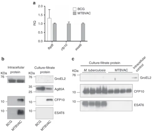
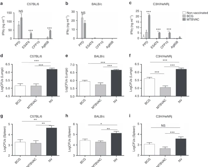
ESAT6/CFP10 reactogenicity depends on host genetic back-ground. Unlike BCG, MTBVAC contains the RD1 region and as expected, we found that it differentially expressed esat6 (esxA -Rv3875) andcfp10(esxB-Rv3874) (Fig. 1a). Accordingly, ESAT6 and CFP10 proteins were present in the intracellular fraction of MTBVAC but not BCG (Fig. 1b). Analysis of the secreted protein fraction of MTBVAC revealed that ESAT6 was not released by MTBVAC, which is in agreement with our previous results9. Conversely, we found CFP10 in the MTBVAC extracellular fraction (Fig. 1b,c), which was unexpected as these two proteins are described to be co-secreted18. We next sought to evaluate ESAT6- and CFP10-specific reactogenicity in different mouse genetic backgrounds following MTBVAC vaccination. To do that, we subcutaneously immunized with MTBVAC C57BL/6, BALB/c or C3H/HeNRj mouse strains, each expressing a different haplotype of the major histocompatibility complex (MHC); H-2b, H-2d and H-2k, respectively. After vaccination, splenocytes were stimulated with either purified protein derivative (PPD) or single antigens ESAT6, CFP10 or Ag85B, with the objective to elucidate the possible influence of the MHC haplotype in the specific response against these proteins. Our data revealed substantial differences among the three different mouse strains (Fig. 2). MTBVAC vaccination conferred immunogenicity to ESAT6 and Ag85B in C57BL/6 (Fig. 2a); to none of the single antigens tested in this work in the BALB/c background (Fig. 2b); and to ESAT6 and CFP10, but not to Ag85B, in the C3H/HeNRj mouse strain (Fig. 2c). PPD-positive IFNgresponse in the BALB/ c mice suggested recognition by the H-2d haplotype of other undefined exported mycobacterial proteins different from the single antigens studied here, but present in the PPD. We found that BCG did not induce any response to ESAT6 or CFP10 stimulation irrespective of the MHC alleles. Our results are in accordance with previous work showing the exclusive capacity of the H-2k haplotype of the C3H mouse strain to recognize CFP10-derived peptides, in comparison to H-2b or H-2d of C57BL/6 and BALB/c strains, respectively19.
Flow cytometry analysis of CD4þIFNgþ splenocytes confirmed the IFNg secretion profile observed by ELISA (Supplementary Fig. 1a–c). In addition, analysis of IFNg-positive splenocytes indicated that around 70% of this population corresponded to CD4þ cells, whereas o5% were CD8þ cell, suggesting that CD4þ T cells are major contributors of IFNg production on antigen stimulation ex vivo (Supplementary Fig. 1d,e).
Since our data revealed lack of Ag85B-specific response in BCG-vaccinated C57BL/6 mice (Fig. 2a), we comparedfbpBgene expression and Ag85B secretion between BCG and MTBVAC. Even thoughfbpBgene was similarly expressed by both vaccines (Fig. 1a), Ag85B protein was not detected in the secreted fraction of BCG (Supplementary Fig. 2), which could explain ourin vivo
observations. Absence of Ag85B in the secreted fraction of BCG could be justified by a recently described Phe140Leu polymorphism in thefbpBgene of all BCG substrains predicting an unstable protein20.
CFP10 and ESAT6 reactogenicity contributes to protection. Eight weeks following BCG or MTBVAC subcutaneous vaccina-tion, mice from the three strains were intranasally infected with a low-dose challenge ofM. tuberculosisH37Rv (infectivity 20 CFU). Four weeks later, bacterial load in lungs (Fig. 2d–f) and spleen
(Fig. 2g–i) revealed protection of both vaccines as assessed by bacterial load reduction relative to unvaccinated controls. Nevertheless, whereas both BCG and MTBVAC protected to a similar extent in C57BL/6 (Fig. 2d,g) and BALB/c (Fig. 2e,h) mice, we observed a more pronounced bacterial load reduction in C3H mice vaccinated with MTBVAC, as compared to BCG (Fig. 2f,i).
As an additional control, we studied the replication and dissemination profile of MTBVAC and BCG in the draining lymph nodes and spleen of C3H mice (Supplementary Fig. 3). The pattern observed resulted comparable, with no profound differences between both vaccines, similar to that published previously in BALB/c mice9. Altogether, these results suggest that differences in protection between BCG and MTBVAC in C3H mice were not due to a different biological behaviour of the two vaccines.
To evaluate the contribution to protection of MTBVAC-induced immune responses against CFP10 and ESAT6, we constructed an MTBVAC substrain with deleted cfp10 and
esat6 genes (MTBVACDE6C10) (Fig. 3a). To construct this mutant, we used a BAC-rec strategy that permitted us to substantially accelerate the generation of deletion mutants in
M. tuberculosis (Supplementary Fig. 4). We found that mice vaccinated with MTBVACDE6C10 showed inability to elicit specific responses to CFP10 and ESAT6 (Fig. 3b,c). Interestingly, IFNg production in C3H mice following PPD stimulation was lower in the MTBVACDE6C10-vaccinated group compared to MTBVAC-vaccinated control, suggesting that ESAT6 and CFP10 are likely the main immunodominant antigens in this genetic background. Importantly, MTBVACDE6C10 triggered an
Ag85B-specific response equivalent to the parental strain (Fig. 3b), indicating that genetic manipulation did not alter the ability of MTBVAC knockout to elicit antigen-specific immunity. Protective efficacy studies in C57BL/6 mice (Fig. 3d,g), reactive to ESAT6 but not CFP10, showed that immunization with MTBVACDE6C10 or parental MTBVAC-conferred similar protection, suggesting that ESAT6-specific immune response is not sufficient for optimal efficacy of MTBVAC. Remarkably, MTBVACDE6C10 vaccination of C3H mice, the only genetic background reactive to both antigens studied in the present work, conferred lower protection than MTBVAC vaccination and comparable to BCG (Fig. 3f,i).
Confirming the capacity of I-Ak, I-Ek molecules to present CFP10-derived peptides to CD4þ cells, we observed in vitro
that CD4þ cells purified from C3H mice vaccinated with MTBVAC, but not with MTBVACDE6C10, produced IFNgwhen incubated with CFP10-pulsed syngeneic bone-marrow-derived macrophages in an antigen concentration-dependent fashion (Supplementary Fig. 5).
In addition, an MTBVAC knockout for Ag85B was constructed to evaluate the role of this antigen in MTBVAC-conferred protection. Results showed that in the absence of Ag85B-specific response, MTBVAC efficacy remained unaffected in any of the three genetic backgrounds tested (Fig. 4).
Increasedcfp10andesat6gene expressionin vivo. Even though we observed that MTBVAC induced a strong response specific for CFP10, ESAT6 and Ag85B, our results indicate that only reactogenicity against RD1-containing antigens, but not Ag85B, BCG MTBVAC esat6 cfp10 fbpB 2.0 1.5 1.0 0.5 0.0 RQ Intracellular protein protein KDa 76 KDa 76 GroEL2 Ag85A CFP10 ESAT6 35 25 10 10 10 10 BCG MTBV AC BCG MTBV AC KDa Culture-filtrate protein M. tuberculosis MTBVAC 76 10 10 Intr acellular control GroEL2 CFP10 ESAT6 Culture-filtrate a b c
Figure 1 | Expression and secretion of ESAT6 and CFP10 by MTBVAC.(a) Normalized expression (usingsigAas housekeeping gene) offbpB,esat6and
cfp10genes in log-phase broth-cultured BCG and MTBVAC. Data in the graph are represented as the relative quantity (RQ) using MTBVAC as comparator. Results are the average from triplicate experiments. Data are mean±s.d. (b,c) Immunoblot analysis of GroEL2, Ag85A, ESAT6 and CFP10 in BCG, MTBVAC andM. tuberculosisprotein extracts. (b) Intracellular (left) and supernatant (right) fractions of BCG and MTBVAC were assessed. (c) Comparison between
M. tuberculosisand MTBVAC-secreted fractions. A BCG cell lysate sample was used as positive control for GroEL2 detection. Four independent protein extractions of each strain were included in the analysis. Full blots are shown in Supplementary Fig. 7a–c.
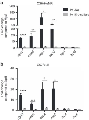
contributed to protection. We hypothesized that this difference could be related to the expression level of each of these antigens by M. tuberculosis on early lung infection. Indeed, it has been described that M. tuberculosis downregulates fbpB
(Ag85B-codifying gene) expression in vivo, as a mechanism to impair host recognition21. Thus, we comparedin vivoexpression of fbpA and fbpB with esat6, cfp10, espA and espC genes. EspA and EspC are essential factors for ESAT6 and CFP10 secretion22–24. Thus, we intranasally infected C3H (Fig. 5a) and C57BL/6 (Fig. 5b) mice with H37Rv, and 4 weeks later we isolated intrapulmonary bacteria and analysed gene expression by RT–qPCR. Expression levels were compared under in vivo
conditions and in culture using standard 7H9 media. Our data revealed a comparable expression level for all the genes studied under in vitro culture. Conversely, when studied in vivo, we detected in both mouse strains an enriched expression of esat6 and cfp10 genes, as well as espA and espC, relative to
fbpAandfbpB.
Of note, higher in vivo expression of esat6 relative to fbpB
has been previously described6,25. Our results extend these
observations to other genes from the ESX-1 secretion system, including cfp10and those involved in the secretion of ESAT6.
MTBVAC induces a CFP10-specific response in human vaccinees. As our data showed a link between CFP10-/ESAT6-induced reactogenicity following MTBVAC immunization and improved protection compared to BCG, we sought to evaluate the induction of responses against these antigens in MTBVAC-vaccinated humans. We used the ESAT6- and CFP10-specific ELISPOT data from the MTBVAC Phase 1 trial performed in adults in Switzerland, which had concluded that none of the MTBVAC vaccinees exceeded the spot threshold for positive latent
M. tuberculosisinfection at the end of active follow-up study17. We thus compared results from each individual before (day 0) and after (day 210) vaccination with MTBVAC or BCG (5105 CFU vaccination dose), finding a significant increase in the CFP10-specific response in the MTBVAC vaccinees (Fig. 6a). For ESAT6, though results showed a rising tendency in the group of MTBVAC, the increment was not significant (Fig. 6c).
PPD ESA T6 CFP10 Ag85B BALB/c IFN γ (ng ml –1 ) NS 30 20 10 0 b PPD ESA T6 CFP10 Ag85B IFN γ (ng ml –1) C3H/HeNRj 25 20 15 10 5 0 *** *** *** Non-vaccinated BCG MTBVAC c 150 100 50 0 PPD ESA T6 CFP10 Ag85B C57BL/6 IFN γ (ng ml –1) NS *** *** a C57BL/6 6.5 6.0 5.5 5.0 4.5 LogCFUs (Lungs) BCG MTBV AC NV *** *** d BALB/c LogCFUs (Lungs) BCG MTBV AC NV *** *** 7.0 6.5 6.0 5.5 5.0 e C3H/HeNRj 6.5 6.0 5.5 5.0 4.5 LogCFUs (Lungs) BCG MTBV AC NV *** *** *** f C57BL/6 LogCFUs (Spleen) BCG MTBV AC NV 5 4 3 2 ** ** g BALB/c LogCFUs (Spleen) BCG MTBV AC NV 6 5 4 3 ** * h C3H/HeNRj LogCFUs (Spleen) BCG MTBV AC NV 5 4 3 2 *** NS i
Figure 2 | Improved protection of MTBVAC compared to BCG is dependent on the host genetics.(a,b,c) Antigen-specific IFNgproduction following stimulation with PPD (5mg ml1), ESAT6 (2mg ml1), CFP10 (2mg ml1) and Ag85B (2mg ml1) during 48 h of splenocytes from mock-, BCG- and MTBVAC-vaccinated C57BL/6, BALB/c and C3H/HeNRj mice. (d–i) Lung (d,e,f) and spleen (g,h,i) bacterial load 4 weeks post low-dose H37Rv intranasal challenge. C57BL/6 (d,g), BALB/c (e,h) and C3H/HeNRj (f,i) were vaccinated with BCG, MTBVAC or unvaccinated eight weeks before challenge. (a,b,c) Data are representative from one of two independent experiments (n¼5 mice per group per experiment). (d–i) Data in the graphs represent a pool of two independent experiments (n¼12 mice per group). All data are mean±s.e.m. (a,b,c) NS, non-significant; ***Po0.001 by unpairedt-student test. (d–i) NS, non-significant; *Po0.05; **Po0.01; ***Po0.001 by one-way ANOVA andBonferronipost-test.
76 KDa GroEL2 CFP10 ESAT6 10 10 MTBV AC ΔE6C10 MTBV AC a C57BL/6 75 50 20 10 0 PPD ESA T6 CFP10 Ag85B PPD ESA T6 CFP10 Ag85B NS NS *** *** *** ** IFN γ (ng ml –1 ) IFN γ (ng ml –1 ) b c C57BL/6 *** *** *** 6.5 6.0 5.5 5.0 4.5 LogCFUs (Lungs) BCG MTBV AC MTBV ACΔ E6C10 NV d BALB/c *** *** *** LogCFUs (Lungs) BCG MTBV AC MTBV ACΔ E6C10 NV 7.0 6.5 6.0 5.5 5.0 e C3H/HeNRj C3H/HeNRj *** *** ** *** *** 6.5 6.0 5.5 5.0 4.5 LogCFUs (Lungs) BCG MTBV AC MTBV ACΔ E6C10 NV NS f C57BL/6 ** ** ** LogCFUs (Spleen) BCG MTBV AC MTBV ACΔ E6C10 NV 5 4 3 2 g BALB/c ** *** * LogCFUs (Spleen) BCG MTBV AC MTBV ACΔ E6C10 NV 6 5 4 3 h C3H/HeNRj * LogCFUs (Spleen) BCG MTBV AC MTBV ACΔ E6C10 NV NS NS 5 4 3 2 i 70 60 50 40 10 0 Non-vaccinated MTBVAC MTBVAC ΔE6C10
Figure 3 | MTBVAC-induced immune response specific to ESAT6 and CFP10 is protective.(a) Immunoblot analysis of GroEL2, ESAT6 and CFP10 in MTBVAC and MTBVACDE6C10 lysate samples. Full blot is shown in Supplementary Fig. 7d. (b,c) Antigen-specific IFNgproduction following stimulation with PPD (5mg ml1), ESAT6 (2mg ml1), CFP10 (2mg ml1) and Ag85B (2mg ml1) during 48 h of splenocytes from mock-, MTBVAC- and MTBVACDE6C10-vaccinated C57BL/6 (left) and C3H/HeNRj (right) mice. (d–i) Lung (d,e,f) and spleen (g,h,i) bacterial load 4 weeks post low-dose H37Rv intranasal challenge. C57BL/6 (d,g), BALB/c (e,h) and C3H/HeNRj (f,i) were vaccinated with BCG, MTBVAC, MTBVACDE6C10 or unvaccinated 8 weeks before challenge. (b,c) Data are representative from one of two independent experiments (n¼5 mice per group per experiment).
(d,e,g,h) Data are derived fromn¼6. (f,i) Data represent a pool of two independent experiments (n¼12 mice per group). All data are mean±s.e.m. (b,c) NS, non-significant; **Po0.01; ***Po0.001 by unpairedt-student test. (d–i) NS, non-significant; *Po0.05; **Po0.01; ***Po0.001 by one-way ANOVA andBonferronipost-test.
Confirming assay specificity, none of the BCG-vaccinated participants showed any increment in CFP10- and ESAT6-specific responses at the end of the study (Fig. 6b,d). Therefore, MTBVAC induces a CFP10-specific immune response in humans.
Discussion
The MHC is composed of several highly polymorphic loci resulting in a wide interindividual variability of MHC haplotypes, which can have great influence in host-specific responsiveness to vaccines, as individual haplotype may determine antigen immunodominance associated with protection26. To date, there are no defined BCG-specific immunodominant antigens associated with efficacy against TB. The identification of such correlates of protection for new TB vaccines, as MTBVAC, would greatly accelerate clinical development to efficacy trials.
A plausible explanation for the poor protection of BCG against pulmonary TB is the loss of several genomic regions, including RD1 (refs 5,8), which encodes ESAT6, CFP10 and part of their secretory machinery contained in the ESX-1 secretion system. Despite their low molecular weight, ESAT6 and CFP10 are the top two antigens with the highest proportion of peptides recognized by human MHC haplotypes20,27. Importantly, loss of RD1 has also been reported as the main basis for attenuation of BCG5. Our results indicated that, unlike other major antigens not related with virulence, such as those belonging to the Ag85 complex, ESAT6, CFP10 and the machinery involved in their secretion are highly over expressed on early infection, which is probably the result of their role in virulence and infection establishment. Indeed, in agreement with these observations, the
phagosomal escape of M. tuberculosis, which is mediated by ESAT6, occurs as early as day 3 after in vitro macrophage infection28. Thus, a vaccine inducing an effective response against ESAT6 and CFP10, such as MTBVAC, may have the advantage to target antigens highly expressed during early M. tuberculosis
infection. This would probably lead to a better recognition of
M. tuberculosis-infected cells, whose MHC molecules should be coated with epitopes derived from dominant antigens. Conversely, vaccination-induced immunogenicity against other antigens, such as Ag85A or Ag85B, underexpressed and therefore less represented in the antigen repertoire at early infection stages, would be less efficient against initial infection as a consequence of a poorer recognition of pathogen-infected cells. In line with this, other authors have demonstrated downregulation of Ag85B expression during earlyM. tuberculosisinfection21.
Our efficacy data in mice seem to contrast with previous results using a recombinant BCG vaccine overexpressing Ag85B (rBCG30), which showed an improved protection relative to BCG in an outbred guinea pig model29. However, the different animal model used in that study (more sensitive to TB) and the different readout utilized to measure vaccine efficacy (survival) could account for such differences.
An important proportion of human MHC class II haplotypes recognizes ESAT6- and CFP10-derived peptides30. This could be explained by the important roles played by both proteins in virulence, eliciting an evolutionary pressure leading to the selection of MHC human haplotypes that recognize highly conserved peptides derived from these antigens. Our results suggest that the immunodominance of ESAT6 and CFP10 in humans is not reflected in mice, as only one of the three haplotypes tested simultaneously presents both antigens. 70 60 50 40 30 20 10 0 PPD ESA T6 CFP10 Ag85B C3H/HeNRj IFN γ (ng ml –1) Non-vaccinated MTBVAC MTBVAC ΔAg85B b 90 80 70 60 50 40 30 20 10 0 PPD ESA T6 CFP10 Ag85B C57BL/6 IFN γ (ng ml –1) * *** a BALB/c *** *** *** MTBV AC ΔAg85B NV BCG MTBV AC LogCFUs 7 6 5 4 d C3H/HeNRj *** *** * * *** MTBV AC ΔAg85B NV BCG MTBV AC LogCFUs 6.5 6.0 5.5 5.0 4.5 e C57BL/6 *** *** *** MTBV AC ΔAg85B NV BCG MTBV AC LogCFUs 7 6 5 4 c NS
Figure 4 | Ag85B-specific immunogenicity induced by MTBVAC is not protective.(a,b) Antigen-specific IFNgproduction following stimulation with PPD (5mg ml), ESAT6 (2mg ml1), CFP10 (2mg ml1) and Ag85B (2mg ml1) during 48 h of splenocytes from mock-, MTBVAC- and
MTBVACDAg85B-vaccinated C57BL/6 (left) and C3H/HeNRj (right) mice. (c–e) Lung bacterial load 4 weeks post low-dose H37Rv intranasal challenge. C57BL/6 (c), BALB/ c (d) and C3H/HeNRj (e) were vaccinated with BCG, MTBVAC, MTBVACDAg85B or unvaccinated 8 weeks before challenge. (a,b) Data are representative from one of two independent experiments (n¼5 mice per group per experiment). (c–e) Data are derived fromn¼6. All data are mean±s.e.m. (a,b) NS, non-significant; **Po0.01; ***Po0.001 by unpairedt-student test. (c–e) *Po0.05; **Po0.01; ***Po0.001 by one-way ANOVA and
This difference could be due to lack of co-evolution of the mouse MHC loci with ESAT6- and CFP10-expressing mycobacteria. Indeed, Mycobacterium microti, the mycobacterial species that naturally infects rodents, lacks the RD1mic region that includes both genes5. Conversely, those species of the M. tuberculosis
complex able to infect humans (M. canettii, M. africanumand
M. tuberculosis) have retained intactesat6andcfp10genes as well as their secretory machinery31,32.
Our previously published results in MTBVAC-vaccinated newborn C57BL/6 mice showed a slight but significant improvement of MTBVAC-conferred protection relative to BCG in lungs of infected pups with an aerosol challenge dose of H37Rv similar to the one used in the present work16. This previous work also demonstrated that, unlike the comparable immunogenicity of MTBVAC and BCG observed in adult mice, MTBVAC was much more immunogenic (following PPD stimulation) than BCG when animals were vaccinated at birth, which could account for such difference in protection. In two other studies9,33, where we vaccinated C57BL/6 adult mice, we also found an improved protection in the MTBVAC group, although in those works we used a much higher initial H37Rv challenge dose (100 and 1,000 CFU) than the one we used in the present work (20 CFU). In line with this, previous data in a guinea pig model revealed that MTBVAC only improved BCG protection in high-dose challenge experiments14, suggesting the contribution of undefined
mechanism(s) of protection occurring at different experimental conditions than the tested in this work.
Our data indicate that both H-2b (C57BL/6) and H-2k (C3H) haplotypes recognized ESAT6-derived epitopes following MTBVAC immunization. However, MTBVAC only improves BCG-conferred protection in C3H mice, the only of the three tested genetic backgrounds whose MHC haplotype presents CFP10-derived peptides. This result could suggest a particular contribution of CFP10-specific reactogenicity to MTBVAC protection. Interestingly, we found that MTBVAC secretes CFP10 but not ESAT6. Provided secreted proteins are differentially processed by the host immune system in comparison to intracellular proteins34, we speculate that ESAT6 and CFP10 might induce different responses in MTBVAC vaccinees, which might lead to differential protection. In this regard, it could be relevant in the future to construct MTBVAC mutant strains for ESAT6 and CFP10 separately, to study these potential mechanisms in more detail.
It has been reported that both ESAT6 and CFP10 are co-secreted by M. tuberculosis18. Therefore, further work is needed to elucidate the molecular mechanisms behind the CFP10-independent secretion of ESAT6 in MTBVAC. Two previous publications reported that uncoupled ESAT6 and CFP10 secretion can occur under some conditions35,36. In these studies, authors found that CFP10 is secreted independently of ESAT6 in the presence of an aberrant ESX-1 system. Thus, we speculate that since PhoP is a regulator of different genes encoding ESX-1 components12,37,38, MTBVAC may express a dysfunctional ESX-1 system responsible of the observed phenotype.
Our study underlines the role of host genetics in vaccine-induced protection and how genetic background determines antigen immunodominance, as shown with CFP10 in the C3H mouse strain. In line with this finding, the importance of host genotype for tuberculosis susceptibility and vaccine protection in the mouse model has been recently reported39. Thus, our results indicate that MTBVAC-vaccinated mice reactogenic for CFP10 and ESAT6 are more protected against tuberculosis than non-reactogenic strains. In humans, responders to these antigens comprise around 60–80% of individuals30,40. We speculate that a vaccine expressing ESAT6 and CFP10 might confer improved protection in these individuals (in comparison with BCG); however, further clinical data would be needed to test this hypothesis.
One of the handicaps of a vaccine whose protective mechanism is based on ESAT6- and/or CFP10 antigens remains in their potential interference with the latent TB diagnostic test. Data provided by the first-in-human clinical trial of MTBVAC in adults indicated that the ELISPOT response elicited by MTBVAC following ESAT6 or CFP10 stimulation was below the cutoff established for tuberculosis infection17, suggesting no interference of MTBVAC at least in this target population. Interestingly, it has been reported that individuals with a quantiFERON value greater than 4 IU ml1 have a higher probability of developing active TB, while individuals with a value between 0.35 and 4 (cutoff for positivity is 0.35) have a similar probability than negative individuals41. Thus, this study opens the possibility to reconsider the cutoff of this technique to identify individuals with a high risk of developing active TB. Current clinical trials in TB-endemic countries in neonates (NCT02729571) are further evaluating quantiFERON conversion following MTBVAC vaccination to evaluate this question in more detail.
MTBVAC is the only vaccine in the TB vaccine pipeline of candidates in clinical evaluation that primes both CFP10- and ESAT6-specific immune responses42. Our data suggest that this strategy might be effective in protecting from pulmonary TB, 200 150 100 50 30 20 10 0 F o ld-change compared to fbpB
cfp10 esat6 espA espC fbpA fbpB
In vivo In vitro culture C3H/HeNRj * ** ** **** a F old-change compared to fbpB
cfp10 esat6 espA espC fbpA fbpB C57BL/6 40 30 20 10 0 *** * * **** b
Figure 5 | Enrichedin vivoexpression of genes from ESX-1 secretion system.(a,b) Expression ofesat6,cfp10,espA,espC,fbpAandfbpBgenes (normalized with16sgene expression) from H37Rv isolated from lungs from C3H/HeNRj (a) and C57BL/6 (b) 4 weeks after high-dose (103CFU) intranasal challenge, in comparison to expression obtained under 7H9-culturein vitroconditions. Normalized results are represented for each gene and experimental condition as the fold-change induction in comparison tofbpBexpression level.In vivodata are derived fromn¼4 mice (a) andn¼6 mice (b).In vitrodata represent a pool of four independent RNA extractions. All data are mean±s.e.m. *Po0.05;
which would have an evident impact on TB transmission. Supporting this hypothesis, prospective cohort studies of persons exposed to individuals with active TB have indicated that latently infected individuals (reactive to CFP10 and ESAT6 stimulation) are more protected against reinfection than non-infected people43. Our data support the potential usefulness of the analysis of CFP10- and ESAT6-positive cells after vaccination in future clinical efficacy trials, in the search for a possible biomarker of vaccine-induced protection.
Methods
Bacteria.M. bovisBCG Danish (Statens Serum Institute),M. tuberculosis
MTBVAC (University of Zaragoza) andM. tuberculosisH37Rv (Institut Pasteur Paris) strains were grown at 37°C in Middlebrook 7H9 broth (Difco) supplemented with ADC 10% (Difco) and 0.05% (v/v) Tween-80 (Sigma) or on solid Middlebrook 7H11 (Difco) supplemented with ADC 10%. Bacterial suspensions for vaccination or infection were prepared in PBS from glycerol stocks previously quantified by plating serial dilutions.
Mouse experiments.All mice were kept under controlled conditions and observed for any sign of disease. Experimental work was conducted in agreement with European and national directives for protection of experimental animals and with approval from the Ethics Committee from University of Zaragoza (approved protocol PI46/14). No randomization specific methodology was applied to this study.
Female, 8–10 weeks old C57BL/6, BALB/c and C3H/HeNRj mice
(Janvier Biolabs) were vaccinated subcutaneously (100ml) with 106CFU of vaccine strains in PBS. Eight weeks post vaccination, mice were intranasally challenged with 150 CFU of H37Rv in 40ml of PBS. Bacterial burden was assessed 4 weeks post challenge by plating homogenized lungs and spleen on solid medium. A group of infected mice was killed 1 day after challenge to determine the initial bacterial load in lungs, which resulted to beB20 CFU in all experiments (Supplementary Fig. 6). For immunogenicity studies, mice were vaccinated subcutaneously with 106 CFU of vaccine strains in PBS, and 8 weeks later animals were killed and splenocytes collected. 106cells were stimulated in 96-well U bottom plates with PPD (Statens Serum Institute, SSI) 5mg ml1, or 2mg ml1of ESAT6, CFP10 or Ag85B (LIONEX GmbH, Braunschweig, Germany) during 48 h for supernatant collection and cytokine detection by ELISA. Cytokine concentration in supernatants was determined with an IFNg-specific ELISA kit (MabTECH). For
intracellular staining, splenocytes were incubated with antigens for 24 h, and 10mg ml1Brefeldin A (Sigma) was added during the last 6 h of incubation. For surface staining, cells were labelled with anti-CD4-FITC (553047, BD Biosciences) and anti-CD3-PerCPVio700 (130-109-883, Miltenyi Biotec), diluted 1:500 and 1:50, respectively, in culture medium with 10% FCS. Then, cells were fixed and permeabilized with the Cytofix/Cytoperm Fixation/Permeabilization Kit (BD Biosciences) following the manufacturer’s instructions, and stained with anti-IFNg-APC (554413, BD Biosciences), diluted 1:200 in permeabilization buffer. Cells were acquired with a Gallios Flow Cytometer (Beckman).
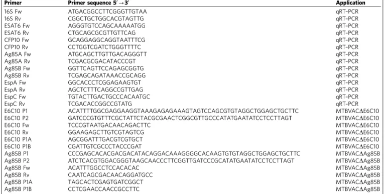
Construction of MTBVAC mutant substrains.We developed a BAC-rec strategy to accelerate knockout construction inM. tuberculosis, which allowed to generate the desired mutant strains inB1 month. To construct the allelic exchange substrate (AES), we used aM. tuberculosisH37Rv bacterial artificial chromosome (BAC) library constructed in the pBeloBAC11 vector and introduced into
Escherichia coliDH10B44(a kind gift from Roland Brosch, Institut Pasteur Paris, France). TheE. coliclone carrying the BAC containing the target gene was identified (Rv221 forfbpBand Rv414 forcfp10-esat6). Bacteria were made electrocompetent and transformed with pKD46 carrying the red recombinase from lambda phage45. The resultingE. colistrain was transformed with a PCR product, which contains the kanamycin-resistant marker (Kmr) from pKD4 (ref. 45) flanked by 40 bp homology arms to the target gene located in the BAC (using E6C10 P1/E6C10 P2 or Ag85B P1/Ag85B P2 primer pairs, Table 1). After induction of the lambda red recombinase by incubation in the presence of 1 mM L-arabinose, recombinant colonies were selected by plating in the presence of kanamycin. Gene disruption in the BAC was confirmed by PCR, resulting in a highly efficient process with nearly 100% positive clones. The BAC carrying the disrupted allele(s) was used as a template for a PCR reaction using the Pwo high fidelity DNA polymerase. The final PCR product was subsequently used as AES and contains the Kmr cassette with 1,500 bp homology arms flanking the target gene(s) (using E6C10 P1A/E6C10 P2A or Ag85B P1A/Ag85B P2A primer pairs, Table 1). In parallel, MTBVAC transformed with pJV53H (a kind gift from Christophe Guilhot, IPBS, Toulouse, France), which consists on the replacement of the original Kmrwith a hygromycin-resistance marker in pJV53 (ref. 46), was grown in the presence of 0.2% acetamide to allow expression of the recombinase system from this plasmid. The AES was transformed in these cells by electroporation. Recombinant MTBVAC colonies containing the desired mutation were selected by plating on kanamycin and confirmed by PCR amplified with primers flanking the deleted gene (using E6C10 Fw/E6C10 Rv or Ag85B Fw/Ag85B Rv primer pairs, Table 1). Some of the colonies analysed contained both wild-type and mutant amplicons suggesting an unspecific recombination of the AES. We selected colonies where only the mutant amplicon was amplified (Supplementary Fig. 4 and Table 2).
50 40 30 20 10 0 SFC (CFP10) 0 210 MTBVAC P= 0.043 a Days post-vaccination 50 40 30 20 10 0 SFC (CFP10) 0 210 BCG b Days post-vaccination MTBVAC 50 40 30 20 10 0 SFC (ESA T6) 0 210 P= 0.1641 c Days post-vaccination 50 40 30 20 10 0 SFC (ESA T6) 0 210 BCG d Days post-vaccination
Figure 6 | Vaccination with MTBVAC induces a CFP10-specific immune response in humans.Specific IFNgELISPOT results from the first-in-human MTBVAC clinical trial are shown for CFP10 (a,b) and ESAT6 (c,d), comparing for each volunteer the number of spots pre- and post-BCG (b,d) and MTBVAC (a,c) vaccination with 5105CFU. Wilcoxon matched-pairs signed rank test was used to compare pre- and post-vaccination status for each vaccine and
In vitropresentation assay.Bone marrow cells collected from mouse femurs and tibias were plated on sterile Petri dishes and incubated for 7 days in Dulbecco’s modified Eagle’s medium (Gibco) containing 10% (v/v) heat-inactivated FCS, 100 units ml1penicillin/100 mg ml1streptomycin (Sigma) and 10% (vol/vol) conditioned medium from mouse L929 fibroblasts (a kind gift from Dr Esther Perez, GSK, Tres Cantos, Madrid, Spain). 105bone-marrow-derived macrophages were seeded in 96 flat-well plates and incubated for 4 h with CFP10 antigen at the indicated concentrations in triplicates. After three washing steps with PBS, 106 purified CD4þsplenocytes obtained by MACS separation (Miltenyi Biotec) (purity higher than 90 %) were added in 200ml of RPMI 1640 (Sigma) containing 10% (v/v) heat-inactivated FBS, 100 units ml1penicillin, 100 mg ml1 streptomycin, 50mM 2-mercaptoethanol (Sigma) and cells were incubated for 24 h. The plate was then centrifuged at 800g, and the supernatant recovered to determine IFNgby ELISA (MabTECH).
Protein analysis.To analyse proteins from extracellular and intracellular compartments, BCG, MTBVAC or H37Rv were cultured in 7H9 liquid medium with 0.05% (v/v) Tween-80 and supplemented with 0.2% (w/v) dextrose and 0.085% (w/v) NaCl, to avoid albumin contamination from ADC in the supernatant fraction. After 3 weeks of incubation, cultures were pelleted by centrifugation and the supernatants were passed through 0.2mm-pore filters to remove any residual bacteria. Supernatant proteins were precipitated with 10% (v/v) trichloroacetic acid (Sigma) during 1 h in ice and centrifugation at 3,200gduring 30 min at 4°C. Pelleted proteins were rinsed with cold acetone and, after decanting acetone, air dried pellets were resuspended in 150 mM Tris/HCl pH 8. Bacterial pellet was washed twice and resuspended in 1 ml PBS containing a protease inhibitor cocktail (Roche). Bacteria were disrupted by sonication for 30 min at 4°C using a Bioruptor (Diagenode). Samples were centrifuged 5 min at 18 000gand the supernatant collected for further analysis.
Intracellular and secreted protein concentrations were determined with the QuantiPro BCA Assay kit (Sigma). Ten micrograms of protein per well were loaded and separated by SDS–PAGE. Following protein transference to PVDF membranes, immunodetection was carried out using mouse monoclonal antibodies anti-ESAT6 (1:1,000; clone 11G4; Abcam ref.: ab26246) and anti-GROEL (1:500; clone BDI578; Abcam ref.: ab20045), rabbit polyclonal anti-CFP10 (1:2,000; Thermo Scientific ref.: PA1-19445) and chicken polyclonal anti-Ag85A (1:1,000; Abcam ref.: ab14073). Membranes were incubated with the corresponding HRP-conjugated secondary antibodies (1:20,000; Sigma) and signal developed with the ECL Plus Western Blotting System (GE HealthCare). To reprobe each blot with different antibodies, membranes were incubated with ReBlot Plus Strong Antibody Stripping Solution (Millipore) according to the manufacturer’s instructions.
For visualization of proteins following electrophoresis, polyacrylamide gels were stained using a commercial colloidal blue staining kit (Invitrogen) according to the manufacturer’s instructions. Bands of interest were split and sent for identification by MALDI-TOF MS to Proteomics Services of the University of Zaragoza.
qRT–PCR.Groups of C3H/HeNRj or C57BL/6 8–10 weeks old, female mice were intranasally challenged with 103CFU of H37Rv in 40ml of PBS. Four weeks later, mice were killed individually and lungs collected and cut in small pieces, which were homogenized in 4 ml of TRIZOL by mechanical disruption using a Dounce tissue grinder. Lung homogenates were split in four 1-ml aliquots and placed in dry ice until further processing. Then, 200ml of chloroform were added per ml of TRIZOL and after vigorous vortexing, tubes were centrifuged at 18,000gduring 1 h at 4°C. Aqueous upper phase containing eukaryotic RNA was recovered and stored at 80°C. Remaining supernatant was discarded and the four pellets containing mycobacteria were resuspended and collected with one additional millilitre of TRIZOL and 200ml of chloroform. Bacteria were disrupted adding glass beads using a Fast Prep device, with two 4500cycles at highest speed. Then, tubes were
centrifuged 10 min at 18,000g(4°C) and aqueous phase (500ml approx.) containing mycobacterial RNA (and also remaining eukaryotic RNA) were recovered. A measure of 700ml of isopropanol was added and tubes were incubated at room temperature during 15 min to favour RNA precipitation. Precipitated nucleic acids were collected by centrifugation. The pellets were rinsed with 70% ethanol and air dried before being re-dissolved in RNase-free water. DNA was removed from RNA samples with TurboDNAfree (Ambion) by incubation at 37°C for 1 h. RNA integrity was assessed by agarose gel electrophoresis, and absence of contaminating DNA was checked by lack of amplification products after 30 PCR cycles. Lungs from uninfected animals were processed as above to be used as non-infected control in the qPCR.
For qRT–PCR, retro-transcription of 1mg of RNA was performed following a standard reverse transcription reaction with the SuperScript Reverse Transcriptase (Invitrogen). The generated cDNA served as a template for qPCR in the presence of gene-specific primers (Table 1) and SYBR Green Master-Mix (Roche). Four Table 1 | Primer list.
Primer Primer sequence 50-30 Application
16S Fw ATGACGGCCTTCGGGTTGTAA qRT–PCR 16S Rv CGGCTGCTGGCACGTAGTTG qRT–PCR ESAT6 Fw AGGGTGTCCAGCAAAAATGG qRT–PCR ESAT6 Rv CTGCAGCGCGTTGTTCAG qRT–PCR CFP10 Fw GCAGGAGGCAGGTAATTTCG qRT–PCR CFP10 Rv CCTGGTCGATCTGGGTTTTC qRT–PCR Ag85A Fw ATGCAGCTTGTTGACAGGGTT qRT–PCR Ag85A Rv TCGACGCGACATACCCGT qRT–PCR Ag85B Fw GGTTCAGTTCCAGAGCGGTG qRT–PCR Ag85B Rv TCGAGCAGATAAACCGCAGG qRT–PCR EspA Fw GGCACCCTCGGAGAAGTGT qRT–PCR EspA Rv AGCTCTTTCAGGCCGTTGAG qRT–PCR EspC Fw TGTACTTGACTGCCCACAATGC qRT–PCR EspC Rv TCGACACCGGCCGTATG qRT–PCR
E6C10 P1 ACATTTTGGCGAGGAAGGTAAAGAGAGAAAGTAGTCCAGCGTGTAGGCTGGAGCTGCTTC MTBVACDE6C10 E6C10 P2 GATCCCGTGTTTCGCTATTCTACGCGAACTCGGCGTTGCCCATATGAATATCCTCCTTAGT MTBVACDE6C10
E6C10 Fw TCCCGTAATGACAACAGACTTC MTBVACDE6C10
E6C10 Rv GGAAGAGCTTGTCGTAGTCG MTBVACDE6C10
E6C10 P1A AGCGGATTTGACGTCGTGCT MTBVACDE6C10
E6C10 P1B CGATTGTCGCCCTACCCGAT MTBVACDE6C10
Ag85B P1 CCCGAGCACACGACGACATACAGGACAAAGGGGCACAAGTGTGTAGGCTGGAGCTGCTTC MTBVACDAg85B Ag85B P2 ATCTCACGTGGACGGGTAAGCAACCCTTCGGTTGATCCCGCATATGAATATCCTCCTTAGT MTBVACDAg85B
Ag85B Fw ACATTTGGCCTCCACACAC MTBVACDAg85B
Ag85B Rv CAATCAGCGACAACAGGATGCC MTBVACDAg85B
Ag85B P1A TAGCACTCGAGTGATCGGCT MTBVACDAg85B
Ag85B P1B CCTCGAACCAACCGCCTTC MTBVACDAg85B
Table 2 | Plasmid and BAC information.
BAC or plasmids Source Reference
H37Rv BAC Library Institut Pasteur Paris (France) 44 pKD46 Coli Genetic Stock Center
(Yale University)
45
pKD4 Coli Genetic Stock Center (Yale University)
45
replicates of each geneCTvalue were obtained in the StepOne Plus Instrument
(Applied Biosystems) and were normalized to theCTof the16srRNA gene
(amplified from the same samples), obtaining aDCT¼CT,j–CT,16S, wherejis a gene
different from16s. Our observations indicated unspecific gene amplification in the non-infected cDNA controls. As a result, following a procedure previously reported47, we calculated aDDC
Tvalue specific for infection (DDCT(inf)),
subtracting theDCTmean value for each gene obtained from non-infected controls
from theDCTobtained for each sample from infected mice. Finally, change was
calculated with the equation 2DDCTðinfÞ. Gene expression analysis fromin vitro samples was performed using conventional comparativeCTmethod. Normalized
gene expression data (using the16srRNA as housekeeping gene) were represented as the fold-change compared tofbpBexpression for each of thein vivoandin vitro
conditions tested. The melting curves for all gene-specific primer pairs (sequences used in Supplementary Experimental Procedures) were examined to identify primer-dimer formation and to ensure the uniformity of the amplicons.
Statistical analysis.Results from this study were not blinded for analysis. No statistical method was used to calculate sample size in animal experiments. GraphPrism software was used for statistical analysis. Brown–Forsythe test was used to assess variances homogeneity. Variances were similar among compared groups. Statistical tests used for each experiment are indicated in the figure legends. All statistical tests used were two-tailed. Outlier values were determined applying the Grubb’s test to all data sets, and were discarded from the final statistical analysis. Differences were considered significant atPo0.05.
Data availability.The authors declare that the data supporting the findings of this study are available in this article and its Supplementary Information Files, or from the corresponding authors on request.
References
1. Marinova, D., Gonzalo-Asensio, J., Aguilo, N. & Martin, C. Recent developments in tuberculosis vaccines.Expert Rev. Vaccines12,1431–1448 (2013).
2. WHO. Global tuberculosis report 2016. http://www.who.int/tb/publications/ global_report/en/ (2016).
3. Fine, P. E. Variation in protection by BCG: implications of and for heterologous immunity.Lancet346,1339–1345 (1995).
4. Brosch, R.et al.Genome plasticity of BCG and impact on vaccine efficacy.Proc. Natl Acad. Sci. USA104,5596–5601 (2007).
5. Pym, A. S., Brodin, P., Brosch, R., Huerre, M. & Cole, S. T. Loss of RD1 contributed to the attenuation of the live tuberculosis vaccinesMycobacterium bovisBCG andMycobacterium microti.Mol. Microbiol.46,709–717 (2002). 6. Aagaard, C.et al.A multistage tuberculosis vaccine that confers efficient
protection before and after exposure.Nat. Med.17,189–194 (2011). 7. Groschel, M. I.et al.Recombinant BCG expressing ESX-1 ofMycobacterium
marinumcombines low virulence with cytosolic immune signaling and improved TB protection.Cell Rep.18,2752–2765 (2017).
8. Pym, A. S.et al.Recombinant BCG exporting ESAT-6 confers enhanced protection against tuberculosis.Nat. Med.9,533–539 (2003).
9. Arbues, A.et al.Construction, characterization and preclinical evaluation of MTBVAC, the first live-attenuatedM. tuberculosis-based vaccine to enter clinical trials.Vaccine31,4867–4873 (2013).
10. Stucki, D.et al. Mycobacterium tuberculosislineage 4 comprises globally distributed and geographically restricted sublineages.Nat. Genet.48,
1535–1543 (2016).
11. Kamath, A. T.et al.New live mycobacterial vaccines: the Geneva consensus on essential steps towards clinical development.Vaccine23,3753–3761 (2005). 12. Frigui, W.et al.Control of M. tuberculosis ESAT-6 secretion and specific T cell
recognition by PhoP.PLoS Pathog.4,e33 (2008).
13. Camacho, L. R., Ensergueix, D., Perez, E., Gicquel, B. & Guilhot, C. Identification of a virulence gene cluster ofMycobacterium tuberculosisby signature-tagged transposon mutagenesis.Mol. Microbiol.34,257–267 (1999). 14. Martin, C.et al.The liveMycobacterium tuberculosisphoP mutant strain is
more attenuated than BCG and confers protective immunity against tuberculosis in mice and guinea pigs.Vaccine24,3408–3419 (2006). 15. Verreck, F. A.et al.MVA.85A boosting of BCG and an attenuated, phoP
deficient M. tuberculosis vaccine both show protective efficacy against tuberculosis in rhesus macaques.PLoS ONE4,e5264 (2009).
16. Aguilo, N.et al.MTBVAC vaccine is safe, immunogenic and confers protective efficacy againstMycobacterium tuberculosisin newborn mice.Tuberculosis96,
71–74 (2016).
17. Spertini, F.et al.Safety of human immunisation with a live-attenuated
Mycobacterium tuberculosisvaccine: a randomised, double-blind, controlled phase I trial.Lancet Respir. Med.3,953–962 (2015).
18. Renshaw, P. S.et al.Conclusive evidence that the major T-cell antigens of the
Mycobacterium tuberculosiscomplex ESAT-6 and CFP-10 form a tight, 1:1 complex and characterization of the structural properties of ESAT-6, CFP-10,
and the ESAT-6*CFP-10 complex. Implications for pathogenesis and virulence.
J. Biol. Chem.277,21598–21603 (2002).
19. Kamath, A. B.et al.Cytolytic CD8þT cells recognizing CFP10 are recruited to the lung afterMycobacterium tuberculosisinfection.J. Exp. Med.200,
1479–1489 (2004).
20. Copin, R., Coscolla, M., Efstathiadis, E., Gagneux, S. & Ernst, J. D. Impact of
in vitroevolution on antigenic diversity ofMycobacterium bovisbacillus Calmette-Guerin (BCG).Vaccine32,5998–6004 (2014).
21. Bold, T. D., Banaei, N., Wolf, A. J. & Ernst, J. D. Suboptimal activation of antigen-specific CD4þ effector cells enables persistence ofM. tuberculosis in vivo.PLoS Pathog.7,e1002063 (2011).
22. Fortune, S. M.et al.Mutually dependent secretion of proteins required for mycobacterial virulence.Proc. Natl Acad. Sci. USA102,10676–10681 (2005).
23. Lou, Y., Rybniker, J., Sala, C. & Cole, S. T. EspC forms a filamentous structure in the cell envelope ofMycobacterium tuberculosisand impacts ESX-1 secretion.Mol. Microbiol.103,26–38 (2017).
24. MacGurn, J. A., Raghavan, S., Stanley, S. A. & Cox, J. S. A non-RD1 gene cluster is required for Snm secretion inMycobacterium tuberculosis.Mol. Microbiol.
57,1653–1663 (2005).
25. Rogerson, B. J.et al.Expression levels ofMycobacterium tuberculosis
antigen-encoding genes versus production levels of antigen-specific T cells during stationary level lung infection in mice.Immunology118,195–201 (2006).
26. Mehra, N. K. & Kaur, G. MHC-based vaccination approaches: progress and perspectives.Expert Rev. Mol. Med.5,1–17 (2003).
27. Zhang, W.et al.Genome sequencing and analysis of BCG vaccine strains.
PLoS ONE8,e71243 (2013).
28. Simeone, R.et al.Phagosomal rupture byMycobacterium tuberculosisresults in toxicity and host cell death.PLoS Pathog.8,e1002507 (2012).
29. Horwitz, M. A. & Harth, G. A new vaccine against tuberculosis affords greater survival after challenge than the current vaccine in the guinea pig model of pulmonary tuberculosis.Infect. Immun.71,1672–1679 (2003).
30. Lindestam Arlehamn, C. S.et al.A quantitative analysis of complexity of human pathogen-specific CD4 T cell responses in healthyM. tuberculosis
infected South Africans.PLoS Pathog.12,e1005760 (2016).
31. Gonzalo-Asensio, J.et al.Evolutionary history of tuberculosis shaped by conserved mutations in the PhoPR virulence regulator.Proc. Natl Acad. Sci. USA111,11491–11496 (2014).
32. Supply, P.et al.Genomic analysis of smooth tubercle bacilli provides insights into ancestry and pathoadaptation ofMycobacterium tuberculosis.Nat. Genet.
45,172–179 (2013).
33. Solans, L.et al.Hyper-attenuated MTBVAC erp mutant protects against tuberculosis in mice.Vaccine32,5192–5197 (2014).
34. Grotzke, J. E., Siler, A. C., Lewinsohn, D. A. & Lewinsohn, D. M. Secreted immunodominantMycobacterium tuberculosisantigens are processed by the cytosolic pathway.J. Immunol.185,4336–4343 (2010).
35. Gao, L. Y.et al.A mycobacterial virulence gene cluster extending RD1 is required for cytolysis, bacterial spreading and ESAT-6 secretion.Mol. Microbiol.53,1677–1693 (2004).
36. Pang, X.et al.MprAB regulates the espA operon inMycobacterium tuberculosis
and modulates ESX-1 function and host cytokine response.J. Bacteriol.195,
66–75 (2013).
37. Solans, L.et al.A specific polymorphism inMycobacterium tuberculosisH37Rv causes differential ESAT-6 expression and identifies WhiB6 as a novel ESX-1 component.Infect. Immun.82,3446–3456 (2014).
38. Solans, L.et al.The PhoP-dependent ncRNA Mcr7 modulates the TAT secretion system inMycobacterium tuberculosis.PLoS Pathog.10,e1004183 (2014).
39. Smith, C. M.et al.Tuberculosis susceptibility and vaccine protection are independently controlled by host genotype.MBio7,e01516-16 (2016). 40. Millington, K. A.et al.Rv3615c is a highly immunodominant RD1 (region of
difference 1)-dependent secreted antigen specific forMycobacterium tuberculosisinfection.Proc. Natl Acad. Sci. USA108,5730–5735 (2011). 41. Andrews, J. R.et al.Serial QuantiFERON testing and tuberculosis disease risk
among young children: an observational cohort study.Lancet Respir. Med.5,
282–290 (2017).
42. Kaufmann, S. H., Weiner, J. & von Reyn, C. F. Novel approaches to tuberculosis vaccine development.Int. J. Infect. Dis.56,263–267 (2017).
43. Andrews, J. R.et al.Risk of progression to active tuberculosis following reinfection withMycobacterium tuberculosis.Clin. Infect. Dis.54,784–791 (2012).
44. Brosch, R.et al.Use of aMycobacterium tuberculosisH37Rv bacterial artificial chromosome library for genome mapping, sequencing, and comparative genomics.Infect. Immun.66,2221–2229 (1998).
45. Datsenko, K. A. & Wanner, B. L. One-step inactivation of chromosomal genes inEscherichia coliK-12 using PCR products.Proc. Natl Acad. Sci. USA97,
46. van Kessel, J. C. & Hatfull, G. F. Recombineering inMycobacterium tuberculosis.Nat. Methods4,147–152 (2007).
47. Zhao, M.et al.Systems infection biology: a compartmentalized immune network of pig spleen challenged withHaemophilus parasuis.BMC Genomics
14,46 (2013). Acknowledgements
The authors acknowledge the Scientific and Technical Services from Instituto Aragone´s de Ciencias de la Salud and Universidad de Zaragoza. This work was supported by the Spanish Ministry of Economy and Competitiveness (Grant number BIO2014-5258P), the European Commission H2020 program (Grant number TBVAC2020 643381) and ‘Gobierno de Arago´n/Fondo Social Europeo’.
Author contributions
N.A., F.S. and C.M. designed the experiments and directed the study. N.A., J.G.-A., S.A.A., A.B.G., S.U. and R.A. performed the experiments. R.S. and M.S. provided the ESAT6 and CFP10 recombinant antigens. N.A., D.M. and C.M. wrote the manuscript. The funders had no role in study design, data collection and analysis, decision to publish or preparation of the manuscript.
Additional information
Supplementary Informationaccompanies this paper at http://www.nature.com/ naturecommunications
Competing interests:C.M. and J.G.-A. are co-inventors in a patent application entitled ‘‘Tuberculosis vaccine’’ filled by the University of Zaragoza (application number:
PCT/ES 2007/070051). The remaining authors declare no competing financial interests.
Reprints and permissioninformation is available online at http://npg.nature.com/ reprintsandpermissions/
How to cite this article:Aguilo, N.et al.Reactogenicity to major tuberculosis antigens absent in BCG is linked to improved protection againstMycobacterium tuberculosis.
Nat. Commun.8,16085 doi: 10.1038/ncomms16085 (2017).
Publisher’s note:Springer Nature remains neutral with regard to jurisdictional claims in published maps and institutional affiliations.
Open Access This article is licensed under a Creative Commons Attribution 4.0 International License, which permits use, sharing, adaptation, distribution and reproduction in any medium or format, as long as you give appropriate credit to the original author(s) and the source, provide a link to the Creative Commons license, and indicate if changes were made. The images or other third party material in this article are included in the article’s Creative Commons license, unless indicated otherwise in a credit line to the material. If material is not included in the article’s Creative Commons license and your intended use is not permitted by statutory regulation or exceeds the permitted use, you will need to obtain permission directly from the copyright holder. To view a copy of this license, visit http://creativecommons.org/ licenses/by/4.0/