EuropeanJournalofChemistry
ISSN 2153‐2249 (Print) / ISSN 2153‐2257 (Online) 2015 Atlanta Publishing House LLC ‐ All rights reserved ‐ Printed in the USA http://dx.doi.org/10.5155/eurjchem.6.2.157‐162.1219
European Journal of Chemistry
Journal webpage:
www.eurjchem.com
Design, synthesis, antimicrobial activity and anticancer screening of some new
1,3‐thiazolidin‐4‐ones derivatives
Wesam Saber Shehab
1,* and Samar Mohamed Mouneir
2 1 DepartmentofChemistry,FacultyofScience,ZagazigUniversity,Zagazig,44511,Egypt 2 DepartmentofPharmacology,FacultyofVeterinaryMedicine,CairoUniversity,Cairo,12211,Egypt*Correspondingauthorat:DepartmentofChemistry,FacultyofScience,ZagazigUniversity,Zagazig,44511,Egypt.
Tel.:+2.01023599259.Fax:+2.055.2285374.E‐mailaddress:dr_wesam123@yahoo.com(W.S.Shehab).
ARTICLE INFORMATION ABSTRACT
DOI: 10.5155/eurjchem.6.2.157‐162.1219 Received: 17 November 2014
Received in revised form: 11 January 2015 Accepted: 01 February 2015
Published online: 30 June 2015 Printed: 30 June 2015
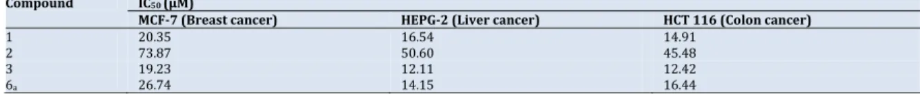
A series of new thiazolidin‐4‐ones have been synthesized by the reaction of 3‐acetylindole with thiourea to yield 2‐amino‐arylthiazole (1) which, reacted with 2‐chloroacetyl chloride to produce 2‐chloroacetamido‐4‐arylthiazoles (2). The later was treated with potassium thiocyanate to afford the related 2‐amino‐3‐(4‐arylthiazol‐2‐yl) thiazolidin‐4‐ones (3). Condensation of compounds 1 and 3 with different aromatic aldehydes give Schiff’s bases (4a‐ c) and (5a‐c) reaction of compound 5a‐c with thioglycollic acids furnishes the target thiazolidin‐4‐one molecules (6a‐c). Further, condensation of compound 6a with benzaldehyde affords benzylidenethiazolo derivative (7) which on refluxing with malononitrile, acetylacetone afforded thiazolopyridine derivatives (8,9). Structure elucidation of the products has been accomplished on the basis of elemental analysis, IR, 1H NMR data.
Compound 3exhibited the most potent antibacterial and anticancer activity.
KEYWORDS Anticancer Schiff’s bases 3‐Acetylindol Antimicrobial 1,3‐Thiazolidin‐4‐ones
Thiazolopyridine derivatives Citethis:Eur.J.Chem.2015, 6(2), 157‐162
1.Introduction
Thiazolidinones are derivatives of thiazolidine and they also constitute an important group of heterocyclic compounds
[1]. Thiazolidin‐4‐one derivatives exhibit various biological
activities such as anti‐microbial [2], anti‐inflammatory [3,4],
antihistaminic [5], anti‐hypertensive, analgesic [6] and anti‐
bacterial activities [7‐9].
Thiazolidinones, with a carbonyl group in position 2, 4‐ or
just 4‐, have been extensively studied [10,11] and literature
surveys showed that thiazolidin‐4‐ones are important compounds due to their broad range of biological activities
[12‐18]. 2‐Substituted 4‐thiazolidinones derivatives exhibit
unusually high activity against mycobacterium tuberculosis
when tested in vitro [19]. Overviews of their synthesis,
properties, reactions and applications have been published [10,11].
Indoles have been reported to possess a wide variety of
biological activities like anti‐inflammatory [20], anticancer
[21], antifungal [22] and were used in the treatment of
gastrointestinal, cardiovascular and central nervous system (CNS) disorders, HIV‐1 integrase inhibitors for antitumor activity, inhibitors of hepatitis, as antibacterial and as anti‐
malarial agents [23‐28]. Therefore, the aim of the present
work was to prepare thiazolidin‐4‐one‐derivatives using 3‐ acetylindol in order to find out new biologically active compounds.
2.Experimental
2.1.Instrumentation
All melting points are uncorrected and were determined on GallenKamp electric melting point apparatus. IR spectra (KBr discs) were recorded on of FT/IR‐400 spectrophoto‐
meter (Perkin Elmer). 1HNMR spectra were recorded on
ovarian 300 MHz (DMSO‐d6) solutions. Chemical shifts were
reported as δ values relative to tetramethylsilan (TMS) as internal reference. The analyses were carried out at Micro‐ Analytical Center, Cairo University.
2.2.Synthesisof4‐(1H‐indol‐3‐yl)thiazol‐2‐amine(1) 3‐Acetylindole (0.05 mole) and thiourea (0.05 mole) were taken in round bottom flask and dissolved in propanol (35 mL) and refluxed for 2 h. The solid obtained was triturated with ethanol to remove unreacted acetylindole. To this pyridine (5 mL) was added continued refluxed for 5 h.
Scheme1
Scheme2
The reaction completion was monitored by TLC. The solid
separated is filtered recrystallized from ethanol, 1(Scheme 1).
Color: Pale yellow. Yield: 69%. M.p.: 158‐160 °C. FT‐IR (KBr, ,
cm‐1): 3340 (N‐H), 3012 (Ar‐CH), 1582 (C=N), 1457 (C=C). 1H
NMR (300 MHz, DMSO‐d6, δ, ppm): 4.32 (s, 2H, NH2), 7.08 (s,
1H, thiazole‐H), 7.10‐7.60 (m, 5H, Ar‐H), 14.01 (s, 1H, NH).
Anal. calcd. for C11H9N3S: C, 61.37; H, 4.21; N, 19.52. Found: C,
61.31; H, 4.20; N, 19.49%.
2.3.SynthesisofN‐(4‐(1H‐indol‐3‐yl)thiazol‐2‐yl)‐2‐chloro acetamide(2)
In conical flask, 0.01 mole of 2‐amino‐4‐aryl thiazole (1) in
25 mL benzene was stirred for 30 min in ice‐bath till the temperature becomes below 0‐5 °C then add 0.01 mole chloroacetyle chloride was add drop by drop in conical flask within 2h. After complete addition the reaction mixture was refluxed it for2h in water bath then the solvent was evaporates. The product that separated was recrystallization
from ethanol (Scheme 2). Color: Pale yellow. Yield: 82%. M.p.:
145‐147 C. FT‐IR (KBr, , cm‐1): 3160 (NH), 3012 (Ar‐H),1633
(C=O). 1H NMR (300 MHz, DMSO‐d6, δ, ppm): 4.22 (s, 2H,
COCH2), 7.14‐7.55 (m, 6H, Ar‐H), 14.00 (s, 1H, NH). Anal. calcd.
for C13H10ClN3OS: C, 53.52; H, 3.45; N, 14.40. Found: C, 53.50;
H, 3.41; N, 14.39%.
2.4.Synthesisof3‐(4‐(1H‐indol‐3‐yl)thiazol‐2‐yl)‐2‐imino thiazolidin‐4‐one(3)
A mixture of 2‐chloro acetamido‐4‐aryl thiazole (2) (0.03
mole), KSCN (0.06 mole) in dry acetone (100 mL) was refluxed for 3 h. The reaction mixture was kept for evaporated under vacuum to obtain crude product. The residue was stirred with water. The solid product was filtered, washed with water,
dried and recrystallized with ethanol (Scheme 2). Color: Pale
brown. Yield: 93%. M.p.: 162‐164 °C. FT‐IR (KBr, , cm‐1):
3497 (=NH), 3197 (Ar‐H), 1671 (C=O), 1602 (C=N), 705 (C‐S).
1H NMR (300 MHz, DMSO‐d6, δ, ppm): 3.80 (s, 1H, =NH), 4.10
(s, 2H, CH2), 7.12‐8.40 (m, 6H, Ar‐H), 11.9 (bs, 1H, NH). Anal.
calcd. for C14H10N4OS2: C, 53.49; H, 3.21; N, 17.82. Found: C,
53.45; H, 3.19; N, 17.80%.
2.5.Generalprocedureforpreparation3‐(4‐(1‐indol‐3‐yl) thiazol‐2‐yl)‐5‐benzylidene‐2‐iminothiazolidine‐4‐one(4a‐
c)
A mixture iminothiazolidin‐4‐one (3) (1 mmol), aldehyde
and sodium acetate (1.5 mmol) in glacial acetic acid was refluxed for 2 to 4 h. Till completion of the reaction (TLC check), the reaction mixture was poured onto ice‐cold water the solid thus separated was filtered and the crude product was recrystallized using absolute ethanol to get compounds
4a‐c(Scheme 2).
3‐(4‐(1H‐Indol‐3‐yl)thiazol‐2‐yl)‐5‐benzylidene‐2‐iminothia
zolidin‐4‐one(4a): Color: Brown. Yield: 94%. M.p.: 220‐223 °C.
FT‐IR (KBr, , cm‐1): 3488 (NH), 1700 (C=O), 1640 (C=CH‐),
1558 (C=N). 1H NMR (300 MHz, DMSO‐d6, δ, ppm): 3.9 (s, 1H,
=NH), 7.24‐7.37 (d, 1H, ‐C=CH), 7.38‐8.19 (m, 11H, Ar‐H),
10.16 (br, 1H, exchangeable with D2O). Anal. calcd. for
C21H14N4OS2: C, 62.67; H, 3.51; N, 13.92. Found: C, 62.64; H,
3.42; N, 13.80%.
3‐(4‐(1H‐Indol‐3‐yl)thiazol‐2‐yl)‐2‐imino‐5‐(4‐methoxy
benzylidene)thiazolidin‐4‐one(4b): Color: Brown. Yield: 96%.
M.p.: 210‐212 °C. FT‐IR (KBr, , cm‐1): 3497 (NH), 1670 (C=O),
1660 (‐C=CH‐), 1570 (C=N). 1H NMR (300 MHz, DMSO‐d6, δ,
ppm): 3.8 (s, 3H, ‐CH3), 7.24‐7.27 (d, 1H, ‐C=CH‐), 7.38‐8.41 (m,
10H, Ar‐H), 13.90 (s, 1H, NH).
3‐(4‐(1H‐indol‐3‐yl)thiazol‐2‐yl)‐5‐(4‐chlorobenzylidene)‐2‐
iminothiazolidin‐4‐one (4c): Color: Pale yellow. Yield: 94%.
M.p.: 200‐204 °C. FT‐IR (KBr, , cm‐1): 3460 (NH), 1680 (C=O),
1560 (‐C=CH‐). 1H NMR (300 MHz, DMSO‐d6, δ, ppm): 3.9 (s,
1H, =NH), 7.24‐7.37 (d, 1H, ‐C=CH‐), 7.38‐8.41 (m, 10H, Ar‐H), 14.02 (s, 1H, NH).
Scheme3 2.6.GeneralprocedureforthepreparationofN‐(substitute benzylidene)‐4‐(1H‐indol‐3‐yl)thiazol‐2‐amine(5a‐c)
To an equimolar methanolic solution of 2‐amino‐4‐aryl thiazole (0.1 mol) and substituted benzaldehyde (0.1 mol), a few drops of glacial acetic acid were added. The mixture was then refluxed on water bath 5‐6 h. It was then allowed to cool, poured into crushed ice and recrystallized from methanol (Scheme 3).
N‐(Benzylidene)‐4‐(1H‐indol‐3‐yl) thiazol‐2‐amine (5a):
Color: Yellow. Yield: 82%. M.p.: 166‐168 °C. FT‐IR (KBr, , cm‐
1): 3400 (N‐H), 1620 (‐N=CH‐). 1H NMR (300 MHz, DMSO‐d6, δ,
ppm): 7.0‐7.6 (m, 11H, Ar‐H), 8.1 (s, 1H, ‐N=CH), 10.1 (s, 1H,
NH). Anal. calcd. for C18H13N3S (303.08): C, 71.26; H, 4.32; N,
13.85. Found: C, 71.22; H, 4.31; N, 13.82%.
N‐(4‐Chlorobenzylidene)‐4‐(1H‐indol‐3‐yl)thiazol‐2‐amine
(5b): Color: Pale yellow. Yield: 85%. M.p.: 160‐165 °C. FT‐IR
(KBr, , cm‐1): 3310 (N‐H), 1600 (‐N=H). 1H NMR (300 MHz,
DMSO‐d6, δ, ppm): 7.12‐7.68 (m, 10H, Ar‐H), 8.20 (s, 1H, ‐
N=CH), 10.10 (s, 1H, NH). Anal. calcd. for C18H12ClN3S: C, 64.00;
H, 3.58; N,12.44. Found: C, 64.12; H, 3.52; N, 12.39%.
N‐(4‐Methoxybenzylidene)‐4‐(1H‐indol‐3‐yl)thiazol‐2‐amin
(5c): Color: Yellow. Yield: 84%. M.p.: 170‐172 °C. FT‐IR (KBr, ,
cm‐1): 3390 (NH), 2920 (Ar‐CH), 2840 (Aliph‐CH), 1600
(‐N=CH). 1H NMR (300 MHz, DMSO‐d6, δ, ppm): 3.53 (s, 3H,
‐OCH3), 6.87‐7.61 (m, 10H, Ar‐H), 8.01 (s, 1H, N=CH), 10.10 (s,
1H, NH). Anal. calcd. for C19H15N3OS (333.41): C, 68.45; H, 4.53;
N, 12.60. Found: C, 64.42; H, 4.52; N, 12.59%.
2.7.Generalprocedureforthepreparationofthe3‐(4‐1H‐
indol‐3‐yl)thiazol‐2‐yl)‐2‐(4‐substitutedphenyl)thiazolidin‐
4‐one(6a‐c)
A mixture of Schiff's base (5a‐c) (0.01 mol) and
thioglycolic acid (0.01 mol) was refluxed in dimethyl formamide (15 mL) for 6 h. The reaction mixture was cooled and poured into crushed ice. The solid obtained was filtered
and recrystallized from ethanol to give compounds 6a‐c
(Scheme 3).
3‐(4‐(1H‐Indol‐3‐yl)thiazol‐2‐yl)‐2‐phenyl thiazolidin‐4‐one
(6a): Color: Pale brown. Yield: 82%. M.p.: 200‐204 °C. FT‐IR
(KBr, , cm‐1): 3225 (NH), 1700 (C=O), 1631(C=N), 1583 (C=C).
1H NMR (300 MHz, DMSO‐d6, δ, ppm): 4.00 (s, 2H, COCH2S), 5.9
(s, 1H, ‐NCHS), 6.6‐7.6 (m, 11H, Ar‐H), 10.1 (s, 1H, NH). Anal.
calcd. for C20H15N3OS2: C, 63.64; H, 4.01; N, 11.13. Found: C,
63.62; H, 4.00; N, 11.10%.
3‐(4‐(1H‐Indol‐3‐yl)thiazol‐2‐yl)‐2‐(4‐chlorophenyl)thiazo
lidin‐4‐one(6b): Color: Pale brown. Yield: 84%. M.p.: 210‐212
°C. FT‐IR (KBr, , cm‐1): 3300 (NH), 1700 (C=O), 1600 (‐N=C‐).
1H NMR (300 MHz, DMSO‐d6, δ, ppm): 3.83 (s, 2H, COCH2S), 5.8
(s, 1H, ‐NCHS), 7.1‐7.8 (m, 10H, Ar‐H), 10.1 (s, 1H, NH). Anal.
calcd. for C20H14ClN3OS2: C, 58.31; H, 3.43; N, 10.20. Found: C,
58.29; H, 3.40; N, 10.19%.
3‐(4‐(1H‐Indol‐3‐yl)thiazol‐2‐yl)‐2‐(4‐methoxyphenyl)thia
zolidin‐4‐one(6c): Color: Brown. Yield: 84%. M.p.: 205‐210 °C.
FT‐IR (KBr, , cm‐1): 3290 (NH), 2920 (Ar‐CH), 2840 (Aliph‐
CH), 1690 (C=O), 1600 (‐N=C). 1H NMR (300 MHz, DMSO‐d6, δ,
ppm): 3.4 (s, 2H, COCH2S), 3.70 (s, 3H, ‐OCH3), 5.90 (s, 1H,
NCHS), 6.60‐7.90 (m, 10H, Ar‐H), 10.10 (s, 1H, NH). Anal. calcd.
for C21H17N3O2S2: C, 61.89; H, 4.20; N, 10.31. Found: C, 61.88; H,
4.19; N, 10.30%.
2.8.Synthesisof3‐(4‐(1H‐Indol‐3‐yl)thiazol‐2yl)‐5‐
benzylidiene‐2‐phenylthiazolidin‐4‐one(7)
A mixture of compound 6a(0.01 mole) and benzaldehyde
(0.01 mole) was refluxed in absolute ethanol (30 mL) and catalyzed with few drops of TEA for 5 h. After cooling the obtained solid was filtered, washed, dried and recrystallized
from ethanol (Scheme 4). Color: Brown. Yield: 89%. M.p.: 220‐
222 °C. FT‐IR (KBr, , cm‐1): 3420 (NH), 3040 (Ar‐CH), 1700
(C=O), 1600 (C=C). 1H NMR (300 MHz, DMSO‐d6, δ, ppm): 5.90
(s, 1H, NCHS), 6.10 (s, 1H, ‐CH‐), 7.10‐7.90 (m, 16H, Ar‐H),
10.40 (s, 1H, NH). Anal. calcd. for C27H19N3OS2: C, 69.65; H,
4.11; N, 9.03. Found: C, 69.64; H, 4.10; N, 9.04 %.
2.9.Synthesisof3‐(4‐(1H‐indol‐3‐yl)thiazol‐2‐yl)‐5‐amino‐
2,3‐dihydro‐2,7‐diphenylthiazolo[4,5‐b]pyridine‐6‐
carbonitrile(8)
A mixture of compound 7 (0.01 mole), malononitrile (0.01
mole) and ammonium acetate (1 g) in 30 mL acetic acid was refluxed for 3 h. The solid formed upon cooling was collected by filtration, washed with water and recrystallized from
ethanol (Scheme 4). Color: Pale orange. Yield: 72%. M.p.: 256‐
258 °C. FT‐IR (KBr, , cm‐1): 3410, 3285 (NH, NH2), 3040 (Ar‐
CH),2200 (CN), 1600 (C=C). 1H NMR (300 MHz, DMSO‐d6, δ,
ppm): 4.00 (s, 2H, NH2), 4.90 (s, 1H, ‐NCHS), 7.00‐7.90 (m, 16H,
Ar‐H), 10.01 (s, 1H, NH). Anal. calcd. for C30H20N6S2: C, 68.16;
H, 3.81; N, 15.90. Found: C, 68.14; H, 3.80; N, 15.89%. 2.10.Synthesisof1‐(3‐(4‐(1H‐indol‐3‐yl)thiazol‐2‐yl)‐2,3‐
dihydro‐5‐methyl‐2,7‐Diphenylthiazolo[4,5‐b]pyridine‐6‐
yl)ethanone(9)
A mixture of compound 7 (0.01 mole), acetylacetone (0.01
mole) and ammonium acetate (1 g) in 30 mL acetic acid was refluxed for 3 h. The solid formed upon cooling was collected by filtration, washed with water and recrystallized from
ethanol (Scheme 4). Color: Pale brown. Yield: 64%. M.p.: 200‐
204 °C. FT‐IR (KBr, , cm‐1): 3410 (NH), 3040 (Ar‐CH), 2840
(Aliph‐CH),1660 (CO), 1595 (C=C). 1H NMR (300 MHz, DMSO‐
d6, δ, ppm): 3.20 (s, 3H, COCH3), 3.80 (s, 3H, ‐N=C‐CH3), 4.95 (s,
1H, ‐NCHS), 6.90‐7.90 (m, 16H, Ar‐H), 10.20 (s, 1H, NH). Anal.
calcd. for C32H24N4OS2: C, 70.56; H, 4.44; N, 10.29. Found: C,
70.50; H, 4.41; N, 10.27%. 2.11.Antimicrobialactivity
Antimicrobial activity of the tested compounds was determined using a modified Kirby‐Bauer disc diffusion
method [29]. Briefly, 100 µL of the test bacteria/fungi were
grown in 10 mL of fresh media until they reached a count of approximately108 cells/mL for bacteria 105 cells/mL for fungi
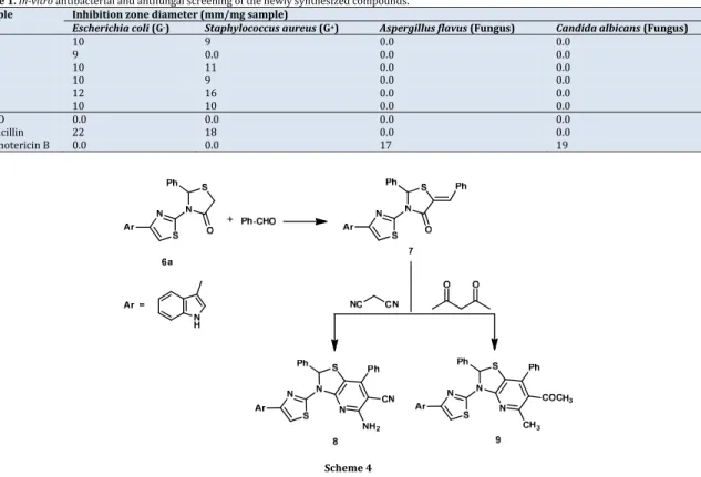
Table1.In‐vitro antibacterial and antifungal screening of the newly synthesized compounds.
Sample Inhibitionzonediameter(mm/mgsample)
Escherichiacoli(G‐) Staphylococcusaureus(G+) Aspergillusflavus(Fungus) Candidaalbicans(Fungus)
1 10 9 0.0 0.0 2 9 0.0 0.0 0.0 3 10 11 0.0 0.0 6a 10 9 0.0 0.0 6b 12 16 0.0 0.0 6c 10 10 0.0 0.0 DMSO 0.0 0.0 0.0 0.0 Ampicillin 22 18 0.0 0.0 Amphotericin B 0.0 0.0 17 19 Scheme4
100 µL of microbial suspension was spread onto agar plates corresponding to the broth in which they were maintained. Isolated colonies of each organism that might be playing a pathogenic role should be selected from primary agar plates and tested for susceptibility by disc diffusion
method [31‐33]. Of the many media available, National
Committee for Clinical Laboratory Standards (NCCLS) recommends Mueller‐Hinton agar due to itsgood results in batch‐to‐batch reproducibility. Disc diffusion method for filamentous fungi tested by using approved standard method
(M38‐A) [34] for evaluating the susceptibilities of filamentous
fungi to antifungal agents. Disc method for yeasts developed
by using approved standard method (M44‐P) [35]. Plates
inoculated with filamentous fungi as Aspergillusflavus at 25 °C
for 48 h; Gram (+) bacteria as Staphylococcusaureus; Gram (‐)
bacteria as Escherichiacoli, they were incubated at 35‐37 °C
for 24‐48 h and yeast as Candidaalbicans incubated at 3 °C for
24‐48 h and, then the diameters of the inhibition zones were
measured in millimeters [27]. Standard discs of Ampicillin
(Antibacterial agent), Amphotericin B (Antifungal agent) served as positive controls for antimicrobial activity but filter discs impregnated with 10 µL of solvent (Distilled water, chloroform, DMSO) were used as a negative control. The agar used is Meuller‐Hinton agar that is rigorously tested for composition and pH. Furthermore, the depth of the agar in the plate is a factor to be considered in the disc diffusion method. This method is well documented and standard. A zone of inhibition has been determined for susceptible and resistant values. Blank paper disks (Schleicher & Schuell, Spain) with a diameter of 8.0 mm were impregnated with 10 µL of tested concentration of the stock solutions. When a filter paper disc impregnated with a tested chemical is placed on agar the chemical will diffuse from the disc into the agar. This diffusion will place the chemical in the agar only around the disc. The solubility of the chemical and its molecular size will determine the size of the area of chemical infiltration around the disc. If an organism is placed on the agar it will not grow in the area
around the disc if it is susceptible to the chemical. This area of
no growth around the disc is known as a “Zoneofinhibition”
or" Clearzone”. For the disc diffusion, the zone diameters were
measured with slipping calipers of the National Committee for
Clinical Laboratory Standards [31]. Agar‐based methods such
as test and disk diffusion can be good alternatives because
they are simpler and faster than broth‐based methods [35‐37]
(Table 1).
2.12.Anticanceractivity 2.12.1.Cellculture
The cells were obtained from Egyptian Holding Company for Biological Products & Vaccines (VACSERA), Giza, Egypt and then maintained in the tissue culture unit .The cells were grown in RBMI‐1640 medium, supplemented with 10% heat inactivated FBS, 50 units/mL of penicillin and 50 mg/mL of
streptomycin and maintained at [37] in a humidified
atmosphere containing 5% CO2. The cells were maintained as
monolayer culture by serial sub‐culturing. Cell culture reagents were obtained from Lonza (Basel, Switzerland). The anticancer activity of the rested compounds was evaluated against MCF‐7 cells (Breast cancer), HEPG‐2 cells (Liver
cancer) and HCT 116 (Colon cancer) (Table 2).
2.12.2.ThesulforhodamineB(SRB)cytotoxicityassay Cytotoxicity was determined using the sulforhodamine B
(SRB) assay method as previously described by Skehan etal.
[35]. Exponentially growing cells were collected using 0.25%
Trypsin‐EDTA and seeded in 96‐well plates at 1000‐2000 cells/well in RBMI‐1640 supplemented medium. After 24 h, cells were incubated for 72 h with various concentrations of the tested compounds. Following 72 h treatments, the cells would be fixed with 10% trichloroacetic acid for 1 h at 4 °C.
Table2.In‐vitro anticancer screening of the newly synthesized compounds against different cell lines.
Compound IC50(µM)
MCF‐7(Breastcancer) HEPG‐2(Livercancer) HCT116(Coloncancer)
1 20.35 16.54 14.91
2 73.87 50.60 45.48
3 19.23 12.11 12.42
6a 26.74 14.15 16.44
Wells were stained for 10 minutes at room temperature with 0.4% SRBC (Sulphorhodamine B) dissolved in 1% acetic acid. The plates were air dried for 24 h and the dye was solubilized with Tris‐HCl for 5 min on a shaker at 1600 rpm. The optical density (OD) of each well was measured spectrophotometrically at 564 nm with an ELISA microplate
reader (ChroMate‐4300, FL, USA). The IC50 values were
calculated according to the equation for Boltzman sigmoidal concentration response curve using the nonlinear regression fitting models (Graph Pad, Prism Version 5)
3.Resultsanddiscussion
3.1.Synthesis
Our synthetic strategy for 1, 3‐thiazolidin‐4‐one deriva‐ tives starts with refluxing of 3‐ acetyl indol and thiourea in
propanol to afforded 4(1H‐indol‐3‐yl) thiazol‐2‐amine (1)
which formed by the attack of sulphur nucleophile on imine carbon followed by intermolecular cyclization on elimination
of water, Scheme 1.
Chloroacetamide (2) was obtained by reacting of 2‐amino
thiazoles (1) with chloroacetyl chloride in presence of
pyridine. The IR of compound 2showed the presence of bands
characteristic for C=N at 1631‐1642 cm‐1 and an amide
function at 1661 (C=O) and 3160‐3184 cm‐1 (NH). The 1H NMR
of compound 2 revealed a broad singlet at δ 10.32 ppm
characteristic for a secondary amine group, a multiplet at δ
7.14‐7.55 ppm for aromatic protons and COCH2 as a singlet at
δ 4.22 ppm.
When chloroacetamide (2) was refluxed with potassium
thiocyanate in dry acetone, 2‐iminothiazolidine‐4‐one (3) was
obtained in a moderate to good yields. The structures of the isolated compounds were determined by spectral methods.
The IR of compound 3 revealed characteristic bands for C=N at
1602cm‐1, C=O at 1671cm‐1, primary and secondary amines at
3224‐3278cm‐1and 3166‐3178 cm‐1. The 1H NMR spectra
showed the presence of broad exchangeable singlets at δ 11.90
ppm NH protons, while O=C‐CH2 protons appears at δ 4.1, 4.3
ppm for C=NH and the aromatic protons of compound 3appear
as a multiplet at δ 7.12‐8.57 ppm.
The iminothiazolidine‐4‐one (3) on Knoevenagel conden‐
sation with different substituted aryl aldehydes in presence of sodium acetate as a base in glacial acetic acid afforded the 3‐ (4‐(1‐indol‐3‐yl)thiazol‐2‐yl)‐ 5‐benzylidene‐2‐iminothiazoli
dine‐4‐one (4a‐c) which established on the basis of IR, 1H NMR
spectra did not only show the absence of CH2 protons at δ 4.1
ppm, but also the presence of HC=C proton at δ 7.24‐7.37 ppm, Scheme 2.
New compounds (5a‐c)were prepared by refluxing 2‐
amino‐4‐aryl thiazole (1) and substituted benzaldehyde, a few
drops of glacial acetic acid in methanol for 5‐6 h. These Schiff’s
base (5a‐c) and thioglycolic acid (0.01 mol) when refluxed in
dimethyl formamide (15 mL) for 6 h afforded the 3‐(4‐1H‐ indol‐3‐yl)thiazol‐2‐yl)‐2‐(4‐substituted phenyl)thiazolidin‐4‐
one (6a‐c) in good yield which determined by IR and 1H NMR
spectra. IR spectra showed the characteristic bands for C=N at
1630‐1600 cm‐1 and amide function at 1690‐1700 (C=O) and
3255‐3300 cm‐1 (NH). The 1H NMR of showed the presence of
one broad exchangeable singlet at δ 10.1 ppm (NH), a multiplet at δ 7.1‐7.8 ppm (Ar‐H's), a singlet at δ 5.8 ppm
(thiazolyl‐C5‐H), and a singlet at δ 3.83 ppm (O=C‐CH2),
Scheme 3.
Condensation of compound (6a) with benzaldehyde
affords 5‐benzylidenethiazolo derivatives (7), later, refluxing
of compound 7 with malononitrile in the presence of
ammonium acetate resulted in cycloaddition affording
thiazolopyridine derivative (8) which showed IR spectra at
3410, 3285 (NH, NH2) and 2200 cm‐1 (CN). The 1H NMR
spectra showed the presence of broad exchangeable singlets at δ 10.01 ppm NH protons, while NCHS protons appears at δ 4.9 ppm and the aromatic protons appears as a multiplet at δ 7.00‐ 7.90 ppm.
Also, thiazolopyridine derivative (9) was obtained by
using acetylacetonein the same manner the structure was
confirmed by 1H NMR spectra showed signal at δ 3.20 ppm for
OCH3, δ 3.80 ppm for ‐N=C‐CH3, δ 4.95 ppm ‐NCHS and singlets
at δ 10.20 ppm for NH. The structure of all newly isolated compounds was fully confirmed by spectral and elemental
analyses methods, Scheme 4.
3.2.Antibacterialactivity
All the tested compounds have antibacterial activity
against Gram negative (Escherichiacoli) and Gram positive
(Staphylococcusaureus), bacteria except compound 2 which
had antibacterial activity against Gram negative bacteria only.
Compound 6bshowed the highest antibacterial activity. All the
tested compounds had no antifungal activity against
Aspergillus flavus and Candida albicans. The antibacterial
activity of the newly synthesized compounds is due to the
presence of thiazolidine ring [6,7].
All the synthesized compounds were tested for in vitro anticancer activity using SRB cytotoxicity assay method. The anticancer screening of the tested compounds revealed that all the synthesized compounds exhibited a significant cytotoxic activity against MCF‐7 Breast cancer, HEPG‐2 Liver cancer and HCT116 Colon cancer cell lines in variable degrees. The anticancer activity of these newly synthesized compounds may be due to the presence of indoles. Previous studies revealed that indoles possess a high anticancer activity against different
cell lines [19].
From Table 2, we can notice that compound 3 exhibited
the most potent anticancer activity against MCF‐7 Breast cancer, HEPG‐2 Liver cancer and HCT116 Colon cancer cell
lines by IC50 19.23, 12.11 and 12.42 µM, respectively, while
compound 2 showed the least anticancer activity.
4.Conclusion
Our present investigation is centered on the studies of synthesis, reactions, spectral analysis and biological activities of 1,3‐thiazolidin‐4‐ones derivatives. The procedure proved more beneficial than those previously reported in the
literature. Compound 3 exhibited the most potent antibacterial
and anticancer activity. All the compounds had no antifungal
activity against Aspergillusflavus and Candidaalbicans.
References
[1]. Agarwal, O. P. Textbook of Organic Chemistry, 4th edition, Goel
Publisher, p730, 2006.
[3]. Mahd, A. K.; Zaman, M. S. IndianJ.Chem.B. 2004, 43,2189‐2194.
[4]. Verma, L.; Wakode, S. PharmaInnov.J.2013, 2,41‐50.
[5]. Raghuram, R. A.; Rajan, K. S.; Devsingh, S.; Bhagavan, R. M. IndianJ.
Chem.B2001, 40, 813‐816 .
[6]. Reddy, P. S. N.; Manmahan, R.; Pradap, R. P. IndianJ.Chem.B2003,
40,2119‐2120.
[7]. Mulwad, W.; Choudhari, B. Indian.J.Chem.B2005, 42,1074‐1078.
[8]. Mhogale, V. A. IndianJ.Chem.Sci.1990, 102(4),535‐540.
[9]. Sharshira, E. M.; Hamada, N. M. M. Am.J.Org.Chem. 2012, 2(3), 69‐
73.
[10]. Brown, F. C. Chem.Rev. 1961, 61,463‐463.
[11]. Singh, S. P.; Parmar, S. S.; Raman, K.; Stenberg, V. I. Chjem.Rev. 1981,
61,175‐203.
[12]. Doran, W. J.; Shonle, H. A. J.Org.Chem. 1938, 3,193‐197.
[13]. Troutman, H. D.; Long, L. M. J.Am.Chem.Soc.1948, 70,3436‐3439.
[14]. Rout, M. K.; Mahapatra; G. N. J.Am.Chem.Soc.1955, 77,2427‐2428.
[15]. Gaikwad, N. J.; Trupude, R. N.IndianDrugs1994, 31,593‐594.
[16]. El‐Gendy, Z.; Abdel‐Rahman, R. M.; Fawzy, M. M; Mahmoud, M. B. J.
Ind.Chem.Soc.1994, 67,927‐929.
[17]. Diumo, M. V.; Mazzoni, O.; Piscopo, E.; Calignano, A.; Giordano, F.;
Bolognese, A. J.Med.Chem.1992, 35,2190‐2912.
[18]. Shah, V.; Pant, C. K.; Joshi, P. C. AsianJ.Chem. 1993, 5,83‐88.
[19]. Mukhatar, S.; Majeebar, R. V. P.; Ansari, W. H.; Lemiere, G.; De Groot,
A.; Dommisse, R. J.Ind.Chem.Soc. 1999, 4,232‐237.
[20]. Radwan, M. A. A.; Ragab, E. A.; Nermien, M. Bioorg.Med.Chem. 2007,
15,3832‐3841.
[21]. Singh, P.; Mittal, A.; Kaur, S. Bioorg.Med.Chem.Lett. 2008, 18,85‐89.
[22]. Ryu, C. K. K.; Lee, J. Y.; Park, R.; Ma, N. Bioorg.Med.Chem.Lett. 2007,
5,127‐31.
[23]. Liu, K.; Sun, J.; Lou, H.; Dai, S. , Xu, H. Phytochem.2008, 69, 1405‐
1410.
[24]. Abdel‐Rahman, A. A. H.; Abdel‐Megied, A. E. S.; Hawata, M. A. M.;
Kasem, E. R.; Shaaban, M. T. Monatsh.Chem. 2007, 138,889‐897.
[25]. Geroge, S.; Waran, M. P.; Chakraborty, A.; Ravi, T. K. Acta.Pharm.
2008, 58,119‐129.
[26]. Metawally, M. A.; Shaaban, S.; Abdel‐Wahab, B. F.; Gamal, A. Curr. Org.
Chem. 2009, 13,1475‐1495.
[27]. Winter, C. A.; Risley, E. A.; Nuss, G. W. Soc.Exp.Biol.Med.1962, 111,
544‐547.
[28]. Loveleen, V.; Sharad, W. PharmaInnov.J.2013, 2(5), 41‐50
[29]. Bauer, A. W.; Kirby, W. M.; Sherris, C.; Turck, M. Am.J.Clin.Path.
1966, 45,493‐496.
[30]. Pfaller, M. A.; Burmeister, L.; Bartlett, M. A.; Rinaldi, M. G. J.Clin.
Microbiol.1988, 26, 1437‐1441.
[31]. National Committee for Clinical Laboratory Standards. Performance
Vol. 41, Antimicrobial susceptibility of Flavobacteria, 1997.
[32]. National Committee for Clinical Laboratory Standards. Methods for
dilutionantimicrobial susceptibility tests for bacteria that grow aerobically. Approved standard M7‐A3. National Committee for Clinical Laboratory Standards, Villanova, PA, USA, 1997.
[33]. National Committee for Clinical Laboratory Standards, Reference
Methodfor Broth Dilution Antifungal Susceptibility Testing of Conidium‐Forming Filamentous Fungi: Proposed Standard M38‐A, NCCLS, Wayne, PA, USA, 2002.
[34]. National Committee for Clinical Laboratory Standards, Method
forAntifungal Disk Diffusion Susceptibility Testing of Yeast: Proposed Guideline M44‐P, NCCLS, Wayne, PA, USA, 2003.
[35]. Liebowitz, L. D.; Ashbee, H. R.; Evans, E. G. V.; Chong, Y.; Mallatova, N.;
Zaidi, M.; Gibbs, M. Diagn.Microbiol.Infect.Dis. 2002, 4, 27‐33.
[36]. Matar, M. J.; Ostrosky‐Zeichner, L.; Paetznick, V. L.; Rodriguez, J. R.;
Chen, E.; Rex, J. H. Antimicrob. Agents Chemother. 2003, 47, 1647‐
1651.
[37]. Skhen, P.; Storeng, R.; Scudiero, D.; Monks, A.; McMahon, J.; Vistica, D.;
Warren, J. T.; Bokesch, H.; Kenney, S.; Oyd, M. R. J.Nat.CancerInst.