STIMULATION AND SUBSTITUTION
OF BONE ALLOGRAFT AROUND
NON-CEMENTED IMPLANTS
Ph.D. thesis
Thomas Bo Jensen
Faculty of Health Sciences
University of Aarhus
Abbreviations ...4
Abstract ...6
Introduction...7
Background ...8
Grafting in reconstructive surgery of the hip ...8
Biomechanics of impacted morselized bone allograft ...9
Incorporation of impacted bone allografts ...10
Risks and disadvantages associated with the use of bone graft ...9
Bone graft substitutes based on osteoconductivity ...15
Bone growth factors and implant healing ...12
Enhancement of bone grafts and osteoconductive bone substitutes ...15
Materials and methodological conciderations...17
Ethical conciderations ...17 Animals ...17 Observation time ...17 Implants...17 Experimental model ...17 Design ...18 Grafting materials ...18 Mechanical evaluation ...20 Histomorphometry ...21 Reproducibility...21 Statistics ...22 Results...22 Exclusions ...22 Study II...23 Study III ...25 Study V ...26 Discussion ...27
This thesis is based on the following papers and manuscripts:
I: Jensen-TB, Overgaard-S, Rahbek-O, Lind-M, Bünger-C, Søballe-K. Osteogenic Protein-1
Device Increases Bone Graft Resorption and Bone Formation Around Cementless Implants. Acta Scandinavica Orthopaedica,2002, 73(1) p31-39
II: Jensen-TB, Overgaard-S, Rahbek-O, Lind-M, Bünger-C, Søballe-K. Osteogenic Protein-1
Device Increases Fixation of Implants Grafted with Morsellized Bone Allgraft and ProOsteon. An experimental study in dogs. Resubmitted J. Bone Joint Surg. (Br), 2002
III: Jensen-TB, Overgaard-S, Rahbek-O, Lind-M, Bünger-C, Søballe-K. The influence of OP-1 d
Device on Mechanical Properties of Impacted Morsellized Bone Allograft after 3 Weeks. Manuscript 2003
IV: Jensen-TB, Overgaard-S, Rahbek-O, Søballe-K, The Influence of Platelet Rich Plasma on
Osteointegration of Fresh Frozen and Processed Bone Allograft. Submitted J. Arthro. 2002
V: Jensen-TB, Overgaard-S, Rahbek-O, Søballe-K, Does Platelet Rich Plasma Influence Fixation
Abbreviations
AGF: Autologous growth factor
BMP: Bone morphogenic protein
EGF: Epidermal growth factor
FGF: Fibroblast growth factor
HA: Hydroxyapatite
IGF: Insulinlike growth factor
OP-1: Osteogenic protein-1
PC: Platelet concentrate
PDGF: Platelet derived growth factor
PRP: Platelet rich plasma
TCP: Tricalciumphosphate
THA: Total hip arthroplasties
Ti-6Al-4V: Titanium-6 aluminium-4
vanadium
Definitions
Bioactive materials: Materials which elicit or
modulate biological activity
Biocompatible: The ability of a material to
perform with an appropriate host in a specific application
Cytokine: Poly peptide regulator of
cell-to-cell interaction in relation to the immunological system
Graft:
-autograft:Bone tissue harvested from and implanted in the same individual
-allograft: Bone tissue harvested from one individual and implanted in another individual, same species
-Xenograf: Harvested from one species and implanted in another
Growth factors: Polypetides which act as
signalling agents for cells
Histomorphometry: Quantitative evaluation
of tissue
Implant: A device made of a biomaterial that
is intentionally placed in the body covered totally or partly by an epithelial surface
Implant in- or ongrowth: Bone corverage of
the implant surface in percentage
Bone incorporation: Integration of a
biomaterial by bone
Osteoconduction: A process where tissue
involved in bone formation is lead conducted on the surface of the biomaterial
Osteogenesis: Local bone formation occuring
when bone forming cells are transplanted from one site to another
Osteoinduction: Biochemical stimaulation of
new bone formation at an ectopical place
Resorption: Reduction of a material due to
dissolution or cellular activity
Shear: Force or stress occurring under
displacement of two parallel surfaces
Strain: Relative deformation of an object
Stress: The forces that develop within a
Preface
This thesis is based on experimental studies performed at Orthopaedic Research Group, Department of Orthopaedics, Aarhus University Hospital during my employment as a Diploma student in 1997 (grant from Aarhus University) and during my enrolement as a research fellow in 1999-2003 at the Department of Orthopaedics, Amtssygehuset, Aarhus University Hospital (grants from Danish Rheumatism Assiciation and Aarhus University).
All surgical procedures and animal handling was done at the facilities of the Institute of
Experimental Clinical Research. Preparation and sectioning of tissue and following evaluation was done at Orthopaedic Research Lab.
My supervisors were professor Kjeld Søballe, M.D., D.M.SC., professor Søren Overgaard, M.D., D.M.SC. and professor Cody Bünger, M.D., D.M.SC. who took valuable time of to solve problems and kindly commented my work. A special thank to Søren Overgaard for support during the
operationspending weekends in the operation room.. I owe specialA sincere gratitude is expressed toto Kjeld Søballe for his astonishing enthusiasm and his involving me in studies and arranging my stay at theintroducing me to Orthopaedic Biomechanics Laboratory, Hennepin County Medical Center, Minneapolis, MN, US in 2001-2002. A special thank is directed to Joan Bechttold, Ph.D. for comments to the manuscripts.
Professor Fleming Melsen, M.D., D.M.Sc. is thanked for is friendly attitude and his expertise in the evaluation of the bone-implant specimen.
My co-workers Ole Rahbek, M.D., Ph.D. and Martin Lind, M.D., Ph.D., D.M.Sc. are thanked for good assistance and comments to the articles.
These studies could not have been done without the knowledge and skills of lab technicians Anette Milton and Jane Pauli.
Acknowledgements
The studies were financially supported by The Danish Rheumatism Association, the Danish Medical Research Council, Institute of Experimental Clinical Research, the Velux Foundation, Beckett foundation and the Aarhus University.
The following companies contributed with materials: Biomet: Implants
Stryker Biotech: OP-1 device Interpore-Redcross: ProOsteon 200
Abstract
Bone grafting is important in the revision of failed total joint replacements. However the clinical results on revisions still do not match those of the primary inserted arthroplasties. Histological examinations of impacted bone in revision surgery often show incomplete incorporation and significant subsidence of the prosthesis is often seen. One aim of the thesis was to investigate if bone growth factor osteogenic protein-1 (OP-1) or platelet rich plasma (PRP) increases fixation and bone incorporation of bone allografted implants.
Bone allograft, especially fresh frozen, is associated with a number of potential disadvantages and risks however. Risks of viral transmission, bacterial contamination and insufficient supply might limit the use of bone allograft. Different processing techniques almost eliminate the risk of viral transmission or bacterial contamination. Substitutes such as granular ceramics might replace or expand the volume of bone allograft in the future.
The thesis is based on five experimental in vivo studies in canines. In all studies, we used non-loaded, stable, hydroxyapatite (HA) coated implants surrounded by a gap. Observation time was three weeks. Evaluation was based on histomorphometry and mechanical push-out tests.
In study I and II we examined the effect of recombinant human OP-1 in combination with bone allograft, HA granules or a composite of those. We showed, that OP-1 not only stimulates bone formation but also accelerated bone graft resorption dramatically. As a consequence, OP-1 did not increase fixation of bone allografted implants. Since ProOsteon is very slowly resorbed, OP-1 enhanced mechanical stability of implants grafted with HA granules.
In study III we investigated the influence of OP-1 on the mechanical properties of bone allograft and HA-granules after three weeks. OP-1 increased all mechanical parameters of bone allograft and HA-granules.
In study IV and V we focused on platelet rich plasma (PRP) as a source of growth factor. We found no effect of PRP alone or in combination with bone allograft. We found no influence of processing by defatting, irradiation and freeze drying on the incorporation of impacted bone allograft.
Introduction
An increasing number of THA is done annually 81. The 10 year survival is now reported up to 97 % and mean age at the primary replacement is 68 years 49. Therefore the majority of patients receiving their first replacement at an old age will never need a revision. Survival of an arthroplasty depends on age, gender and diagnosis and with a three years survival of 94% and 10 year survival down to 80% among men younger than 55 years, the number of revisions are expected to increase in the future 49,81. Aseptic loosening is estimated to be the cause of failure in 79% to 89% of all cases in Scandinavian countries 49,115. In general, revision THA have poorer clinical results, higher costs, longer recreation time and shorter longevity92,115. One major problem in revisions is periprosthetic osteolysis and the success of revision arthroplasty is highly depended on restoration of the bone stock and creation of a stable implant.
The use of bone allograft (morsellized or/and structural) has become routine for many surgeons. Under optimal conditions the impacted morsellized bone graft provides initial mechanical support and creates a newly formed living bone stock which support the prosthesis and might make future re-revisions easier. In vitro studies show, that revision of the femoral component using morsellised bone graft followed by cementing with a collarless prosthesis with a polished tapered stem restores the integrity of the proximal femur and provides immediate stability of the implant83. However histological analysis of retrieved impacted bone allograft often show incomplete bone incorporation
17,79,93,161
. Follow up on cemented prosthesis inserted with impaction have not been conclusive. Reports from the innovators have shown good short- and middle term results at a level similar to primary arthroplasties 80. However high incidence of subsidence especially after massive loss of bone stock has been reported in later studies 35,36,41,91,160 (table I).
A large number of bone growth factors has now been isolated and studied in vitro and in vivo. Among the BMP’s, most focus has been put on BMP-2 and -7 which are now available for clinical use. The beneficial effect on fracture healing has now been documented in humans 39, but the effect on grafted and non-grafted implants is still unclear.
PRP is a concentrate óf platelets in plasma containing a cascade of different growth factors and other cytokines capable of bone stimulation. By mixing bone allograft with purified growth factors or PRP bone invasion might be increased 88 and implant fixation and thereby possibly long term result could be improved.
Fresh frozen bone allograft for impaction is preferred in Denmark and other countries. The use of fresh frozen bone allograft is associated with possible immunological reactions, infections and limited supply however. Several processing techniques such as defatting, irradiation, treatment with antibiotics and freeze-drying have been suggested to decrease the potential disadvantages associated with the use of fresh frozen bone allograf 26. Bone substitutes are widely used in orthopaedic
surgery, however no substitute has so far been used in larger scale to replace bone allograft in revision of failed THA 100. Ceramics such as HA or TCP might substitute bone allograft. However since the bone stimulating effect is mainly osteoconductive, a combination between a ceramic and a growth factor might be necessary to replace bone graft.
Aim (bliver udvidet)
The aims of this Ph.D. thesis is to answer the following questions:
-Can ProOsteon substitute or extend the volume of fresh frozen bone allograft?
-Is there any positive or negative effect of adding growth factor OP-1 to bone allograft or ProOsteon?
-Does PRP increase fixation and incorporation of implants grafted or non-grafted?
-How does processing of bone allograft effect implant fixation and bone incorporation?was to test the following hypothesis:
-In study I and II we tested the hypothesis that HA granules or a mixture of HA granules and bone
graft can replace bone graft alone. Also we hypothesised, that the addition of OP-1 to the grafting materials increased the bioactivity of the graft and thereby increased stability of the implant.
-In study III we tested the hypothesis that OP-1 increases the mechanical properties of impacted
HA granules whereas it impairs the mechanical properties of bone allograft due to accelaretd bone graft resorption.
-In study IV we tested the hypothesis, that processing by defatting, freeze drying and irradiation
impairs the bioactivity of fresh frozen bone allograft. Also we tested the hypothesis that PRP increases bioactivity of processed and fresh frozen bone allograft.
-In study V we tested the the hypothesis that PRP increases implant bone incorporation and gap
healing alone or in combination with bone allograft.
Background
Grafting in reconstructive hip surgery
Aseptic loosening of THA is often associated with losed bone stock on the femoral and acetabular site. Four different strategies can be used to obtain stability of the revised femoral stem.
1) A short stem designed to fill out the proximal osteolytic defect with proximal fixations is one possibility with minor bone loss.
2) Another approach is to use a long cementless stem with distal fixation leaving an empty proximal defect . This empty proximal defect may be left ungrafted.
3) Simple recementation with a long stem is a third possibility especially in older patients 92. 4) A last possibility is reconstruction by impacted bone allograft around cemented or
cementless stems.
Restoration of the bone defects by bone grafting may make future revisions easier. Therefore it is recommended to younger patients and to patients with major bone deficiencies.
Morsellized impacted bone grafting for cemented revision was introduced by Sloof in 1984 on the acetabular site based on Hastings and Parkes operation technique on protrusio acetabuli using autograft and cement 132. On the femoral site, the use of impacted morselized bone allograft was first introduced without cement in Exeter in 1985 41. Due to high subsidence, cemented technique was introduced in 1987 and subsequent development of operation technique and instrumentation was done as an cooperation between Exeter and Nijmegen. The first publications based on short term results were excellent. However, unpredictable early subsidence of the stem was later reported
35,91
. At the beginning, impaction bone grafting mainly included less severe bone loss41, whereas later studies included more severe bone defects 110,160 (table I). In one study, subsidence was found in 38% of the patients with an average subsidence of 10.1 mm 91. High subsidence has been
associated increased risk of loosening of primary inserted prosthesis 59 and also with pain 60. Recent studies have on revision THA have shown similar results when comparing cemented and
Subsidence and re-revision rates are dependent on multiple factors of which the individual surgeons experience and skills, prosthesis design and the size of the femoral defect seems to be the most important predictors 94,110,160,162.
Allograft is more often used in revision surgery than autograft due to required high volume of graft. Cortical or corticocancellous struts harvested from cadavers are mostly used to reconstruct
uncontained bone defects. Special attention has been put on cortical struts to strengthen the cortical shell prior to impaction or cementation.
Table I: Results of femoral revisions using cemented impaction technique and collared stems
Author Number
hips Patients
Bone defect a Follow up Number of rerevisions Subsidence Gie, 199341 56 Grade I: 9% Grade II: 48% Grade III: 43 Grade IV: 0 18-49 months 2 20% >5 mm 4%>10 mm Elting 1995 36 56 Grade I 34% Grade II 40% Grade III 21% Grade IV 5% 2 years 2 48 % subsided
Eldridge35 79 No data 1 year 0 11% >10 mm
Melding91 34 No data 30 months 2 35% 4-31
mm Van Biezen160 21 Grade III: 43% Grade IV:57% 41-85 months 0 5% >10 mm
a: bonedefects quantified according to Endo-Klinik
Risks and disadvantages associated with the use of bone graft
The use of bone allograft does carry potential risks of transmitting virus such as HIV, HBV and HCV 2,10,86,95,124. Even though the load of HIV in bone is low, experimental studies and clinical cases show, that HIV transmission is possible 10,16,86,95,124. One clinical case has been reported where bone graft was harvested from a cadaver in the U S. Fifty-three persons received bone grafts of whom four persons received fresh frozen bone allograft. Three of four patients receiving fresh frozen allograft were with HIV whereas none of the patients receiving freeze dried bone allograft were transmitted 124. Different processing techniques decreases the risk of transmitting virus. Ethanol destroys HIV. However, there is concerns if ethanol penetrates cortical bone sufficiently. Bone marrow has a heavy load of HIV which is decreased by defatting and lavage10. Todays practise is screening the donor for hepatitis and HIV and no cases of HIV transmission have so far been reported using graft from tested donors.
Bone grafts also carry the risk of bacterial contamination. Even though cultures taken at the time of harvesting are negative, they might occasionally be positive when it is later thawed and result in
deep infection 2,141,146. Again, processing of the allograft by soaking in antibiotics, ethanol or irradiation decreases the risk of contamination. However, even though various methods of
preserving, processing and sterilization might decrease the risk of transmission, the most important prevention is sufficient medical and social history of the donor and donor screening. Even though certain donors are excluded, the mineral density of the donated femorla head varies with the general bone quality and hence the quality of banked bone allograft is not consistent 47,109.
The demand of bone allograft depends on the number of revisions and also the number of femoral heads needed in each operation. Both parameters are expected to rise 40 and the supply of femoral heads might not be sufficient in the future. Consequently alternative operation techniques to impaction such as distal fixation or substitutes to bone allograft might be used in the future.
Biomechanics of impacted morselized bone allograft
The biomechanical properties of morsellized bone allograft alone have been investigated in different experimental set-ups 14,42,155. Impacted morselized bone allograft has the mechanical characteristics of an elasto-viscoplastic material 42. The impacted bone recoils immediately after impaction which explains the relatively high initial stability the prosthesis. Stability of a grafted implant is dependent on a number of parameters such as impaction pressure, chips size and bonegraft properties and type of prostheses 153,155.
Freeze-drying of trabecular bone increases strength and stiffness, however rehydration makes freeze dried bone similar to fresh frozen bone allograft19. The combination of freezedrying and irradiation seems to weaken the trabecular bone 24 and freeze drying prior to irradiation is worse than the irradiation prior to freezedrying 114.
Bigger size and partly defatted bone chips increase stability of cemented cups 153. Usually bone chips with diameters from 3-5 mm are recommended. Also higher impaction force increases initial fixation however cautions should be taken to prevent fractures 91. In cadavers, the initial stability of cemented stems inserted with impacted bone allograft is lower than cemented stems inserted without impacted graft but better than uncemented stems with no graft11,83.
The biomechanical properties of impacted bone graft changes when tissue invade the graft. Fibrous ingrowth alone increases strength 148. The biomechanical properties of bone chips impacted into rabbit condyles were similar to the original bone after three weeks 173. However the bone ingrowth was better than would be expected in bone allograft in humans.
Incorporation of impacted bone allografts
Lamerigt described the events of bone incorporation of impacted bone allograft as “invasion of the bone graft by a front of vascular fibrous tissue after which osteoclasts resorbed the dead bone graft, followed by woven bone apposition on the graft remnants” 70. This front does not go all the way from the
periphery to the cement or implant surface. Usually three layers in the grafted area around cemented implants can be identified histologically: an inner zone with fibrous tissue and pieces of allograft chips, a middle zone with bone formation and neocortex and an outer zone consisting of cortex 93. Bone incorporation of impacted bone allograft is fastest during the first six months 79 and
radiographic examinations show little change after two years 36.
Specimens of impacted graft have been taken as biopsies or autopsis in humans around cemented acetabular and femoral components (table II). Usually a higher degree of bone ingrowth into the graft is seen on the acetabular side compared to the femoral site. Incorporation of impacted morselized bone allograft has also been tested in various animal models. However since bone turnover is higher in the usually young animals and because the anatomy often allows only smaller gaps, bone incorporation and graft remodelling are often reported to be greater than the findings from human studies 73,126,173.
Many factors influence the rate and extent of bone graft incorporation including immunological response, processing of the graft, size of the bone chips and porosity of the impacted graft particles, thickness and tightness of the graft, load and stability and the bone forming capacity of the
recipient.
Table II: Histological findings in incorporated impacted bone allograft in humans around implants
Implant Source of specimen Number of specimen Observation time
Ingrowth Graft Author
Cemented femur Component
Autopsy 1 6 months Fibrous invasion in the proximal part Graft mostly embedded in fibrous tissue Ullmark 154 Cemented femoral component Biopsies 4 11-27 months Fibrous tissue in the innerzone, new bone in the periphery Bone chips embedded in fibrous tissue Nelissen 93 Cemented femoral component Biopsies and autopsies 14 3-96 months Usually fibrous invasion
Still chips after 8 years Linder 79 Acetabular Component Biopsies 8 1-72 months Fibrous tissue dominated in the innerzone, bone in the periphery Bone chips remodelled in the periphery, few chips in the innerzone Buma 17 Acetabular component Biopsies 24 3-180 months Non-incorporated graft in some areas regardless of time Van der Donk 161
Allografts are rarely tested for histocompalibility eventhough frozen bone allograft contains cell debris with antigens. Studies have shown, that recipients are being sensitised towards the HLA-type of the graft 144. Mismatched fresh bone allografts are poorly integrated 56,144 and host immune response might play a role in incorporation 43,44. One approach to impair the rejection could be systemic treatment with immunosupressive drugs used for transplantation of organs 43,44. However taken the side effects into account, that is not clinical relevant. Another approach to decrease the immunological response is to decrease the load of antigens. Freezing alone decreases the
immunological response 144. Removing the bone marrow cells by lavage and defatting improve bone incorporation 9,56,151 probably because the load of cells is decreased. Removal of cartilage from the femoral head is of great importance of bone incorporation 161.
The impact of load on incorporation of bone graft has been proved in a number of experimental studies163. Wang showed in a rabbit tibia that a loaded stem increased graft resorption and bone formation compared to an unloaded stem167. Moreover Donk S found a bigger area of active bone incorporation in loaded impacted bone compared to unloaded 163. Since bone around a femoral component is not equally loaded, the histological findings of graft around prostheses might differ according to the anatomical location with more graft replaced by bone distally compared to more proximal156.
The speed of bone formation and the mechanical properties during remodelling differs between cortical and trabecular bone due to differences in porosity. Massive cortical grafts are gradually resorbed prior to invasion of vessels and might only be partly substituted by bone 37,46. The fact that cortical graft is resorbed prior to new bone formation might lead to mechanical failure even though it is initially mechanical sufficient 37. On the contrary, cancellous bone allograft serves as a scaffold, where bone is formed on the surface of the trabecular of the bone graft. For that reason, mechanical strength of cancellous graft tends to increase during invasion of bone. The bone allograft is
gradually resorped during remodelling of the construct. By soaking the graft in a solution of bisphosphonates, resorption of the graft can be postphoned 7.
Porosity is decreased by impaction of the morsellized bone allograft, compared to cancellous bone
147
delaying bone incorporation. Whether the mechanical properties of impacted bone allograft during remodelling is similar to cortical or cancelleous bone is not well known.
The importance of BMP’s in non-demineralized bone allograft is still not clear. Processing of bone allograft chemically, by irradiation or heat might inactivate BMP’s and other growth factors contained in the bone matrix. The growth factors can be demasked by demineralising the bone (DMB). Such bone graft is commercial available and has been proven to be osteoinductive and will usually be incorporated with bone faster than non-demineralised allograft. Application of DMB around implants has so far not been encouraging 22.
Bone graft substitutes
A number of bone graft substitutes has been suggested to replace morsellized bone allograft around implants due to the disadvantages associated with bone allograft described above. Although e.g. growth factors and bone marrow aspirates improves gap healing, they do not provide initial
mechanical support and can therefore not replace bone allograft in revisions. Neither can DBM due to the poor mechanical properties.
Mechanical properties of ceramics like HA and TCP granules have been tested 12,152,164,173. They do not have the visco-elastic properties as previously described for bone allograft. Therefore they will not recoil and the initial fixation of implants surrounded by only HA or TCP granules might not be sufficient 164. However stability of cemented femoral prostheses grafted with porous HA/TCP composite and bone allograft have shown promissing results 12,164.
Table III: Animal studies on bone graft and bone graft substitutes around non-cemented implants
Graft Model Observation periode
Animal Resultat Author
Autograft THA-revisions, fiber-Ti implants 12 weeks Canine +++ McDonald9
0
Allograft Revision THA, fiber Ti implants 12 weeks Canine +++ McDonald9
0
Autograft Revision THA, fiber Ti implants 6 months Canine +++ Turner152 Autograft Revision THA, fiber Ti implants 6 months Canine +++ Turner152 HA/TCP
granules
Revision THA, fiber metal Ti implants
6 months Canine 0 Turner152
Allograft 2 mm gap model, Ti implants 6 weeks Canine +++ Soballe, K139 Allograft 2 mm gap model, HA-coatede
implants
6 weeks Canine +++ Soballe, K
139
Allograft THA, HA-coated implants 6 og 12 weeks. Goat ++ Schreurs,B W126
HA-granules
3 mm gap model, HA-coated implants
3 weeks Canine 0 Study I
Allograft/ HA
granulae
Allograft 2.5 mm gap model, HA-coated implants
3 weeks Canine +++ Study IV
Ceramics have not shown same level of bioactivity as bone allograft 27,152 but one recent study has indicated, that ceramics might be used as an extender of bone allograft around femoral stems 111. Resorption of ceramics depends on structure and chemical compound. TCP is resorbed faster than HA 15,31,129. Due to the differences in mechanical properties between ceramics and bone graft and since some HA granules might never resorp, bone incorporated HA granules might never obtain the same mechanical characteristics as bone alone 173. It is not known if implants surrounded by such composite have inferior survival.
Also Bioglass (45S5) has been suggested to replace bone allograft. Bioglass is incorporated at a faster speed compared to HA granules and is faster resorbed 99,101. Bioglass has recently shown promising results as an extender of bone allograft around revision hip prosthesis 34.
Bone growth factors
Bone growth factors are polypeptides secreted by bone- and other cells (table IV) providing a mechanism for altering cell behaviour such as division, differentiation and matrix synthesis. The exploration and identification of bone growth factors have taken more than a century. In 1889 Nicholas Senn used bovine demineralized bone matrix to fill defects after osteomyelitis and found, that new bone developed. Huggins discovered in 1931, that epithelia from bladder and urether resulted in ectopic bone formation when it was implanted in fascies of guinea pigs. A mile stoe was the article in 1965 by Urist discovering that DBM induced ectopic bone bone formation and later he isolated BMP’s which had greater bone inducing effect than DBM 157-159. BMP’s were initially isolated from bovine bone until 1988 when Wosney produced recombinant human BMP-2 171. Bone growth factors have a number of potential surgical applicationsmight be used in the future in a number of clinical situations including treatment of pseudoarthrosis secondary to impaired bone healing, spinal fusions and bone reconstruction after loosened implants and tumor resection.
Table IV: Examples of growth factor sources
Growth factor Source
TGF-β Platelets, leucocytes, osteoblasts, chondrocyttes
BMP Chondrocyttes, osteoblasts, urether- and bladderepithel FGF Monocyttes, macrophages, osteoblasts, chondrocyttes PDGF Platelets, monocyttes, macrophaghes
At the moment bone growth factors BMP-2 and BMP-7 are commercially available. OP-1 (BMP-7) is approved in the treatment of nonunion of the tibia of at least 9 month duration, secondary to trauma in skeletally mature patients, in cases where previous treatment with autograft has failed or use of autograft is infeasible 39. Also DBM and kits for preparing PRP are commercialiced.
Platelet concentrates
Fracture repair is regulated by a number of systemic and local factors. In all stages of fracture repair, growth factors, cytokines and other proteins produced by platelets, leucocytes, and macrophages are believed to play an important role 88,143. A haematoma is formed at the site of a fracture; platelets are activated by collagen exposure leading to fibrin clotting and platelet
aggregation. Thrombin initiates platelet degranulation. A large number of growth factors such as platelet derived growth factors (PDGF), transforming growth factor-β (TGF-β1), insulin-like growth factors (IGF) and epidermal growth factor (EGF) have been isolated from platelet α -granules71,71,71,131,131,131,169. Other factors such as β-thromboglobulin and macrophage inflammatory protein-1 α, which are mediators of inflammation with various effects on white cells including macrophages are also found in platelet granules 65.
Platelet concentrates are usually used in a fraction of autologous plasma. Marx introduced the name “platelet rich plasma”. Later a number of different companies has patented kits for PRP preparation and call the final product AGF, Symphony, PCCS etc.
PRP is basically based on the idea that ”more is better”. Local application of PRP might increase the local concentration of growth factors and thereby stimulate healing of damaged tissue such as bone defects or ulcers66,67,84. Since platelets are active in fracture and tissue repair, a concentrate might be beneficial. Furthermore PC stimulates osteoblast proliferation in vitro 131,131,131. Local application of platelet concentrates (PC) has been suggested to enhance healing of damaged tissue such as bone defects or ulcers{24}{464}{465}.
Experimentally, platelet concentrate increased bone ingrowth in bovine cancellous bone in a rabbit calvarial defect model 62. Also Kim et al found more bone in contact with dental implants in the group treated with DBM and PRP than DBM alone after 6 weeks but not after 12 weeks 63. Jensen et al found effect of AGF®, a platelet concentrate processed with the use of a commercialized filter, in combination with allograft around non-HA coated implants in canines 54. However they found no effect of AGF® with no bone graft around loaded implants 54 indicating, that platelet concentrates need to be mixed with a material to keep it on location preventing it from dilution.
Platelet concentrates iA number of articles describes the use of platelet rich plasma to enhance bone regeneration and healing of soft tissue in humans 4,66,142,165,170 but only trial using PRP to enhance bone healing has been published88. In a prospective randomized clinical study, 88 patients with mandibular defects were randomly treated with morselized autograft alone or autograft+PRP. PRP was increased by 238% compared to base line. After 6 months more bone was found in defects treated with PRP and radiographic examination showed enhanced maturity and bone consolidation of the PRP treated graft.
A number of companies has recently commerzialized kits to prepare PC in autologous plasma. The level of platelets and growthfactors also level of fibrinogen might depend on the commercial kit being used.One important question to answer regarding the use of PRP is the optimal concentration of platelets. It is well known, that growth factors only stimulate bone healing beyond a certain threshold. Since most growth factors are found in bloodcells and not in the plasma, a correlation between level of bloodcells and bone healing is expected. However this has only been proved on TGF-β1 169. 29Since growth factor levels might be individually associated with sex, age, smoking etc., the growth factor levels should ideally be determined in each preparation to ensure activity. But since PRP preparation is often done at the same time as the operation takes place, platelet counts as a quality control is considered good manufacture practise 29. 4
By adding thrombin to the plasma, a gel is formed and platelets are activated 175. Theoretically not only platelets but also the gel might stimulate bone formation. Fibrin adhesives have been used in many surgical areas since early 70’s. It can be prepared from autologous 149, single donor or pooled plasma samples. Fibrinogen products such as Tissel® (Immuno, Austria) are commercially available and is used for numerous applications to seal lungs, trachea, vascular anastomoses and liver- and kidney injuries. Fibrin sealant makes bone chips easier to handle 88,149, however the influence on bone healing is controversial 82. Bone might be formed on the surface of fibrin as reported from biopsies from impacted bone allograft 161.
Table V: Platelet concentrations in a number of platelet concentrate studies
Preparation Animal PRP Platelet conc. (*109/l)
Mean increase (%)
Reference
No commercial kit Human 785 383 Marx RE 88
No commercial kit Human 3990 1700 Dugrillon, A 29
PCCS® Human 2209 761 Weibrich, G 168,169
Curasan Human 1075 371 Weibrich, G 168,169
No commercial kit Canine 1735 391 Kim, ES 61
AGF® Canine 1212 717 Jensen, TB 52
PCCS® Canine 645 496 Jensen, TB 54
No commercial kit Canine 1884 770 study IV and V
Bone growth factors and grafted implants
A large number of purified growth factors has previously been investigated in combination with bone graft and bone substitutes in bone defects. BMP-3 stimulates bone ingrowth in HA in baboons
116,117
. BMP-2 alone increases ingrowth in HA in calvaria defects in rabbits 96,97 and OP-1 has previously increased ingrowth in HA in sheeps 27. bFGF stimulates bone ingrowth and increases vasular invasion into bone grafts and porous HA depending on dose 69,166. Addition of rhBMP-2 to autograft increases the expression of other BMPs and genes associated with bone formation and is capable of increasing bone formation in the central zone of spinal fusion masses in rabbits. In an unloaded bone chamber, the addition of rhBMP-2 increased the number of osteoclasts and amount of woven bone and fibrous tissue.
Since it is well documented, that growth factors increase bone incorporation of bone graft and HA, growth factors might as well increase bone incorporation of grafted titanium implants. Soballe et al, found no effect of OP-1 on fixation of loaded implants in a primary setting. But in a revision with delayed bone healing properties, OP-1 increased fixation, however only at low concentrations 133. This indicates, that the greatest response of growth factors might be found in models and clinical situations with impaired healing capacity. Also OP-1 has been used in a clinical trial in combination with bone allograft in the revision of failed THA. However the trial was stopped due to some early failures 6.
Growth factor
Carrier Graft Model Obs. period
Animal Result Ref.
rhOP-1 No carrier fresh frozen bone allograft
loaded, non-HA coated, gap, trabecular bone
4 weeks canine no effect on fixation Søballe, K133
rhOP-1 No carrier fresh frozen bone allograft
revision, loaded, non-HA coated, gap, trabecular bone
4 weeks canine ffect at lower concentration Søballe, K 133
AGF® No carrier fresh frozen bone allograft
unloaded, non-coated, gap, trabecular bone
3 weeks canine effect on bone ingrowth and fixation Jensen, TB 54
PRP No carrier fresh frozen bone allograft
unloaded, HA coated, gap, trabecular
3 weeks canine no effect on bone ingrowth or implant fixation
Study IV PRP No carrier fresh frozen or
processed bone allograft
unloaded, HA coated, gap, trabecular bone
3 weeks canine no effect on bone ingrowth or fixation StudyV
rhOP-1 Collagen fresh frozen bone allograft
unloaded, HA coated, gap, trabecular
3 weeks canine effect on bone ingrowth Study I rhOP-1 Collagen ProOsteon unloaded, HA coated, gap,
trabecular
3 weeks canine effect on bone ingrowth and implant fixation
Study I rhOP-1 Collagen ProOsteon/fresh
frozen bone allograft
unloaded, HA coated, gap, trabecular
3 weeks canine effect on bone ingrowth and implant fixation
Study II
rhOP-1 Collagen Bone allograft loaded knee prostheses 6 weeks rabbit no effect Jeppsson, C 55
rhOP-1 Collagen Fresh frozen bone allograft
unloaded, HA coated, gap, trabecular
6 weeks canine no effect Lind, M 73
PCCS® No carrier Allograft Non-loaded, non-HA coated, gap, trabecular bone
4 weeks canine effect on bone ongrowth and fixation Jensen, TB 52
Bone growth factors and non-grafted implants
The ability of increasing bone incorporation of ungrafted implants has been investigated in a number of studies. TGF-β absorbed to TCP or HA coatings increases implant fixation and gap healing, however the effect is highest at lower concentrations 25,75,76,140. Cook et al found no effect of OP-1on bone ingrowth in dental implants inserted press fit after 12 weeks 23. OP-1 in a collagen carrier had little effect on implants inserted in trabecular bone surrounded by a gap.
Table VI: Studies on enhancement of implant fixation using growth factors or PRP with no graft
Growth factor
Carrier Model Obs.
period
Animal Result Ref.
Bovine OP*
Collagen Interference fit after tooth ekstraction, non-HA coated 3 weeks Monkey Qualitative evaluation, increased ingrowth Ruther-ford, RB 121
rhOP-1 Collagen Unloaded, +/- HA coating, gap, trabecular bone 6 weeks Canine Effect on fixation bone ingrowth and gap healing compared to empty gap but only on fixation compared to collagen
Lind, M 78
rhOP-1 Collagen Interference fit after tooth ekstraktion, +/- HA coating 12 weeks
Canine No effect Cook, SD 23
TGF-β1 Implant Loadede +/- TCP coating, gap 6 weeks Canine Effect on ingrowth, no effect on fixation Lind, M 75
TGF-β1 Implant Unloaded +/- TCP coating, gap 6 weeks Canine Effect on fixation and bone ingrowth Lind, M 76
TGF-β1 Implant Unloaded, HA coatede, gap 6 weeks Canine Effect on fixation and bone TGF-β1 Lind, M 74
TGF-β1 Implant Unloadede, HA and TCP coatede, gap, 4 weeks Canine Effect on bone ingrowth and gap healing Sumner, RE
145
PRP No carrier Unloaded, HA coated, gap, trabecular bone 3 weeks Canine No effect Study IV
and V AGF No carrier Loaded, non-HA coated, gap, trabecular bone 4 weeks Canine No effect Jensen, TB
52 *bovine OP consist ofBMP-2A and OP-1 125
Materials and methodological conciderations
Ethical conciderations
Canines bred for research were used in all studies. All animal handling was approved by Danish Control Board for Animal Research.
Animals
A long list of animals from mice to baboons are being used in orthopaedic research. We chose canines for several reason. Observation time, animal mobilisation and post operative care depend on species and we have more than 10 years of experience using canines allowing us to compare new data with previous results. Also canine bone have similar composition as human bone1. The size of the animal is critical using the Soballe model and the use of smaller animals such as rabbits is not possible. Animals such as goats or sheeps have been used in implant research and might be used in our research group in future projects3.
We used same breed of dogs, at appr. same age and weight to decrease the biological variation.
Observation time
We used a relatively short observation period of three weeks for several reasons. Remodelling in canine bone has been estimated to be 2-3 times that of humans 64. We used young, but skelletal mature, healthy dogs and did not do any attempts to decrease bone healing.
A gap of 2.5 mm surrounding a HA coated implant inserted in trabecular bone in a healthy canine heals after 6 weeks 139. Therefore all the gaps in our studies, grafted or non grafted, would probably heal after a time period and a short observation time was thus essential. A short observation time is interesting from a clinical point of view since early anchorage of the implant is an important predictor of long term survival 59.
Implants
We used porous coated titanium implants plasma sprayed with HA. Titanium porous surfaces are osteoconductive and the bioactivity is further enhanced by HA coating 102. As a consequence Soballe et al found strong effect of bone grafting in gaps surrounding non-HAcoated implants but no effect of bone grafting when the implants were HA coated 139. Differences between treatment groups might easilier have been demonstrated using non-HA coated implants. HA coatings similar to the one we used in our experimental studies now show good clinical results85,119.
Experimental model
Implants were surrounded by 3.0 mm gaps (study I and II and III) or 2.5 mm gaps (study IV and V). The implants were non-loaded and stable inserted extraarticularly in the femoral condyles 139 (study I, II and III) or the proximal humerus 105 (IV and V).
This model was developed by Soballe and has been used for more than a decade in Orthopaedic Research Group to investigate the influence of various surface coatings and textures, growth factors, bone allografts, operation techniques and bone substitutes 53,74-78,102-105,107,112,113.
The border of the drill hole was surrounded by trabecular bone in the femoral condyles whereas the border of the drill hole in some areas was in contact with cortical bone in the humerus. Primary cementless THA rely on cancellous bone ingrowth in the proximal femur. However in revisions
with extensive bone resorption, only a cortical shell might be left or the defect might even be uncontained.
We used a non loaded model allowing no micromotions to occur. Implants in the clinical settings are loaded eventhough full weight loading is often avoided the first months postoperatively. Load plays a keyrole in incorporation and remodelling of bone allograft (Wollfs law). This model is thus less clinical relevant compared to other models previously been used in experimental orthopaedic research. The main reason for choosing the non-weight loaded model was, that it has less “noise”. We could have used a loaded Soballe model103,104,107,112,113,135-138. However this model only allows a gap of 0.75 mm which might be too small to further increase bone grafted implants by adding growth factors.
THA has previously been inserted in canines and goats 126-128. This provide more clinical relevant data. However important parameters such as the volume of the graft, load and micromotions might not be easy to standardize in such model so the biological variation might increase significantly. Therefore more animals would be needed and only one grafting material would be investigated in each animal increasing the expenses and number of animals.
Design
The size and anatomy of the canines allowed insertion of implants in trabecular bone in each femoral condyle and two implants in each proximal humerus. Two studies were designed in each dog, each study using a paired design. Due to differences in loading and possible differences in mineral densities between humerus and femur, data humerus and femur should not be compared. Also, bone healing in the medial vs the lateral condyle in the femur and proximal vs distal humerus might differ due to differences in bone density. The different treatment groups were block
randomized to the different locations. The paired design within each animal gives a higher statistical power since variation is decreased 68.
Sample size
The error of first kind (2α) was selected to be 5% and error of second error (β) to be 20%. Based on previous studies we estimated SD to be 50% of the mean. The minimal difference between
treatment groups to be detected was set to be 70%. Based on these assumptions, the minimum number of animals was calculated to be seven102. Eight dogs was thus included in each study.
Grafting materials
Fresh frozen bone allograft (study I-V)
Proximal humerus, proximal femur and proximal tibia were harvested from two dogs in study I-II and III and one dog in study IV and V. After three weeks the bone graft was thawed and soft tissue and cartilage was removed. Using the finest grater in a standard bone mill, the graft was milled to chips which could be used in a 2.5 mm gap. These chips sizes are smaller than recommended in revision surgery. Smaller chips incorporate and resorb faster 108. Bacterial cultures ensured, that the graft was not contaminated. The graft was packed as tightly as possible. Impaction does decrease ingrowth, however tight impaction ensures implant stability.
Processed bone allograft (study IV)
Freshly morsellized bone allograft was further processed by lavage, defattening in ethanol, freeze drying and irradiation. These steps are similar to recommendations by American Red Cross.
ProOsteon (study I,II and III)
ProOsteon 200 (Interpore,Irvine,US) is a corraline porous hydroxyapatite bone substitute approved by the FDA which has been on the market for more than two decades. The osteoconductive
properties are well documented. Coral originally consists of 99% calcium carbonat 120 which is chemically transformed into almost non-resorbable HA 120. ProOsteon 200 was delivered as granules with a diameter of 425 µm to 1000 µm and a mean porous diameter of 230 µm and interconnection diameter of . Void fraction of ProOsteon 200 is 63%, mean trabecular thickness is ProOSteon was standardized by weight, put into containers and autoclaved according to
manufactures instructions.
OP-1 device (study I,II and III)
OP-1 was used in study I, II and III. OP-1 is considered one of the most potent bone stimulating growth factors. Furthermore, it is commercially available for clinical use.
Recent studies on OP-1 have not encouraged further use of OP-1 in revision surgery, however at the time of our experiment no studies on the effect of OP-1 on bone allograft was published. We could as well have used BMP-2 which was available through another company.
OP-1 was delivered in a device consisting of 2.5 mg recombinant human OP-1 in 1 gram of bovine type I collagen (Stryker Biotech). Using this formula, we can not conclude whether the effect of OP-1 is due to OP-1 or the collagen carrier. Bovine collagen type I stimulates human osteoblasts in vitro and collagen enhances integration of bone substitutes in vivo. Collagen has osteoconductive properties with high bioactivity probably because it is capable of binding circulating factors such as osteonectin and growth factors. Surprisingly, collagen I alone proved to be just as efficient as collagen+OP-1 to promote bone ongrowth to non-cemented HA coated and non-coated implants emphasizing the high bioactivity of collagen.
The effect of OP-1 depends on concentration 20,21. The dosage of OP-1 in the present study was 300
µg OP-1 in 120 mg collagen carrier used in a 0.75 cc gap. Determination of concentration was based on studies by Cook in non-unions in canines 20,21. He concluded that concentrations beyond a certain threshold did not further increase bone formation.
Platelet Rich Plasma (study IV and V)
The preparation of PRP was done following the same procedure as described by Marx 88.
Platelets and leucocytes counts in the PRP were appr. 770% and 910% compared to venous blood (table VIII). Also erythrocyt count was increased.
Table VIII, analyses of PRP and whole blood. PRP/whole blood was calculated for every single dog. (median(range))
Group Baseline Count PRP PRP/whole blood
Platelets (*109/l) n=8 246 (132-321) 1884 (1156-2742) 7.7 (6.0-8.9) Leukocytes(*109/l) n=8 8.1 (5.8-13.3) 71.7 (45.1-95.5) 9.1 (6.8-11.8) Erythrocytes(*1012/l) n=8 6.0 (5.0-7.3) 8.6 (7.6-11-6) 1.5 (1.2-2.0)
Given our negative results in study IV and V, a test of the level of growth factors or a positive test on a cell culture would have been recommended.
At the time of surgery, the commercial kits for preparation of PRP such as Symphony and AGF were not available.
Mechanical evaluation
The object of the mechanical test in study I, II, IV and V was to evaluate the bone-implant surface interface mechanically. The fixation of an implant is determined by the direct bonding of tissue, of which bone is believed to give the best fixation. Also bone interlock on a porous coated implant might play a role 102. We used a destructive push-out test which has been used in several studies. A pull-out or torque test could have been an alternative. However no mechanical test can mimic the clinical load, which is not only axial but also involves bending, shearing and compression. As an alternative to the destructive push-out test, we could have chosen a cyclic test 51
.
Thickness of implants varied from 2.8-3.6 mm and push-out data was normalized by the surface area of the tested implant. Clearance (distance from the surface of the hole in the support jig to the surface of the implant) was set to 500 µm as suggested by Dhert 28.
Ultimate shear strength, stiffness and energy absorption were determined on the load-displacement curves as previously described 102,134. Energy absorption has been suggested to be the most important mechanical parameter 102. A high stiffness might minimize micromotions, but a low energy absorption could lead to failure 102.
The outcome of push-out test is affected by a number of parameters 102. We reduced the potential risks of variations due to storing, machine calibration, temperature, centralization over the support jig etc. by doing the tests in each study the same day, in random order and done blindly.
The aim of the mechanical test in study III was to test the mechanical properties of incorporated bone graft and ProOsteon. This test was performed by centralizing the grafted 11 mm gap over a hole of 11.3 mm in diameter. A piston 10 mm in diameter applied load on the gap (paper III, figure 1 and 2). This test is a combination of compression of the bone loaded by the piston and the metal platform and a push-out test with stress applied to the interface at the border of the drillhole. The failure was always seen at the border of the drillhole. A similar test has previously been used to test newly formed bone in craniotomies 18.
Histomorphometry
After dehydration, each specimen was embedded in methylmethacrylate (Technovit 7200 VLC, Exakt, Germany). Four sections of 25-30 µm thickness were cut on a microtome (Leiden, Holland) and surface stained with 2 % light green 45. By this staining method, mineralized tissue is stained green other tissue is red.
In study I and II, the sections were done perpendicular to the long axis of the implant. In study III and IV, the vertical section method 106 was followed: Each implant was randomly rotated around a vertical axis of the implant prior to sectioning and serial sections were made parallel to that axis. Quantification was performed using an image-analysis system (Grid, Olympus, Denmark). The microscope fields were transmitted to a computer screen and user-specified grids were
superimposed randomly according to the method for unbiased estimates 106. The vertical method gives an unbiased estimate surfaces. However volume fractions can be estimated unbiased using either methods. Using the vertical section method will only present a true value of the size of the peri-implant gap when the section is done through the centre of the implant 102.
Volume fractions of woven bone, grafting material and other tissue in the gaps were determined in two well defined zones: Respectively 0-1 mm from the implant surface and 0-1 mm from the border of the drill hole at a 100X magnification. 250 points were counted in each of the two zones bone on every section. In order to estimate bone corverage of the implant, 250 intersections between a line grid and the surface of the implant was counted on each section.
The influence of OP-1 on the density of the bone surrounding the drill hole was studied in study I. 420 points were counted in a 1 mm zone outside the border of the drill-hole (zone 3) and volume fraction of bone was determined.
In order to compare volume fractions of grafting materials in study one and two after 3 weeks to those at the time of implantation, eight control implants from each treatment group were inserted into cadaver bone using the same materials as in the in vivo experiment. The control implants and surrounding bone were cut out en bloc and prepared and evaluated as described previously.
Reproducibility
Double measurements on histomorphometry on all sections from the ProOsteon and allografted group (a total of 12 implants) were done in study II with a time interval of approximately 2 years by the same person. Reproducibility (intra-observer variation) was calculated as coefficient of variation (CV) as previously described 174:
S2 = (1/2k) Σd2,
Where k is the number of double measurements (in the present study 6 in each group) and d is the difference between first and second quantification. CV is calculated as
CV= s/x
Where x is the mean value of first and second quantification.
CV was highest on bone ingrowth. CV on bone graft was higher than CV on ProOsteon (table IX).
Table IX, study II: Coefficient of variation (CV) based on double measurements in percentage
Woven bone Soft tissue Graft/ProOsteon
Bone ingrowth Zone 1 Zone 2 Zone 1 Zone 2 Zone 1 Zone 2
Allograft, n=6 6 9 5 3 4 7 12 ProOsteon, n=6 9 6 4 2 5 5 6
Statistics
Most of the data was not normal distributed and non-parametric tests were chosen in all studies. one way ANOVA on ranks was applied to determine any significant differences between four groups. Groups were pairwise compared using Student-Newman-Keul or Wilcoxon signed rank test.
Data are presented as median (range) in study I, II, III and V and median (interquartile range) in study IV. P values less than 0.05 weres considered significant.
Results
Exclusions
Two of eight dogs were killed after two weeks in study I, II and III (same dogs were used in all three studies). In study IV, four implants were excluded prior to evaluation since they were inserted too close to the knee joint cavity. No samples were lost during preparation or analysis.
Table X, study IV: Numbers of dogs in each treatment group after exclusion of displaced implants
Group Fresh frozen allograft Processed allograft
Without PRP n=6 n=7
With PRP n=7 n=8
Study I
Mechanical test
Bone allograft had significantly better fixation compared to ProOsteon without OP-1 device. Adding OP-1 device to bone allograft resulted in an insignificant decrease of ultimate shear strength and stiffness. In contrast, OP-1 device increased fixation in the ProOsteon group. OP-1 device increased ultimate shear strength of ProOsteon by 800% to a level comparable with bone allograft (table XI).
Table XI, study I: Push-out values median (range)
Group
Ultimate shear strength (MPa) Energy abs. (J/m2) Apparent Stiffness (MPa/ mm) Allograft 2.30 (0.49-4.75) 330 (144-719) 16.1 (13.0-26.0) ProOsteon 0.39 (0-0.58) a 83 (0-357) a 1.4 (0-2.1) a Allograft+OP-1 device 1.91 (0.38-4.83) 346 (61-705) 13.1 (7.5-35.9) ProOsteon+OP-1 device 2.56 (0.82-5.67) 382 (205-719) 25.9 (4,3-50.3)
a: p<0.05 compared to the three other groups<
Histology
Qualitative analysis: New bone formation was deposited mainly on the surface of bone allograft (paper I, figure 3a and b) or ProOsteon granules (figur 4a and b). In the ProOsteon group, bone apposition was seen on the HA coating despite absence of bone formation in the gap close to the implant. Remnants of OP-1 collagen carrier were found in a few gaps. Resorption lacunae could be
recognised on the surface of the allograft and ProOsteon. Non-mineralised tissue was mainly soft and cell-rich.
Quantitative analysis: No significant differences in bone ongrowth to the implant between the four groups were found. OP-1 device significantly increased bone formation in the gap in both zones and in combination with both grafting materials (table XII). In zone 1, 6 (0-13)% woven bone was seen in ProOsteon without OP-1 compared to 12 (7-20)% in bone allograft without OP-1. In zone 2, 23 (15-26)% woven bone was deposited in ProOsteon without OP-1 compared to 16 (10-19) % in bone allograft without OP-1 (p<0.05). By adding OP-1 device to the gap, volumefraction of trabecular bone at the border of the drill-hole (zone 3) was increased from 37 (32-43)% to 44 (34-53)% (p=0.04). Resorption of bone allograft was significantly increased resulting in more non-mineralised tissue. No resorption of ProOsteon was detected (table III).
Table XII, study I: Bone ongrowth and gap healing.
Allograft ProOsteon
-OP-1 device +OP-1 device -OP-1 device +OP-1 device
Zone 1 Zone 2 Zone 1 Zone 2 Zone 1 Zone 2 Zone 1 Zone 2
Bone ongrowth 13 (0-13) 13 (3-28) 12 (0-33) 39 (0-69)
Woven bone 12 (7-20) bc 16 (10-19) bc 20 (12-27) ab 32 (24-37) ab 6 (0-13) 23 (15-26) 25 (4-35) a 26 (25-32) a
Non-mineralised tissue 54 (52-64) 60 (55-63) bc 69(68-73) abc 64 (61-75) abc 56 (51-58) 37 (34-45) 42 (39-68) a 41 (38-45)
Values reported as median (range)
a: p<0.05 compared to same grafting material without OP-1 b: p<0.05 compared to ProOsteon without OP-1
c: p<0.05 compared to ProOsteon with OP-1
Table XIII, study I: Fractions of grafting materials and calculation of resorption
Allograft ProOsteon
-OP-1 device +OP-1 device -OP-1 device +OP-1 device
Zone 1 Zone 2 Zone 1 Zone 2 Zone 1 Zone 2 Zone 1 Zone 2
Time zero 33 (31-39) 36 (30-40) 29 (24-37) 28 (27-34) 39 (35-43) 40 (34-44) 31 (28-34) 32 (25-34)
3 weeks 30 (27-38) 24 (20-35) a 9 (1-18) a 2 (1-7) a 39 (36-41) 40 (39-41) 28 (24-33) 33 (28-38)
Resorption 3 (-5-7) 12 (0-16) 20 (11-28) b 28 (22-29) b 0 (-2-3) 0 (-1-1) 4 (-2-7) 0 (-7-4)
Values reported as median (range) a: p<0.05 compared to time zero
b: p<0.05 compared to same material without OP-1
Study II
Mechanical tests
Energy absorption was significantly higher in the OP-1 treated group compared to the other three groups (table XI). The same tendency was seen in the other mechanical parameters. The implants grafted with ProOsteon alone had inferior fixation compared to the three other groups. ANOVA on ranks found no statistical difference in stiffness between the groups (p=0.07).
Table XIV, study II: Push-out data (median(range)) Group
Energy absorption (J/m2)
Ultimate shear strength (MPa) Apparent Stiffness (MPa/ mm) Allograft, n=6 165 (59-543)a 1.77 (0.66-5.64) 14.0 (3.9-64.7) ProOsteon, n=6 36 (6-306)b 0.44 (0.11-2.60)d 3.9 (0.6-18.5) Allograft+ProOsteon, n=6 134 (16-343)c 1.67 (0.28-3.53) 15.3 (0.9-29.5) Allograft+ProOsteon+OP-1, n=6 539 (202-809) 3.48 (2.01-6.19) 23.8 (3.9-58.3) a:Allograft vs ProOsteon and allograft+ProOsteon+OP-1, p<0.05
b:ProOsteon vs allograft+ProOsteon and allograft+ProOsteon+OP-1, p<0.05 c: Allograft+ProOsteon vs allograft+ProOsteon+OP-1, p<0.05
d: ProOsteon vs allograft and allograft+ProOsteon and allograft+ProOsteon+OP-1, p<0.05
Histology
Qualitative analysis: Bone formation and bone graft resorption was mostly found in the periphery of the gap except for the OP-1 treated gaps where bone was formed and graft was resorbed also close to the implant surface (paper I, figure 3). Remnants of OP-1 collagen carrier were found in a few gaps. Resorption lacunae was recognised on the surface of allograft and ProOsteon. Non-mineralized tissue was mainly soft, cell-rich tissue with no signs of infection.
Quantitative analysis: The non-OP-1 treated implants had appr. 33% bone ingrowth whereas the OP-1 treated had ingrowth of 57% (table XV). This difference was not statistical significant (p=0.25). OP-1 device increased new bone formation significantly in zone 1 compared to the three other groups. Significantly more bone was found in zone 1 in the bone allografted group compared with the groups grafted with ProOsteon or bone allograft+ProOsteon. Only minor differences in bone formation were seen in zone 2 (table XV). The volume fraction of bone allograft after 3 weeks in the bone graft+ProOsteon group was dramatically decreased from 10 % to 2 % in zone 1 when OP-1 was added (table XVI).
Table XV, study II: Bone ingrowth and volume fractions of newly formed bone and other tissue in gap in percentage (median(range))
Woven bone Soft tissue
Bone ingrowth Zone 1 Zone 2 Zone 1 Zone 2
Allograft, n=6 34 (0-70) 28 (19-35)a 33 (30-39) 50 (41-62) 44 (32-53)a
ProOsteon, n=6 33 (0-77) 20 (5-24)b 32 (25-39) 50 (41-72) 39 (27-42)b Allograft+ProOsteon, n=6 33 (0-65) 21 (11-29)c 31(25-38) 51 (44-61) 38 (31-41)c Allograft+ProOsteon+OP-1, n=6 57 (11-69) 32 (25-36) 33 (28-36) 56 (48-66) 46 (44-53) a: Allograft vs ProOsteon, allograft+ProOsteon, allograft+ProOsteon+OP-1 p<0.05
b:ProOsteon vs allograft+ProOsteon+OP-1, p<0.05
c: Allograft+ProOsteon vs Allograft+ProOsteon+OP-1, p<0.05
Table XVI: Volume fractions of grafting materials in percentage (median(range)), fractions were not compared statistically
Allograft ProOsteon Allograft+ ProOsteon Allograft+ ProOsteon+ OP-1 device
Zone 1 Zone 2 Zone 1 Zone 2 Zone 1 Zone 2 Zone 1 Zone 2
Allograft (n=6) 22 (17-32) 25 (11-29) - - 10 (6-13) 11 (9-17) 2 (1-3) 0 (0-1) ProOsteon (n=6) - - 33 (14-41) 35 (25-36) 18 (17-21) 21 (15-23) 15 (10-17) 20 (16-24)
Study III
Mechanical tests
ProOsteon alone showed inferior mechanical properties. The differences between the four groups in stiffness was not statistical significant using ANOVA on ranks (p=0.07). Energy absorption showed significant differences between all groups using a pairwise comparison. Eventhough ProOsteon showed high ultimate shear strength, the stiffness of ProOsteon resulted in a significant lower energy absorption compared to allograft+OP-1. However it was higher than allograft alone (p<0.05) (table XVII).
Table XVII, study III: Mechanical data (median (range), n=6
Group
Ultimate shear strength (N/mm) Energy abs. (N) Apparent Stiffness (N/ mm2) Allograft 60 (6-102) 25 (1-38)a 190 (29-328) ProOsteon 35 (10-57)a 6 (2-20)a 165 (11-323) Allograft+OP-1 84 (57-109) 53 (43-81)a 198 (49-292) ProOsteon+OP-1 87 (53-106) 33 (24-76)a 283 (61-372) a: p<0.05 compared to three other groups
Study IV
Mechanical tests
Processing of bone allograft decreased all mechanical parameters however not significantly (table III). PRP had little effect on fixation (table XVIII).
Table XVIII, study IV: Push-out data (median(range))
Group
Ultimate shear strength (MPa)
Energy abs. (J/m2)
Apparent Stiffness (MPa/mm)
Fresh frozen allograft 1.28 (0.65-2.59) 222 (91-598) 5.63 (1.9-10.7) Processed allograft 1.20 (0.04-3.19) 213 (11-643) 5.03 (0.2-15,7) Fresh frozen allograft+PRP 1.13 (0.17-2.63) 223 (39-382) 4.35 (0.6-7.8) Processed allograft+PRP 1.05 (0.09-1.99) 185 (21-368) 4.29 (0.2-9.7) None of the differences in any of the parameters were statistical significant
Histology
Qualitative analysis: There was no difference in the appearance between fresh frozen and processed bone allograft after three weeks. The HA-coating was sometimes covered with bone even when little bone formation was seen in zone 1. Tissue quantified as “non-mineralized” was mostly loose and rich in blood cells.
Quantitative analysis: The implants treated with fresh frozen bone with or without PRP had up to 59% more bone ingrowth compared to implants treated with processed bone allograft (table XIX). Also, more newly formed bone was found in zone 1 in the groups treated with fresh frozen bone allograft compared to processed bone allograft. These findings were not significant. Addition of PRP to the graft had no influence on bone ingrowth or new bone formation.
Table XIX, study IV: Bone ingrowth and volume fractions of woven bone, bone graft and non-mineralised tissue in percantages of total area (median(range))
Woven bone Graft Non-mineralised tissue
Bone ingrowth Zone 1 Zone 2 Zone 1 Zone 2 Zone 1 Zone 2
Fresh frozen allograft 35 (10-64) 13 (6-19) 18 (12-23) 32 (24-38) 24 (16-33) 55 (52-58) 58 (51-69) Processed allograft 22 (8-46) 11 (6-20) 19 (9-28) 32 (27-39) 22 (14-29) 57 (50-56) 59 (50-67) Fresh frozen allograft+PRP 30 (12-49) 15 (8-21) 21 (17-29) 28 (16-38) 22 (11-31) 58 (51-64) 57 (41-66) Processed allograft+PRP 23 (0-58) 11 (0-19) 25 (15-33) 29 (20-40) 22 (13-32) 59 (51-69) 52 (40-63) None of the differences in any of the parameters were statistical significant
Study V
In four of sixteen implants from the non-bone allografted groups, a preload of 2 N could not be achieved. Of thosefour implants, three was from the non-PRP treated (empty) group; one was from the PRP treated group.
By adding PRP to the gap, energy absorption was increased from 6 (0-14) J/m2 to 14 (7-23) J/m2 (NS)(table XX).
Bone allografting resulted in a 27-fold increase in ultimate strength compared to empty gap. Adding PRP to fresh frozen bone allograft did not change mechanical parameters significantly (table XX). Table XX, study V: Push-out data (median values (interquartile ranges))
Group
Ultimate shear strength (MPa) Energy abs. (J/m2) Apparent Stiffness (MPa/mm) Empty gap 0.03 (0.00-0.04) 6 (0-14) 0.05 (0.00-0.10) PRP 0.07 (0.03-0.13) 14 (7-38) 0.15 (0.10-0.70) Allograft 1.59 (1.38-1.76)* 331 (209-512)* 7.50 (4.55-9.65)* Allograft+PRP 1.56 (0.75-2.31)* 275 (88-397)* 6.95 (4.90-10.95)*
*: p<0.05 compared to no empty gap or PRP treated gaps
Histology
Qualitative analysis: In the non-bone grafted groups, woven bone was seen in the gap in both zones. The degree of bone in growth varied a lot from implant to implant, but was not associated with PRP treatment. All bone in the gap was woven. Non-mineralised tissue was mainly cell rich and bone marrow was seen. A membrane with fibres parallel to the implant surface was found in contact with implant surface in some specimen (paper V, figure 5).
In the bone grafted group, a large number of bone chips was found in both zones. They could be distinguished from newly formed bone in the lamellar appearance and also, the green colour was lighter than that of newly formed bone (figure 5). The bone chips were all cancellous bone. Newly formed bone was found in both zones and bone ongrowth into the HA coating was common.
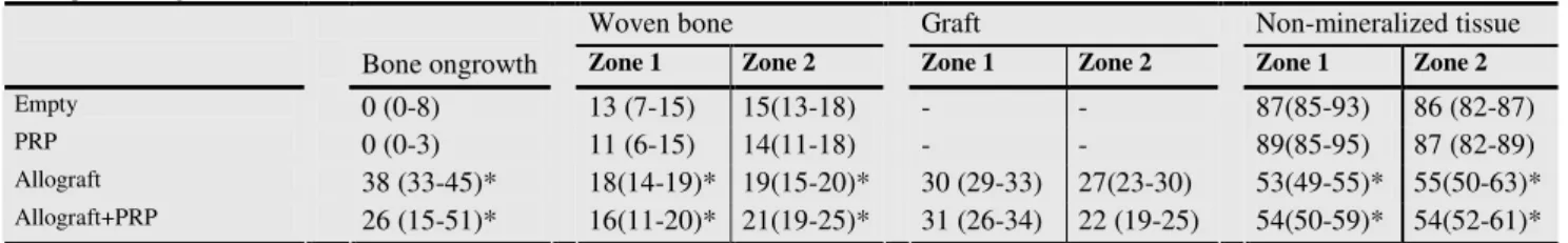
Quantitative analysis: PRP had no influence on bone formation in the grafted or non-grafted group (table XXI). Allografting increased bone ongrowth significantly from 0 (0-8)% to 38 (33-45)% and also bone formation in both zones was increased.
Table XXI , study V: Bone ongrowth and volume fractions of woven bone, bone graft and non-mineralised tissue in percentages of total area (median values (interquartile ranges))
Woven bone Graft Non-mineralized tissue
Bone ongrowth Zone 1 Zone 2 Zone 1 Zone 2 Zone 1 Zone 2
Empty 0 (0-8) 13 (7-15) 15(13-18) - - 87(85-93) 86 (82-87)
PRP 0 (0-3) 11 (6-15) 14(11-18) - - 89(85-95) 87 (82-89)
Allograft 38 (33-45)* 18(14-19)* 19(1