ABSTRACT
WIEGERT, JEFFREY GLENNON. Effects of Practically Increasing Amino Acids and Energy in Late Gestation on Colostrum Intake, Colostrum Composition and Sow Performance (Under the direction of Dr. Mark Knauer).
as fixed effects where appropriate. A meta-analysis was performed by calculating each sow’s total lysine (TLYS), added fat (TFAT), and ME (TME) intake from d 93 of gestation to farrowing. In EXP 1, feeding LACT tended (P<0.10) to increase piglet CI and WWT. No treatment differences were observed in EXP 2. In EXP 3, feeding GEST+FAT tended (P=0.06) to improve piglet BWT, while sows consuming GEST+SBM tended (P=0.08) to produce less colostrum. In EXP 4, piglets born to sows bump fed LACT at d 106 of gestation tended (P=0.07) to have greater CI. Meta-analysis estimates indicated that consuming 1 g additional TLYS in late gestation improved (P<0.02) CI and WWT by 0.12 g and 1.4 g, respectively, while consuming 1 g TFAT improved (P=0.01) CI by 0.03 g. a 1 Mcal increase in TME improved (P=0.02) WWT 4.0 g. A one nipple increase in TEATS improved (P≤0.05) CI, NW, and litter WWT by 13.3 g,
Effects of Practically Increasing Amino Acids and Energy in Late Gestation on Colostrum Intake, Colostrum Composition and Sow Performance
by
Jeffrey Glennon Wiegert
A dissertation submitted to the Graduate Faculty of North Carolina State University
in partial fulfillment of the requirements for the degree of
Doctor of Philosophy
Animal Science
Raleigh, North Carolina 2019
APPROVED BY:
_______________________________ _______________________________ Dr. Mark Knauer Dr. William Flowers
Committee Chair
DEDICATION
BIOGRAPHY
Jeff Wiegert is a native of Weldon Spring, Missouri. He is the son of Elaine and Greg Wiegert, who taught him the core values of community, dignity, and common sense, and that every lesson in life worth learning can be learned from baseball. Jeff has two brothers who, in his own opinion, are significantly braver and better men than he.
ACKNOWLEDGMENTS
Someone once told me that life is easy when you have a good boss. They were right. Accordingly, I would like to thank my advisor, Dr. Mark Knauer, and graduate committee members, Drs. Billy Flowers, Eric van Heugten, and Sung Woo Kim, for their support, criticism, and encouragement. Their guidance and, often, patience, have been integral to my development as an animal scientist. I also thank Dr. Chris DePerno for serving the committee as the Graduate School Representative. His insights have offered a new and valuable perspective to the
proceedings. Finally, I would be remiss to neglect to mention the contributions and leadership of Drs. Todd See and Joan Eisemann, who are both models of integrity and of the effectiveness of leading by example.
I graciously acknowledge Terry Armstrong, Brandon “Big Red” Barnes, Dwight Davenport, Austin Jones, Larry Jordan, Tyler O’Dell, Bo Mobley, and Lee Tyre of the North
TABLE OF CONTENTS
LIST OF TABLES ... vi
LIST OF FIGURES ... vii
CHAPTER 1: Literature review... 1
Introduction ... 1
Section 1. Prenatal piglet development ... 3
1.1. Biology of prenatal piglet development ... 3
1.2. Physiological implications of piglet intrauterine growth restriction... 7
1.3. Production consequences of piglet intrauterine growth restriction... 13
Section 2. Swine mammogenesis and lactogenesis ... 14
2.1. The sow udder ... 14
2.2. Mammogenesis and mammary gland tissue composition ... 17
2.3. Udder and mammary gland vascularization and blood flow ... 22
2.4. Lactogenesis and lactation ... 24
2.5. Nutrient and biochemical uptake into the mammary gland ... 28
2.6. Methods to measure colostrum and milk production in swine ... 36
Section 3. The role of colostrum in piglet development ... 39
3.1. Piglet suckling behavior and teat competition ... 39
3.2. The role of colostrum in piglet energetics ... 41
3.3. The role of colostrum in piglet immunity, survival, and growth ... 43
Section 4. Feeding sows to improve colostrum production ... 47
4.1. Sow nutrient requirements in late gestation ... 47
4.2. Effects of bump feeding on sow reproduction and lactation ... 50
Conclusions ... 55
Literature Cited ... 66
CHAPTER 2. Effects of practically increasing amino acids and energy in late gestation on colostrum intake, colostrum composition and sow performance ... 100
Abstract ... 100
Introduction ... 101
Materials and Methods ... 102
Experiment Design and Treatment Feeding ... 103
Reproductive and Colostrum Measures ... 105
Statistical Analysis ... 106
Results ... 108
Summary Statistics ... 108
Experiment 1 ... 108
Experiment 2 ... 109
Experiment 3 ... 109
Experiment 4 ... 110
Meta-Analysis ... 110
Discussion ... 112
Conclusions ... 120
Acknowledgements ... 121
LIST OF TABLES
Table 1.1 Gastrointestinal organ weights and dimensions in intrauterine growth restricted piglets and normal birthweight littermates at birth. ... 56 Table 1.2 Recommended dietary levels of lysine and metabolizable energy in gestating
sows before and after day 90 of gestation. ... 57 Table 2.1 Ingredient composition, calculated nutrient content, and nutrient analysis of the
gestation (GEST) and lactation (LACT) diets utilized in the experiments. ... 122 Table 2.2 Variables classified as categorical effects in the statistical models of
experiments 1 through 4 ... 123 Table 2.3 The main effects of late-gestation (day 104 to farrowing) sow diet (LSMEANS)
and feeding level (estimates) on sow reproductive performance in EXP 1. ... 124 Table 2.4 Sow reproduction and lactation performance in EXP 2 following transition from
a gestation to a lactation diet at a continuous feeding level (2.05 kg/d) at day 93, 100, or 107 of gestation or not until the day of farrowing ... 125 Table 2.5 Sow reproduction results and lactation results in EXP 3 following daily
supplementation of a gestation diet (GEST) with 120 g of fat (GEST+FAT) or 280 g of soybean meal (GEST+SBM) or with both fat and soybean meal to create lysine and added fat values comparable to a lactation (LACT) diet
beginning at day 107 of gestation. ... 126 Table 2.6 Reproduction and lactation performance of sows in EXP 4 transitioned from a
gestation to a lactation diet at day 102 of gestation and feeding level increased from 2 to 4 kg/d beginning at days 102, 106 or 110 of gestation or not until
farrowing ... 127 Table 2.10 Parameter estimates and probability values corresponding to the main effects of
sow total lysine (TLYS), total added fat (TFAT), and total net energy (TNE) intake from day 93 of gestation to farrowing on sow reproduction and lactation traits. ... 128 Table 2.11 Parameter estimates of colostrum production and colostrum nutrient composition
LIST OF FIGURES
Figure 1.1 Timing of fetal primary and secondary fiber myogenesis relative to the period of uterine crowding in gestation ... 58 Figure 1.2 Maternal, environmental, and piglet-specific factors contributing to piglet
deathloss during and after farrowing ... 59 Figure 1.3 The accumulation of mammary tissue and DNA in serially sacrificed gilts from
birth to 300 days of age ... 60 Figure 1.4 The accumulation of mammary tissue and DNA in pregnant gilts from breeding
to farrowing. ... 61 Figure 1.5 Cardiovascular anatomy of the sow udder ... 62 Figure 1.6 The percent protein, lactose, and fat in A) colostrum produced in the first 24
hours of lactation, and B) milk produced from 1 to 27 days of lactation ... 63 Figure 1.7 Total protein gain of maternal protein pools in a hypothetical second-parity sow
over the course of gestation ... 64 Figure 1.8 Dietary energy partitioning to maternal maintenance, maternal growth, conceptus
growth, and heat production and adipose tissue throughout pregnancy ... 65 Figure 2.1 Piglet colostrum intake was increased (P=0.03) in EXP 1 when increasing levels
of LACT but not GEST were fed from d 104 to farrowing ... 130 Figure 2.2 Litter weaning weight was increased (P=0.02) in EXP 1 when increasing levels
of LACT but not GEST were fed from d 104 to farrowing ... 131 Figure 2.3 The average colostrum yield of sows consuming GEST+SBM in EXP was
reduced (P<0.05) compared to sows consuming GEST, GEST+FAT, or LACT .. 132 Figure 2.4 The average birth weight of piglets born to sows consuming GEST+FAT in
EXP 3 was greater (P<0.02) than sows consuming GEST+SBM or LACT, but was not different (P=0.12) from sows consuming GEST ... 133 Figure 2.5 Effects of increasing sow total added fat intake from day 93 of gestation to
farrowing on piglet colostrum intake (P=0.02) ... 134 Figure 2.6 Effects of increasing sow lysine intake from day 93 of gestation to farrowing
On piglet colostrum intake (P=0.10) ... 135 Figure 2.7 Effects of increasing sow net energy intake from day 93 of gestation
Figure 2.8 The impact of the total number of piglets born on piglet colostrum intake
CHAPTER 1: Literature Review
Introduction
The pig is a litter-bearing species. The average litter size of the European wild boar (Sus scrofa), the ancestral species of the domestic pig (Sus scrofa domesticus), is typically between 3 to 7 piglets per litter, although this may vary greatly between populations according to genetics, geographic location, season of breeding, and nutrient and resource availability (Mauget, 1982; Fernández-Llario and Mateos-Quesada, 1998; Náhlik and Sándor, 2003). The advantage of litter-bearing reproduction in an evolutionary sense is the maximization of a population’s reproductive rate to meet the carrying capacity of a given habitat (Cody, 1965; Spencer and Steinhoff, 1968). The disadvantage of litter-bearing reproduction, however, is high preweaning offspring
mortality. In one study of free-ranging European wild boar, piglet postnatal mortality was estimated at 55% to 60% of the litter (Náhlik and Sándor, 2003).
According to Smith and Fretwell (1974), two general biological patterns explain the balance between parental energy expenditure and offspring fitness at birth: “1. As the energy expended on individual offspring is increased, the number of offspring that parents can produce is decreased; and, 2. As the energy expended on individual offspring increases, the fitness of individual offspring increases.” These patterns imply that as litter size is increased, the maternal resources allocated to each individual fetus is decreased, and the fitness of these offspring is decreased.
et al., 2010). These low birthweight piglets are less viable, at greater risk of pre- and
postweaning mortality, show reduced postweaning growth rate and feed efficiency, and are less likely to be full value market hogs at finishing (De Roth and Downie, 1976; Quiniou et al., 2002; Fix et al., 2010b). In recent years, the US swine industry has suffered an increase in piglet
preweaning deathloss concurrent with genetic selection for increased litter size (Lund et al., 2002). The average total number of piglets born per litter increased from 11.8 in 2005 to 13.9 per litter in 2017, but at the same time, average piglet preweawning mortality increased from 13.7% to 17.8% (Knauer and Hostetler, 2013; Stalder 2018). The average number of piglets weaned per litter increased from 9.3 in 2005 to 10.2 in 2011, but since that time has only increased to 10.3 pigs in 2017 (Knauer and Hostetler, 2013; Stalder, 2018).
Given the present scenario of reduced maternal resource allocation in utero as a consequence of large litter sizes, it follows that greater consideration should be given to the feasibility of increasing postnatal maternal resource allocation through colostrum and milk. The essential role of colostrum in piglet survival and growth cannot be understated. In swine, colostrum serves two fundamental purposes necessary for piglet survival: 1. To provide the piglet with sufficient energy to generate metabolic heat for thermoregulation; and 2. To serve as the vehicle for the passive transfer of maternal antibodies and other immune cells. Without adequate energy or antibodies, the piglet’s likelihood of surviving to weaning is jeopardized
(Quesnel et al., 2012). In addition, colostrum contains numerous bioactive proteins that are either localized in the intestines or communicated intact across the intestinal barrier to contribute to organogenesis and improved pre and postweaning performance (Pluske, 2016).
This literature review will be divided into four areas related to swine colostrum
lactogenesis; 3. The role of colostrum in postnatal piglet development; and 4. Feeding sows to improve colostrum production and composition.
Section 1. Prenatal piglet development
1.1. Biology of prenatal piglet development
The average litter size in the US swine industry in 2017 was 13.9 total piglets born (Stalder, 2018), yet the actual number of pigs born at farrowing is considerably less than the number of eggs originally ovulated. According to studies reviewed by Foxcroft et al. (2006), the ovulation rate (defined here as the number of corpora lutea present on the ovary after ovulation) of gilts and first-parity sows is 15 to 20 ova, while the ovulation rate of multiparous sows is 20 to 25 ova. A small number of multiparous sows are capable of achieving an ovulation rate over 30. The fertilization rate of these ova is high (95%; Kridli et al., 2016), indicating that embryonic litter size at the beginning of pregnancy may be as much as two-fold that of the ultimate litter size at farrowing.
The majority of embryonic loss occurs at or before approximately 30 days in gestation, and this coincides roughly with the timing of placental development (Wright et al., 2016). The pig placenta is epitheliochorial in structure and utilizes clustered areolae for diffuse exchange of blood and biochemical materials between fetal and uterine tissues (Chen et al., 1975).
In a landmark study conducted by Knight et al. (1977), unilaterally hysterectomized-ovariectomized gilts (UHOX; n=44) were compared to intact control gilts (n=44) to determine the effects of experimentally-induced intrauterine crowding on placental and fetal development. Owing to compensatory hypertrophy of the remaining contralateral ovary, the ovulation rate between UHOX and control gilts was similar in the experiment, yet the endometrial surface area available for implantation in UHOX gilts was reduced by half. Total hysterectomy and tissue collection of UHOX and control gilts then occurred at 11 regularly-spaced intervals from days 20 to 110 of gestation (n=4 UHOX and 4 control gilts collected at each time point). Placental weight and length were reduced in UHOX compared to controls at the first sampling point at day 20 of gestation, and these differences persisted throughout pregnancy. Placental surface area and the number and distribution of placenta areolae were not recorded until day 35, as placentae before this time were too brittle to withstand dissection from the uterus. However, both placental surface area and the number of areolae at day 35 were greater in control gilts (281.5±18.0 cm2 and 661.0±59.0 areolae/placenta) than in UHOX gilts (234.1±11.0 cm2 and 333.2±26.9
areolae/placenta), and these patterns also continued throughout pregnancy. The number of live embryos was similar between groups in early pregnancy, yet between days 30 and 35 of gestation, the number of live embryos dissected from UHOX gilts decreased (11.0±0.4 vs. 8.8±1.3, respectively), but did not change in the control gilts (10.3±1.4 vs. 10.8±2.0,
respectively). Finally, a treatment by day of gestation interaction was observed beginning on day 40 of gestation between UHOX and control gilts in both fetal crown-rump length (5.1±0.03 cm vs. 4.8±0.08 cm) and fetal wet weight (9.40±0.14 g vs. 9.10±0.33 g). More pronounced
respectively, in UHOX gilts compared to controls. Collectively, these results suggest that limited uterine capacity stunts placental development by day 20 of pregnancy, causing an increase in embryonic mortality after day 30 of gestation, and a restriction of intrauterine fetal growth beginning at day 40 of gestation. The results of this study, and the underlying theories of uterine capacity limiting fetal growth and litter size through placental insufficiency, have been supported by the more recently conducted studies of Christenson et al. (1987), Biensen et al. (1998), and Freking et al., (2007).
The number of quality pigs produced per sow is the most economically influential trait on the breeding swine farm (De Vries, 1989), and accordingly intense selection pressure has
historically been placed on increasing litter size in the commercial industry (Knauer and Hostetler, 2013). Genetic selection for increased litter size results in increased ovulation rate (Gama and Johnson, 1993). Lamberson et al. (1991) identified that a 1 ova increase in ovulation rate increases litter size at farrowing by approximately 0.25 pigs. However, selection for greater litter size has failed to create appreciable changes in uterine dimensions or volume (Gama and Johnson, 1993). It can be said, then, that selection for increased litter size exacerbates uterine crowding and reduces fetal growth.
(Leymaster and Christenson, 2000). Following the eleventh generation of the experiment, 593 gilts representing the three selection lines (i.e. selection for greater ovulation rate, selection for greater uterine capacity, and the randomly selected control line) were subjected to UHOX under procedures similar to those utilized in Knight et al. (1977) to establish uterine crowding. Gilts were then bred to boars within line and harvested at days 25, 45, 65, 85, or 105 of gestation (n=24 to 30 gilts sampled per time per line; Freking et al., 2007). As expected, ovulation rate was greatest in the line selected for increased ovulation rate, and no differences in the number of live fetuses were observed between lines at day 25 of gestation. By day 45, however, fetal litter size was greater in gilts selected for greater uterine capacity compared to other lines. Average fetal weight, litter fetal weight, average placental weight, litter placental weight, and uterine length were all greater in gilts selected for greater uterine capacity than in gilts selected for ovulation rate from gestational day 45 onwards.
Results of these genetic selection and hysterectomy-ovariectomy studies conclude that restricted fetal growth and development should be expected following genetic selection for increased litter size. Indeed, surveys of commercial sows suggest that a 1 piglet increase in litter size is associated with a 30 to 50 gram decrease in average piglet birth weight (Roehe, 1999; Quiniou et al., 2002; Opschoor et al., 2010). Increasing litter size is also associated with greater within-litter variation in piglet birth weight (Boulot et al., 2008). Large within-litter variation in piglet birth weight suggests that intrauterine embryonic competition for endometrial surface area and maternal resources also creates divergent patterns in fetal growth. Hence, not only is average piglet birth weight decreased, but the number of exceptionally light birth weight pigs is
increased. In one observational study incorporating 12,041 piglets from 965 litters, the
increased from 9 to 17 total number born (calculated in this study as number born alive plus the number stillborn; Quiniou et al., 2002).
Given the considerable economic influence of litter size, continued selection for
increasingly hyperprolific sows in the swine industry is to be expected. Selection on the basis of greater litter size at farrowing will increase ovulation rate, thereby expediting uterine crowding issues, decreasing average piglet birth weight, and increasing the number of light birth weight piglets. A greater understanding of the physiological effects of intrauterine growth restriction in swine thus becomes necessary.
1.2 Physiological implications of piglet intrauterine growth restriction
Intrauterine growth restriction (IUGR) in swine results in reduced piglet birth weight and altered fetal physiology that persists throughout adult life. According to Foxcroft et al. (2006), an IUGR piglet is defined as a piglet whose birth weight is an outlier from the mean birth weight of a given litter. On the other hand, small for gestation age (SGA) piglets are defined as those piglets whose birthweights are greater than 2 standard deviations from the mean, but not statistical outliers. For the purposes of comparison, normal birth weight piglets (NBW) are defined as those piglets whose birth weight closely matches the mean litter birth weight. The exact definition of an IUGR or SGA piglet is variable between researchers and reports. Efforts will be made to delineate the definitions of IUGR and SGA piglets used by researchers in the studies included in this section when differing from the definition presented by Foxcroft et al. (2006).
in turn diminish the pig’s production efficiency, as evidenced by increased postnatal morbidity
and mortality and decreased lifetime growth rate, feed efficiency, and carcass value (Wu et al., 2006; Fix et al., 2010a; Fix et al., 2010b).
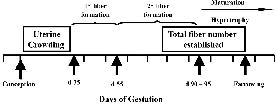
Myogenesis is the formation of primary and secondary skeletal muscle fibers during fetal development (Maltin et al., 2001). Primary muscle fibers are developed prior to day 50 of
gestation through the fusion of primary myoblasts. This process is thought to be generally consistent, with little variation between littermates according to the degree of fetal development (Swatland, 1973). Secondary fibers are then established on top of the existing primary fiber architecture, and secondary fiber myogenesis has been demonstrated to be amenable to such factors as placental insufficiency and maternal under- or over-nutrition in mid-gestation (Fig. 1.1; Swatland, 1973; Wigmore and Stickland, 1983; Dwyer et al., 1994; Foxcroft et al., 2006; Zou et al., 2017). Multiple studies have identified reduced secondary muscle fiber number in light weight pigs compared to normal birth weight littermates (Dwyer and Strickland, 1991; Town et al., 2004; Rehfeldt and Kuhn, 2006; McNamara et al., 2006; Alvarenga et al., 2013). It is important to emphasize that no myogenesis occurs postnatally, although hypertrophy of existing fibers does continue after birth, provided adequate nutrition is available (Lefaucheur et al., 2003). Without doubt, disruptions to the developmental biology in utero create permanent consequences for the animal. According to studies reviewed by Oksbjerg and Therkildsen (2017), the number of muscle fibers at birth in swine is moderately correlated (r = 0.42 to 0.46) with postnatal growth rate, carcass leanness, and feed efficiency.
Diminished intrauterine secondary myofiber formation may be traced to endocrine and transcription factor dysregulation. In particular, the myogenic regulatory factor family
of myoblasts into myotubes, and ultimately into myofibers (Oksbjerg and Therkildsen, 2017). Down regulation of MYOG, the gene coding for the myogenin transcription factor, has been observed in embryos weighing 85% to 95% of control embryos through mid- and late gestation in both Western crossbreed sows (Tse et al., 2008) and Meishan sows (Zou et al., 2017).
Insulin-like growth factor I (IGF-I) and insulin-like growth factor II (IGF-II) also contribute to prenatal muscle development. The birthweight of IGF-I and IGF-II knockout mice is reduced 30% compared to unaltered controls (Baker et al., 1993). Although both IGF-I and IGF-II contribute to fetal development, IGF-II is thought to have the greater role prenatally, while IGF-I is the more significant mediator of musculoskeletal growth in adulthood (Fowden, 2003; Duan et al., 2010; Kent et al., 2012). These growth factors, although working in concert to initiate fetal development, operate under separate stimuli. As stated by Fowden (2003), IGF-II provides the “constitutive drive for intrauterine growth via its placental effects and direct paracrine actions on fetal tissues,” while on the other hand, IGF-I “regulates fetal growth in relation to the nutrient supply.” Therefore, IGF-II expression and activity may be thought of as
biologically fixed by genotype, while IGF-I activity may be relative to environmental
weighing greater than 1.75 kg at birth compared to pigs weighing less than 1.10 kg at birth (Gondret et al., 2005).
Stunted organogenesis is also observed in IUGR fetuses. Town et al. (2004) subjected 30 third-parity sows to oviduct ligation to inhibit fertilization of ova from one ovary, thereby experimentally decreasing litter size and creating uncrowded uterine conditions. Fetal growth in the 30 “non-crowded” sows was then compared to fetal growth in 30 sows with normal
“crowded” uterine conditions. Half of the crowded and non-crowded sows were sacrificed at day
In a similar study, Wang et al. (2004) compared the organ development of NBW piglets (n=5) with true IUGR littermates (n=5; IUGR defined here as piglets weighing less than 2 standard deviations from the litter mean). Body weight and organ weight at birth of NBW and IUGR piglets are presented in Table 1. From these published data, calculations were made of organ weight as a percent of body weight within birth weight classifications and of organ weight of IUGR piglets as a percent of NBW. Expressing organ weight both as a percentage of body weight, and organ weight of IUGR piglets as a percentage of NBW piglets allows greater identification of developmental patterns. Expressed as a percentage of body weight, the relative organ weights of the liver, stomach, and pancreas of IUGR piglets (2.86%, 0.53%, and 0.13%, respectively) were actually greater than the relative organ weights in NBW piglets (2.61%, 0.49%, and 0.12%, respectively). However, the relative weight of the small and large intestines in IUGR piglets (2.36% and 0.65%, respectively) were less than the relative weight in NBW piglets (2.50% and 0.74%, respectively). Significant growth and maturation of the
gastrointestinal tissue is necessary to prepare the piglet for postnatal intestinal nutrient
absorption. Indeed, the small intestine is known to increase in relative weight by 70-80% during the last three weeks of gestation (Sangild et al., 2000). This coincides with a period of rapid fetal development in swine (Ji et al., 2017). Other authors have identified similar patterns of
Wang et al. (2005) and Hu et al. (2015) reported decreased microvilli number at birth in the proximal jejunum and ileum, respectively. On the other hand, D’Inca et al. (2010) reported no differences in small intestine villous height between NBR and IUGR piglets (weighing less than 1.5 standard deviation from the mean) at birth (400±18 µm vs. 402±0.15 µm). Yet by two days of age, villous height had increased to a greater extent in the NBW but not the IUGR piglets (687±26 µm vs 508±21 µm). These data may indicate reduced intestinal responsiveness to milk-borne growth factors in IUGR piglets compared to NBW littermates (a more complete discussion of the effects of milk nutrients and growth factors will follow in section three of the literature review). Differences in intestinal attributes in IUGR piglets compared to NBW littermates persist to adulthood (Alvarenga et al., 2013).
Altered intestinal enzyme activity also indicates immature gastrointestinal tissue
development in IUGR piglets. In an analysis of 12 pairs of 7 day old NBW and IUGR littermates (weighing less than 1.5 standard deviations of NBW littermates at birth), Hu et al. (2015)
bacterial infiltration of the gut mucosal barrier, in IUGR piglets likely indicates intestinal immaturity and increased susceptibility to enteric diseases (Chen et al., 1975).
The altered physical development and enzymatic activity observed in SGA and IUGR piglets suggests that these animals are at a reduced biological age, despite being the same chronological age as normal birth weight littermates. The muscular, metabolic, and digestive issues discussed create production challenges for the growing pig.
1.3 Production consequences of piglet intrauterine growth restriction
The combined effects of reduced muscular development and underdeveloped and ineffectual gastrointestinal tissue create lifetime production consequences for the light
Assuming that the pig survives to weaning, the detrimental consequences of low birth weight persist through life. In an observational study of commercial swine, Fix et al. (2010b) reported a positive linear relationship between pig birth weight and survival in the nursery but not finishing phases of production. Further, Fix et al. (2010a) identified that piglet birth weight is positively associated with average daily gain in both the nursery and finishing phases, and with increasing loin muscle area and back fat thickness at marketing. These results are supported by similar studies correlating the relationships between low muscle fiber number in low birth weight piglets with poor growth rate in the nursery (Dwyer et al., 1993) and finishing phases (Rehfeldt and Kuhn, 2006). Low birth weight pigs are also less likely to be full value at market (full value defined in this instance as pigs that survived to harvest, weighed greater than 100 kg at
marketing, and were free of injuries, health issues, and physical deformities; Fix et al., 2010b), and yield carcasses with lower lean meat percentage, high intramuscular fat percentage, and greater carcass drip loss, indicating poor water holding capacity and, thus, poor meat quality (Rehfeldt and Kuhn, 2006).
Section 2. Swine mammogenesis and lactogenesis
2.1 The sow udder
and second pairs), abdominal glands (third, fourth, and fifth pairs), and inguinal glands (sixth and greater, if existing; Farmer and Hurley, 2015). Mammary glands may also be defined simply as anterior or posterior according to their position relative to the navel (McKay and Rahnefeld, 1990; Kim et al., 2000; Wu et al., 2010; Šamanc et al., 2013). The location of the mammary gland on the body is relevant because differences exist between glands in tissue composition, blood supply, and the nutrient profile of the produced milk.
The actual number of mammary glands is variable between individual pigs. In a survey of 987 Meidam (Meishan x Large White) sows, Balzani et al. (2016b) reported a range of 12 to 19 teats per sow, with a mean of 15.6 ± 1.1 total teats. In a separate survey of 118,267 purebred Yorkshire pigs, Felleki and Lundeheim (2015) reported 14.53 ± 0.92 teats, with a range of 9 to 21 teats. These data suggest variation by breed or within populations in the number of mammary glands per female. The number of mammary glands is a moderately to highly heritable trait. Estimates for heritability of total teat number range from 0.20 on the low end (Clayton et al., 1981) to 0.42 on the upper end (McKay and Rahnefeld, 1990). Analyses of the Swedish Yorkshire herd yields consistent trait heritability between 0.30 and 0.35 (Chalkias et al., 2013; Lundeheim et al., 2013; Felleki and Lundeheim, 2015). Lundeheim et al. (2013) also reported heritability for functional teats (0.31) and non-functional teats (0.09). The heritability of inverted teats has been estimated to be 0.20 (Clayton et al., 1981).
females (Kratochwil, 1971) and this may occur in litter bearing species when female embryos are positioned between two male embryos (vom Saal and Bronson, 1978) or when the sex ratio is skewed at least 2:1 towards male fetuses (Drickamer et al., 1997). The number of males born in a litter and the intrauterine positioning of female swine fetuses between male littermate fetuses has also been shown to modestly reduce age at puberty (Lamberson et al., 1988; Parfet et al., 1990), alter the dynamics of the gilt’s preovulatory LH surge (Seyfang et al., 2018), and decrease conception rates (Drickamer et al., 1997). These experiments collectively indicate intrauterine androgen exposure as consequence of higher male:female sex ratio reduces female reproductive potential later in the life. Skewed male:female sex ratios may result from uterine crowding (Tse et al., 2008), and restricted sow feeding in lactation to elicit a severe negative energy balance at weaning (Vinsky et al., 2006; Oliver et al., 2011). On the other hand, however, prenatal methods to improve mammary number and development have not yet been identified.
Congenital defects and physical injuries to the udder during life may reduce the number of functional teats. Teat functionality is defined as the ability for that teat to excrete milk during lactation. Congenital teat defects include inverted teats (i.e. the mammary gland terminates in a teat that is inverted upwards into the body cavity), blind teats (i.e. no teat exists at the
termination of the gland), and pin nipples (i.e. small bud-like projections on the udder
Physical injuries to the teats and udder may further compromise the number of functional teats available for piglet nursing. Udder injuries may stem from improperly maintained
farrowing crates, the sow stepping on her own udder, or from aggressive piglet nursing. Injuries are most common on the abdominal and inguinal glands (Persson, 2010). Affected teats are also more susceptible to mastitis (Persson, 2010), which may decrease milking ability in the current or subsequent lactation. Udder injuries are not always included in farm management software culling codes, so data on the prevalence of udder injuries is limited. In an observational report of culling criteria of 21 Swedish sow farms (n=14,234 sows culled over a three-year period), udder problems accounted for 18.1% of sow removal (Engblom et al., 2006). In this report, udder problems included “low or no milk production, mastitis, and/or udder abscess” and was most
commonly observed in 4th to 6th parity sows. Removal rate due to udder problems was on par with the removal rate due to “old age” (18.7%), but less than “reproductive disorders” (26.9%) and greater than “lameness and/or foot lesions” (8.6%) and “traumatic injuries” (7.1%).
2.2 Mammogenesis and mammary gland tissue composition
In the sow, two complete lobuloalveolar units each terminating in a distinct gland cistern exist within each mammary gland (Nickerson and Akers, 2011). Accordingly, milk accumulating in each gland cistern exits the gland through a separate teat cistern.
The mammary gland alveolar, ductal, and glandular tissue is collectively referred to as parenchymal tissue. Logically, the gland’s connective and adipose tissue involved in structural support is titled extraparenchymal tissue (Farmer and Hurley, 2015). Milk production in mammals is positively associated with the number of mammary secretory epithelial cells, amount of parenchymal tissue, and the ratio of parenchymal to extraparenchymal tissue in the mammary gland (Knight et al., 1984; Knight and Peaker, 1984; Kim et al., 2000; Nielsen et al., 2001). Quantification of mammary DNA, protein content, and total tissue content may be used to estimate the number of secretory cells and the amount of parenchymal tissue in the gland, and these measures are often used as indicators of mammary development (Tucker, 1987). In one study, Kim et al. (2000) reported moderate to high correlations between piglet preweaning growth rate and the protein content (r=0.67) and DNA amount (r=0.54) of the mammary gland suckled during lactation. Thus, mammary development as defined by greater secretory epithelial cell number is positively correlated with the milking ability of the gland.
Mammary development in gilts is slow through the first 90 days of age. According to Sørensen, et al. (2002), who conducted serial sampling of non-pregnant gilt mammary tissue at 10 day intervals beginning at birth, prepubertal mammary development from birth (0.3 mg DNA per gland) to 90 days of age (7 mg DNA per gland; Fig. 1.3; Fig. 1.4) is negligible and largely isometric with body weight gain. From 90 days of age to puberty, however, the accumulation rate of mammary DNA and mammary tissue was 3.9 and 5.6-fold greater, respectively, compared to accumulation rates before 90 days of age.
The increase in the rate of mammary development beginning at 90 days of age coincides with the timing of ovarian development, including the appearance of steroid-producing antral follicles and the initial establishment of the hypothalamic-pituitary-gonadal axis (Dyck and Swierstra, 1983; Camous et al., 1985; Pressing et al., 1992). Multiple authors have reported intrinsic roles for estrogen and prolactin in prepubertal mammary gland development in swine and other mammals (Purup et al., 1993; Bocchinfuso et al., 2000; Farmer and Palin, 2005; Horigan et al., 2009). Estrogen is involved in lobuloalveolar formation and prolactin is required for mammary epithelial cell differentiation (Barrington et al., 1999; Akers, 2017). Attempts to increase mammogenesis during this time through dietary means have been largely unsuccessful (Farmer et al., 2004; Sørenson et al., 2006; Farmer et al., 2007; Farmer et al., 2012).
Following puberty, little mammary development is observed until the last trimester of pregnancy. Ji et al. (2006) reported a three-fold increase in mammary growth rate beginning after day 75 of gestation. Similarly, between days 75 and 112 of gestation, Sørenson et al. (2002) reported massive accumulation of mammary DNA (40 vs. 838 mg, respectively) and mammary tissue (80 g vs. 373 d, respectively). The increased tissue accretion in these data reflects
accelerated growth rates, while the large accumulation of mammary DNA suggests tissue
specialization into functional lobuloalveolar components. Indeed, the percent crude protein of the gland increases from 11.4% to 38.3% in late gestation, while the percent adipose decreases from 87.6% to 58.8% (Ji et al., 2006). Histological measurements of the gland indicate progressive growth of the terminal ductal lobular unit from day 75 to term (Ji et al., 2006).
Similar to mammary development early-in-life, mammary growth during pregnancy is largely under endocrine control. Circulating estrogen levels in the pregnant female remain low until approximately 60 days in gestation, after which time increase precipitously until parturition. DeHoff et al. (1986) reported 967.6 pg/ml estrogen at day 60 of gestation and 21,439 pg/ml at day 112. Knight et al. (1973) identified that the elevated estrogen found in the sow’s systemic circulation in late gestation is of fetal origin. Interestingly, though, no linear relationship between litter size and mammary development has been observed (Kensinger et al., 1986b). In contrast to estrogen, prolactin’s period of influence on mammogenesis does not begin until later in
pregnancy. Farmer and Petticlerc (2003) observed a 46% decrease in parenchymal tissue mass when bromocriptine, a dopamine agonist that inhibits prolactin secretion, was fed to gilts from 90 to 109 days of gestation, but no effects on mammary development when bromocriptine was fed from 50 to 69 or 70 to 89 days of gestation. Likewise, feeding domperidone, a dopamine antagonist that stimulates prolactin secretion, from day 90 to 110 of pregnancy increased alveolar lumen diameter and sow milk yield throughout lactation (VanKlompenberg et al., 2013).
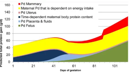
Given the considerable mammary growth, as well as the previously discussed concurrent fetal and placental tissue growth in late gestation, it is logical that the sow’s nutrient
requirements in the third trimester are greater than the first and second. A meta-analysis conducted by Kim et al. (2005) demonstrated the daily lysine requirements of a hypothetical second-parity sow with 14 fetuses and 16 mammary glands in early and late gestation. Daily lysine requirements to support conceptus and mammary growth before day 70 of gestation are 0.27 g/d and 0.17 g/d, respectively, but increase in late gestation to 4.0 g/d and 4.1 g/d,
sow nutrient requirements throughout gestation will be provided in Section 4 of the literature review.
After farrowing, piglet nursing becomes responsible for maintaining lactation and promoting continued mammary development. With no nursing pressure, unsuckled glands quickly begin regressing (Kim et al., 2001), and by 72 hours become incapable of maintaining milk production (Theil et al., 2005). In contrast, intense nursing pressure (e.g. frequent nursing interval or large piglets nursing the sow) is associated with increased prolactin secretion (Spinka et al., 1999), which in turn drives further mammary development (Kim et al., 1999).
2.3 Udder and mammary gland vascularization and blood flow
Blood flow to a mammary gland is commonly estimated using diffusion principles. Briefly, at a sufficient concentration in arterial blood, the rate at which the concentration of a biological marker, such as a nutrient, gas, or growth factor, in venous blood approaches that of arterial blood increases relative to the rate of blood flow to the organ, assuming that gland uptake of the marker is either negligible or accountable (Kety and Schmidt, 1945). This principle was first utilized to estimate mammary blood flow in lactating swine by Linzell et al. (1969) who catheterized the carotid, internal saphenous, or external pudic arteries and the mammary vein on either side of a functioning mammary gland and infused tritriated water (3H2O) into the jugular vein at a constant rate. Synchronized repeated sampling of arterial and venous blood allowed researchers to quantify the concentrations of radiolabeled water through the gland, and hence, estimate blood flow to the mammary. Results of this experiment, and of the more recent studies of Trottier et al. (1997), Guan et al., (2002; 2004a; 2004b), Nielsen et al. (2002a; 2002b), and Renaudeau et al. (2002), utilized these same principles to estimate mammary blood flow and the corresponding plasma:milk (volume:volume) ratio. In these studies, mammary blood flow ranged from 1.9 liters to 5.5 liters of blood flow per minute and 441 liters to 1,050 of blood plasma per liter of milk produced (Farmer et al., 2015). Renaudeau et al. (2002) calculated the coefficient of variation of blood flow through the right pudic artery of a sow in a given day to be 19%.
(Algers and Jensen, 1991; Toner et al., 1996; King et al., 1997; Auldist et al., 1998). No studies to date have measured or elucidated the regulators of late gestation mammary blood flow.
2.4 Lactogenesis and lactation
The lactation fluids produced by the sow are defined as colostrum or milk according to nutrient composition and timing of production relative to farrowing. Colostrum is synthesized in the mammary gland in late gestation and is available to the piglets immediately after farrowing (Quesnel et al., 2015). Colostrum is characterized by a higher percent protein content compared to milk (Hurley, 2015). Commonly, colostrum is defined as the fluids produced for the first 24 hours of lactation. The nutrient profile of colostrum begins to change almost immediately after farrowing. After 24 hrs, the fluid protein content has decreased to an extent to resemble mature milk more than colostrum (Devillers et al., 2007). Subtle changes in nutrient composition continue until approximately 10 days into lactation, after which time the nutrient profile of the milk is quite stable (Fig. 1.6; Hurley, 2015). Compared to milk, the colostrum produced within the first 6 hours after farrowing is more energy dense (4.9 kJ/g vs. 6.4 kJ/g) and contains greater total solids (20.0% vs. 26.2%) and percent protein (5.3% vs. 15.7%), but less lactose (7.4% vs. 2.8%) and fat (7.4% vs. 6.1%). Immunoglobulin G (IgG) is the major protein in colostrum, but IgA and IgM are the major proteins in milk (Hurley, 2015). A meta-analysis comparing sow milk nutrient composition in reports published in the 1980s and 2010s shows that modern sows
produce milk with slightly greater levels of fat and lower levels of lactose compared with
previous generations (Zhang et al., 2018a). Colostrum ejection is continuous and freely available to the piglets for the 8 to 11 hours after farrowing, after which time letdown occurs
established lactation, nursing frequency may vary from 17 to 35 events per day, depending on litter age and nursing intensity (Jensen et al., 1991). Peak lactation in sows occurs at 18.7 days in milk (Hansen et al., 2012b).
Colostrogenesis is the process of colostrum synthesis and begins in the mammary gland in late gestation. The physiological markers used to define colostrogenesis include the
histological changes in the mammary gland structure and the appearance of mammary-specific markers in sow blood, specifically lactose (Farmer et al., 2006). β-Lactoglobulin is the first mammary-specific protein detectable in the sow’s systemic blood, as early as day 80 of gestation (Dodd et al., 1994). Interestingly, other whey proteins, such as α-lactalbumin, are not found in sow systemic blood until the last week of pregnancy (Dodd et al., 1994). The function of
be said that colostrogenesis as defined the morphological changes to mammary structure
necessary to support glucose conversion to milk substrates begins approximately one week prior to farrowing, but colostrogenesis as defined as the accumulation of physical colostrum within the gland does not begin until the days or hours immediately before farrowing.
The developmental changes in the gland that begin in late gestation continue up to and even following farrowing. As previously mentioned, mammary epithelial cell content is
positively correlated with milk production (Kim et al., 2000). Hence, the hormones that stimulate mammogenesis also have lactogenic roles. In particular, prepartum sow prolactin is positively correlated with sow blood lactose content (Martin et al., 1978), total colostrum production (Quesnel et al., 2013), and early lactation piglet growth rate and survival (de Passillé et al., 1993). The essential role of prolactin in swine has been demonstrated experimentally by Farmer et al. (1998), who provided oral doses of bromocriptine, a dopamine agonist, to gilts beginning on day 110 of gestation and noted reduced piglet weight gain in early lactation. In rodents, progesterone inhibits prolactin secretion from the anterior pituitary and reduces prolactin receptor expression in the mammary gland (Haug and Gautvik, 1976). Accordingly, a greater prolactin:progesterone ratio prior to farrowing is a valid marker of greater colostrum production (Devillers et al., 2007; Foisnet et al., 2010, 2011).
induced by the fetus, some have suggested a fetal role in colostrogenesis. For example, Devillers et al. (2007) suggested a modest correlation (r=0.38) between piglet birth weight and sow
colostrum production, although the fetal role in colostrogenesis in this study was only
circumstantial. Potentially, large birthweight piglets in the study may have simply had greater vitality at birth and were able to strip more colostrum from the sow immediately after birth.
In established lactation, the sow produces milk reciprocal to litter nursing pressure. Milk production increases linearly with increasing litter size, and is also associated with the time and intensity of piglet teat massage (Algers and Jensen, 1991; Toner et al., 1996). Indeed, Auldist et al. (1998) utilized cross-fostering to create staggered litter sizes of 6, 10, and 14 piglets, and observed daily sow milk yields of 9.80, 13.05, and 15.52 kg, respectively. Also using cross-fostering strategies, King et al. (1997) observed a 26% increase in sow milk production when two-week old piglets were fostered onto dams that had farrowed only 2 days prior, and a 22% decrease in milk production when 2 day old piglets were placed on sows in peak lactation.
neural signal to release oxytocin to begin milk letdown (Jensen et al., 1991). In one study of fifteen catheterized lactating sows and their nursing litters, milk letdown occurred at
approximately 45 minute intervals following 2.4 minutes of intense piglet udder massage. Increased sow vocalizations (1.9±0.1 grunts per second) began approximately 23 seconds prior to a rapid increase in intramammary pressure, lasting between 8 and 41 seconds (Ellendorf et al., 1982). The actual duration of milk letdown is brief (8 to 15 seconds; Fraser et al., 1980; Pedersen et al., 2011).
2.5 Nutrient and biochemical uptake into the mammary gland
The mammary gland is not a gluconeogenic organ, yet milk production is responsible for 65-70% of a sow’s energy demand in peak lactation (Noblet et al., 1990). Of course, then, significant energetic material is regularly transported into the mammary gland. The major milk precursors utilized by the mammary gland are glucose, triglyceride fatty acids, and amino acids, and these compounds collectively account for approximately 95% of the carbon incorporated into the gland (Boyd et al., 1995). Spincer et al. (1969) used blood vessel catheterization and mammary gland arterio-venous differences to estimate the mammary gland uptake of glucose, triglyceride fat, and amino acids necessary to synthesize 1 dL of milk to be 14.1 g, 2.7 g, and 6.2 g, respectively. Other milk precursors transported into the gland at minor concentrations include acetate, non-esterified fatty acids, β-hydroxybutyrate, lactate, and citrate (Linzell et al., 1969; Spincer et al., 1969).
1995). The majority of the research in this field has been conducted in the bovine and murine species, and the few swine studies available typically focus on the mammary gland in peak lactation. Efforts will be made to delineate species and stage of lactation for transporters or mechanisms not highly conserved across Mammalia. It should also be stated that, with the exception of Renaudeau et al. (2003), who measured glucose and fat uptake in heat stressed sows, the most recent studies measuring sow mammary gland uptake of energetic precursors were conducted fifty years ago (Linzell et al., 1969; Spincer et al., 1969) in sows in their 5th and 6th week of lactation. Considerable management and genetic changes have occurred on swine farms since these studies were conducted. An updated investigation into mammary energetics in the modern sow is long overdue.
Glucose accounts for approximately 40 to 60% of the total mass of substrates transported into the mammary gland of a lactating sow (Farmer et al., 2008). Indeed, as much as 60% of available arterial glucose in a lactating sow may be utilized by the mammary glands (Spincer et al., 1969). Renaudeau et al. (2003) estimated a 1,300 g/d glucose requirement to support an 11 kg/d milk yield.
Glucose is a polar and hydrophilic molecule which cannot cross a plasma membrane through simple diffusion. Multiple facilitated transport mechanisms have been described in the mammary epithelial cells of bovine and rodent species, including glucose transporter (GLUT) 1, GLUT8, and GLUT12 (Zhao, 2014). Facilitated transporters are bidirectional
energy-independent transporters that move glucose according to concentration gradients. Sodium-dependent cotransporters (SGLT) have also been isolated in bovine epithelial cells.
abundance of GLUT1, GLUT8, SGLT1, SGLT3, and SGLT5 in the swine mammary gland in late gestation and in lactation. In this report, GLUT1 mRNA was approximately 10-fold more abundant than other transporters, likely indicating greater physiological significance of this particular transporter. Given the large abundance, one may also infer that most GLUT1 is located on the epithelial cell basolateral membrane (nearest maternal bloodstream), as is the case in other species (Zhao, 2014), which would explain how such a considerable amount of glucose enters the cell from the bloodstream. Other likely locations for GLUT and SGLT glucose transporters are at the Golgi apparatus and the cell apical membrane (nearest the alveolar lumen). Future studies utilizing immunohistochemistry of mammary epithelial cells will be necessary to elucidate glucose transporter location and better understand mammary gland function and metabolism in swine.
gluconeogenesis, glycogenolysis) maintain arterial glucose availability at sufficient levels in all but cases of severe starvation.
Blood glucose is also affected by stage of pregnancy and litter size. Many species, including swine, experience decreased insulin secretion and tissue responsiveness to insulin in late gestation, and this is thought to be an evolutionary mechanism to shunt greater glucose to the gravid uterus (Zhao, 2014). Indeed, Pére et al. (2000) noted greater blood glucose and decreased insulinemia beginning after 85 days in pregnancy. More recent work conducted by Pére and Etienne (2018) indicates that uterus glucose uptake increases with increasing litter size, but potentially not at levels sufficient to diminish arterial glucose concentrations.
Hence, increased mammary glucose uptake at constant arterial glucose concentrations is achieved by increased glucose transport number. Chen et al. (2017) reported increased GLUT1 and SGLT3 mRNA abundance in sow mammary glands at farrowing compared to day 97 of pregnancy. Feeding lactating rodents the dopamine agonist bromocriptine decreased mammary GLUT1 receptor number, indicating a role of prolactin in mammary glucose transporter
expression (Rudolph et al., 2011). In cows, the advancing stages of pregnancy are associated with increased GLUT1, GLUT8, GLUT12, SGLT1, and SGLT2 (Zhao and Keating, 2007), suggesting that all glucose transporters may be upregulated in late gestation to increase mammary glucose flux.
Cockburn, 1986). Lactose is also the primary osmotic agent in the mammary gland and therefore has a large role in determining milk volume (Zhang et al., 2018b).
Lactose synthesis occurs in the Golgi apparatus and the process is highly conserved across domestic species (Huang et al., 2012; Zhang et al., 2018a). Briefly, glucose is first enzymatically converted to the monosaccharide galactose in cytoplasm and then combined with free glucose in the Golgi. The lactose synthase enzyme, consisting of the regulatory subunit α-lactalbumin (LALBA) and the catalytic subunit β-galactosyltransferase (GALT), is required to produce lactose. Milk lactose content is low in colostrum (approximately 2.8%) but increases in mature milk (approximately 5%; Hurley, 2015). Accordingly, GALT and LALBA mRNA abundance and protein expression are relatively low at farrowing, but increase significantly by approximately 3 days in milk (Chen et al., 2017; Zhang et al., 2018b).
The remaining glucose not utilized for milk lactose synthesis is utilized in normal mammary gland metabolism or is converted into glycerol through glycolysis (Zhang et al., 2018a). Glycerol is combined with three fatty acids in the mammary epithelial cell endoplasmic reticulum to produce milk triglycerides. Only 2% of mammalian milk fat is not bound in
triglyceride form (Innis, 2011). Glucose may also be converted directly into fatty acids through de novo glycolysis and the TCA cycle to produce a citrate intermediary. In non-ruminant species, short and medium chain fatty acids are produced through de novo synthesis in the mammary gland, while long chain fatty acids present in milk are from dietary sources (Bauman and
Griinari, 2003). Long chain fatty acids constitute the majority of milk fat in swine colostrum and milk (Zhang et al., 2018a).
fatty acid uptake into the mammary gland (Bionaz and Loor, 2008; Shi et al., 2015) yet FABP3 only appears to be the major transport protein in swine (Lv et al., 2015). Further, FABP3 mRNA and protein expression increases as the sow transitions from late gestation to lactation and in response to incubating epithelial cells with increasing levels of long chain fatty acids in vitro (Lv et al., 2015; Lv et al., 2018).
Mammary gland protein utilization involves uptake of both amino acids and whole proteins. Compared to mature milk, colostrum is characterized by a higher percent protein content and lower percent fat and lactose (Hurley, 2015). Indeed, proteins account for approximately 60% of the total solids in colostrum, and immunoglobulins (Ig) make up approximately 80% of these proteins (Klobasa et al., 1987).
Immunoglobulin (Ig) G is the major protein in swine colostrum. Immunoglobulin refers to any one of five isotypes (Ig A, D, E, G, and M) differing in location and function and
into systemic circulation, IgA is derived within the mammary gland from B cells that migrate from the maternal gut through the lymphatic system. After colostrum consumption, IgA remains localized in the piglet gastrointestinal tract to provide immune services against enteric-specific pathogens (Rooke and Bland, 2002). Hence the location of antibody synthesis and existence in the sow (blood serum vs. gut) becomes the location of antibody residence in the piglet, and therefore provides location-specific protection against location-dependent challenges.
Immunoglobulin concentrations in sow colostrum change during the first 24 hours after farrowing. According to a review of 12 studies conducted by Hurley (2015), the concentration of IgG in swine colostrum at 0, 12, and 24 hours from farrowing is 64.4, 34.7 and 10.3 mg/mL, respectively. By 72 hours post-farrowing, IgG levels have decreased to such an extent that IgA becomes the dominant antibody in milk (3.1 mg/mL IgG vs. 4.1 mg/mL IgA; Hurley, 2015). Immunoglubulin M is also present in swine colostrum (8.4 mg/mL) and milk (3.1 mg/mL) at physiologically relevant concentrations (Klobasa et al., 1987; Hurley, 2015).
Significant variation exists in IgG concentration in swine colostrum, with little
Late gestation mammary structure may impact milk protein composition by altering the efficacy of epithelial cell transport systems. The effects of late gestation prolactin concentrations on mammary epithelial cell development are known (Martin et al., 1978; Farmer et al., 1998; Devillers et al., 2007; Foisnet et al., 2010; 2011). In cattle, late gestation hyperprolactinemia decreases colostrum protein content by decreasing mammary expression of the FcRn receptor involved in transcytosis of IgG molecules from the bloodstream into the alveolar lumen (Barrington et al., 1999). Similarly, sows with experimentally induced hyperprolactinemia produce colostrum with decreased IgG content, and this may be due to decreased relative mammary gland FcRn mRNA in early lactation (VanKlompenberg et al., 2013). Finally, colostrum IgG content may be gland specific, with anterior glands producing colostrum with greater IgG content compared to posterior glands (Wu et al., 2010).
Proteins are also synthesized from amino acids in epithelial cells, particularly those proteins involved in local biochemical reactions (e.g. enzymes, receptors). Amino acid uptake is accomplished through a variety of transporters, and the rate of uptake varies considerably
between individual amino acids (Zhang et al., 2018a). In lactating sows at peak lactation, Trottier et al. (1997) reported the essential amino acids with the greatest rate of extraction (defined as the amino acid arteriovenous difference multiplied by the concentration of the amino acid in arterial blood) are lysine, isoleucine, leucine, arginine, and methionine (53.0%, 39.9%, 37.0%, 34.5%, and 31.2%, respectively) and the amino acids with the lowest extraction rates are phenylalanine, valine, threonine, histidine, and tryptophan (26.4%, 23.4%, 20.9%, 15.7%, and 13.5%,
requirement for incorporation into milk protein. Indeed, lysine, leucine, and isoleucine have the greater presence in bovine LALBA than any other essential amino acids (Brew et al., 1970).
2.6 Methods to measure colostrum and milk production in swine
Estimating sow colostrum and milk yield is a useful assessment of sow health, metabolism, and productivity. Objective measures of milk yield, such as mechanical milking machines, do exist (Fraser et al., 1985; Garst et al., 1999), but are rare and impractical.
Accordingly, it is more common to utilize piglet weight change as an indirect measure of sow colostrum and milk production. Three measures for estimating sow colostrum and milk
production will be discussed in this section, all varying in labor and laboratory costs. Limitations and considerations to maximize the precision and accuracy of each method will be considered.
(r=0.58) between sow milk yield and piglet preweaning growth rate in litters standardized to a common size. No estimates of sow colostrum production may be utilized from this method.
In contrast, deuterium oxide dilution is the most accurate measurement of both colostrum and milk production. However, it is also the most costly and requires the greatest inputs of skilled labor and laboratory materials. In this method (as described by Quesnel et al., 2015), sow colostrum and milk samples are first analyzed for water content, and the piglet is fasted for a defined period of time before receiving an injection of deuterated water. After a period of suckling (generally hours or days, depending on whether colostrum or milk production is being measured), a blood sample is collected from the piglet and the dilution of deuterated water is compared against the ingestion of water from milk consumption. Total milk intake may then be calculated based on the water content of the milk sample. This method provides the most objective measurement of milk availability, yet the requirement for costly laboratory materials and skilled laborers to collected serial blood samples from neonatal piglets make this method rare in the scientific literature.
Weigh-suckle-weigh methods to measure colostrum production are based on changes in piglet body weight between birth and 24 hours of age. Two regression equations have been proposed to estimate piglet colostrum intake based on 24 hour body weight change. The first was developed by Devillers et al. (2004):
Colostrum intake (g) = -217.4 + 0.217t + 1,861,019BW24/t + BWB (54.80-1,861,019/t)(0.9985 - 3.7x10⁻⁴TFS + 6.1x10⁻⁷TFS2)
where t = time (minutes) between first and second weighing; BW24 = body weight (kg) at 24 hours of age; BWB = body weight (kg) at birth; and TFS = the interval (minutes) between birth and first suckle.
A second equation was developed by Theil et al. (2014):
Colostrum intake (g) = -106 + 2.26ΔWT + 200BWB + 0.111t – 1,414ΔWT/t + 0.0182ΔWT/BWB
where BWB = body weight (kg) at birth; ΔWT = weight change (g) between birth and 24 hours
of age; and t = time (minutes) between the first and second weighing.
Both the Devillers et al. (2004) and Theil et al. (2014) equations were developed by comparing the 24 hour growth of piglets nursing a sow to the 24 hour growth of piglets
nursing a sow (e.g. less littermate interaction and no udder competition) and their intake is greater than piglets nursing a sow. In a study comparing the growth rate of piglets nursing a sow to piglets reared on artificial milk of similar nutrient composition, the artificially reared pig average daily gain was 70% greater (395 vs. 232 g/d) and 21 day weaning weight was 53% greater (9.8 vs. 6.4 kg) than piglets nursing a sow (Harrell et al., 1993). Indeed, Theil et al. (2014) also compared actual piglet colostrum intake as defined by deuterium oxide dilution to the estimated value provided by Devillers et al. (2004) and found that the original prediction equation underestimated piglet colostrum intake by 43%. Accordingly, the Theil et al. (2014) regression equation provides a more biologically accurate estimation of true piglet colostrum intake and total sow colostrum production. The choice of which equation to use is particularly impactful if the author’s goal is to justify a recommended minimum amount of colostrum intake
required to ensure satisfactory piglet performance (Le Dividich et al., 2005; Quesnel et al., 2012)
Section 3. The role of colostrum in postnatal piglet development
3.1 Piglet sucking behavior and teat competition
born in the second-half of the litter therefore have unfettered udder access while the first-born piglets sleep (Castrén et al., 1989). Hence, authors have reported that piglet birth order in non-dystocic farrowings may have relatively little effect on the piglet’s ability to consume colostrum (Devillers et al., 2011; Le Dividich et al., 2017).
The number of piglets born in the litter has a substantial effect on individual pig colostrum intake. Large litter sizes result in a longer latency between birth and first suckle, increased udder competition, and a greater number of failed nursing attempts (Milligan et al., 2001; Andersen et al., 2011; Balzani et al., 2016b). Indeed, piglet colostrum intake decreases by 11 to 20 g for each additional piglet born (Decaluwé et al., 2014b; Keilland et al., 2015).
Investigations into the role of litter dynamics and udder access in modern hyperprolific sows is justified.
Considering the effects of litter size on piglet colostrum intake, placing greater emphasis on udder characteristics and piglet nursing ability would be logical. Few studies to date,
however, have considered the effects of functional teat number or teat location in relation to piglet colostrum intake. Vasdal and Andersen (2012) noted that newborn piglets prefer to suckle teats from one row of mammary glands, and Balzani et al. (2016a) reported a tendency for piglets to suckle teats closest to the sow’s dorsal midline. These findings are logical because the
sow lies on her side during and following farrowing and piglets may not be strong enough to lift the sow’s udder or back leg to expose the bottom row or the most posterior teats. These data also
indicate that a greater number of functional teats per sow is associated with increased piglet suckling opportunities after farrowing.
the number of piglets weaned per litter. Greater functional teat number should logically increase the number of pigs weaned by decreasing udder competition. Previously, authors have reported moderate correlations between teat number and the litter size at 21 days in lactation (r=0.35, Allen et al., 1959; r=0.19, Skjervold, 1963). On the other hand, however, Balzani et al. (2016b) reported a low phenotypic correlation (r=0.03) between sow total teat number and the number of piglets alive at 10 days in lactation, yet this may be due to the sows used in this study having a larger number of teats (15.6±1.1) than number of piglets born alive (11.7±3.1). Non-significant phenotypic correlations between teat number and the number of piglets weaned per litter have also been observed when cross-fostering is used to standardize litters within 24 hours of birth (Pumfrey et al., 1980). Perhaps these results indicate that functional teat number is an important factor for litter performance in large litters or when intensive farrowing room management strategies such as cross-fostering are not used.
Piglet-specific factors have greater impacts on piglet colostrum intake. Indeed, piglet colostrum intake is heavily dependent on the piglet’s ability to approach the udder and compete
with littermates after birth. Devillers et al. (2007) reported a moderate correlation (r=0.38) between mean litter birth weight and 24 hour sow colostrum yield. Other factors that contribute to decreased piglet vitality at birth, such as a ruptured umbilical cord, splayed legs, and hypoxia, further impair piglet colostrum intake (Devillers et al., 2007).
3.2 The role of colostrum in piglet energetics
metabolizable energy present in the fluid is retained and utilized by the piglet (Le Dividich et al., 1994b). This is vital, as piglets are born with sparse hepatic glycogen stores (approximately 14.4% of liver weight and 0.14% of body weight, respectively) and muscle glycogen stores (approximately 9.0% of muscle weight and 2.6% of body weight, respectively), little white body fat (approximately 1.5% of body weight), no thermogenic brown fat, thin hair coats, and a large surface area relative to body mass (Herpin et al., 2002; Lay et al., 2002; Theil et al., 2011; Edwards and Baxter, 2015). The majority of white body fat at birth is structural and not readily utilizable, leaving glycogen oxidation as the primary energy source in the neonatal pig (Herpin et al., 2002; Theil et al., 2011). Without colostrum intake, glycogen content is fatally depleted within 16 hours of birth (Theil et al., 2011). The energy mobilized from the two neonatal glycogen pools (liver and muscle) have different purposes. According to Theil et al. (2011), hepatic glycogen stores are oxidized to support the biochemical reactions associated with
systemic glucose homeostasis (e.g. gluconeogenesis and glycogenolysis), while muscle glycogen stores are consumed within the muscle to support locomotion and shivering thermogenesis.
Farrowing is associated with a sudden decrease in ambient temperature as the piglet passes from the warm birth canal and into the colder natural world. This drastic (at least 15-25oF) change forces shivering behavior to generate body heat, which stresses the piglet’s already
stretched energy preserves. Berthorn et al. (1994) reported the piglet’s lower critical temperature to be 33.85oC (92.9oF), defined as the ambient temperature at which muscle contractile activity (i.e. shivering) as measured by electromyography commences and increases linearly with decreasing ambient temperature. Shivering is an inefficient means of heat production: approximately 10% of body energy utilized for shivering is converted to heat (Herpin and
LeDividich, 1995). Achieving such heat production requires hefty energetic inputs. In the first 48 hours of life, muscle glycogen content decreases as much as 50% in pigs reared in thermoneutral environments and to an even greater extent in cold-stressed pigs (Berthorn et al., 1996; Theil et al., 2011). Piglets born into environments without supplemental insulation (e.g. nesting
materials) or heat sources (e.g. heating lamps) will quickly deplete energy reserves, grow slower, and are at greater risk of pre-weaning mortality (Adams et al., 1980; Edwards and Baxter, 2015).
3.3 The role of colostrum in piglet immunity, survival, and growth
That colostrum has an essential role in the piglet as an energy source is clear, yet proteins clearly constitute the major macronutrient class of the fluid (Hurley, 2015). Le Dividich et al. (1994b) estimated the efficiency of piglet utilization of colostrum nitrogen content to be 89%. Similarly, Lin et al. (2009) reported colostrum crude protein and dry matter digestibilities of 97% and 98%, respectively. Such high efficiency is possible because large macro-proteins are