_____________________________________________________________________________________________________
*Corresponding author: E-mail: ashishsaini2005@yahoo.co.in;
www.sciencedomain.org
In-vitro Shear Bond Strength of a Nano-composite
Resin in Comparison with Microfilled and
Microhybrid Composite Resins
Meghna Singh
1*, Neerja Singh
1, Ashish Saini
2, Pranav Kumar Singh
2,
Tanu Tewari
3and Charu Tandon
21
Department of Pedodontics, BBD College of Dental Sciences, Lucknow, India.
2
Department of Periodontics, BBD College of Dental Sciences, Lucknow, India.
3
Department of Conservative Dentistry and Endodontics, BBD College of Dental Sciences, Lucknow, India.
Authors’ contributions
This work was carried out in collaboration between all authors. Authors MS and NS designed the study, wrote the protocol and wrote the first draft of the manuscript. Author AS managed the literature searches. Authors PKS, TT and CT wrote the final draft of manuscript. All authors read and approved the final manuscript.
Article Information
DOI: 10.9734/BJMMR/2017/28161 Editor(s): (1) Emad Tawfik Mahmoud Daif, Professor of Oral & Maxillofacial Surgery, Cairo University, Egypt. (2)Chan Shen, Department of Biostatistics, MD Anderson Cancer Center, University of Texas, USA. Reviewers: (1) Anonymous, A. J. Institute of Dental Sciences, India. (2)Rafael Menezes Silva, University of São Paulo-USP, Brazil. (3)Konda Karthik Roy, MNR Dental College and Hospital, India. (4)Hamit S. Cotert, Ege University, Turkey. (5)Ana Sofia Coelho, University of Coimbra, Portugal. Complete Peer review History:http://www.sciencedomain.org/review-history/17276
Received 4th July 2016 Accepted 11th November 2016 Published 19th December 2016
ABSTRACT
Background: Nanotechnology has revolutionized all aspects of research and development, and dentistry has not been an exception. Nanocomposites are a gift of this nanotechnology. Extensive work has been done on its various physical properties, but few studies have been conducted on its shear bond strength.
Aims: The present study was undertaken to determine the shear bond strength of nanocomposites and to compare it with that of the microfilled composites and the microhybrid composites.
Materials and Methods: The study specimen consisted of 45 extracted human teeth which were divided into 3 groups [A, B and C] of 15 specimens each. The teeth were embedded in resin blocks
and a flat dentin surface was prepared on buccal aspect. Bonding agent was applied and light cured. The specimens of group A, B and C were restored by using microfilled, nanofilled and microhybrid composites respectively. The cured samples were placed in distilled water and stored in incubator at 37 degree Celsius for 48 hrs. Shear bond strength was measured using Universal Testing Machine (Unitek No 9450).
Statistical Analysis: ANOVA was used and ‘p’≤0.05 was considered to be statistically significant. Results and Conclusion: Mean Shear bond strength of nanocomposites was less than that of microhybrid composites, but the difference was not statistically significant. The Mean shear bond strength of the nanocomposites was more than that of microfilled composites and the difference was statistically significant.
Keywords: Nanocomposites; microfilled; microhybrid; shear bond strength.
1. INTRODUCTION
A vast variety of restorative materials are available in the market today with composites in the lead. The composites are available in great variety of forms like microfilled, macrofilled, hybrids, microhybrids etc. Until recently, research was on to develop a composite which could satisfy both the requisites of an ideal restorative material i.e. usage in highly stress bearing areas in the posterior region as well as in esthetically demanding anterior region [1].
Nanotechnology, molecular nanotechnology or molecular engineering is the science dealing with the production of substances on a nanometric scale with the particle size ranging from 0.1 nm to 100 nm [2]. Hence, the properties and the structure of materials can be altered at the nanometer level to bring about dramatic improvements [3]. In keeping pace with this novel nanotechnology, dentistry has maintained a contemporary outlook and has applied this
technology in the development of
nanocomposites.
In oral cavity, compressive, tensile and shear forces are constantly present, influencing the dental restorations during mastication and parafunctional activities which may fracture or debond them. The nanocomposites have vowed to be the savior in this area as claimed by different authors [4-9]. These materials have excellent esthetics, durability and strength with a combination of scientific principles for increased longevity. The improved properties may be owed to increased interfacial interaction between resin and fillers [10].
Shear stresses produced by functional and parafunctional forces lead to breakdown of bonds between hydroxyapatite crystals and may result in bulk enamel loss, similarly this can lead to the
bond fracture between the enamel and
composite restoration.
Although ample amount of work has been done on the compressive strength, tensile strength and fracture resistance of nanocomposites [5,8,9,11-15], but little research has been done to assess their shear bond strength that plays a significant role in the longevity of these restorations [16-18].
Therefore, a study was designed to examine the shear bond strength of nanocomposites and later compared to those of microfilled and microhybrid composites to validate the clinical applicability of these materials.
2. MATERIALS AND METHODS
The study specimen consisted of 45 extracted human permanent upper and lower molars. The patients were selected from the outpatient department of Oral & Maxillofacial surgery and department of Periodontics, King George’s Medical College, Lucknow, India.
The sample teeth were chosen based on the following inclusion criteria: the teeth were freshly extracted; the teeth had sound enamel and dentin; the teeth were caries free and unrestored. The study was conducted in accordance with the Helsinki Declaration of 1975, as revised in 2000 [19].
Teeth were cleaned thoroughly and stored in physiological saline solution [16]. Each tooth was held in 30 mm× 8 mm× 10 mm block of autopolymerizing acrylic resin.
The Primer, Unifil bond (Light cured bonding system, GC Corporation, Tokyo, Japan) was applied on to the dentin surface of the prepared tooth with the applicator. It was left undisturbed for 20 seconds and then gently dried with an air syringe for 5 seconds followed by the application of bonding agent, Unifil bond (Light cured bonding system, GC Corporation, Tokyo, Japan) to the primed dentin surface. It was blown gently with an air syringe to form a thin film and light cured for 10 seconds with curing tip distance of 2 mm from the specimen.
Specimens were divided into 3 groups of 15 each on the basis of type of composite used:
Group A: Microfilled composite (Durafill VS, Heraus-Kulzer, Weihrheim, Germany) Shade A2
Group B: Nanofilled composite (Ceram X, Dentsply Caulk) Shade A2
Group C: Microhybrid composite (EsthetX, Dentsply Caulk) Shade A2
A translucent plastic cylinder of 3.2 mm diameter and 6 mm height was used to build the
composite. Three layers of composite
approximately 2 mm in height were applied and individually light cured so as to simulate the incremental technique that is followed in a clinically large composite restoration [20] (Fig. 1). The curing time was as per the specifications of the manufacturer. The cured samples were placed in distilled water and stored in an incubator at 37 degree centigrade for 48 hours.
Fig. 1. Prepared sample
Shear Bond strength was tested with the help of Universal testing machine (Unitek No. 9450, PC, FIE, India) at Research, Design and Standard Organization (RDSO) Lucknow, India. A portion of the specimen block was attached to a specially formed jig, which was affixed to the holding arm of the machine. Shear force was applied with the help of wire loop perpendicular
to the vertical axis of the composite cylinder at a distance of 0.2 mm from the bond interface (Fig. 2). The crosshead speed was 1 mm/min. The load at which the sample got debonded was
recorded and shear bond strength was
calculated (Fig. 3).
Fig. 2. Sample clamped in Universal Testing Machine
Fig. 3. Debonded sample
2.1 Statistical Analysis
ANOVA was applied to the data to calculate the level of significance. Data was represented in the form of Mean±SD and ‘p’ <0.05 was taken as the level of statistical significance.
3. RESULTS
Shear bond strength of nanocomposites was higher (18.97±1.73 MPa) than that of microfilled composites (15.56±1.77MPa) and the difference was found to be statistically significant (p < 0.001) [Table 1].
(20.02±1.07 MPa) than that of nanofilled composite (18.97±1.73MPa) [Table 2].
4. DISCUSSION
The functional and parafunctional forces produce shear stresses which may lead to debonding of composite restoration in a manner similar to the tensile and shear stresses generated in the cervical region of a tooth that cause breakdown of the bonds between the hydroxyapatite crystals. This can be explained on the basis of a biomechanical theory postulated by Levitch et al
[21], which states that the mechanical
overloading of cervical enamel caused by cuspal flexure may contribute to non-carious cervical tooth loss. As the tooth flexes, the cusps are subjected to axial compressive load, resulting in tensile and shear stresses acting at right angles to the axial load in a manner similar to a diametral compression test [22]. This causes
breakdown of the bonds between the
hydroxyapatite crystals leading to crack initiation, and the resultant crack growth resulting in bulk enamel loss [23,24].
Therefore, it becomes imperative for an apt clinician to know which composite material can suitably tolerate the shear forces acting against them and be esthetic at the same time.
Nanotechnology has a significant contribution in resin composite research. Due to the reduced dimension of particle size and a variable size distribution, an increased filler load can be achieved with the consequence of reducing the polymerization shrinkage and improving the mechanical properties such as tensile strength, compressive strength and resistance to fracture. These may be equivalent to or higher
than those of universal composites and
significantly higher than those of microfilled composites [8].
In the present study, caries free, unrestored human permanent molars were used as per the guidelines of the International Standards Organization (ISO/technical committee 106/ SC1/ WGII, 1991) [25]. Durafill VS, Ceram X and Esthet X were used as the microfilled, nanofilled and microhybrid composites respectively as these materials are easily available in the local market and are manufactured by companies of international repute. The shear bond strength testing was done with the help of computerized Universal Testing Machine (Unitek No. 9450, PC, FIE, India) using wire loop method at a crosshead speed of 1 millimeter per minute. This is similar to the method employed in previous studies [16].
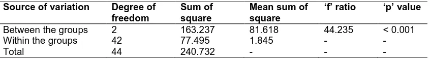
Table 1. Analysis of variance of shear bond strength in various groups
Source of variation Degree of freedom
Sum of square
Mean sum of square
‘f’ ratio ‘p’ value
Between the groups 2 163.237 81.618 44.235 < 0.001
Within the groups 42 77.495 1.845 - -
Total 44 240.732 - - -
Table 2. Comparison of Shear Bond strength of Group-A & B
Group-A Group-B
Number of sample 15 15
Mean shear bond strength (in MPa) 15.56 18.97
Standard Deviation (S.D.) 1.772 1.7327
Standard Error (S.E.) 0.2402 0.3536
Range (in MPa) 13.68 - 17.67 16.06 – 21.65
(‘t’ = 6.31, ‘p’< 0.001)
Table 3. Comparison of Shear Bond Strength of Group-B & C
Group-B Group-C
Number of sample 15 15
Mean shear bond strength (in MPa) 18.97 20.02
Standard Deviation (S.D.) 1.7327 1.0712
Standard Error (S.E.) 0.3536 0.2186
Range (in MPa) 16.06 – 21.65 18.04 – 21.77
The investigations revealed that the mean shear bond strength of microfilled composite was 15.56±1.77 MPa approximating the results of Roh and Chung [16] and Almuammar et al. [26]. The mean Shear Bond strengths of nanocomposites was 18.96±1.73 MPa and that of microhybrid was 20.02±1.07 MPa.
Every effort to minimize the formation smear layer was taken such as the preparation of the facial surface of crown was done under water cooling, as smear layer has an impact on bond strength.
Absence of thermocycling can be a drawback of the present study, however there is a question that arises over the validity of this procedure
since the temperatures used to stress
restorations may not be the real temperatures of cold and hot food/beverage tolerated by patients [27].
Mode of failure during bond strength
determination was also not determined, which was another drawback of the study.
Therefore, based on the results of the study, it can be said that nanocomposites are ideal for both anterior and posterior restorations as they possess excellent esthetic and mechanical
properties. This is analogous to the
investigations done by previous researchers who demonstrated that microfilled composite resin exhibited the lowest mechanical properties and nanofilled resin composites showed mechanical properties as good as those of universal hybrids and therefore can be used in similar clinical situations [8].
Unfortunately, restorations are often done in conditions that are far from ideal i.e., in a complex oral environment. Hence, the physical
and mechanical properties of composite
restorations may practically differ in a clinical environment.
5. CONCLUSION
Within the limitations of the study it can be concluded that Microfilled composite resin exhibited the lowest mechanical properties. Nanofilled resin composites showed mechanical properties atleast as good as those of the universal hybrids. So based on the strength and
esthetic properties of the resin based
nanocomposites should allow the clinican to use it for both anterior and posterior restorations.
Further studies to test the clinical performance of these new generation composites should to be conducted for universal acceptance.
CONSENT
It is not applicable.
ETHICAL APPROVAL
All authors hereby declare that all experiments have been examined and approved by the appropriate ethics committee and have therefore been performed in accordance with the ethical standards laid down in the 1964 Declaration of Helsinki.
COMPETING INTERESTS
Authors have declared that no competing interests exist.
REFERENCES
1. Denehy GE. A direct approach to restore anterior teeth. Am J Dent. 2000;13:55-9. 2. Kirk RE, Othmer DF, Kroschwitz J,
Howe-Grant M. Encyclopedia of chemical
technology. 4th Ed. New York, Wiley; 1991;397.
3. Whitesides GM, Christopher Love J. The art of building small. Sci. Am. 2001;285(3): 38-47.
4. Condon JR, Ferracane JL. Reduced
polymerization stress through non-bonded nanofiller particles. Biomaterials. 2002; 23(18):3807-15.
5. Moszner Norbert, Klapdohr Simone.
Nanotechnology for dental composites. Int J of Nanotechnology. 2004;1(1-2).
6. Kim JS, Cho BH, Lee IB, Um CM, Lim BS,
Oh MH, Chong CG, Son HH. Effect of hydrophilic loading on the mechanical properties and the microtensile bond strength of an ethanol-based one-bottle dentin adhesive. J Biomed Mater Res B Appl Biomaterials. 2005;72(2):284-91. 7. Yap AU, Lim LY, Yang TY, Ali A, Chung
SM. Influence of dietary solvents on
strength of nanofill and ormocer
composites. Oper Dent. 2005;30(1):129-133.
9. Chen Min-Huey, Chen Ci-Rong, Haw Hsu Seng, Shih-Po Sun, Wei-Fang Su. Low shrinkage light curable nanocomposite for dental restorative material. Dent Mater. 2006;22:138-145.
10. Giannelis EP. Polymer layered silicate nanocomposites. Adv Mater. 1996;8:29-35.
11. Mitra SB, Wu D, Holmes BN. An
application of nanotechnology in advanced dental materials. J Am Dent Assoc. 2003; 134(10):1382-90.
12. Xu HHK, Quinn JB, Giuseppetti AA. Wear and mechanical properties of nano-silica-fused whisker composites. J Dent Res. 2004;83(12):930-35.
13. Kim JS, Cho BH, Lee IB, Um CM, Lim BS,
oh MH, Chong CG, Son HH. Effect of hydrophilic loading on the mechanical properties and the microtensile bond strength of an ethanol-based one-bottle dentin adhesive. J Biomed Mater Res B Appl Biomaterials. 2005;72(2):284-91. 14. Rodrigues SA Jr, Scherrer SS, Ferracane
JL, Della Bona A. Microstructural
characterization and fracture behavior of microhybrid and a nanofill composite. Dent Mater. 2008;24(9):1281-8.
15. Endo T, Finger WJ, Kanehira M, Utterodt A, Komatsu M. Surface texture and roughness of polished nanofill and nanohybrid resin composites. Dental Mater J. 2010;29(2):213-23.
16. Roh OD, Chung JH. Micro-shear bond strength of five resin-based composites to dentin with five different dentin adhesives. Am J Dent. 2005;18(6):333-7.
17. Yap AU, Lim LY, Yang TY, Ali A, Chung SM. Influence of dietary solvents on
strength of nanofill and ormocer
composites. Oper Dent. 2005;30(1);129-133.
18. Korkmaz Y, Gurgan S, firat E, Nathanson D. Shear bond strength of three different nano-restorative materials to dentin. Oper Dent. 2010;35(1):50-7.
19. Adopted by the 18th WMA General
Assembly, Helsinki, Finland, June 1964, and amended by the: 29th WMA General Assembly, Tokyo, Japan, October 1975
and 52nd WMA General Assembly,
Edinburgh, Scotland, October 2000.
20. Schneider BT, Baumann MA, Watanabe
LG, Marshall Jr GW. Dentin shear bond strength of compomers and composites. Dent. Mater. 2000;16:15-19.
21. Levitch LC, Bader JD, Shugar DA,
Heyman HO. Non-carious cervical lesions. J Dent. 1994;22:195-207.
22. Craig RG. Mechanical Properties. In: Craig RG, ed. Restorative dental materials. 6th edn. St. Louis, USA: CV Mosby Co. 1980;76-78.
23. McCoy G. The aetiology of gingival erosion. J Oral Implantol. 1982;10:361-362.
24. Lee WC, Eakle WS. Possible role of tensile stress in the aetiology of cervical erosive lesions of teeth. J Prosthet Dent. 1984;52: 374-380.
25. ISO/Technical committee. 106/SC1/WG11.
Dental Material Guidance on testing of adhesion to tooth structure. Committee draft: ISO/TC 106/SC1 N 236, resolution 6, Trieste; 1991.
26. Almuammar MF, Schulman A, Salama FS.
Shear bond strength of six restorative materials. J Cli Pediatr Dent. Spring. 2001; 25(3):221-5.
27. Pazinatto FB, Campos BB, Costa LC, Atta
MT. Effect of thermocycles on
microleakage of resin composite
restorations. Pesqui Odontol Bras.
2003;17(4):337-41.
_________________________________________________________________________________ © 2017 Singh et al.; This is an Open Access article distributed under the terms of the Creative Commons Attribution License (http://creativecommons.org/licenses/by/4.0), which permits unrestricted use, distribution, and reproduction in any medium, provided the original work is properly cited.
Peer-review history: