Clinical Ophthalmology 2016:10 1853–1858
Clinical Ophthalmology
Dove
press
submit your manuscript | www.dovepress.com 1853
C l i n i C a l T r i a l r e p O rT
open access to scientific and medical research
Open access Full Text article
Intravitreal ziv-aflibercept for macular edema
following retinal vein occlusion
remya paulose1
Jay Chhablani1
Chintan J Dedhia1
Michael W Stewart2
ahmad M Mansour3,4
1Smt. Kanuri Santhamma Centre for
Vitreoretinal Diseases, lV prasad
Eye Institute, Hyderabad, India;
2Department of Ophthalmology,
Mayo Clinic, Jacksonville, FL, USA;
3Department of Ophthalmology,
American University of Beirut,
4Department of Ophthalmology, Rafic
Hariri University Hospital, Beirut, Lebanon
Aim: To report the efficacy of intravitreal ziv-aflibercept injections in eyes with macular edema due to retinal vein occlusions (RVOs).
Methods: Consecutive patients with persistent or recurrent macular edema (central macula
thickness .250 µm) due to RVO were enrolled in this prospective study. Study eyes received intravitreal injections of ziv-aflibercept (1.25 mg/0.05 mL) at baseline. Patients were reassessed monthly for 4 months and given additional injections pro re nata for worsening best-corrected visual acuity (BCVA), intraretinal edema or subretinal fluid seen on spectral domain optical coherence tomography, or central macular thickness (CMT) measurements .250 µm. The primary endpoint was improvement in mean CMT at 4 months. Secondary endpoints included improvement in mean BCVA, and ocular and systemic safety signals.
Results: Nine eyes (five central and four branch RVOs) of nine patients were enrolled. The
mean ± standard deviation CMT decreased from 604±199 µm at baseline to 319±115 µm (P=0.001) at 1 month and to 351±205 µm (P=0.026) at 4 months. The mean BCVA did not improve significantly from baseline (1.00 LogMAR) to the 1-month (0.74 LogMAR; P=0.2) and 4-month (0.71 LogMAR; P=0.13) visits. No safety signals were noted.
Conclusion: In this small prospective study, intravitreal ziv-aflibercept significantly improved
mean CMT in eyes with persistent or recurrent macular edema due to RVOs. Prospective, ran-domized trials comparing ziv-aflibercept with standard pharmacotherapy are needed to better define efficacy and safety.
Keywords: ziv-aflibercept, retinal vein occlusion, cystoid macular edema
Introduction
Macular edema due to retinal vein occlusions (RVOs), the second most common retinal vascular condition, frequently results in significant loss of best-corrected visual acuity (BCVA). Intravitreal pharmacotherapy with corticosteroids and drugs that bind vascular endothelial growth factor (VEGF) usually resolves edema and improves BCVA better than observation in eyes with central retinal vein occlusion (CRVO)1–4
and better than laser photocoagulation in eyes with branch retinal vein occlusion (BRVO).5,6 Although most of these eyes respond favorably to first-line intravitreal
pharmacotherapy, improvements are usually transient7,8 and some eyes experience no
improvement with available therapy. Achieving and maintaining a satisfactory clini-cal response usually becomes an expensive, long-term process that often challenges patient compliance.
Aflibercept (Eylea®, Regeneron, Tarrytown, NY, USA) is the newest anti-VEGF
drug that has been approved for the treatment of macular edema due to RVO. This recombinant fusion protein, composed of extra-cytoplasmic, native-receptor VEGF-binding sequences from VEGF receptor (VEGFR) 1 and VEGFR2,9 binds VEGF
165
Correspondence: ahmad M Mansour
Department of Ophthalmology, American University of Beirut, POB 113-6044, Beirut, Lebanon
Tel +961 1 337 7633
email ammansourmd@gmail.com
Journal name: Clinical Ophthalmology Article Designation: Clinical Trial Report Year: 2016
Volume: 10
Running head verso: Paulose et al
Running head recto: Intravitreal ziv-aflibercept for macular edema DOI: http://dx.doi.org/10.2147/OPTH.S116343
Clinical Ophthalmology downloaded from https://www.dovepress.com/ by 118.70.13.36 on 21-Aug-2020
For personal use only.
Number of times this article has been viewed
This article was published in the following Dove Press journal: Clinical Ophthalmology
Dovepress
paulose et al
100 times tighter than either bevacizumab or ranibizumab, and also binds isoforms of VEGF-B and placental growth factor.10 The VIBRANT trial demonstrated that aflibercept
was superior to laser photocoagulation in eyes with BRVO,6
and the COPERNICUS and GALILEO trials showed that aflibercept was superior to sham in eyes with CRVO.3,4
Aflibercept’s strong VEGF-binding affinity may provide a therapeutic advantage over other VEGF-binding drugs when treating vascular conditions with high intravitreal VEGF concentrations such as diabetic macular edema (DME), proliferative diabetic retinopathy, and RVOs, but very few randomized, multicenter, double-masked trials have directly compared aflibercept with bevacizumab and ranibizumab. DRCR.net Protocol T showed that for eyes with DME, aflibercept produced superior 1-year improvements in BCVA compared to bevacizumab and ranibizumab,11 though
statistical superiority was not maintained through 2 years.12
The ongoing SCORE-2 trial compares aflibercept with bevacizumab for primary treatment of macular edema due to RVOs, with aflibercept also serving as rescue therapy for eyes that fail to respond favorably to bevacizumab (https://
clinicaltrials.gov/ct2/show/NCT01969708).
Aflibercept use in developing countries is limited due to its high cost and narrow regulatory approvals. Our group recently reported the short-term safety of intravitreal ziv-aflibercept (Zaltrap®; Regeneron, Tarrytown, NY, USA),
which has the identical molecular structure as aflibercept, in eyes with neovascular age-related macular degeneration (nAMD).13–15 The purpose of the study described in this
manuscript was to evaluate the 4-month safety and efficacy of intravitreal ziv-aflibercept in eyes with RVO that had previously received intravitreal pharmacotherapy.
Methods
This two-center, open-label, nonrandomized, single-arm, prospective study enrolled patients between April 2015 and October 2016. Institutional Review Board approval was granted at LV Prasad Eye Institute, Hyderabad, India and Rafic Hariri University Hospital, Beirut, Lebanon and the study adhered to the tenets of the Declaration of Helsinki. Written informed consent to participate in the study was given by each patient after learning about the study method-ology and the possible risks and benefits. The clinical trial registry number is NCT02486484.
inclusion and exclusion criteria
Key inclusion criteria included previously treated (intra-vitreal bevacizumab, ranibizumab, dexamethasone insert, or laser photocoagulation) macular edema due to RVO that
either recurred since the last treatment or never satisfactorily resolved after at least 3 months of therapy. A central macular thickness (CMT) of at least 250 µm on spectral-domain optical coherence tomography (SD-OCT) was required for enrollment.
Exclusion criteria included uncontrolled systemic vascular disease (including systemic arterial hypertension and diabetes mellitus), glaucoma, vitreomacular interface disorders in the study eye that could limit resolution of edema and improvement in BCVA, and previous vitrectomy.
Clinical evaluations and treatments
Clinical examinations included determination of best- corrected Snellen visual acuity, intraocular pressure measure-ment with applanation tonometry, slit-lamp examination of the anterior segment, biomicroscopic and indirect ophthal-moscopic evaluation of the fundus, and SD-OCT scanning of the central macula. CMT was measured with the 3D OCT-2000 FA plus Topcon® (Topcon, Tokyo, Japan) in
Lebanon and with the Cirrus® (Carl Zeiss Meditec, Dublin,
CA, USA) in India. All CMT measurements taken with the Topcon scanner were converted to Cirrus measurements by adding 23.6 µm, consistent with the normalization proposed by Lammer et al.16 Intravenous fluorescein angiography was
performed at baseline.
Ziv-aflibercept was prepared and injected according to standardized protocols.13 After instillation of topical
anes-thesia and povidone–iodine solution onto the conjunctiva, a sterile eyelid retractor was positioned. Ziv-aflibercept (1.25 mg/0.05 mL) was injected through the sclera (3.5–4 mm posterior to the limbus) into the mid-vitreous. All study eyes received ziv-aflibercept injections at baseline.
Patients received comprehensive eye examinations and SD-OCT macula scans monthly for 4 months. Additional injections of ziv–aflibercept were performed on a pro re nata basis if the BCVA worsened, intraretinal edema or subretinal fluid was noted, or the CMT measured .250 µm.
Outcomes
The primary outcome measure was the change in mean CMT from baseline to month 4. Secondary outcomes included any change in the mean BCVA at 4 months and ocular and systemic safety signals.
Statistical analyses
Statistical analyses were performed using SPSS version 22 (IBM Corporation, Chicago, IL, USA). Snellen BCVA val-ues were converted into LogMAR for statistical analysis. Changes in CMT and BCVA were assessed with the
Clinical Ophthalmology downloaded from https://www.dovepress.com/ by 118.70.13.36 on 21-Aug-2020
Dovepress Intravitreal ziv-aflibercept for macular edema
Wilcoxon-signed rank test and P-values of ,0.05 were deemed statistically significant.
Results
Baseline characteristics
Nine eyes (five eyes with CRVO and four eyes with BRVO) of nine patients were enrolled in this study. Baseline charac-teristics of the patients and study eyes are listed in Table 1. The mean ± standard deviation age of patients in the cohort was 64±8 years and eight of the nine (89%) patients were males. Diabetes mellitus and systemic arterial hyper-tension were each found in six patients (67%), coronary artery disease in two (22%), and hyperhomocysteinemia in one (11%). None of the patients had features of diabetic retinopathy in the non-study eyes.
Prior to enrollment in this study, the patients had fluores-cein angiography and had previously been treated with laser photocoagulation and intravitreal injections of bevacizumab, ranibizumab, triamcinolone, or dexamethasone implant (Table 1). The median number of prior intravitreal injections was 5 (range: 2–24) and the mean duration since the last injection was 11±20 months (range: 1–63 months).
Study treatments
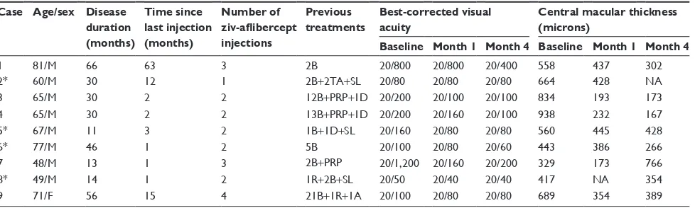
The mean number of ziv-aflibercept injections administered during the 4-month study period was 2.3±0.9 (range: 1–4). The mean treatment-free interval after switching to ziv-aflibercept was 1.9±0.2 months (range: 1–2 months). Figure 1 shows the treatment response of a representative case.
Visual acuity
The mean BCVA at study entry was 1.0 LogMAR (Snellen equivalent: 20/200) with a range of 0.4 to 1.7 LogMAR (Snellen equivalent: 20/50 to 20/1,200). At 1 month, the
mean BCVA improved to 0.74 LogMAR (Snellen equivalent: 20/120; P=0.2) and at 4 months to 0.71 LogMAR (Snellen equivalent: 20/100; P=0.13). Visual acuity improved in most eyes (eight of nine) at 4 months (median improvement from 20/160 to 20/80), with greater changes seen in the eyes that were last treated within 3 months (improvement in median BCVA from 20/180 to 20/90) compared to those last treated .12 months previously (median improvement from 20/100 to 20/80).
Macular thickness
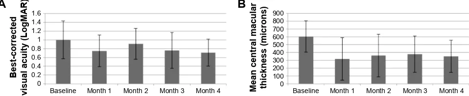
The mean CMT at baseline (603±199 µm) decreased to 319±115 µm (P=0.001) at 1 month and to 351±205 µm (P=0.026) at 4 months. Significant improvements in CMT occurred both in eyes that had received intravitreal pharmacotherapy only 1 to 3 months previously (mean improvement in CMT from 587 µm to 370 µm) and also in eyes that had last been treated over 1 year prior (mean improvement in CMT from 637 to 361 µm). Changes in BCVA and CMT throughout the study period are shown in Figure 2.
Safety
One eye (Case 9) developed minimal anterior chamber flare 2 days after the fourth injection. Cases 1–8 did not develop intraocular inflammation. None of the patients experi-enced systemic adverse events that could be attributed to intravitreal ziv-aflibercept. The mean intraocular pressure did not change from baseline (13.2±3.3 mmHg) to month 4 (13.4±2.3 mmHg).
Discussion
In this study of patients with previously treated macular edema due to RVOs, we found that switching to ziv-aflibercept
Table 1 Clinical characteristics of study patients
Case Age/sex Disease duration (months)
Time since last injection (months)
Number of ziv-aflibercept injections
Previous treatments
Best-corrected visual acuity
Central macular thickness (microns)
Baseline Month 1 Month 4 Baseline Month 1 Month 4
1 81/M 66 63 3 2B 20/800 20/800 20/400 558 437 302
2* 60/M 30 12 1 2B+2Ta+Sl 20/80 20/80 20/80 664 428 na
3 65/M 30 2 2 12B+prp+1D 20/200 20/100 20/100 834 193 173
4 65/M 30 2 2 13B+prp+1D 20/200 20/160 20/100 938 232 167
5* 67/M 11 3 2 1B+1D+Sl 20/160 20/80 20/80 560 445 428
6* 77/M 46 1 2 5B 20/100 20/80 20/60 443 386 266
7 48/M 13 1 3 2B+prp 20/1,200 20/160 20/200 329 173 766
8* 49/M 14 1 2 1r+2B+Sl 20/50 20/40 20/40 417 na 354
9 71/F 56 15 4 21B+1r+1a 20/100 20/80 20/80 689 354 389
Note: *Eyes with branch retinal vein occlusion, others were central retinal vein occlusion.
Abbreviations: A, intravitreal aflibercept (2 mg/0.05 mL); B, intravitreal bevacizumab (1.25 mg/0.05 mL); D, intravitreal dexamethasone insert; F, female; M, male; NA, not available; PRP, panretinal photocoagulation; R, intravitreal ranibizumab (0.3 mg/0.05 mL); SL, sectoral laser; TA, intravitreal triamcinolone (2 mg/0.1 mL).
Clinical Ophthalmology downloaded from https://www.dovepress.com/ by 118.70.13.36 on 21-Aug-2020
Dovepress
paulose et al
resulted in significant improvements in macular edema and modest improvements in BCVA, most likely due to the chronicity of the disease at presentation.
The Phase III registration trials demonstrated that intra-vitreal ranibizumab and aflibercept are superior to standard-of-care for the treatment of macular edema due to BRVO and CRVO.2–6 Bevacizumab has not been studied with the
same rigor as ranibizumab and aflibercept but bevacizumab regimens appear to produce comparable improvements in macular edema and BCVA.17,18 Most eyes with RVO respond
well to bevacizumab or ranibizumab but a small proportion fail to achieve satisfactory responses with initial therapy or experience an unacceptably early or frequent recurrence of edema. Small retrospective studies reported visual gains after switching to aflibercept but since standardized procedures and control arms were lacking, the significance of these findings is unclear.19,20 Nonetheless, switching incomplete
responders to aflibercept appears to be a reasonable treat-ment option.
Surgeons in many developing countries cannot use aflibercept to treat RVOs because it may be excluded from the formularies of insurance plans or national health care plans and its high cost ($1,850 US per dose) prevents many patients from paying out-of-pocket. In countries such as India, aflibercept has been approved for the treatment of nAMD but not yet for RVOs.
Just as off-label bevacizumab became an accepted, affordable treatment for chorioretinal vascular disorders, ziv-aflibercept could become a similar option in countries with limited access to aflibercept. Ziv-aflibercept was approved by the US Food and Drug Administration in August 2012 for the intravenous treatment of advanced colorectal carcinoma.21 Ziv-aflibercept is packaged in a single use 4 mL
vial (1.25 mg/0.05 mL) and costs ($512 US) substantially less than aflibercept and even slightly less than bevacizumab (4 mL vial costs $660).
Buffering solutions in ziv-aflibercept produce a high osmolality (1,000 mOsm/kg) that is toxic to most cells.22
Concerns regarding hyperosmolality-induced retinal tox-icity due to intravitreal ziv-aflibercept have been largely dispelled by recent publications.23 Case reports describing
ziv-aflibercept treatment of nAMD, polypoidal choroidal vasculopathy, and CRVO identified no safety concerns.13,14,24
Mansour et al noted no ocular toxicity with intravitreal ziv-aflibercept in patients with DME and nAMD, and they pos-ited that the small volume of high osmolality ziv-aflibercept (0.05 mL) is adequately diluted by the relatively large vitreous cavity (4 mL).14 Chhablani et al noted no clinical
$
$WEDVHOLQH$WPRQWK
$WPRQWK
$WPRQWK
$WPRQWK
6
, 1 7
6
1 7
6
, 1 7
6
, 1 7
6
, 1 7
%
&
'
(
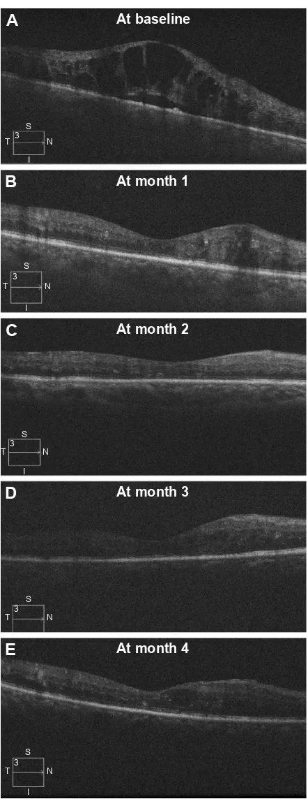
Figure 1 Serial linear OCT scan after intravitreal ziv-aflibercept injections.
Notes: a 65-year-old male developed macular edema due to a CrVO. Over a
2.5-year period, he received 12 anti-vascular endothelial growth factor injections,
intravitreal triamcinolone, and peripheral retinal photocoagulation. Upon entering
this study, his visual acuity measured 20/200 with a central macular thickness of
938 µm (A). Intravitreal ziv-aflibercept injections at baseline and month 1 resolved the macular edema (B and C), after which he was observed until the study’s primary temporal endpoint (D and E). His visual acuity improved to 20/60.
Abbreviations: CRVO, central retinal vein occlusion; OCT, optical coherence tomography; T, temporal; S, superior; N, nasal; I, inferior.
Clinical Ophthalmology downloaded from https://www.dovepress.com/ by 118.70.13.36 on 21-Aug-2020
Dovepress Intravitreal ziv-aflibercept for macular edema
toxicity and detected no electroretinographic abnormalities in 12 eyes that received single injections of ziv-aflibercept for treatment-resistant nAMD.13
Most eyes in our study, whether last treated within the previous 3 months or more than a year prior, experienced decrease in macular edema. Visual acuity improvements favored eyes treated within the past 3 months but our small cohort is insufficiently powered to draw firm conclusions. Larger RVO trials suggest that even brief periods of under-treatment (as short as 6 months) may result in poorer long-term visual outcomes.2,4 Nonetheless, our results support the
notion that RVO patients should be followed closely so that recurrent edema can be treated promptly.
The optimal dose of intravitreal ziv-aflibercept is not known. We injected 0.05 mL (1.25 mg) of undiluted ziv-aflibercept so that the small volume would minimize acute elevations in intraocular pressure. The approved dose of aflibercept (2 mg/0.05 mL) delivers 60% more drug than was used in our trial but smaller doses of aflibercept have not been reported in eyes with RVOs. Patients treated with 0.5 mg aflibercept in the VIEW trials achieved comparable results to those receiving the 2 mg dose, suggesting that dose size may not be a critical determinant of clinical response.25
Dose range determinations with ziv-aflibercept continue as a higher dose (2 mg/0.08 mL) is being evaluated.
Our study has several strengths and weaknesses. To the best of our knowledge, this is the first prospective study to evaluate the safety and efficacy of ziv-aflibercept for macular edema due to RVOs. The study reports data gathered from more than one site but the small cohort prevents us from detecting statistically significant visual acuity changes. Because of the small cohort, we elected to analyze eyes with central RVO and branch RVO together, even though these conditions often have different baseline characteristics and responses to treatment. The mixed composition of the cohort – patients who were actively being treated for RVO and others whose last injections were at
least 1 year prior – makes it difficult to use the data to create firm indications for switching patients to ziv-aflibercept. The 4-month primary endpoint is brief and longer follow-up periods are needed.
Despite the shortcomings of this pilot study, we believe that these data support the use of ziv-aflibercept in selected patients with RVOs. This study adds to the growing body of data supporting the safety of intravitreal ziv-aflibercept in eyes with chorioretinal vascular conditions. Whether intravitreal ziv-aflibercept would be an acceptable option in countries where aflibercept has already been approved for the treatment of RVOs is unclear, but in lower income countries that have not approved aflibercept, ziv-aflibercept could become a cost-effective alternative to other anti-VEGF drugs. Larger, multicenter, prospective, controlled studies are needed to better define the efficacy of ziv-aflibercept in patients with macular edema due to RVOs.
Disclosure
The authors report no conflicts of interest in this work.
References
1. Haller JA, Bandello F, Belfort R Jr, et al. Dexamethasone intravitreal implant in patients with macular edema related to branch or central retinal vein occlusion twelve-month study results. Ophthalmology. 2011;118(12): 2453–2460.
2. Campochiaro PA, Brown DM, Awh CC, et al. Sustained benefits from ranibizumab for macular edema following central retinal vein occlusion: twelve-month outcomes of a phase III study. Ophthalmology. 2011; 118(10):2041–2049.
3. Heier JS, Clark WL, Boyer DS, et al. Intravitreal aflibercept injection for macular edema due to central retinal vein occlusion: two-year results from the COPERNICUS study. Ophthalmology. 2014;121(17):1414–1420. 4. Ogura Y, Roider J, Korobelnik JF. et al. Intravitreal aflibercept for macular
edema secondary to central retinal vein occlusion: 18-month results of the phase 3 GALILEO study. Am J Ophthalmol. 2014;158(5):1032–1038.
5. Brown DM, Campochiaro PA, Bhisitkul RB, et al. Sustained benefits from ranibizumab for macular edema following branch retinal vein occlusion: 12-month outcomes of a phase III study. Ophthalmology. 2011; 118(8):1594–1602.
6. Campochiaro PA, Clark WL, Boyer DS, et al. Intravitreal aflibercept for macular edema following branch retinal vein occlusion: the 24-week results of the VIBRANT study. Ophthalmology. 2015;122(3):538–544.
%DVHOLQH 0RQWK 0RQWK 0RQWK 0RQWK
%DVHOLQH 0RQWK 0RQWK 0RQWK 0RQWK
$
%
%HVWFRUUHFWHG
YLVXDODFXLW\/RJ0$5 0HDQFHQWUDOPDFXODU WKLFNQHVVPLFURQV
Figure 2 Mean best-corrected visual acuity (A) and mean central macular thickness (B) with standard deviation limits at each study visit.
Notes: Mean best-corrected visual acuity improved through 4 months but changes were not statistically significant. Compared to baseline, mean central macular thickness was significantly better at each follow-up visit.
Clinical Ophthalmology downloaded from https://www.dovepress.com/ by 118.70.13.36 on 21-Aug-2020
Clinical Ophthalmology
Publish your work in this journal
Submit your manuscript here: http://www.dovepress.com/clinical-ophthalmology-journal
Clinical Ophthalmology is an international, peer-reviewed journal covering all subspecialties within ophthalmology. Key topics include: Optometry; Visual science; Pharmacology and drug therapy in eye diseases; Basic Sciences; Primary and Secondary eye care; Patient Safety and Quality of Care Improvements. This journal is indexed on
PubMed Central and CAS, and is the official journal of The Society of Clinical Ophthalmology (SCO). The manuscript management system is completely online and includes a very quick and fair peer-review system, which is all easy to use. Visit http://www.dovepress.com/ testimonials.php to read real quotes from published authors.
Dovepress
Dove
press
paulose et al
7. Heier JS, Campochiaro PA, Yau L, et al. Ranibizumab for macular edema due to retinal vein occlusions: long-term follow-up in the HORIZON trial. Ophthalmology. 2012;119(4):802–809.
8. Campochiaro PA, Sophie R, Pearlman J, et al; RETAIN Study Group. Long-term outcomes in patients with retinal vein occlusion treated with ranibizumab. the RETAIN study. Ophthalmology. 2014;121(1): 209–219.
9. Holash J, Davis S, Papadopoulos N, et al. VEGF-Trap: a VEGF blocker with potent antitumor effects. Proc Natl Acad Sci U S A. 2002;99(17): 11393–11398.
10. Papadopoulos N, Martin, Ruan Q, et al. Binding and neutralization of vascular endothelial growth factor (VEGF) and related ligands by VEGF Trap, ranibizumab and bevacizumab. Angiogenesis. 2012;15(2): 171–185.
11. Diabetic Retinopathy Clinical Research Network, Wells JA, Glassman AR, Ayala AR, et al. Aflibercept, bevacizumab, or ranibizumab for diabetic macular edema. N Engl J Med. 2015;372(13):1193–1203.
12. Wells JA, Glassman AR, Ayala AR, et al; Diabetic Retinopathy Clinical Research Network. Aflibercept, bevacizumab, or ranibizumab for diabetic macular edema: Two-year results from a comparative effectiveness randomized clinical trial. Ophthalmology. 2016;123(6): 1351–1359.
13. Chhablani J, Narayanan R, Mathai A, Yogi R, Stewart M. Short-term safety profile of intravitreal ziv-Aflibercept. Retina. 2016;36(6):1126–1131. 14. Mansour AM, Al-Ghadban SI, Yunis MH, El-Sabban ME. Ziv-aflibercept
in macular disease. Br J Ophthalmol. 2015;99(8):1055–1059. 15. Mansour AM, Chhablani J, Antonios RS, et al. Three-month outcome
of ziv-aflibercept for exudative age-related macular degeneration.
Br J Ophthalmol. Epub 2016 Mar 30.
16. Lammer J, Scholda C, Prunte C, Benesch T, Schmidt-Erfurth U, Bolz M. Retinal thickness and volume measurements in diabetic macular edema: a comparison of four, optical coherence tomography systems. Retina. 2011;31(1):48–55.
17. Prager F, Michels S, Kriechbaum K, et al. Intravitreal bevacizumab (Avastin) for macular oedema secondary to retinal vein occlusion: 12-month results of a prospective clinical trial. Br J Ophthalmol. 2009; 93(4):452–456.
18. Epstein DL, Algvere PV, von Wendt G, Seregard S, Kvanta A. Benefit from bevacizumab for macular edema in central retinal vein occlu-sion: twelve-month results of a prospective, randomized study.
Ophthalmology. 2012;119(12):2587–2591.
19. Papakostas TD, Lim L, van Zyl T, et al. Intravitreal aflibercept for macular oedema secondary to central retinal vein occlusion in patients with prior treatment with bevacizumab or ranibizumab. Eye (Lond). 2016;30(1):79–84.
20. Lehmann-Clarke L, Dirani A, Mantel I, Ambresin A. The effect of switching ranibizumab to aflibercept in refractory cases of macular edema secondary to ischemic central vein occlusion. Klin Monbl
Augenheilkd. 2015;232(4):552–555.
21. Chung C, Pherwani N. Ziv-aflibercept: a novel angiogenesis inhibitor for the treatment of metastatic colorectal cancer. Am J Health Syst
Pharm. 2013;70(21):1887–1896.
22. Marmor MF, Martin LJ, Tharpe S. Osmotically induced retinal detach-ment in the rabbit and primate. electron miscoscopy of the pigdetach-ment epithelium. Invest Ophthalmol Vis Sci. 1980;19(9):1016–1029. 23. Malik D, Tarek M, Caceres del Carpio J, et al. Safety profiles of
anti-VEGF drugs: bevacizumab, ranibizumab, aflibercept and ziv-aflibercept on human retinal pigment epithelium cells in culture. Br J Ophthalmol. 2014;98 Suppl 1:11–16.
24. Chhablani J. Intravitreal ziv-aflibercept for recurrent macular edema secondary to central retinal venous occlusion. Indian J Ophthalmol. 2015; 63(5):469–470.
25. Schmidt-Erfurth U, Kaiser PK, Korobelnik JF, et al. Intravitreal aflibercept injection for neovascular age-related macular degeneration: ninety-six-week results of the VIEW studies. Ophthalmology. 2014; 121(1):193–201.
Clinical Ophthalmology downloaded from https://www.dovepress.com/ by 118.70.13.36 on 21-Aug-2020