_____________________________________________________________________________________________________
www.sciencedomain.org
Superficial Mycoses in Relation to Age and Gender
O. A. Oninla
1*and S. O. Oninla
21Department of Dermatology and Venereology, Obafemi Awolowo University, Ile-Ife,
Osun State, Nigeria. 2
Department of Paediatrics and Child Health, Ladoke Akintola University of Technology, Osogbo, Osun State, Nigeria.
Authors’ contributions This work was carried out in collaboration between both authors. Author OAO conceived and designed the study, analyzed the data and wrote the first draft of the manuscript. Author SOO contributed to the writing of the manuscript and analyzed the data. Authors OAO and SOO agree with manuscript results and conclusions and jointly developed the structure and arguments for the paper.
Both authors read and approved the final manuscript. Article Information
DOI: 10.9734/BJMMR/2016/23455
Editor(s):
(1)Roberto Manfredi, Department of Medical and Surgical Sciences, University of Bologna, Bologna, Italy.
Reviewers:
(1) Mutlu Cayirli, Mevki Military Hospital, Ankara, Turkey. (2)Ajith Prasanna Kannangara, Ministry of Health, Sri Lanka. (3)Farhana Tahseen Taj, KLE University, Karnataka, India. Complete Peer review History:http://sciencedomain.org/review-history/13032
Received 1st December 2015 Accepted 5th January 2016 Published 20th January 2016
ABSTRACT
Aims: To determine the type of superficial fungi infections of the skin affecting different ages and gender at a dermatology clinic in a developing country.
Study Design: A descriptive cross-sectional study.
Place and Duration of Study: Dermatology Clinics of Obafemi Awolowo University Teaching Hospitals’ Complex, (OAUTHC), Ile-Ife, Osun State, Nigeria between October 2009 and September 2012.
Methodology: Demographic information, history and clinical examination of consecutive patients with fungi infections of the skin were documented. The diagnoses were mainly clinical, and necessary laboratory procedures were performed for confirmation. Statistical analysis was done using SPSS 16 and odds ratio with 95% confidence interval obtained using WINPEPI version 11.
Results: The total number of new cases of dermatoses presenting during the study period were 1454, from few weeks to 90 years of age, and 55% were females. Patients affected constituted
39.5%. Superficial fungi infections found were Dermatophytes (13.1%), Pityriasis versicolor (4.3%), and Candidiasis (2.1%). Children had more infections than adults (P = 0.000; OR = 9.31; 95% C.I. = 6.93; 12.50). The most common type was tinea capitis (children), pityriasis versicolor (adults), tinea unguium (females), and tinea cruris (males). Dermatophytic infections and candidiasis were significantly associated with childhood and female gender respectively.
Conclusion: Superficial fungi diseases are the most common dermatoses presenting to dermatologists in Nigeria and affect different ages and both gender invariably. Tinea capitis occurred predominantly in children, and pityriasis versicolor, tinea unguium and tinea cruris in adults, females and males respectively. Adequate control will lead to drastic fall in the number of skin problems confronting the general populace.
Keywords: Superficial fungi; skin infections; dermatoses; childhood infections. 1. INTRODUCTION
Skin conditions affecting the Nigeria populace are mostly infective skin diseases [1-3]. Majority of these infections are fungal diseases [4,5]. Superficial fungi skin infections are fungal diseases affecting primarily, the skin, the mucous membranes (oropharyngeal, esophageal, vulvovaginal and urethral), nails, and hair [6,7,8]. They have been found to be prevalent and account for most of the skin diseases in schools, prisons, community and among patients presenting to the hospitals [9-14]. The superficial fungi infections mainly consist of dermatophyte infections caused by Trichophyton species, Epidermophyton species and Microsporum species, candidiasis due to Candida albicans, glabrata etc, and pityriasis versicolor due to Malassezia furfur [15].
Superficial fungi infections still need to be addressed as a public health problem among the growing populace of Nigeria. The favorable environment of hot and humid climate, poverty, poor sanitary conditions and overcrowding are well known factors that favor these fungi growth. These conditions well abound in Nigeria. The aim of the study was to determine the superficial fungi infections presenting in a specialist hospital setting. The objectives are 1) to find out the types of superficial fungi infections predominant here and 2) assess the relationship of these infections with age and gender.
2. METHODOLOGY
2.1 Study Design and Setting
A prospective and descriptive cross-sectional study of consecutive patients referred to the Der-matology and Venereology Clinics of Obafemi Awolowo University Teaching Hospitals’
Complex, Ile-Ife, Osun State was done from October 2009 to September.
2.2 Patients and Methods
Information on age, gender, residential address, and presenting complaints were recorded. A thorough physical examination of the patient and the skin presentations was done in broad daylight. The diagnoses were mainly clinical, and where necessary laboratory procedures were performed for confirmation using skin scrapping with 10-20% KOH solution and culture in Sabouraud Dextrose Agar with chloramphenicol. All consenting patients with history and clinical presentations of lesions suggestive of superficial fungi infections were eligible for inclusion. Excluded from the study are those without clear clinical and / or laboratory diagnosis.
Data was analyzed with Statistical Package for Social Sciences Statistics version 16.0 (SPSS Inc. Released 2007. SPSS for Windows Version 16.0, Chicago, SPSS Inc.) (SPSS) was used to obtain percentage frequencies, and cross-tabulation for data presentation. Fishers exact test (with a P value < 0.05 taken as statistically significant), odds ratio (OR), and confidence interval (CI) were performed using WINPEPI version 11.15 [16] to determine relationship of these infections to age and gender.
3. RESULTS AND DISCUSSION
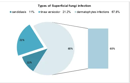
The superficial fungi infections found comprises of 3 major groups- dermatophytes, pityriasis versicolor, and candidiasis (intertrigo and paronychia). Dermatophytic infections were the most common affecting 13.4% of studied population. Pityriasis versicolor and candidiasis were found in 4.2 and 2.2% respectively. Among infected patients, 67.8% had dermatophytic infections while 21.2% had pityriasis versicolor and 11.0% had candidiasis (Fig. 1).
Dermatophytes constituted the majority of infections in both adults and children (Table 1).
The most common type in children was tinea capitis followed by tinea corporis, and then tinea unguium. In adults, the predominant type was tinea corporis, then tinea pedis, and tinea unguium. Tinea capitis was significantly associated with childhood (Table 2). Tinea unguium was very common in both adult and children. There was a significant association of pityriasis versicolor with adulthood. Although candidiasis was more in adults, there was no significant difference in both children and adults.
Fig. 1. Percentage distribution of superficial fungi infections
Table 1. Superficial fungi infections according to age and gender
Superficial fungi infections
Children Adult Total % of total fungi (273)
% in 1,380 patients Males Females Males Females
Candidiasis 3 5 7 15 30 11.0 2.2
Tinea versicolor 2 6 24 26 58 21.2 4.2
Tinea capitis 19 11 2 0 32 11.7 2.3
Tinea corporis 15 8 18 25 66 24.2 4.8
Tinea cruris 4 0 4 1 9 3.3 0.7
Tinea faciae 0 1 1 1 3 1.1 0.2
Tinea manuum 1 0 2 3 6 2.2 0.4
Tinea pedis 3 4 11 17 35 12.8 2.5
Tinea unguium 1 11 8 14 34 12.5 2.5
Total 48 46 77 102 273 100% 19.8%
11% 21%
68% 68%
Types of Superficial fungi infection
4
Table 2. Odds ratio of superficial fungi infections by age and gender
Superficial fungi infections Gender Odds ratio
Males Females
Candidiasis 10 20 P = .11; OR = 0.58; 95% C.I. = 0.27; 1.24
Tinea versicolor 26 32 P = .47; OR = 0.95; 95% C.I. = 0.56; 1.60
Tinea capitis 21 11 P = .02; OR = 2.27; 95% C.I. = 1.09; 4.74*
Tinea corporis 33 33 P = .30; OR = 1.18; 95% C.I. = 0.72; 1.93
Tinea cruris 8 1 P = .01; OR = 9.44; 95% C.I. = 1.18; 75.55*
Tinea faciae 1 2 P = .56; OR = 0.58; 95% C.I. = 0.05; 6.43
Tinea manuum 3 3 P = .58; OR = 1.17; 95% C.I. = 0.24; 5.80
Tinea pedis 14 21 P = .29; OR = 0.77; 95% C.I. = 0.39; 1.53
Tinea unguium 9 25 P = .01; OR = 0.41; 95% C.I. = 0.19; 0.89*
Total (273) 125(45.8%)
19.6% of all males
148(54.2%)
19.9% of all females
All males = 637; all females = 743; Total = 1,380 P = .47; OR = 0.98; 95% C.I. = 0.75; 1.28
Superficial fungi infections Age Odds ratio
Children Adults
Candidiasis 8 22 P = .57; OR = 1.00; 95% C.I. = 0.44; 2.26
Tinea versicolor 8 50 P = .000; OR = 0.17; 95% C.I. = 0.08; 0.37*
Tinea capitis 30 2 P = .000; OR = 45.00; 95% C.I. = 10.71; 189.15*
Tinea corporis 23 43 P = .08; OR = 1.51; 95% C.I. = 0.90; 2.54
Tinea cruris 4 5 P = .20; OR = 2.22; 95% C.I. = 0.59; 8.31
Tinea faciae 1 2 P = .61; OR = 1.38; 95% C.I. = 0.13; 15.24
Tinea manuum 1 5 P = .20; OR = 2.22; 95% C.I. = 0.06; 4.72
Tinea pedis 7 28 P = .25; OR = 0.68; 95% C.I. = 0.30; 1.58
Tinea unguium 12 22 P = .17; OR = 1.55; 95% C.I. = 0.75; 3.11
Total (273) 94(34.4%)
25.6% of all children
179(65.6%) 17.7% of all adults
All children = 367; all adults = 1,013; Total = 1,380 P= .001; OR = 1.60; 95% C.I. = 1.21; 2.13
Candidiasis was found mostly in females though not significantly so. This was also found to be the same for pityriasis versicolor (Fig. 2). Tinea capitis and tinea cruris were significantly associated with the male gender, and tinea unguium with the female gender (Table 2). There was an equal preponderance of tinea corporis in both genders. Tinea faciae and manuum showed no relationship with gender.
4. DISCUSSION
Majority of skin diseases presenting to referral centers particularly dermatology centers are superficial fungal dermatoses [7,17,18]. In some centers where eczemas are the most common, fungi diseases are next in line [18]. Dermatophytes are most often the prevailing fungal diseases in many schools, hospital and community [18-23]. A similar result was obtained in this study.
The finding of pityriasis versicolor and candidiasis in sequence also corresponds to other tertiary hospital reports by Ogunbiyi et al. [21] and Onayemi et al. [22]. The prevalence of 13.4% was, however, much higher than the 4.5% found at Ibadan [21] which is a larger city in an adjoining South-western State. This may be because the study centre subserves many
surrounding rural places. The prevalence of 4.2% and 2.2% for Pityriasis versicolor and candidiasis respectively, were similar to the 4.5% and 1.9% reported by Ogunbiyi et al. at Ibadan. In the Northern Nigeria tertiary hospital study by Onayemi et al., prevalence of dermatophyte infection was 13.4% while Pityriasis versicolor and candidiasis were 6.7% and 4.5% respectively.
A higher percentage of the infections were found in adults than children. However, the proportion of children that had fungi infection was more than adults, and this was significant. This means that the infections were more associated with childhood (P= 0.001). In a study of schoolchildren by Amoran et al. [23], 83.7% of all skin disorders seen in 480 pupils were infective dermatoses with superficial fungal infections (dermatophytoses and pityriasis versicolor) constituting 74.1%. Ogunbiyi et al. [19] reported skin diseases in 35% of school children with 20.6% having fungi infections (dermatophytes- 15.9%; pityriasis versicolor- 4.7%) that are mostly tinea capitis (14.5%). Odueko et al. [20]in a prevalence survey of 5001 Nigerian children aged 0-12 years at the Urban Comprehensive Health Centre, Ile-Ife, Nigeria revealed that 492 children (9.8%) had dermatological conditions with dermatophytic infections in 17.1%.
Tinea capitis was found mostly in children. This is similar to earlier reports of fungal studies in children [24-30]. Amoran et al. reported a prevalence of 16.5% in 480 children [23] while Ogunbiyi et al. found the infection in 14.5% of 375 children [19]. Enemuor et al. [28], also showed that scalp infection (85.4%) was more frequent than infection of the glabrous skin (14.6%). Nweze screened domestic animals for the presence of dermatophytes both clinically and mycologically. An incidence of 39.8% of dermatophytes was obtained in the 538 animals studied. The species found were those pathogenic to man [24]. Children often play with domestic animals and soil, and these could account for the higher prevalence of T. capitis in them. The age predilection of tinea capitis is thought to be due to the fungistatic properties of fatty acids in post-pubertal sebum.
The populations of female and male pupils in the study by Enemuor et al. were 969 and 1215, respectively. The respective prevalence rates of superficial mycoses were 1.5% and 10.6%. Comparison of the prevalence rates of females and males pupils showed a significantly higher (P = 0.0005) prevalence rate in male pupils [28]. A lower prevalent rates of T. capitiswas found in girls than boys in this study. Other reports by Mirmirani et al. [27], Enemuor et al. [28] and Adefemi et al.[29] showed greater affectation of boys by dermatophyte infections, and most especially, tinea capitis.However, Anosike et al. [30] found that girls were affected more though this was not significant. The higher prevalence in male in this environment may be as a result of increased contact between boys during play, sharing of combs and cutting of hair at local barber shops, and possibly more contact with pets during play.
Tinea unguium was more common in adults than children and it was reported as uncommon in children [31]. In this study, it was associated significantly with the female gender. A study by Asadi et al.[32] also revealed the same finding. Females in this environment are predisposed as they are usually involved in house-keeping and cooking resulting in frequent immersion of hands in water which may not be properly dried.
Tinea cruris was more in the male gender and the difference was significant. It is a dermatophytoses that mainly occurs in males [33] affecting men (particularly young) three times more than women [34]. This may be the result of occlusive dressing caused by the male
clothing. It has become more common in post-pubertal females who are overweight or wear tight clothing such as pantyhose [31].
Most of the cases of Tinea pedis found occurred in adults, and in the male gender. There was no significant difference, however, in the prevalent rates of children and adults, and gender. Exposure to a moist environment provided by wearing of occlusive foot-wears and macerated skin predisposes to this infection. It has been observed to be less in communities that do not wear shoes.
Pityriasis versicolor was more prevalent in adults than children [35]. This was also observed in this study. More females than males were, however, affected among both adult and children in contrast to male predominance in the report by Jena et al. [36], Zarrin et al. [37], and Abdul-Hussein [38]. A survey in school children by Uneke et al. revealed a female preponderance [26]. Candidiasis was more in adults and in females. Candidiasis was more in girls than in boys. Altraide et al. [39] also found candida intertrigo/ paronychia to be more in adults and females. This could be a result of domestic work often performed by the female gender involving regular immersion of hands in water. If the hands are not properly dry, the wetness provides good culture medium for candida species particularly
Candida albicans.
5. CONCLUSION
Superficial fungi infections are still very common in this environment affecting about 20% of the study population. They were significantly associated with childhood. Dermatophytic infections were the most predominant in all ages and gender. Tinea capitis and pityriasis versicolor were the most significant types in children and adults respectively. Tinea capitis and tinea cruris were more associated with male gender, and tinea unguium with the female gender.
ETHICAL APPROVAL
COMPETING INTERESTS
Authors have declared that no competing interests exist.
REFERENCES
1. WHO/CAH. Epidemiology and
management of common skin diseases in children in developing countries; 2005. [Cited 2012 Sep 5].
Available:http://whqlibdoc.who.int/hq/2005/ WHO_FCH_CAH_05.12_eng.pdf
2. Alabi GO. Trends in the pattern of skin diseases in Nigeria. Nig Med J. 1980;10:163–8.
3. Fekete E. The pattern of diseases of the skin in the Nigerian Guinea savanna. Int J Dermatol. 1978;17:331–8.
4. Shrank AB, Harman RRM. The incidence of skin diseases in a Nigerian teaching hospital dermatology clinic. Br J Dermatol. 1966;78:235.
5. Oninla OA, Olasode OA, Onayemi O, Ajani AA. The prevalence and pattern of skin disorders at a university teaching hospital in Ile-Ife and Ilesha, Nigeria. Clinical
Medicine Insights: Dermatology.
2014;7:25–31.
DOI: 10.4137/CMD.S14422.
6. Hainer BL. Dermatophyte infections. Am Fam Physician. 2003;67:101-9.
Available:http://www.aafp.org/afp/2003/010 1/p101.html
7. Okafor JI. Fungal diseases: A serious threat to human existence in recent times. [Cited 2014 May 21].
Available:http://www.nuc.edu.ng/nucsite/Fil e/UNN%20Inaugural%20Lectures/41st%2 0Lecture.pdf
8. Gupta S, Gupta BL. Evaluation of the incidences of dermatophillic infection in Rajastahan: Case studies from Rajasthan, India. Int. J. Med. Med. Sci. 2013;5:229-32. DOI: 10.5897/IJMMS2013.0929
Available:http://www.academicjournals.org/ IJMMS
9. Oyeka CA, Eze I. Fungal skin infections among prison inmates in Abakaliki, Nigeria. Mycoses. 2008;51:50–4.
10. Ayanlowo O, Akinkugbe A, Oladele R, Balogun M. Prevalence of Tinea capitis infection among primary school children in a rural setting in south-west Nigeria. JPHIA. 2014;5:349.
DOI: 10.4081/jphia.2014.349
11. Sanuth HA, Efuntoye MO. Distribution and microbiological characterization of dermatophytes infection among primary school children in Ago Iwoye, Ogun State, Nigeria. Researcher; 2010;2(6):95‐9. [Cited 2014 Nov 14].
Available: http://www.sciencepub.net/resea rcher/research0206/12_3202research0206 _95_99.pdf
12. Oninla OA, Onayemi O. Skin infections and infestations in prison inmates. Int J Dermatol. 2012;51:178–81.
13. Okafor OO, Akinbami FO, Orimadegun AE, Okafor CM, Ogunbiyi AO. Prevalence of dermatological lesions in hospitalized children at the University College Hospital, Ibadan, Nigeria. Niger J Clin Pract [serial online]. 2011;14:287-92. [cited 2014 Nov 6] Available:http://www.njcponline.com/text.a sp?2011/14/3/287/86769
14. Nweze EI. Dermatophytosis in Western Africa: A review. Pakistan Journal of Biological Sciences. 2010;13:649-56.
[Cited 2014 Nov 6].
Available:http://scialert.net/abstract/?doi=pj bs.2010.649.656
15. Okafor JI. Fungal diseases: A serious threat to human existence in recent times. [Cited 2014 May 21].
Available:http://www.nuc.edu.ng/nucsite/Fil e/UNN%20Inaugural%20Lectures/41st%2 0Lecture.pdf
16. Abramson JH. WINPEPI updated:
Computer programs for epidemiologists, and their teaching potential. Epidemiol Perspect Innov. 2011;8:1.
17. Emodi IJ, Ikefuna AN, Duru UA. Skin diseases among children attending the children out- patient clinic at the University of Nigeria teaching hospital Enugu, Nigeria. Afr Health Sci. 2010;10:362–66. [PMC free article] [PubMed].
18. Nnoruka EN. Skin diseases in south-east Nigeria: A current perspective. Int J Dermatol. 2005;44:29–33.
19. Ogunbiyi OA, Owoaje E, Ndahi A. Prevalence of skin disorders in school children in Ibadan, Nigeria. Pediatr Dermatol. 2005;22:6-10.
20. Odueko OM, Onayemi O, Oyedeji GA. A prevalence survey of skin diseases in Nigerian children. Niger J Med. 2001;10:64-7.
22. Onayemi O, Isezuo SA, Njoku CH. Prevalence of different skin conditions in an outpatients’ setting in north-western Nigeria. Int J Dermatol. 2005;44:7–11.
23. Amoran OE, Runsewe-Abiodun OO,
Mautin AO, Amoran IO. Determinants of dermatological disorders among school children in Sagamu, Nigeria. Educ Res. 2010;2:1743-8.
24. Nweze EI. Etiology of dermatophytoses amongst children in north eastern Nigeria. Med Mycol. 2001;39:181-4.
25. Ayanbimpe GH, Bello CSS, Gugnani HC. The aetiological agents of superficial cutaneous mycoses in Jos, Plateau State of Nigeria. Mycoses. 1995;38:235-7. 26. Uneke CJ, Ngwu BA, Egemba O. Tinea
capitis and Pityriasis versicolor infections among school children in the South-Eastern Nigeria: The public health implications. Internet J Dermatol. 2006;4:2. 27. Mirmirani P, Tucker L. Epidemiologic trends in pediatric tinea capitis: A population-based study from Kaiser Permanente Northern California. JAAD. 2013;69:916-21.
28. Enemuor SC, Amedu AS. Prevalence of superficial mycoses in primary school children in Anyigba, Kogi State, Nigeria. Afr. J. Microbiol. Res. 2009;3:062-5. 29. Adefemi SA, Odeigah LO, Alabi KM.
Prevalence of dermatophytosis among primary school children in Oke-oyi community of Kwara state. Niger J Clin Pract. 2011;14:23-28.
30. Anosike JC, Keke IR, Uwaezuoke JC, Anozie JC, Obiukwu CE, Nwoke BEB, Amajuoyi OU. Prevalence and distribution of ringworm infections in Primary School
Children in parts of Eastern, Nigeria. JASEM. 2005;9:21-5. [Cited 2014 Nov 6]. Available:http://www.bioline.org.br/request ?ja05053
31. Andrews MD. Common Tinea infections in children. Am Fam Physician. 2008;77: 1415-20.
32. Asadi MA, Dehghani R, Sharif MR. Epidemiologic study of onychomycosis and tinea pedis in Kashan, Iran. Jundishapur J Microbiol. 2009;2:61-4.
33. Otero L, Palacio V, Vázquez F. Tinea
cruris in female prostitutes.
Mycopathologia. 2002;153:29-31.
34. Patel GA, Wiederkehr M, Schwartz RA. Cutis. 2009;84:133-7.
35. Rao GS, Kuruvilla M, Kumar P, Vinod V. Clinico-epidermiological studies on tinea versicolor. Indian J Dermatol Venereol Leprol. 2002;68:208-9.
36. Jena DK, Sengupta S, Dwari BC, Ram MK. Pityriasis versicolor in the pediatric age group. Indian J Dermatol Venereol Leprol. 2005;71:259-61.
37. Zarrin M, Poosashkan M, Mahmoudabadi AZ, Mapar MA. Prevalence of superficial fungal infection in primary school children in Ahvaz, Iran. Maced J Med Sci. 2011;4:89-92.
DOI: 10.3889/MJMS.1957-5773.2011.0148 38. Abdul-Hussein AA. Clinical and pigmentary variation of pityriasis versicolor in Al-Muthana Government's Patients. Medical Journal of Babylon. 2010;7:383-8.
PICTURES OF SUPERFICIAL FUNGI INFECTIONS
Tinea capitis
Tinea unguium
OF SUPERFICIAL FUNGI INFECTIONS
Picture 1
Tinea corporis
Candida paronychia
Pityriasis versicolor
_________________________________________________________________________________ © 2016 Oninla and Oninla; This is an Open Access article distributed under the terms of the Creative
License (http://creativecommons.org/licenses/by/4.0
medium, provided the original work is properly cited.
The peer review history for this paper can be accessed here: http://scien
Picture 2
Candida paronychia
Candida intertrigo (poorly treated) in a young child
Pityriasis versicolor Tinea pedis with fissures and lichenification
_________________________________________________________________________________ This is an Open Access article distributed under the terms of the Creative Commons Attribution
http://creativecommons.org/licenses/by/4.0), which permits unrestricted use, distribution, and reproduction in any medium, provided the original work is properly cited.
Peer-review history:
The peer review history for this paper can be accessed here: http://sciencedomain.org/review-history/13032
Candida intertrigo (poorly treated) in a
Tinea pedis with fissures and lichenification