_____________________________________________________________________________________________________ www.sciencedomain.org
Evaluation and Comparison of the Transverse
Strength of Two Heat Cure Denture Base Resins
Repaired with Auto Polymerizing Resin by Wetting
with Methyl Methacrylate (MMA) at Different Time
Intervals - An
In vitro
Study
S. K. Shakeel
1*, Rama Krishna Alla
2, Shammas Mohammed
3and Tajammul Ahmed
41
Department of Prosthodontist, Gulf Medical University, Ajman, UAE.
2Department of Dental Materials, Vishnu Dental College, Bhimavaram, Andhra Pradesh, India.
3
Department of Prosthodontics, IBN SINA National College for Medical Studies, Jeddah, KSA.
4
Department of Prosthodontics, AL-JABAL AL-GHARBI University, Gharyan, Libya.
Authors’ contributions
This work was carried out in collaboration between all authors. Author SKS designed the study, wrote the protocol, and wrote the first draft of the manuscript. Authors SKS and RKA managed the literature searches, analyses of the study, performed the flexural strength analysis. Authors SM and TA
managed the SEM analysis and authors RKA and SM managed the statistical analyses. All authors read
and approved the final manuscript.
Article Information
DOI: 10.9734/BJMMR/2015/15774 Editor(s): (1)Mieszko Wieckiewicz, Division of Dental Materials, Wroclaw Medical University, Poland. Reviewers: (1)Anonymous, Pakistan. (2)Anonymous, Brazil. Complete Peer review History:http://www.sciencedomain.org/review-history.php?iid=944&id=12&aid=8171
Received 17th December 2014 Accepted 30th January 2015 Published 18th February 2015
ABSTRACT
Aims: Fracture of dentures is a common clinical finding in daily prosthodontic practice, resulting in great inconvenience to both the patient and the dentist. A satisfactory repair should be cost-effective, simple to perform, and quick. This study evaluated and compared the transverse strength of two heat cure denture base resins repaired with auto polymerizing resin by wetting with methyl methacrylate (MMA) at different time intervals.
Materials and Methods: Stellon and Trevalon denture base materials were used in the study. A total of 200 heat cure acrylic resin specimens (100 specimens each of Stellon and Trevalon acrylic material) with the dimensions of (65 mm x 10 mm x 3 mm) were prepared. The specimens were divided with 10 specimens for each of the test groups (n =10). The test groups were designated as Group A through J. Repair gap of 2 mm was prepared in the centre of the specimen. The repair surface of the specimens were wetted with MMA at different time intervals (1, 2, 3, 4, 5, 10, 15 and 30 minutes) and repaired by using auto polymerizing resin. The transverse strength of the repaired specimens was tested by a 3 point bending test. All data was statistically analyzed with one-way ANOVA, differences within the groups were analyzed by independent sample t -test.
Results: The results showed that the significant difference between the specimens wetted with MMA at different time intervals. A gradual increase was shown in the mean transverse strength of repaired specimens wetted from 1 minute to 5 minutes in Stellon and from 1 minute to 10 minutes in Trevalon.
Conclusion: Wetting the repair surfaces with MMA for a period of more than 5 minutes and 10 minutes in case of Stellon and Trevalon respectively increases the incidence of adhesive failure in the repaired specimens.
Keywords: Denture base materials; repair process; transverse strength; methyl methacrylate.
1. INTRODUCTION
Poly methyl methacrylate (PMMA) resin has been the most widely used denture base material ever since it was introduced in 1937 [1-4]. An inherent disadvantage of the acrylic resin denture bases is their liability to fracture during service as a result of fatigue failure in the mouth or due to impact forces outside the oral cavity [5,6]. Acrylic resin dentures fracture due to stress intensification and increased ridge resorption leading to unsupported dentures, deep incisal notching at the labial frenum, sharp changes at the contours of the denture inclusions, previous repair and residual methyl methacrylate (MMA) stresses [3,7-14].Fracture of dentures by impact forces, on the other hand, is due to the accidental dropping of dentures [3,8,15]. Research in improving flexural and impact strength is aimed at modifying the composition or reinforcing the PMMA with stronger materials and developing new materials with better properties [3,13,1-19].
Use of heat polymerized resins for denture repair tends to warp the denture base because of the release of internal stresses in the original denture base when exposed to higher temperatures during processing. This disadvantage has been largely overcome by the use of auto-polymerizing acrylic resins which does not require heat application [20,21]. The type of surface contours of the broken denture base fragments to be repaired has a direct influence on the transverse strength of the repair joint. Various studieshave stated that the rounded joints were clearly superior to the rabett and butt joints and also
suggested that the gap between the fragments should be 3 mm [22]. The studies also support the theory that a properly made joint is as strong as the geometric mean of the strength of the repaired material and the strength of the material being repaired [22-24].
Fracture of the repaired specimens often occurs at the junction of old and new materials which clearly indicates that the interface of old and new materials is the location of stress concentration [20]. If the failure is adhesive, the interface between the heat polymerized and the auto-polymerizing acrylic resin lacks strength, where as cohesive failure indicates that bonding to the repair surface was adequate [25]. The transverse strength of auto-polymerizing acrylic resin is less than that of heat activated acrylic resin [26]. Therefore cohesive fracture occurs in the auto polymerizing acrylic resin at the repaired site. In essence the success of denture repair relies upon the phenomenon of bonding; hence adequate bond between the resin materials is essential. Therefore surface preparation of the sites to be joined is of paramount importance to assure a long life for the repaired denture base.
strength material Trevalon heat acrylic resins after wetting the repair surfaces with MMA for various time intervals starting from 1 minute to a period of 30 minutes.
2. MATERIALS AND METHODS
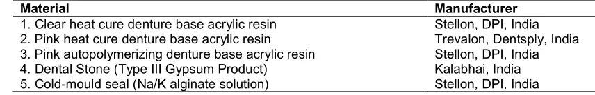
The materials used in this study are listed in Table 1.
2.1 Specimen Preparation
Standardized metal dies with dimensions of 65 mm×10 mm×3 mm were fabricated to prepare the gypsum mold [28,29]. The specimens were fabricated using standard techniques along with a mixture of monomer and polymer in ratio of 1:2.4 by weight, as per the manufacture recommendations. The flask was immersed in an acrylizer (Unident, Mumbai, India) at room temperature for curing. The temperature was raised slowly up to 70ºC and held for 1 hour, and then raised to 100ºC and was maintained for half an hour as it reduces the porosity [30]. Acrylic specimens were finished and polished. By this procedure 100 specimens each of Stellon and Trevalon acrylic material were prepared and divided into 10 groups with 10 specimens each (n =10). The test groups were designated in the following manner.
Group A: Unrepaired test specimens Group B: Untreated test specimens.
Group C: Specimens wetted with Methyl methacrylate for 1 minute.
Group D: Specimens wetted with MMA for 2 minutes.
Group E: Specimens wetted with MMA for 3 minutes.
Group F: Specimens wetted with MMA for 4 minutes.
Group G: Specimens wetted with MMA for 5 minutes.
Group H: Specimens wetted with MMA for 10 minutes.
Group I: Specimens wetted with MMA for 15 minutes.
Group J: Specimens wetted with MMA for 30 minutes.
2.2 Repair Process of Specimens
All specimens were stored in distilled water at room temperature for 48 hours before testing. The test specimens of each group except those in Group A were invested horizontally in a repair index prepared using dental stone. The sample and the index were marked on the corresponding ends to allow realignment in the original position. A centering mark was made exactly at the centre of each specimen with a marker pen and two lines were drawn perpendicular to this marking on either side of it. The sample was cut at these lines with a separating disc, and the center section was discarded. A carbide bur was used to prepare a round joint contour by measuring and drawing a line parallel to the prepared edge at a distance equal to 1 mm from the prepared edge on the top and bottom of the sample. For all samples, the space between the repair edges when placed in the repair index was 2 mm and the prepared joint surface was round without any sharp angles.
A plastic bowl of 10 cm diameter containing MMA was used for wetting the fractured surfaces of the specimens. A strip of adhesive plaster was placed on the open end of the bowl and the specimens were stuck to the plaster with only the repair surface of the specimens coming in contact with the MMA. The bowl was closed with a lid and the specimens were wetted for periods of 1, 2, 3, 4, 5, 10, 15 and 30 minutes.
The repair procedure was done in the same mould namely the repair index, in which the test specimens were prepared. After wetting the repair surfaces of specimens in MMA, the specimens were placed back into the repair index. Alginate separating medium was applied carefully to the walls of the repair index around the repair site. The specimens were repaired with auto-polymerizing acrylic resin.
Table 1. The materials used in the study
Material Manufacturer
Auto-polymerizing acrylic resin in the powder: liquid ratio of 20.5 gm to 10 ml was mixed to obtain a fluid consistency. The mix was poured into the repair site until it filled the repair gap without any entrapment of air voids. The joint space was slightly overfilled to compensate polymerization shrinkage and finishing.
As soon as the repair material lost its surface gloss, the specimens were placed in a hydroflask under warm water (40ºC); pressure was applied up to 2 bars and was maintained for 15 minutes. After 15 minutes the specimens were recovered from the hydroflask. All specimens were carefully restored to their original dimensions by polishing with 600-grit silicon carbide paper and the specimens were stored in distilled water at 37ºC for 4 days before testing [24].
2.3 Transverse Strength Testing
All specimens were stored in distilled water at room temperature for 48 hours before testing. A three point load tester (Instron 3366, Instron Corporation, UK) with a proving ring was used to evaluate the transverse strength. The testing jig was positioned on the platform of the testing device. The proving ring was attached on the top of the jig to the main frame-work. The applied force was shown by the proving ring. The test specimens were placed horizontally on the jig, the support on the jig being 50 mm apart. The load was applied exactly on a line drawn in the centre of the repaired joint with a hardened steel rod of 2.5 mm diameter having a round end. The transverse strength testing was carried out using a constant cross head speed of 1.2 mm/minute. The amount of load applied was noted. A total of 200 specimens were tested in this manner.
The transverse strength of the specimen was calculated using the following formula:
S = 3WL / bd2
Where,
S = Transverse strength
W = Maximum load before fracture L = Distance between the support b = Width of the specimen d = Thickness of the specimen
The repaired surfaces of these specimens were then examined under Scanning Electron Microscope (SEM). All data was statistically analyzed with one-way ANOVA, differences
within the groups were analyzed by independent sample t –test (Significance was set at P<0.05).
3. RESULTS
The results obtained from the transverse strength test were presented in Tables 2, 3 and 4.Table 2 shows the comparative statistics of mean transverse strength of Stellon observed in different groups at different time intervals. Group A showed the highest transverse strength (77.27±2.41 MPa) and group B showed very low transverse strength (48.90±4.26 MPa).
The transverse strength of the specimens wetted with MMA showed a gradual increase from 1 minute to 5 minutes of wetting. Specimens wetted for 5 minutes showed the highest strength (64.82±1.94 MPa). Specimens wetted with MMA for 10 minutes exhibited a reduced strength (52.25±2.90 MPa) with a concurrent decrease in transverse strength of the specimens wetted for 15 - 30 minutes. The specimens wetted for 30 minutes showed the least strength (35.20±2.53 MPa) among the groups (Table 2). ANOVA 'F' test revealed that the transverse strength exhibited by different groups was significantly different. Minimum significant difference between each group was found to be 4.23 MPa (Table 2). With this value when group A was compared with other groups the difference was found to be statistically very significant (P = 0.0l) (Table 2). When group B was compared with group C, H and I the difference was not significant, but when group B was compared with D, E, F, G and J the difference was found to be statistically very significant (P = 0.0l) (Table 2).
Table 3 shows the comparative statistics of mean transverse strength observed in different groups at different time intervals for Trevalon. Group A showed the highest transverse strength (102.30±1.74 MPa) and group B showed very low transverse strength (54.92±4.32 MPa).
the transverse strength exhibited by different groups was significantly different. Minimum significant difference between each group was found to be 3.49 MPa (Table 3). With this value when group A was compared with other groups
the difference was found to be statistically very significant (P = 0.0l) (Table 3). When group B is compared with other groups it was found to be statistically very significant (P = 0.01) except with group A.
Table 2. Comparative statistics of transverse strength (MPa) of stellon specimens wetted with MMA at different time intervals
Group Transverse
Strength (MPa) ‘F’* Value
Min. Sign. Diff.(MPa)
Significance of Difference
Mean SD B C D E F G H I J
A 77.27 2.41 1.992
P<0.01
4.23 VS VS VS VS VS VS VS VS VS
B 48.90 4.26 - NS VS VS VS VS NS NS VS
C 51.45 2.45 - - VS VS VS VS NS NS VS
D 61.55 2.04 - - - NS NS NS VS VS VS
E 61.60 1.42 - - - - NS NS VS VS VS
F 62.10 2.96 - - - NS VS VS VS
G 64.62 1.94 - - - VS VS VS
H 52.25 2.90 - - - NS VS
I 50.60 2.66 - - - VS
J 35.20 2.53 - - - -
SD – Standard Deviation; * - ANOVA F test; VS – Very Significant (P = 0.01); NS – Not Significant
Table 3. Comparative statistics of transverse strength (MPa) of trevalon specimens wetted with MMA at different time intervals
Group Transverse
Strength (MPa) ‘F’* Value
Min. Sign. Diff. (MPa)
Significance of Difference
Mean SD B C D E F G H I J
A 102.30 1.74 546.0
P=0.01
3.49 VS VS VS VS VS VS VS VS VS
B 54.92 4.32 - VS VS VS VS VS VS NS VS
C 60.50 2.24 - - VS VS VS VS VS VS VS
D 65.45 2.53 - - - VS VS VS VS VS VS
E 73.70 1.74 - - - - VS VS VS VS VS
F 80.02 1.56 - - - NS VS VS VS
G 82.22 1.56 - - - VS VS VS
H 89.93 1.86 - - - VS VS
I 56.70 2.82 - - - VS
J 45.10 1.92 - - - -
* - Unpaired ‘t’ Test
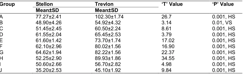
Table 4. Comparison of Transverse Strength (MPa) between stellon and trevalon
Group Stellon Trevlon ‘T’ Value ‘P’ Value
Mean±SD Mean±SD
A 77.27±2.41 102.30±1.74 26.7 0.001, HS
B 48.90±4.26 54.92±4.32 3.14 0.01, VS
C 51.45±2.45 60.50±2.24 8.61 0.001, HS
D 61.55±2.04 65.45±2.53 3.79 0.001, HS
E 61.60±1.42 73.70±1.74 17.02 0.001, HS
F 62.10±2.96 80.02±1.56 16.90 0.001, HS
G 64.62±1.94 82.22±1.56 22.37 0.001, HS
H 52.25±2.90 89.93±1.86 34.55 0.001, HS
I 50.60±2.66 56.70±2.82 4.98 0.001, HS
Table 4 shows the comparison of mean transverse strength between the materials (Stellon and Trevalon) using ‘t’ test. The difference between the Stellon and Trevalon in Group A, C, D, E, F, G, H, I, and J were found to be statistically highly significant (P = 0.001) and in group B it was statistically very significant (P = 0.01) (Table 4).
4. DISCUSSION
The interface between the heat polymerized acrylic resin and the repair material is usually the weakest point of the repaired denture base [24]. Different opinions about the appropriate shape of the joint surface have been presented. Several studies have indicated different edge design, such as a butt joint, 45º bevels, 30º bevels joint, rounded and rabbet joint [23]. The choice of material for repair depends on working time, strength to be obtained with the repair material and the degree of dimensional stability maintained during and after the repair. Most of the fractured dentures are repaired using a resin, which generally allows a simple and quick repair; therefore auto polymerizing resin was used as one of the repair materials [25,31,32].
The results of this study demonstrated that the mean transverse strength of Stellon and Trevalon specimens in group A (unrepaired specimen) showed more transverse strength than group B specimens (untreated specimens). This decrease in the transverse strength of untreated specimens was because the repair joints were not treated with MMA. So the PMMA resin surfaces of the repair joint were not adequately dissolved resulting in decreased transverse strength in the untreated specimens.
There was a gradual increase in transverse strength of the Stellon and Trevalon specimens which were wetted for 1 minute to 5 minutes (Group C to G). The Stellon specimens wetted for 5 minutes (Group G) and Trevalon specimens for 10 minutes (Group H) showed the highest transverse strength of 64.82±1.94 MPa and 89.93±1.86 MPa respectively. The findings in the present study were in accordance with the previous studies conducted by Ward et al. [25] Vallittu et al. [27], Sharma et al. [33] and Pereira Rde et al. [34]. On contrary to Sharma et al. [32], Mahajan et al. [22] studies, the present study demonstrated more strength. This increase in transverse strength is probably due to adequate MMA wetting and also solubility of heat polymerized PMMA resin surface was more strongly bonded to the auto-polymerizing acrylic resin, compared to their study. Moreover the round geometry increases the strength as it reduces the stress concentration [22].
The SEM photomicrographs demonstrated morphologic features of PMMA resin surface that differed when wetted with MMA for various intervals of time (Figs. 1-4). The Stellon specimens wetted for 1 to 5 minutes and Trevalon specimens wetted for 10 minutes showed a smoother structure (Figs. 1 and 2). All the specimens which were wetted with MMA for a time period of 3 minutes showed a definite increase in the transverse strength. These findings were in accordance with a previous study conducted by Vallittu et al. [27], Pereira Rde et al. [34], and Thunyakitpisal et al. [35], where the repaired specimens were wetted with MMA and tested for transverse strength.
Fig. 2. Trevalonspecimens wetted for ten minutes shows well rounded margins
Fig. 3. Stellon specimens wetted for thirty minutes shows more solubility of PMMA resin at the repair site results in large marginal gaps
The Stellon specimens wetted for a period of 10 minutes and Trevalon specimens wetted for a period of 15 minutes showed a decrease in transverse strength. The strength of the Stellon and Trevalon specimens after 30 minutes of wetting decreased respectively. This decrease in transverse strength is due to increased dissolution of MMA in PMMA resin. The SEM photomicrographs showing an increase in the dimension of the spaces has probably resulted in decreased transverse strength (Figs. 3 and 4). Thus when repairing a fractured heat cure acrylic resin, the repair surfaces have to be wetted with auto-polymerizing MMA to improve the strength of the repaired heat polymerized acrylic resin. But care should be taken not to wet the surface beyond a period of 5 minutes while using Stellon material and 10 minutes while using Trevalon material because this will cause a decrease in transverse strength in the area of repair.
5. CONCLUSION
From the results obtained in this study, the following conclusions can be drawn:
1. There is a definite increase in the transverse strength of repaired heat polymerized acrylic resins when the repair surfaces were wetted with MMA for an adequate period before the repair process was carried out.
2. The MMA wetting time should not exceed 5 minutes while using Stellon acrylic material and not more than 10 minutes in the case of Trevalon acrylic material. 3. Wetting of the repair surfaces with MMA
dissolves the surface structure of PMMA.
CONSENT
It is not applicable.
ETHICAL APPROVAL
It is not applicable.
COMPETING INTERESTS
Authors have declared that no competing interests exist.
REFERENCES
1. Peyton F. History of resins in dentistry. Dent Clin North America. 1975;19:2011. 2. Alla RK. Dental materials science, 1st ed.
Jaypee Brothers Medical Publishers (P) Ltd. New Delhi, India. 2013;248-84.
3. Alla RK, Sajjan S, Alluri VR, Ginjupalli K, Upadhya N. Influence of fibre reinforcement on the properties of denture base resins. J Biomater. Nanobiotech. 2013;4(1):91-7.
4. Bhola R, Bhola SM, Liang H, Mishra B. Biocompatible denture polymers – A review. Trends Biomater. Artif. Organs. 2010;23:129-36.
5. Drabar UR, Huggett R, Harrison A. Denture fracture – a survey. Br Dent J. 1994;176(9):342-5
6. Arikan A, Ozkan YK, Arda T, Akalın B. Effect of 180 days of water storage on the transverse strength of acetal resin denture base material. J of Prosthodont. 2010;19(1):47–51.
7. Beyli MS, Von Fraunhofer JA. Repair of fractured acrylic resin. J Prosthet Dent. 1980;44(5):497-503.
8. Meng TR Jr, Latta MA. Physical properties of four acrylic denture base resins. J Contemp Dent Pract. 2005;6(4):93-100. 9. Hirajima Y, Takahashi H, Minakuchi S.
Influence of a denture strengthener on the deformation of complete denture. Dent Mater J. 2009;28(4):507-12.
10. Vallittu PK. Fracture surface characteristics of damaged acrylic-resin-based dentures as analysed by SEM-replica technique. J Oral Rehabil. 1996;23(8):524-9.
11. Wiskott HWA, Nicholls JI, Belser UC. Stress fatigue: Basic principles and prosthodontic implications. Int J of Prosthodont. 1995;8(2):105–16.
12. Bhaskar RM, Davenport JC. Prosthetic treatment of the edentulous patient, 4th ed., Blackwell Munksgaard, Oxford, UK. 2003;290.
13. Asar NV, Albayrak H, Korkmaz T, Turkyilmaz I. Influence of various metal oxides on mechanical and physical properties of heat-cured polymethyl methacrylate denture base resins. J Adv Prosthodont. 2013;5(3):241-7.
14. El-Sheikh AM, Al-Zahrani SB. Causes of denture fracture: A survey. Saudi Dent J. 2006;18:149-54.
15. Zarb G, Hobkirk JA, Eckert SE, Jacob RF. Prosthodontic treatment for edentulous patients: Complete denture and implant supported prosthesis, 13th ed. Reed Elsevier (P) Limited, New Delhi, India. 2013;312.
17. Mathew M, Shenoy K, Ravishankar KS. Vickers Hardness and Specific Wear Rate of Poly Propylene Reinforced PMMA. Int J Sci Stud. 2014;2:71-5.
18. Soygun K, Bolayir G, Boztug A. Mechanical and thermal properties of polyamide versus reinforced PMMA denture base materials. J Adv Prosthodont. 2013;5:153-60.
19. Ayaz EA, Durkan R, Bagis B. The effect of acrylamide incorporation on the thermal and physical properties of denture resins. J Adv Prosthodont. 2013;5:110-7.
20. Shen C, Franc CA, Birns B. Strength of denture repairs as influenced by surface treatment. J Prosthet Dent. 1984; 52(6):844-8.
21. Rhan AO, Ivanhoe JR, Plummer KD. Text book of complete dentures, 6th ed. Peoples Medical Publishing House, Connecticut, USA. 2009;14.
22. Mahajan H, Chandu GS, Mishra SK. An in vitro study of the effect of design of repair surface on the transverse strength of repaired acrylic resin using autopolymerizing resin. Niger J Clin Pract. 2014;17(1):38-42.
23. Harrison WM, Stansbury BE. The effect of joint surface contours on the transverse strength of repaired acrylic resin. J Prosthet Dent. 1970;23:464-71.
24. Kostoulas I, Kavoura VT, Frangou MJ, Polyzois GL. Fracture Force, Deflection, and Toughness of Acrylic Denture Repairs Involving Glass Fiber Reinforcement. J. Prosthodont. 2008;17:257–61.
25. Ward JE, Moon PC, Levine RA, Behrendt CL. Effect of repair surface design, repair material, and processing method on the transverse strength of repaired acrylic resin. J Prosthet Dent. 1992;67(6):815-20. 26. Phillips RW. Phillips’ science of dental
materials, 11th ed. Elsevier, Reed Elsevier Private Limited, New Delhi, India. 2004;721-58.
27. Vallittu PK, Lassila VP, Lappalainen R. Wetting the repair surface with methyl methacrylate affects the transverse
strength of repaired heat-polymerized resin. J Prosthet Dent. 1994;72(6):639-43. 28. Ellakwa AE, Morsy MA, El-Sheikh AM.
Effect of aluminium oxide addition on the flexural strength and thermal diffusivity of heat polymerized acrylic resin. J Prosthodont. 2008;17(6):439-44.
29. Mumcu E, Cilingir A, Gencel B, Sulun T. Flexural properties of a light-cure and a self-cure denture base materials compared to conventional alternatives. J Adv Prosthodont. 2011;3(3):136-9.
30. Sunint Singh, Jayant N. Palaskar, Sanjeev Mittal. Comparative evaluation of surface porosities in conventional heat polymerized acrylic resin cured by water bath and microwave energy with microwavable acrylic resin cured by microwave energy. Contemp Clin Dent. 2013;4(2):147–151. 31. Lewienstein I, Zeltser C, Mayer CM, Tal Y.
Transverse bond strength of repaired acrylic resin strips and temperature rise of dentures relined with visible light cured reline resin. J Prosthet Dent. 1995;74(4):392-9.
32. Sharma K, Dhakshaini MR, Gujjari AK. Evaluation of transverse bond strength of heat cured acrylic denture base resin repaired using heat polymerizing, autopolymerizing and fiber reinforced composite resin - an in vitro study. Int J Clinic Cases Invest. 2012;4:33-43.
33. Sharma A, Batra P, Vasudeva K, Kaur R, Influence of repair materials, surface design and chemical treatment on the transverse strength of repaired denture base – An in vitro study. Ind J Dent Sci. 2012;4(4):23-26.
34. Pereira Rde P, Delfino CS, Butignon LE, Vaz MA, Arioli-Filho JN. Influence of surface treatments on the flexural strength of denture base repair. Gerodontology. 2012;29(2):e234-8.
35. Thunyakitpisal N, Thunyakitpisal P, Wiwatwarapan C. The effect of chemical surface treatments on the flexural strength of repaired acrylic denture base resin. J Prosthodont. 2011;20(3):195-9.
© 2015 Shakeel et al.; This is an Open Access article distributed under the terms of the Creative Commons Attribution License (http://creativecommons.org/licenses/by/4.0), which permits unrestricted use, distribution, and reproduction in any medium, provided the original work is properly cited.
Peer-review history: