Life-giving caspases: revealing new roles during
mouse embryo preimplantation development
DOLORES BUSSO*
,1, CALIXTO DOMINGUEZ
2, TOMAS PEREZ-ACLE
2,3and RICARDO D. MORENO
11Department of Physiology, 2Centre for Bioinformatics (CBUC), Facultad de Ciencias Biológicas, Pontificia Universidad Católica de Chile and 3Fundación Ciencia para la Vida, Ñuñoa, Santiago, Chile
ABSTRACT Caspases, cystein proteases traditionally related to programmed cell death, have recently been found to be involved in vital processes such as cell proliferation, adhesion and differentiation. Although caspases are expressed in mouse embryos before the blastocyst stage, their role is unclear, since apoptosis does not occur significantly before implantation. In this work, we have used mouse preimplantation development as a model to evaluate the existence of non-lethal caspase activities. The use of specific caspase inhibitors during in vitro embryo culture showed that caspase 8 activity, but not caspase 2 or 9, was relevant for development. The inhibition of caspase 8 affected the compaction of morulae and the progression to the blastocyst stage. In agreement with these results, caspase 8 was expressed in mouse embryos, as shown by indirect immunofluorescence and RT-PCR. An in silico approach was used to find putative caspase targets expressed in mouse preimplantation embryos. Large-scale management of sequence data from mouse embryos was used to predict caspase substrates by tools matrix-based on known cleavage sites. A total of 510 potential caspase targets expressed in mouse embryos were identified by this procedure. The functional characterization of these proteins by Gene Onthology associations showed that many of these putative caspase targets were previously related to non-apoptotic functions and only 63 had been previously reported to be actually cleaved by caspases. Interestingly, eleven knockout mice models for caspase substrates identified in our work, i.e. catenin alpha and beta, geminin, pescadillo, calpain-2, have preimplantation lethal phenotypes. This work supports the involvement of caspases in vital functions during mouse preimplantation development and proposes a model in which the regulated cleavage of caspase substrates could account for this role.
KEY WORDS: mouse development, caspase, caspase substrate, compaction
Introduction
Caspases are members of a family of cysteine proteases that, once activated, cleave a variety of intracellular polypeptides. Although traditionally related to programmed cell death, recent evidence shows that there are many instances where caspases fail to promote apoptosis and instead play roles in various vital processes such as cell proliferation, motility and differentiation. In Drosophila melanogaster, caspases are involved in the removal of the cytoplasm of spermatids during their differentiation into sperm (Arama et al., 2003). In mammals, the activity of caspases has also been related to the process of differentiation of numerous cell types such as myoblasts, megakaryocytes, lens cells, keratinocytes, monocytes, osteoblasts and colon cancer cells
BIOLOGY
www.intjdevbiol.com*Address correspondence to: Dolores Busso. Departamento de Ciencias Fisiológicas, Facultad de Ciencias Biológicas, Pontificia Universidad Católica de Chile. Alameda 340. (6513492) Santiago, Chile. Fax: +56 (2) 222-5515. e-mail: dbusso@bio.puc.cl
Accepted: 17 April 2009. Final author-corrected PDF published online: 9 March 2010. Edited by: Roberto Mayor
ISSN: Online 1696-3547, Print 0214-6282 © 2010 UBC Press
Printed in Spain
Abbreviations used in this paper: EST, expressed sequence tag; IIF, indirect immunofluorescence.
(Abraham et al., 2004; Launay et al., 2005). As well, caspases have been shown to contribute to inflammation, lymphocyte proliferation, and cell migration during tumour invasiveness (Launay et al., 2005; Helfer et al., 2006).
non-lethal functions during preimplantation development (Pampfer et al., 1999). Recenty, Zakeri et al., showed that the culture of mouse embryos with the generalized caspase inibitor zVAD did not only fail to prevent the death of ICM cells in culture but, conversely, disrupted the normal development of embryos to the blastocyst stage (Zakeri et al., 2005).
Mouse preimplantation development is an attractive model to study events of early differentiation in mammalian cells. Mouse cultured embryos have a well-defined number of cells in a period that is consistent and relatively short, and develop independently of external factors (i.e. serum suplements) that frequetly interfere with analyses. Mammalian development begings with the succes-sive cleavage of fertilized eggs into 2-cell, 4-cell and 8-cell embryos. The first morphologically distinguishable differentiation event in the dividing embryo is the compaction of cleaving blastomeres into a coherent mass of cells, the morula. The process of compaction starts at the 8-cell stage, and is a
conse-used with the aim of evaluating the relevance of caspase activitiy during mouse preimplantation embryo development and under-standing the possible mechanisms of action of these proteases. The first objective was addressed by analyzing the effect of specific caspase inhibitors during mouse in vitro embryo develop-ment. The second goal was undertaken using an in silico ap-proach by large-scale management of biological data obtained from mouse embryos and the prediction of putative caspase targets using bioinformatic tools. The evidence obtained in this work support the involvement of caspases in vital functions during mouse embryo preimplantation development, proposing a model where the cleavage of caspase substrates could account for these non-lethal roles.
Results
In an attempt to determine which caspase(s) were active in mouse preimplantation embryos, two-cell stage embryos were cultured with different caspase inhibitors and the progression of development was monitored daily. Our results showed that the general inhibition of caspases using zVAD affected early develop-ment of mouse embryos (Fig. 1). Those embryos never reached the morula stage and remained arrested at the four- or the eight-cell stage. Inhibitors for caspase 2 or 9 were inocuous at a 100 μM concentration, as embryos in those treatment underwent their
66,7 ± 33,3 (3) 80,0 ± 20,0 (3)
96,7 ± 3,3 (3)
inh C9
46,7 ± 26,0 (3) 73,0 ± 13,6 (4)
78,8 ± 9,1 (5)
inh C2
4,6 ± 2,9* (5) 77,4 ± 15,5 (5)
89,8 ± 8,8 (6)
inh C8
52,0 ± 18,3 (4) 92,3 ± 4,5 (4)
87,8 ± 3,2 (5)
DMSO
0,0 ± 0,0 (3) 2,7 ± 2,7* (3)
57,0 ± 16,6 (3)
zVAD
81,0 ± 7,0 (4) 93,3 ± 4,2 (4)
92,0 ± 3,4 (5)
control
mean ± SE (n) mean ± SE (n)
mean ± SE (n) treatment
E4 E3
E2 E1
Fig. 1. Development in the presence of caspase inhibitors. Two-cell embryos (E1) were incubated in medium alone (control), or containing either 1 μl DMSO or 1 μl of a stock solution (20 mM) of inhibitors for general caspase activity (zVAD), caspase 8 (inh C8), caspase 2 (inh C2) or caspase 9 (inh C9) dissolved in DMSO. The percentage ± ES of embryos that reached the expected developmental stage at day 2 (4-cell to 8-cell: E2), day 3 (morula: E3) and day 4 (blastocyst: E4) after the beginning of the experiment are indicated for each treatment. Significant values are shown on a gray background (*p≤ 0.05). The number of experiments (using 8-15 embryos/drop) is indicated between brackets.
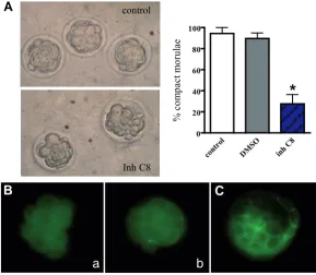
Fig. 2. Effect of caspase 8 inhibitor on morulae compaction. (A)Morphological determination of compaction. Embryos at E3 were observed under an inverted microscope, and the number of compacted morulae was determined. The percentage of com-pact embryos was significanly reduced in the presence of caspase 8 inhibitor compared to both controls, i.e. in the absense of inhibitor or with DMSO (*p≤ 0.05). (B,C) Immunoreactivity for tight junction protein ZO-1. Embryos were fixed, permeabilized and subjected to immunofluorescence using anti-ZO-1. (B) Non-compacted morulae lacked immunoreactivity for anti-ZO-1; (a) non-compacted morula lacking ZO-1; (b) partially compacted morula showing ZO-1 in some blastomeres only. (C) Blastocysts used in parallel as positive controls showed specific staining in trophoectoderm cell-cell contact regions.
control
Inh C8
a
b
contr ol
DMS O
inh C8 0
20 40 60 80 100
%
c
o
mp
a
ct
mo
ru
la
e
*
B
C
A
quence of the polarization of external blastomeres (Ziomek et al., 1980) and the formation of cell-cell adhesion struc-tures such as adherens junctions (Shirayoshi et al., 1983; De Vries et al., 2004), tight junctions (Sheth et al., 1997; Wang et al., 2008) and gap junctions (Houghton et al., 2005). Compaction is followed by the formation of a central cavity, process known as cavitation, and the differentiation of two distinct cell types: the trophoectoderm and the inner cell mass (ICM), to form the blastocyst.developmental programme up to the blastocyst stage similarly to those in drops containing DMSO alone. Of the three specific caspase inhibitors included in this study, only caspase 8 inhibitor clearly arrested development. It was surprising to note that, differently to the results observed with zVAD, caspase 8 inhibitor did not affect the number of embryos reaching the morula stage, but reduced significantly the number of embryos progressing from the morula to the blastocyst stage.
In caspase 8 inhibitor-treated embryos, the morphology of morulae was different to that of morulae in control drops. While untreated embryos at E3 showed a mass of cohesive cells, characteristic of morulae that have undergone compaction, ar-rested embryos had mostly rounded blastomeres showing no morphological signs of compaction (Fig. 2A). The quantification of this effect indicated that the percentage of compact morulae was significantly reduced by caspase 8 inhibitor. In agreement with these observations, indirect immunoflouorescence experiments showed that caspase 8 inhibitor-treated morulae lacked the characteristic membrane localization of tight junction protein 1 (ZO-1), involved in the formation of tight juntions in the embryo at compaction (Fig. 2B a,b) (Sheth et al., 1997). Confirming the specificity of our immunofluorescence experiments, embryos that had progressed to blastocyst and were incubated with anti-ZO-1 showed the characteristic staining, i.e present at the cell-cell contact region of trophoectoderm cells and absent in ICM cells (Fig. 2C) (Sheth et al., 1997). At E3, when a defect in compaction was observed, there were no significant differences in the
num-NAs were detected in 4-cell embryos, morulae and blastocysts and not in oocytes or 2-cell embryos (Fig. 4B). Thus, the expres-sion pattern found for caspase 8 was in agreement with the timing of the effect observed for caspase-8 inhibition, as this only took place after the 8-cell stage.
Caspases extert their apoptotic function by cleaving a number of subtrates that lead to cell death. However, the mechanism of action of non-lethal caspase functions is less clear. Given the scarcity of the biological material that can be obtained from mammalian embryos, a database of expressed sequence tags (ESTs), published from high-throughput studies applied to mouse preimplantation embryos, was compiled using the mouse library browser from UniGene database so that a single library was DMSO inh C8
16-c
ell
32-c
ell
Mean nº
of nuclei 22 ±2 21 ± 3
DMSO inh C8
blastoc
y
sts
55 ±5 34 ± 3*
E3
E4
Fig. 3. Effect of caspase 8 inhibitor on blastomere proliferation. Embryos at E3 and E4 were fixed and their nuclei stained with Hoestch, and counted to determine cell numbers. Representative pictures and the mean numbers of cells are shown per stage (E3 and E4) and treatment (DMSO and caspase 8 inhibitor). A decrease in cell numbers was only found in E4. *p≤ 0.05.
bers of cells in DMSO and caspase 8 inhibitor-treated embryos (Fig. 3). The normal proliferation of 16 to 32-cell embryos in the presence of caspase 8 inhibitor suggests that this treatment did not affect the cell cycle progression until that stage. Control embyos in DMSO proliferated normally until the following day (E4), whereas caspase 8-treated embryos showed a signifi-cant decrease in the number of blastomeres.
Although the expression and activity of some mem-bers of the caspase family in mouse embryos has already been demonstrated (Exley et al., 1999; Hinck et al., 2001; Hinck et al., 2003; Little et al., 2003), to our knowledge this is the first report showing the presence of caspase 8. Both 4-cell embryos (results not shown) and morulae subjected to indirect immunofluores-cence using anti-caspase 8 specific antibody showed strong fluorescent staining (Fig. 4A). The absence of staining in embryos incubated with anti-caspase 3 or without primary antibody (results not shown) con-firmed the specificity of the results. RT-PCR experi-ments confirmed the expression of caspase 8 in pre-implantation embryos. Interestingly, caspase 8
mR-Fig. 4. Expression of caspase 8 in mouse preimplantation embryos. (A) Immunolocalization of caspase 8. Permeabilized morulae incubated with anti-caspase 8 and a FITC-conjugated secondary antibody showed strong immunoreactivity (a) while those incubated with anti-caspase 3 were negative (b). (a‘,b‘) Hoestch nuclear staining of embryos in (a,b), respectively. (B) RT-PCR analysis of caspase 8 expression. RNA was extracted from embryos at different stages, and subjected to RT-PCR using caspase 8-specific primers. Expression of caspase 8 was detected as a unique band of 280 bp in 4-cell stage embryos, morulae and blastocysts. Liver RNA was used as a positive control.
a
b
a’
b’
300 bp200 bp
ooc yte
2-ce ll
4-ce ll
morula blastoc
yst liver
B
obtained for each preimplantation stage (Table 1). Using UniGene identifiers, the associated direct protein products were extracted from the UniProt database (see Methods). Fig. 5 shows the parsing process result with the number of EST sequences used to obtain the putative protein products occurring at each embryo stage. As shown, the EST library of one-cell embryo stage contains 36,036 sequences. The list with unique UniGene IDs is reduced to 5,506 with only 4,173 paired with Gene IDs. Finally, UniProt database was queried to acquire those proteins which are relative to those 4,173 gene IDs achieving 2,930 protein ID codes. For the rest of the developmental stage libraries the parsing procedure was the same, achieving the following results: Two-cell stage library 24,509 EST sequences, 6,217 UniGene IDs, 4,705 Genes and 3,341 UniProt proteins; Four-cell stage library 16,878 EST sequences, 3,934 UniGene IDs, 3,394 Genes and 2,456 UniProt proteins; Eight-cell stage library 24,502 EST sequences, 4,200 UniGene IDs, 3,388 Genes and 2,401 UniProt proteins; Morula stage library 11,770 EST sequences, 2,534 UniGene IDs, 2,105 Genes and 1,659 UniProt proteins; Blastocyst stage library 64,803 EST sequences, 7,612 UniGene IDs, 6,034 Genes and 4,484 UniProt proteins.
UniProt protein IDs from each library stage were compared to those cleavage candidates IDs predicted by Caspredictor, a bioinformatic tool which uses scored amino acid substitutions, residue frequency and similarity with PEST-like sequences to predict proteolytical cleavage sites with a high accuracy (Rogers et al., 1986; Rechsteiner et al., 1996; Garay-Malpartida et al., 2005). The parsed list of caspase candidate targets for each preimplantational stage was functionally annotated by exploring Gene Ontology (GO) associations for mouse. The numbers of proteins with GO annotations were: 280 proteins for one-cell, 293 for two-cells, 229 for four-cells, 235 for eight-cells, 134 and 345 for morula and blastocyst, respectively (Fig. 5). In total, the
non-redundant caspase substrates expressed along the preimplanta-tion mouse embryos predicted by the bioinformatic approach were 510 proteins.
The functional annotation obtained by GO for the 510 putative caspase substrates in embryos indicated their association to diverse subcellular localizations, molecular functions and biologi-cal processes. Considering the results of our in vitro experiments using caspase inhibitor-treated embryos, special attention was paid to those proteins related to functions affected the inhibitors. As described previously, zVAD affected early events of develop-ment, related to blastomere proliferation, and caspase 8 inhibitor had an effect at later stages, where the processes of cell adhe-sion/communication and differentiation occur. The functional
char-1-ce
ll
2-cell 4-cell 8-ce
ll M B
0 1000 2000 3000 4000 5000 6000 7000 8000
ESTs Unigene IDs Genes Proteins Caspase Targets
0 2500 5000 15000 25000 35000 45000 55000 65000
seque
nc
es ES
Ts
36036 24509 16878
24502 11770 64803
5506 6217 3934
4200 2534 7612
4173 4705 3394
3388 2105 6034
2930 3341 2456 2401
1659 4484
280 293 229 235 134 345
Fig. 5. Quantitative sequence analysis for each preimplantation stage during parsing process to identify caspase substrates. The number of ESTs, transcript loci identified by UniGene IDs, Genes, Proteins and putative Caspase Targets is shown for each preimplantation stage. The bars represent the numbers in the table for a better graphic visualization of the proportion of each molecular entity in every stage. EST numbers are indicated in the right Y axis.
Stage IDs of compiled libraries Reference(s)
One-cell 2589, 17903, 17960
1119
Ko, MS et al, 2000 Okazaki, Y et al, 2002 Two-cell 1382
5391, 17932 862, 7223
Ko, MS et al, 2000. Okazaki, Y et al, 2002. NCBI clone registry.
Four-cell 17905, 12243
14709 1524 and 7328
Okazaki, Y et al, 2002. Sharov, AA et al, 2003 NCBI clone registry Eight-cell 17906
15708
1381, 871 and 9845
Okazaki, Y et al, 2002 Sharov, AA et al, 2003 NCBI clone registry Morula 1532
18029
Ko, MS et al, 2000 Okazaki, Y et al, 2002 Blastocyst 1310
1021 10026
12245, 18063, 18021, 12246 18027
850, 875, 13911
Sasaki, N et al, 1998 Ko, MS et al, 2000. Piao, Y et al, 2001. Okazaki, Y et al, 2002. Carninci, P et al, 2005 NCBI clone registry TABLE 1
acterization by GO of CasPredictor-identified caspase targets indicated that 9,8% of the sequences were related to the progress of the cell cycle, 6,5% to cell proliferation and 3,3% to cell division, while 24% of the sequences were related to cell communication, 6,7% to cell adhesion and around 2% to cell fate commitment or differentiation.
Recently, a database known as CASBAH (CAspase Substrate DataBAse) has been published, where all the proteins shown to be caspase substrates by biochemical methods are compiled (Luthi et al., 2007). From a total of 412 CASBAH proteins, only 30 belong to mouse, 356 to human, another 13 to rat and the remaining 13 to other organisms. We used the Mouse Genome Informatics (MGI) database (Eppig et al., 2007) to achieve mouse CASBAH caspase substrates orthologous to human and rat, and then compared them to those obtained in mouse embryos by CasPredictor. This comparison showed that of a total of 510 proteins obtained as putative caspase substrates in mouse em-bryos by CasPredictor, 63 orthologous sequences were demon-strated in the literature to be caspase targets in other systems. Some interesting examples related to non-apoptotic functions are the cell divison control protein 6 (Cdc6) (Hateboer et al., 1998), the negative regulator of mitosis wee1 (Wee1) (Honda et al., 1995) and geminin (Gmnn) (McGarry et al., 1998) involved in proliferation, Beta-catenin (Ctnnb1) (Xu et al., 2007) and presenilin (Psen1) (Georgakopoulos et al., 2000) related to cell adhesion and myeloid leukemia cell differentiation protein MCL-1 homolog (Mcl1) (Okita et al., 1998), Beta-catenin (Xu et al., 2007) and Serum Response Factor (SRF) (Arsenian et al., 1998) with roles in differentiation.
As another approach to determine the possible participation of caspase activity for the progression of preimplantation develop-ment, the CasPredictor-derived substrates were compared to a list of proteins whose deficiency in knockout (KO) mouse models produces embryonic lethality before implantation (Eppig et al., 2007). The result of this analysis showed that 11 out of the 108 embryo lethal knockout genes are transcribed into proteins that can be proteolytically cleaved by caspases. Fig. 6 lists these proteins showing the phenotype of their knockouts, and the GO functional assignation and stage where they are present. As shown, two of the embryo lethal KO genes that were identified as
caspase substrates by CasPredictor, beta-catenin and geminin, were also found in the CASBAH database.
Discussion
Besides the traditional idea where caspase function executes apoptosis, recent evidence indicate that caspase activation does not systematically trigger cell death but can promote processes relevant for life (Abraham et al., 2004; Launay et al., 2005). In this work, we have used mouse embryo development in vitro to evaluate vital roles of caspases. Several reasons have lead us to use this model. First, mouse embryo culture is a reproducible and efficient system. Starting from a embryo with a pair of morphologi-cally identical blastomeres, after three days in culture in the absence of serum or growth factors, a more complex and differ-entiated entity, the blastocyst, forms in the culture dish. Second, although caspases and other molecules related to apoptosis have been shown to be present in embryos (Exley et al., 1999; Hinck et al., 2001; Hinck et al., 2003; Little et al., 2003), significant levels of programmed cell death does not take place before implantation (Pampfer et al., 1999), suggesting that those molecules might be involved in non-lethal processes. Third, the progression of an early embryo from the 2-cell stage to the blastocyst stage involves a series of observable events that occur in a cell-specific and time-dependant manner.
The main biological processes occuring in the blastomeres are cell division or proliferation, cell adhesion or communication, at compaction, and cell differentiation to form two cell types in the blastocyst. As mentioned previously, caspase activities have been related to these processes in different somatic and cancer cells. In embryos, Zakeri et al., showed for the first time that a non-lethal caspase activity is present before implantation (Zakeri et al., 2005). In agreement with those sudies, our results showed that the general caspase inhibitor zVAD significantly affected development, with 2-cell embryos showing an arrest at the 4- to 8-cell stage and thus never reaching the blastocyst stage. The effect of zVAD in early embryos, before compaction or differentia-tion take place, suggests that the main process affected is the proliferation of blastomeres. In an attempt to identify individual caspase activities required for the progression of development, Phenotype of
Geminin* GEMI_MOUSE arrest at 8-cell stage, premature differentiation into TE proliferation (inhibitor)
Catenin beta-1* CTNB1_MOUSE normal compaction, replaced by plakoglobin proliferation, adhesion, differentiation Actin-related protein 3 ARP3_MOUSE arrest at morula stage, no differentiation into TE cell structure (cytoskeleton) Pescadillo homolog 1 PESC_MOUSE arrest at morula stage, unevenly sized blastomeres proliferation, transcription Transcription factor ATF-1 ATF1_MOUSE arrest at morula stage, no inner cell mass (ICM) transcription
n
Non-lethal function Expression (presence of ESTs) Protein
inhibitors for the caspases 2, 8 and 9 were selected. All of these proteases are initiators of the caspase cascade, and are not directly responsible for the cleavage of substrates promoting cell death but have been previously related to events of differentiation (Launay et al., 2005). The results of these experiments showed that caspase 2 and 9 activities were dispensable for embryo development. On the other hand, caspase 8 activity was impor-tant for the progression from the morula to the blastocyst stage. A clear effect on compaction was indicated by the morphological aspect of blastomeres and the absence of the tight junction protein ZO-1 in cell-cell contact areas. Caspase 8-treated em-bryos proliferated normally until the defect on compaction took place. Thus, the reduction in blastomere numbers observed later was probably due to the altered developmental program of non-compacted embryos rather than to an effect of the inhibitor on the cell cycle itself.
Caspase-8 knock mice do not show embryonic lethality before implantation, as null embryos can implant but die in utero due to defects attributable to circulatory failure (Varfolomeev et al., 1998; Sakamaki et al., 2002). Although this evidence might seem contradictory with our results, the unaffected preimplantation development in those embryos might be due to compensation by other proteins. One clear example of this functional compensa-tory mechanism has been demonstrated in beta-catenin null mice, where protein plakoglobin can take over the function of beta-catenin during blastomere compaction (Huelsken et al., 2000).
One curious observation of this work is that the effect of general inhibitor zVAD was earlier than that of caspase 8 inhibitor. An issue worth considering is that chemical inhibitors can produce unintended effects on other proteases with similar substrate specificities, i.e. Granzyme B and Cathepsin L (Thornberry et al., 1997; Schotte et al., 1999). This side-inhibition could account for the additional effects of zVAD over caspase 8 inhibitor. A more interesting possibility is that other caspases besides caspase 8 are involved in early stages of embryo development.
In agreement with the effect of its inhibitor, caspase 8 was detected by immunofluorescence and RT-PCR in mouse em-bryos around the time of compaction. Although a deeper analysis is required to determine what processes or molecular pathways were inhibited in caspase 8-treated arrested morulae, our results suggest that, among other events, molecules involved in cell adhesion and cell differentiation are the most likely to be regulated by that caspase activity.
One of the disadvantages of the mouse embryo model is that the material available for protein analyses is scarse compared to that available using other cell types, or even embryos from other models (i.e. Xenopus or sea urchin). However, the existence of a growing inventory of mouse genes annotated in public databases, expressed in different developmental stages, can still be used to explain events taking place in embryos at the molecular level. In this work, a bioinformatic approach facilitated by large-scale management of biological data was used to evaluate the role of caspases in mouse preimplantation development. In the first place, the total mouse preimplantation embryo ESTs from differ-ent publications were compiled into a single library for each developmental stage. In general, the amount of EST sequences for the different stages was similar, near 20,000, with the excep-tions of morula, with half that amount, and blastocyst, where a
higher number of ESTs (more than 60,000) has been published. The difference in the amount of ESTs for each embryo stage is worth considering when quantitative comparisons are made from these data. The posterior crosstalk among sequence databases allowed the indentification of the cohort of the genes and protein products represented in the ESTs. The number of proteins pre-dicted to be expressed in embryos was around 10% of the number of ESTs identified (range 7 to 14%, depending on the different stages). A large-scale analysis of those proteins using the CasPredictor software allowed the identification of feasible caspase cleavage sites present in those proteins. The functional assigna-tion of these putative caspase substrates in embryos by GO indicated their involvement in a broad variety of biological pro-cesses in different cell types. Interestingly, for each of the devel-opmental stages analyzed, an 8 to 10% of the expressed proteins with assigned functions were putative substrates for caspases, suggesting that these proteases could be involved in the regula-tion of numerous molecular pathways related to different pro-cesses, not only apoptosis, within the cell.
A list of 510 putative caspase substrates found in mouse preimplantation embryos was obtained by the bioinformatic pro-cedures described above. The fact that near half of those caspase substrates are related to vital functions such as cell proliferation, communication or differentiation is in agreement with our results showing that the inhibition of caspase activity in mouse embryos affects the progression of embryo development. One interesting observation from our results is that many caspase substrates present in embryos belong to the Wnt signal transduction cas-cade, involved in numerous events of embryo development and in the control of homeostatic cell renewal in several adult tissues (Clevers et al., 2006; Harwood et al., 2008). Some of these proteins are LRP6 and LRP1 co-receptors of Frizzled (Wnt Receptor), Axin and Daple, positive and negative regulators of Beta-catenin stabilization, Alpha-catenin, Beta-catenin, Delta-catenin and Lim-domain binding protein 1 (Ldb1). Only 63 of the
Degradation of substrates
Cell cycle inhibitors (geminin, wee) Adhesion molecules (catenins) Differentiation factors(ATF1, Xab2)
Activation of substrates
Differentiation factors (MSTl) Signaling proteins (beta-catenin, LRP6)
APOPTOSIS
VITAL FUNCTIONS
CASPASE ACTIVATION
Degradation of substrates
Structural proteins (lamin, fodrin) DNArepair elements (PARP) Caspase inhibitors (IAP)
Activation of substrates
Pro-caspases
Apoptotic proteins (Bcl2, BID)
510 putative substrates were previously described in the literature as caspase targets. Whether the 447 remaining sequences are in fact physiological caspase substrates in embryos or other cell types is still to be determined. In this sense, the bioinformatic procedure undertaken in this work may be useful to provide substantial material for future investigations on the non-lethal role of caspases in mammalian cells.
The relevance of caspase substrates for preimplantation de-velopment was also suggested by the preimplantation lethality described in knockout mice deficient for the coding genes of 11 of the putative caspase substrates identified in this work. In general, the phenotype of the KO embryos is in agreement with a defi-ciency in the function assigned by GO for each of the proteins. Deficiencies in caspase substrates involved in cell adhesion or differentiation in other cell types, produce an arrest at the morula stage, with defects in compaction and differentiation of TE and ICM cells, while deficiencies in proteins involved in proliferation produce in the embryo a failure to undergo the developmental program at earlier stages, when cell division seems to be the main process involved. It was surprising to note that in calpain-2 KO mice, embryos are arrested during early development while the presence of EST for calpain-2 is only detected past the morula stage. In that case, the heterogeneity in the source and the quantity of EST sequences available among different embryo stages may have limited the detection of calpain-2 in earlier embryos. Supporting our idea, Calpain-2 mRNAs have been detected by immunofluorescence (Raynaud et al., 2008) and RT-PCR (Dutt et al., 2006) in mouse embryos at the 8-cell stage, earlier than Calpain-2 ESTs, detected at the morula stage. As in the case of calpain, the limited amount of material from embryos is a caviot of our EST analyses that could affect the interpretation of results. However, in this work we have succeeded in identifying two proteins that are both essential for embryo development and caspase-proven substrates published in the CASBAH database: Beta-catenin and Geminin.
Although the mechanisms responsible for regulating the dual function of caspases in apoptosis and vital processes are un-known, the selective cleavage of specific substrates involved in cell death and life events could explain this duality (Fig. 7). As well, despite the ortodox notion where caspase activation promotes the proteolytical degradation of target proteins, in some cases these proteases can induce the activation of proteins to promote non-lethal functions. One example of this activating function of caspases takes place during the differentiation of skeletal muscle, where the cleavage of a protein known as Mammalian Sterile Twenty-like Kinase (MST1) promotes differentiation of myoblasts (Fernando et al., 2002). In a similar manner, the regulated cleavage of substrates could determine their involvement in one or another function. As an example, the cleavage of Beta-catenin could provide a mechanism regulating its involvement in signal-ling (canonical pathway) or cell adhesion (non-canonical). This might be specially relevant in embryos, where Beta catenin was recently shown to be an important molecule for blastomere compaction (Na et al., 2007) besides its well-known participation in the establishment of the three embryo layers shortly after implantation (De Vries et al., 2004). There is also evidence indicating that Geminin, a multifunctional protein that dominates the balance between cell proliferation and differentiation, can be cleaved by caspases in two sites; the two resulting
caspase-cleavage forms interact with different regulators and, as a conse-quence, promote differential effects on the cell (Hara et al., 2006; Roukos et al., 2007). Another possible mechanism explaining life giving caspase functions is the degradation of proteins that serve as inhibitors of cellular processes such as cell cycle or differentia-tion. In this case, caspases might cleave and inactivate cell cycle checkpoint proteins or inhibitors of reprogramming mechanisms that need to be removed for cells to reach their appropiate fate (i.e. cell division, cell differentiation). One example of this mechanism has been described in T-cells, where the cleavage of the cell cycle inhibitor Wee during caspase activation is correlated to prolifera-tion (Zhou et al., 1998). Interestingly, our results showed that protein Wee is expressed in mouse embryos before implantation (results not shown).
In summary, our results using a combination of experimental and bioinformatic approaches support the participation of caspases in the development of mouse preimplantation embryos, and allow us to propose a mechanism where these proteases can regulate the presence or activity of substrates involved in vital cell func-tions such as proliferation and differentiation. Alhough the in-volvement of caspases in both cell death and non-lethal functions is intriguing, it is probable that these proteins are players of a complex network of molecules acting in synchrony to regulate the number, differentiation state and survival of cells.
Materials & Methods
Animals
Young adult (30-60 days) female CF1 mice and adult (3-12 months) male mice of the same strain were used for the experiments described in this work. Animals were maintained at 23°C with a 12L:12D cycle. Experiments were conducted in accordance with the Guide for Care and Use of Laboratory Animals approved by the National Institute of Health (NIH) and the Biosafety Guide provided by the Chilean Research Council (CONICYT).
Embryo retrieval and treatment with caspase inhibitors
Mouse females were superovulated by an injection (i.p.) of 5 IU equine Chorionic Gonadotropin (eCG) (Sigma Chemical Co., St. Louis, MO) followed by the administration (i.p.) of 5 IU human Chorionic Gonadotro-pin (hCG) (Sigma) 48 hs later and caged with males overnight. Those females showing vaginal plugs, as an indicator of mating, were separated for the retrieval of embryos. The next day (E1) oviducts were placed under a dissecting microscope, and two-cell embryos were recovered in KSOM medium (Summers et al., 2000), washed and placed in 200 μl drops of medium alone or containing 100 μM of caspase inhibitors (Calbiochem, San Diego, CA), under mineral oil (Sigma). The general caspase inhibitor Z-VAD-FMK, caspase 2 inhibitor Z-VDVAD-FMK, caspase 9 inhibitor AAVALLPAVLLALLAP-LEHD-CHO and caspase 8 inhibitor Ac-AAVALLPAVLLALLAP-IETD-CHO stocks were prepared at a 20 mM concentration in DMSO (Sigma) and diluted 1/200 in KSOM before use. Drops containing one microliter of DMSO alone were included in every experiment to discard an effect of the solvent per se. Treated and untreated embryos were cultured in a 37°C incubator with 5% CO2, and the progression of embryo development was monitored daily for the following 3 days (E2, E3, E4). Embryos at E3 were observed under an inverted microscope (Model CKXY1, Olympus Co., Japan) for the quan-tification of morulae compaction.
Indirect immunofluorescence (IIF) of mouse embryos
mg/ml BSA (PBS-BSA4), and permeabilized by incubation for 1 h in 1% Triton X-100 in PBS at RT. After an incubation of 30 min at 37°C in blocking solution (1% BSA Glycine 130 mM in PBS) embryos were exposed for 1 h at 37°C to anti-ZO-1 (1/50 in blocking solution) (Zymed Laboratories, Invitrogen Co, Carlsbad, CA) or anti-caspase 8 antibody (US Biological, Swampscott, MA). Embryos in the absence of primary antibody were subjected to IIF in paralel as negative controls. After washing in PBS-BSA4, eggs were incubated for 30 min at 37°C in the appropriate FITC-conjugated secondary IgG (1:50 in PBS-BSA4, Sigma), washed, and finally mounted in VectaShield medium (Vector Laborato-ries, Burlingame, CA). Eggs were examined under phase contrast and fluorescence microscopy (Optiphot-2, Nikon, Japan). Pictures were ac-quired with a digital camera (CoolPix 4500, Nikon, Japan).
RT-PCR analysis of mouse embryos
RNA was extracted from 40-50 oocytes and embryos from each embryo stage: 2-cell, 4-cell, morulae and blastocysts using Trizol® Reagent (Invitrogen Corp, Carlsbad, CA, US) using the protocol indicated by the manufacturer. RNAse-free glycogen (Invitrogen) (10 μg/tube) was added as carrier for the precipitation of small quantities of RNA. Reverse transcription was perfomed using Reverse Trancriptase from Promega (Madison, WI, US), and the resulting cDNA was used for PCR using Taq Polimerase (Invitrogen) as indicated by the manufacturer, for 40 cycles. Specific primers for caspase 8, designed based on public sequences obtained from GenBank (Wheeler et al., 2003), were as follows:
Sense 5' CAAGCACAGAGAGAAGAATG,
Antisense 5' AGGTCTTACTCAGAGCCTC.
As a control, caspase 8 was also amplified from liver cDNA equivalent to 1 μg RNA input, prepared as previously described. RT (-) reactions, corresponding to samples from liver and oocyte processed in parallel the absence of the RT enzyme, were completely negative. RT-PCR products were visualized in 1,5% agarose gels containing etidium bromide.
Compilation of EST libraries from mouse embryo preimplantation stages
In order to obtain the preeminent transcript data for Mus musculus the
UniGene database Build #171 (Wheeler et al., 2003) was queried.
UniGene is a comprehensible publically available database for mouse EST sequences, grouped in libraries according to the preimplantation stage where the transcripts belong (one-cell, two-cell, four-cell, eight-cell, morula and blastocyst). Using the library browser, data from each embry-onic developmental stage were explored and downloaded. A merged sequence library resulted for each stage and the unique UniGene iden-tifiers were filtered so that one UniGene ID represented several of these EST clustered to one expressed locus or transcript. The UniGene data-base was scanned with the aforementioned UniGene IDs to extract their relative gene IDs (the conversion table can be accessed from ftp:// ftp.ncbi.nih.gov/repository/UniGene/). Then, using these set of gene IDs, the UniProt database for Mus musculus was queried to obtain the
associated protein present at each preimplantation stage.
Identification of putative caspase targets using CasPredictor soft-ware
Caspases are cysteine proteases that cleave substrates at specific tretapeptide sites (denoted as P4P3P2P1) featured by remarkable speci-ficity to aspartate (D) in P1 position. In this work, CasPredictor (Garay-Malpartida et al., 2005) was used to search sequence patterns of caspase
cleavage sites along the Mus musculus UniProt protein database.
CasPredictor is a bioinformatics tool which uses scored amino acid substitutions, residue frequency and similarity with PEST-like sequences to predict proteolytical cleavage sites in proteins with high accuracy (Rogers et al., 1986; Rechsteiner et al., 1996; Garay-Malpartida et al.,
2005). This algorithm allows searching, comparing and evaluating protein segments where an aspartate residue is found. For each library, UniProt protein identifiers found within the list of CasPredictor predicted
se-quences were declared probable caspase cleavage candidates.. The resulted list of candidate proteins for caspase cleavage was filtered using a threshold score higher than 0,6 value. The Gene Ontology Annotation database provides annotation for genes and genes products of over 50 species (Ashburner et al., 2000). To address the biological question in the
context of the preimplantation embryo process, Gene Ontology terms were extracted from proteins Caspredictor selected proteins; see GO Consortium (2000). Those suspicious candidates were queried among the validated caspases target database CASBAH (Luthi et al., 2007) and
the knockout mouse databases in MGI (Eppig et al., 2007).
Statistical analyses
Results were expressed as mean values ± SEM for each series of experiments. Two-way Anova was used to analyze the significance of differences found between percentages of embryos reaching the ex-pected developmental stage for each day in each treatment. One way Anova was used to analyze the significance of differences in compaction among morulae in control, DMSO or caspase-8 treated embryos, and in cell numbers between caspase 8 inhibitor-treated and DMSO-treated embryos. Bonferroni post-tests were used in both analyses to compare values among treatments. The tests were considered significant at p values <0.05.
Acknowledgements
This work was supported by the Chilean Foundation for Cellular Biology and the Life for Science Foundation to T.P-A. and by the National Fund for Development in Science and Technology (FONDECYT) 1070360 to R.D.M. supported by FONDECYT Grant 308002. C.D. is a Ph.D. Fellow from the National Comission of Research in Science and Technology (CONICYT).
References
ABRAHAM, M. C. and SHAHAM, S. (2004). Death without caspases, caspases
without death. Trends Cell Biol 14: 184-193.
ARAMA, E., AGAPITE, J., and STELLER, H. (2003). Caspase activity and a specific
cytochrome C are required for sperm differentiation in Drosophila. Dev Cell 4:
687-697.
ARSENIAN, S., WEINHOLD, B., OELGESCHLAGER, M., RUTHER, U., and NORDHEIM, A. (1998). Serum response factor is essential for mesoderm
formation during mouse embryogenesis. EMBO J 17: 6289-6299.
ASHBURNER, M., BALL, C. A., BLAKE, J. A., BOTSTEIN, D., BUTLER, H., CHERRY, J. M., DAVIS, A. P., DOLINSKI, K., DWIGHT, S. S., EPPIG, J. T., HARRIS, M. A., HILL, D. P., ISSEL-TARVER, L., KASARSKIS, A., LEWIS, S., MATESE, J. C., RICHARDSON, J. E., RINGWALD, M., RUBIN, G. M., and SHERLOCK, G. (2000). Gene ontology: tool for the unification of biology. The
Gene Ontology Consortium. Nat Genet 25: 25-29.
CLEVERS, H. (2006). Wnt/beta-catenin signaling in development and disease. Cell
127: 469-480.
DE VRIES, W. N., EVSIKOV, A. V., HAAC, B. E., FANCHER, K. S., HOLBROOK, A. E., KEMLER, R., SOLTER, D., and KNOWLES, B. B. (2004). Maternal
beta-catenin and E-cadherin in mouse development. Development 131: 4435-4445.
DUTT, P., CROALL, D. E., ARTHUR, J. S., VEYRA, T. D., WILLIAMS, K., ELCE, J. S., and GREER, P. A. (2006). m-Calpain is required for preimplantation
embryonic development in mice. BMC Dev Biol 6: 3.
EPPIG, J. T., BLAKE, J. A., BULT, C. J., RICHARDSON, J. E., KADIN, J. A., and RINGWALD, M. (2007). Mouse genome informatics (MGI) resources for
pathol-ogy and toxicolpathol-ogy. Toxicol Pathol 35: 456-457.
EXLEY, G. E., TANG, C., MCELHINNY, A. S., and WARNER, C. M. (1999). Expression of caspase and BCL-2 apoptotic family members in mouse
pre-implantation embryos. Biol Reprod 61: 231-239.
FERNANDO, P., KELLY, J. F., BALAZSI, K., SLACK, R. S., and MEGENEY, L. A. (2002). Caspase 3 activity is required for skeletal muscle differentiation. Proc Natl Acad Sci USA 99: 11025-11030.
E. (2005). CaSPredictor: a new computer-based tool for caspase substrate prediction. Bioinformatics 21 Suppl 1: i169-i176.
GEORGAKOPOULOS, A., MARAMBAUD, P., FRIEDRICH, V. L., JR., SHIOI, J., EFTHIMIOPOULOS, S., and ROBAKIS, N. K. (2000). Presenilin-1: a compo-nent of synaptic and endothelial adherens junctions. Ann N Y Acad Sci 920:
209-214.
HARA, K., NAKAYAMA, K. I., and NAKAYAMA, K. (2006). Geminin is essential for
the development of preimplantation mouse embryos. Genes Cells 11:
1281-1293.
HARWOOD, B. N., CROSS, S. K., RADFORD, E. E., HAAC, B. E., and DE VRIES, W. N. (2008). Members of the WNT signaling pathways are widely expressed
in mouse ovaries, oocytes, and cleavage stage embryos. Dev Dyn 237:
1099-1111.
HATEBOER, G., WOBST, A., PETERSEN, B. O., LE CAM, L., VIGO, E., SARDET, C., and HELIN, K. (1998). Cell cycle-regulated expression of mammalian CDC6
is dependent on E2F. Mol Cell Biol 18: 6679-6697.
HELFER, B., BOSWELL, B. C., FINLAY, D., CIPRES, A., VUORI, K., BONG, K. T., WALLACH, D., DORFLEUTNER, A., LAHTI, J. M., FLYNN, D. C., and FRISCH, S. M. (2006). Caspase-8 promotes cell motility and calpain activity under
nonapoptotic conditions. Cancer Res 66: 4273-4278.
HINCK, L., THISSEN, J. P., and DE HERTOGH, R. (2003). Identification of caspase-6 in rat blastocysts and its implication in the induction of apoptosis by
high glucose. Biol Reprod 68: 1808-1812.
HINCK, L., VAN DER, S. P., HEUSTERPREUTE, M., DONNAY, I., DE HERTOGH, R., and PAMPFER, S. (2001). Identification of caspase-3 and caspase-acti-vated deoxyribonuclease in rat blastocysts and their implication in the induction
of chromatin degradation (but not nuclear fragmentation) by high glucose. Biol
Reprod 64: 555-562.
HONDA, R., TANAKA, H., OHBA, Y., and YASUDA, H. (1995). Mouse p87wee1
kinase is regulated by M-phase specific phosphorylation. Chromosome Res 3:
300-308.
HOUGHTON, F. D. (2005). Role of gap junctions during early embryo development.
Reproduction 129: 129-135.
HUELSKEN, J., VOGEL, R., BRINKMANN, V., ERDMANN, B., BIRCHMEIER, C., and BIRCHMEIER, W. (2000). Requirement for beta-catenin in anterior-poste-rior axis formation in mice. J Cell Biol 148: 567-578.
LAUNAY, S., HERMINE, O., FONTENAY, M., KROEMER, G., SOLARY, E., and
GARRIDO, C. (2005). Vital functions for lethal caspases. Oncogene 24:
5137-5148.
LITTLE, S. A., KIM, W. K., and MIRKES, P. E. (2003). Teratogen-induced activation
of caspase-6 and caspase-7 in early postimplantation mouse embryos. Cell Biol
Toxicol 19: 215-226.
LUTHI, A. U. and MARTIN, S. J. (2007). The CASBAH: a searchable database of
caspase substrates. Cell Death Differ 14: 641-650.
MCGARRY, T. J. and KIRSCHNER, M. W. (1998). Geminin, an inhibitor of DNA replication, is degraded during mitosis. Cell 93: 1043-1053.
NA, J., LYKKE-ANDERSEN, K., TORRES PADILLA, M. E., and ZERNICKA-GOETZ, M. (2007). Dishevelled proteins regulate cell adhesion in mouse
blastocyst and serve to monitor changes in Wnt signaling. Dev Biol 302: 40-49.
OKITA, H., UMEZAWA, A., SUZUKI, A., and HATA, J. (1998). Up-regulated expression of murine Mcl1/EAT, a bcl-2 related gene, in the early stage of differentiation of murine embryonal carcinoma cells and embryonic stem cells.
Biochim Biophys Acta 1398: 335-341.
PAMPFER, S. and DONNAY, I. (1999). Apoptosis at the time of embryo implanta-tion in mouse and rat. Cell Death Differ 6: 533-545.
RAYNAUD, F., MARCILHAC, A., CHEBLI, K., BENYAMIN, Y., and ROSSEL, M. (2008). Calpain 2 expression pattern and sub-cellular localization during mouse
embryogenesis. Int J Dev Biol 52: 383-388.
RECHSTEINER, M. and ROGERS, S. W. (1996). PEST sequences and regulation
by proteolysis. Trends Biochem Sci 21: 267-271.
ROGERS, S., WELLS, R., and RECHSTEINER, M. (1986). Amino acid sequences
common to rapidly degraded proteins: the PEST hypothesis. Science 234:
364-368.
ROUKOS, V., ILIOU, M. S., NISHITANI, H., GENTZEL, M., WILM, M., TARAVIRAS, S., and LYGEROU, Z. (2007). Geminin cleavage during apoptosis by
caspase-3 alters its binding ability to the SWI/SNF subunit Brahma. J Biol Chem 282:
9346-9357.
SAKAMAKI, K., INOUE, T., ASANO, M., SUDO, K., KAZAMA, H., SAKAGAMI, J., SAKATA, S., OZAKI, M., NAKAMURA, S., TOYOKUNI, S., OSUMI, N., IWAKURA, Y., and YONEHARA, S. (2002). Ex vivo whole-embryo culture of caspase-8-deficient embryos normalize their aberrant phenotypes in the
devel-oping neural tube and heart. Cell Death Differ 9: 1196-1206.
SCHOTTE, P., DECLERCQ, W., VAN HUFFEL, S., VANDENABEELE, P., and BEYAERT, R. (1999). Non-specific effects of methyl ketone peptide inhibitors
of caspases. FEBS Lett 442: 117-121.
SHETH, B., FESENKO, I., COLLINS, J. E., MORAN, B., WILD, A. E., ANDERSON, J. M., and FLEMING, T. P. (1997). Tight junction assembly during mouse blastocyst formation is regulated by late expression of ZO-1 alpha+ isoform.
Development 124: 2027-2037.
SHIRAYOSHI, Y., OKADA, T. S., and TAKEICHI, M. (1983). The calcium-depen-dent cell-cell adhesion system regulates inner cell mass formation and cell
surface polarization in early mouse development. Cell 35: 631-638.
SUMMERS, M. C., MCGINNIS, L. K., LAWITTS, J. A., RAFFIN, M., and BIGGERS, J. D. (2000). IVF of mouse ova in a simplex optimized medium supplemented
with amino acids. Hum Reprod 15: 1791-1801.
THORNBERRY, N. A., RANO, T. A., PETERSON, E. P., RASPER, D. M., TIMKEY, T., GARCIA-CALVO, M., HOUTZAGER, V. M., NORDSTROM, P. A., ROY, S., VAILLANCOURT, J. P., CHAPMAN, K. T., and NICHOLSON, D. W. (1997). A combinatorial approach defines specificities of members of the caspase family and granzyme B. Functional relationships established for key mediators of
apoptosis. J Biol Chem 272: 17907-17911.
VARFOLOMEEV, E. E., SCHUCHMANN, M., LURIA, V., CHIANNILKULCHAI, N., BECKMANN, J. S., METT, I. L., REBRIKOV, D., BRODIANSKI, V. M., KEMPER, O. C., KOLLET, O., LAPIDOT, T., SOFFER, D., SOBE, T., AVRAHAM, K. B., GONCHAROV, T., HOLTMANN, H., LONAI, P., and WALLACH, D. (1998). Targeted disruption of the mouse Caspase 8 gene ablates cell death induction
by the TNF receptors, Fas/Apo1, and DR3 and is lethal prenatally. Immunity 9:
267-276.
WANG, H., DING, T., BROWN, N., YAMAMOTO, Y., PRINCE, L. S., REESE, J., and PARIA, B. C. (2008). Zonula occludens-1 (ZO-1) is involved in morula to
blastocyst transformation in the mouse. Dev Biol 318: 112-125.
WHEELER, D. L., CHURCH, D. M., FEDERHEN, S., LASH, A. E., MADDEN, T. L., PONTIUS, J. U., SCHULER, G. D., SCHRIML, L. M., SEQUEIRA, E., TATUSOVA, T. A., and WAGNER, L. (2003). Database resources of the National Center for
Biotechnology. Nucleic Acids Res 31: 28-33.
XU, W. and KIMELMAN, D. (2007). Mechanistic insights from structural studies of
beta-catenin and its binding partners. J Cell Sci 120: 3337-3344.
ZAKERI, Z., LOCKSHIN, R. A., CRIADO-RODRIGUEZ, L. M., and MARTINEZ, A. (2005). A generalized caspase inhibitor disrupts early mammalian develop-ment. Int J Dev Biol 49: 43-47.
ZHOU, B. B., LI, H., YUAN, J., and KIRSCHNER, M. W. (1998). Caspase-dependent activation of cyclin-Caspase-dependent kinases during Fas-induced
apopto-sis in Jurkat cells. Proc Natl Acad Sci U S A 95: 6785-6790.
ZIOMEK, C. A. and JOHNSON, M. H. (1980). Cell surface interaction induces
Further Related Reading, published previously in the Int. J. Dev. Biol.
See Special Issue Pattern Formation edited by Michael K. Richardson and Cheng-Ming Chuong at:
http://www.ijdb.ehu.es/web/contents.php?vol=53&issue=5-6
Apoptosis in Drosophila: compensatory proliferation and undead cells
Francisco A. Martín, Ainhoa Peréz-Garijo and Ginés Morata Int. J. Dev. Biol. doi: 10.1387/ijdb.072447fm (in press)
Key apoptosis regulating proteins are down-regulated during postnatal tissue development
Shane D. Madden, Maryanne Donovan and Thomas G. Cotter Int. J. Dev. Biol. (2007) 51: 415-424
Stage-specific regulation of programmed cell death during oogenesis of the medfly Ceratitis capitata (Diptera, Tephritidae)
Athanassios D. Velentzas, Ioannis P. Nezis, Dimitrios J. Stravopodis, Issidora S. Papassideri and Lukas H. Margaritis
Int. J. Dev. Biol. (2007) 51: 57-66
Molar tooth development in caspase-3 deficient mice
Eva Matalova, Paul T. Sharpe, Saquib A. Lakhani, Kevin A. Roth, Richard A. Flavell, Jana Setkova, Ivan Misek and Abigail S. Tucker
Int. J. Dev. Biol. (2006) 50: 491-497
Metamorphosis of Hydractinia echinata (Cnidaria) is caspase-dependent
Stefanie Seipp, Karola Wittig, Beate Stiening, Angelika Böttger and Thomas Leitz Int. J. Dev. Biol. (2006) 50: 63-70
A generalized caspase inhibitor disrupts early mammalian development
Zahra Zakeri Richard A. Lockshin, Luis-Miguel Criado-Rodríguez and Carlos Martínez-A Int. J. Dev. Biol. (2005) 49: 43-51
Programmed cell death is not a necessary prerequisite for fusion of the fetal mouse palate
Sachiko Takahara, Toshiya Takigawa and Kohei Shiota Int. J. Dev. Biol. (2004) 48: 39-46
Inhibition of apoptosis in the primary enamel knot does not affect specific tooth crown morphogenesis in the mouse
R Coin, S Kieffer, H Lesot, J L Vonesch and J V Ruch Int. J. Dev. Biol. (2000) 44: 389-396