Developing a Genetic System in
Deinococcus radiodurans
for Analyzing Mutations
Mandy Kim,* Erika Wolff,* Tiffany Huang,* Lilit Garibyan,* Ashlee M. Earl,
†John R. Battista
†and Jeffrey H. Miller*
,1*Department of Microbiology, Immunology and Molecular Genetics and the Molecular Biology Institute, University of California, Los Angeles, California 90095 and†Department of Biological Sciences, Louisiana State University, Baton Rouge, Louisiana 70803
Manuscript received September 3, 2003 Accepted for publication October 16, 2003
ABSTRACT
We have applied a genetic system for analyzing mutations inEscherichia colitoDeinococcus radiodurans, an extremeophile with an astonishingly high resistance to UV- and ionizing-radiation-induced mutagenesis. Taking advantage of the conservation of the-subunit of RNA polymerase among most prokaryotes, we derived again inD. radioduranstherpoB/Rifrsystem that we developed inE. colito monitor base substitutions, defining 33 base change substitutions at 22 different base pairs. We sequenced⬎250 mutations leading to RifrinD. radioduransderived spontaneously in wild-type anduvrD (mismatch-repair-deficient) back-grounds and after treatment withN-methyl-N⬘-nitro-N-nitrosoguanidine (NTG) and 5-azacytidine (5AZ). The specificities of NTG and 5AZ inD. radioduransare the same as those found forE. coliand other orga-nisms. There are prominent base substitution hotspots inrpoBin bothD. radioduransandE. coli. In several cases these are at different points in each organism, even though the DNA sequences surrounding the hotspots and their corresponding sites are very similar in bothD. radioduransandE. coli. In one case the hotspots occur at the same site in both organisms.
A
S we continue to explore the vast diversity of micro- two small regions ofrpoB, they can be analyzed by using only two primer pairs for amplification and sequencing. organisms growing in extreme environments, weneed to develop genetic systems to study biological pro- The rpoB-encoded -subunit of RNA polymerase is highly conserved among prokaryotes (Musser 1995; cesses and to help interpret the information from
ge-nome-sequencing projects. But how can we carry out Campbellet al. 2001). Rifrmutants have been analyzed in a number of microorganisms, including several genetic studies in organisms with no characterized
ge-netic systems? One approach is to adapt methods from pathogens (see review byMusser1995), and the muta-tions resulting in Rifrhave been determined. The muta-studying mutations that have been developed in
well-studied microorganisms and to derive them again in tions cluster in the same region as those forE. coliand mostly affect the corresponding residues. Thus, the the new organism of interest. We have recently
com-pleted the development of a system for analyzing base rpoB/Rifrsystem offers an opportunity for developing a genetic system to analyze mutations even in organisms substitutions inEscherichia colibased on sequencingrpoB
mutations that generate the rifampicin-resistant (Rifr) that have had little genetic analysis. To evaluate the feasibility of applying this system to other bacteria, we phenotype (Garibyanet al. 2003). This system extends
the previous work of several investigators (Ovchinni- initiated an investigation of mutagenesis inDeinococcus radioduransR1 (BattistaandRainey2001), a species
kovet al. 1983 and references therein; JinandGross
1988;Severinovet al. 1993) and, together with other within one of the deeply branching phyla of the domain Bacteria. Genetic methods available for the study ofD.
recent work (Rangarajanet al. 1997;Reynolds2000;
Petersen-Mahrtet al. 2002;Wolffet al.2004), moni- radioduransare relatively primitive (Battista1997), but since the regions of rpoB containing the sites for the tors at least 77 mutations at 37⬚. It has been used to
analyze⬎1500 mutations derived from a series of muta- mutations leading to Rifrhave close to 80% amino acid identity betweenE. coliand D. radiodurans(Figure 1), tors and mutagens, as well as from untreated wild-type
controls (Garibyanet al. 2003;Kimet al. 2003;Wolff we felt that we could construct a mutagenesis assay sys-tem for use inD. radioduranssimilar to that constructed
et al.2004). This work has revealed several prominent
hotspots in the spontaneous spectrum and different for E. coli.D. radiodurans is recognized for its ability to tolerate the lethal and mutagenic effects of DNA damage, hotspots in each of the mutagen-induced sets. Because
the mutations that result in Rifr are clustered within exhibiting unusually high resistance to ionizing radia-tion and ultraviolet (UV) light (Moseley and Mat-tingly1971;Udupaet al.1994), but the biochemical
1Corresponding author: Department of Microbiology, Immunology
details of the response ofD. radioduransto DNA damage and Molecular Genetics and the Molecular Biology Institute,
Univer-are poorly understood. Being able to analyze the speci-sity of California, 609 Charles E. Young Dr. #1602, Los Angeles, CA
90095. E-mail: [email protected] ficity of mutators and mutagens with a system similar
Figure 1.—Homologies in the por-tion of therpoB-encoded-subunits al-tered in Rifrmutants inD. radiodurans (D. rad.) andE. coli(E. coli).
tionary phase. Dilutions were plated on TGY agar containing to theE. coli rpoB/Rifrsystem may lead to insights into
10g/ml chloramphenicol. Transposon insertions intouvrD the nature of mutagenesis and repair in this organism.
were verified using PCR. The set of primers designed to am-plifyuvrD, uvrDLP and uvrDRP1, was combined with a primer (primer S, 5⬘-ATAATCCTTAAAAACTCCATTTCCACCCCT-3⬘)
MATERIALS AND METHODS that anneals within the transposon as described previously.
The 1165-bp fragment corresponding to the amplifieduvrD
Bacterial strains:We usedD. radioduransstrain R1, ATCC sequence could not be detected when all three primers were 13939, as the wild type (Andersonet al. 1956). We constructed present. DNA sequencing using the uvrDRP1 primer estab-auvrDderivative, NS3113, as detailed below. lished that the transposon inserted between nucleotides 886
Construction of pNS1165:A PCR fragment encoding the and 887 of the
uvrDcoding sequence. The strain containing
uvrD gene (DR1775) of D. radioduransR1 was amplified di- the disruption was designated NS3113. Since the disruption rectly from purified R1 chromosomal DNA using a pair of
ofuvrDwould result in a mutator phenotype, we used the Rifr primers derived from the published sequence of the R1
assay to test for a mutator character. The frequency of Rifr genome (http://www.tigr.org/tigr-scripts/CMR2/GenomePage3.
mutants in NS3113 was determined to be 12 times that of the spl?database⫽gdr). Primers uvrDLP (5⬘-TCACGCCTAGCCCA
wild type, R1, indicating that strain NS3113 is a mutator. ACTTCCTCT-3⬘) and uvrDRP1 (5⬘-TACAGGATCGCCATCTC
Isolating Rifrmutants:Spontaneous Rifrmutants were ob-CGACCA-3⬘) were designed for amplification of the first 1165 bp
tained by inoculating 5-ml cultures with 100–300 freshly grow-of theuvrDcoding sequence. This PCR fragment was inserted
ing cells of R1 and growing for 48 hr on a rotor at 37⬚ to directly into the vector pGEM-T (Promega, Madison, WI) to
saturation. Dilutions of the cultures were plated on TGY plates generate the construct pNS1165. The insert was sequenced
to determine the cell titer. The cultures were also concentrated and found to be identical to that of locus DR1775 in The
10-fold by centrifugation and 100l was plated on TGY con-Institute of Genomic Research database.
taining 50g/ml rifampicin (Sigma Chemical, St. Louis) to
Construction of NS3113:Anin vitrotransposition protocol
determine the frequency of Rifrmutations. Mutant frequen-(Earlet al. 2002) developed specifically for use inD.
radio-cies were determined, and the median frequency (ƒ) from a
duranswas used to disrupt theuvrDcoding sequence. Twenty
set of cultures (29) was used to calculate the mutation rate nanograms of purified, circular pGTC101, a derivative of
() per replication by the method ofDrake (1991), using pGPS3 carrying a transposon that is functional in D.
radio-the formula ⫽ƒ/lnN, whereNis the number of cells in
durans, was combined with commercially available TnsABC*
the culture. Ninety-five percent confidence limits were deter-transposase (New England Biolabs, Beverly, MA) and pNS1165
mined according toDixonandMassey(1969). Once rifampi-in a 4:1 molar ratio. The transposition reaction mixture was
cin-resistant colonies were obtained, either spontaneously or transformed by heat shock intoⵑ5⫻105CFU (colony-forming
through the use of a mutagen (see protocols below), they units) of DH5␣MCR. Successful transposon insertions into
were purified on TGY plates and incubated for 48 hr at 37⬚ the target were selected by plating the transformed cells onto
and then double picked onto TGY and TGY ⫹ rifampicin Luria broth medium containing 25g/ml chloramphenicol.
plates to confirm the Rifrphenotype. Colonies from these TGY Fifty of the chloramphenicol-resistant colonies were picked
plates were used to inoculate cultures for DNA isolation and and the plasmids they carried were isolated. These plasmids
subsequent sequencing. were digested with a combination ofApaI andPstI to release
D. radioduransgenomic DNA: Genomic DNA was isolated the gene of interest from the vector. Digestions were separated
from saturated cultures. Briefly, using the Invitrogen (Carls-on 1% agarose gel and stained to c(Carls-onfirm that the transpos(Carls-on
bad, CA) DNAzol protocol, cells were pelleted, resuspended had inserted intouvrD.
in 500l of 95% ethanol, and incubated at room tempera-One microgram ofApaI-linearized plasmid was added to
ture for 5 min to remove the outer membrane. The ethanol-competent cultures ofD. radioduransR1 (ⵑ1⫻107CFU/ml).
stripped cells were collected by centrifugation at 4⬚for 5 min After an 8-hr incubation, 300l of the transformation mixture
at 10,000⫻gand resuspended in 200l of TE buffer (10 mm was plated onto TGY agar plates containing 5g/ml
chlor-Tris, 1 mmEDTA, pH 8.0). The stripped cells were incubated amphenicol. Thirty-six colonies were used to inoculate TGY
with 3l of 50 mg/ml lysozyme (Sigma Chemical) at room broth containing 5g/ml chloramphenicol and cultures were
temperature for 8 min, followed by another incubation for grown to stationary phase. One hundred microliters of this
1 min with 1 ml of DNAzol reagent (Invitrogen). The cells broth culture was used to inoculate TGY broth containing
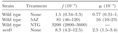
the supernatant was transferred to a new tube with 500l of TABLE 1
100% EtOH. The samples were mixed by inverting and kept
Mutation frequencies (f) and rates () inrpoBresulting in Rifr
at room temperature for 2 min and then centrifuged at 4⬚for 10 min at 10,000⫻gto precipitate the DNA. The DNA pellet
Strain Treatment f(10⫺8) (10⫺8)
was washed twice with 200l of 75% EtOH and allowed to air dry for 30 min. When the ethanol had evaporated, 50l
Wild type None 1.5 (0.34–3.3) 0.77 (0.31–1.3)
of TE buffer was added to the tube and the DNA was allowed
Wild type 5AZ 81 (46–120) 16 (10–23)
to dissolve overnight at room temperature.
Wild type NTG 3200 (2800–3600) —
Sequencing therpoBgene for mutations:Once the
chromo-uvrD None 8.3 (4.2–12.5) 2.5 (1.5–3.4)
somal DNA was isolated, one of two primer pairs was used to amplify the DNA for direct sequencing. Most of the mutations
The mutation rates of strains R1 (wild type) and NS3113 occurred in the region obtained with 5⬘-AAACTGTGCCGAT
(uvrD) were determined by the method ofDrake(1991; see GGTGGAC-3⬘(5⬘position 1058) and 5⬘-TAGCTCACGCGGC
materials and methodsfor rates and frequencies and mu-CATTCAC-3⬘(5⬘position 1945). The rest of the Rifrmutations
tant isolation procedures). Values in parentheses are 95% were found using the primer pair 5⬘-TCTTTCCCATCGACG
confidence limits. AGTCC-3⬘ (5⬘ position 173) and 5⬘-CACGATGGGGCGGTT
GTT-3⬘ (5⬘ position 1224). The PCR reaction included 1⫻ PCR buffer (Bio-Rad, Hercules, CA), 50 pmol of each PCR
primer, 2 mm MgCl2 (Bio-Rad), 40 nmol dNTP, 2.5% for- plates containing 50g/ml rifampicin (seematerials mamide, 1.5 units Taq DNA polymerase (Bio-Rad), 1l of andmethods) that occurred spontaneously inD. radio-DNA, and double-distilled H2O. The DNA was denatured at
duranswild-type or uvrD strains or that were induced 95⬚for 4 min, amplified for 30 cycles of 95⬚for 30 sec, 57⬚
by NTG or 5AZ. Table 1 shows the mutation frequencies for 30 sec, and 72⬚for 1 min and extended for 7 min at 72⬚. PCR
products were purified with the MinElute PCR purification kit and rates that we obtained. Although NTG was a potent (QIAGEN, Valencia, CA) and manually sequenced with the mutagen for D. radiodurans, ethyl methanesulfonate SequiTherm EXCEL II DNA sequencing kit (Epicentre Tech- (EMS) failed to mutagenize this organism at all. Also, nologies, Madison, WI) using one of the two sequencing
prim-although the cytosine analog 5AZ gave positive results, ers, 5⬘-CATGCTGCTCGGCAACCC-3⬘(5⬘position 1221) and
the cytosine analog zebularine, as well as the adenine 5⬘-TGATTCACAAAGACACTGGCGT-3⬘(5⬘position 323),
re-spectively. analog 2-aminopurine, failed to mutagenize D.
radio-N-methyl-Nⴕ-nitro-N-nitrosoguanidine mutagenesis: Six tubes durans. We isolated one mutant per culture and pre-of 5 ml TGY broth were inoculated with R1 and aerated for pared DNA for sequence analysis of the relevant regions 48 hr at 37⬚. The cultures were pooled together into an
Erlen-of therpoBgene (seematerials and methods). Table meyer flask and diluted 1:1 with TGY. A 1-mg/mlN
-methyl-2 shows the results for⬎250 mutations in theD.
radio-N⬘-nitro-N-nitrosoguanidine (NTG) solution was made in 1:1
acetone and 0.1mNaCitrate buffer (pH 5.5). Two microliters durans rpoBgene that result in the Rifrphenotype, in-of culture was aliquoted into tubes, and NTG was added to a cluding 185 spontaneous mutations derived in a wild-final concentration of 0, 30, or 100 g/ml. After a 90-min type background, 33 NTG-induced mutations, and 19 incubation in a 37⬚water bath, the mutagenized cultures were
5AZ-induced mutations, as well as 17 spontaneous muta-washed three times: first with 5 ml of 0.01mMgSO4, followed
tions occurring in auvrDbackground. From these data by 5 ml of TGY, and finally with 2 ml of TGY (Miller1992).
To determine the percentage of cells surviving exposure to we can already define 33 different base substitution mu-NTG, 50l of a 10⫺4dilution of each culture was plated onto
tations at 22 sites (base pairs). Each of the 6 base substi-TGY and incubated at 37⬚for 48 hr. Outgrowth cultures were tutions can be monitored at a set of 3–7 sites.
made by adding 500l of each mutagenized culture to 5 ml
Spontaneous mutations—deletion and base
substitu-TGY and incubating for 48 hr at 37⬚. The outgrowth cultures
tion hotspots:We found that 35 of the 185 (19%) spon-were concentrated 10-fold and 100l was plated on TGY⫹
rifampicin plates to yield Rifrcolonies for sequencing. Two Rifr taneous mutations detected in the wild-type background colonies from each of the 10, 30, and 100g/ml NTG TGY⫹ are deletions of 9 bp at or adjacent to a 7-bp direct rifampicin plates were sequenced. The experiment was repeated repeat separated by 2 bp and probably are the result of with more cultures at a concentration of 100g/ml NTG.
slipped mispairing stimulated at this site (see Figure 2).
5-Azacytidine mutagenesis:An overnight culture was diluted
We detected three different deletions, although one of and used to seed overnight cultures with 100–300 cells in TGY
with 100g/ml of 5-azacytidine (5AZ) and grown for 48 hr these (type III in Figure 2) is represented by only 1 on a rotor (Miller 1992). The mutational frequency was occurrence, while type I and type II deletions are repre-determined by plating 100l of a 10⫺5dilution of each culture
sented by 19 and 15 occurrences, respectively (see also on TGY and 50l directly on TGY⫹rifampicin.
Table 2). Deletion type II (Figure 2) is of the exact
uvrDRifrmutants: D. radiodurans uvrDRifrmutants were
form found for many deletions in bothE. coliand other obtained in the same manner as spontaneous mutants, with
the exception that each overnight culture was inoculated with organisms (seeFarabaughet al. 1978;Albertiniet al. a singleuvrDcolony instead of seeding the cultures with 100– 1982 and references therein). Deletion type I is shifted
300 cells. just 1 bp in one direction. InE. coli, one or two small
deletions inrpoBthat result in Rifrmutants have been reported (JinandGross1988), but these are relatively RESULTS
rare compared with the base substitutions that result in Rifr in that organism.
Isolation of Rifr mutants and sequence analysis of
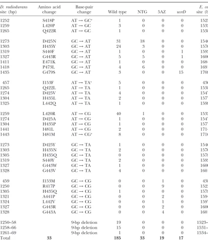
TABLE 2
Distribution of mutations leading to RifrinD. radiodurans
D. radiodurans Amino acid Base-pair E. coli
site (bp) change change Wild type NTG 5AZ uvrD site (bp)
1252 S418P AT→GCa 1 0 0 0 1525
1259 L420P AT→GC 3 0 0 0 1532
1265 Q422R AT→GC 1 0 0 0 1538
1273 D425N GC→AT 31 18 0 0 1546
1303 H435Y GC→AT 24 3 0 0 1576
1319 S440F GC→AT 1 0 0 1 1592
1327 G443R GC→AT 5 5 0 1 1600
1411 E471K GC→AT 1 0 0 0 1684
1418 P473L GC→AT 4 6 0 0 1691
1435 G479S GC→AT 3 0 0 15 1708
457 I153F AT→TAa 5 0 0 0 436
1265 Q422L AT→TA 1 0 0 0 1538
1274 D425V AT→TA 4 0 0 0 1547
1304 H435L AT→TA 2 0 0 0 1577
1325 L442Q AT→TA 1 0 0 0 1598
1259 L420R AT→CG 40 1 0 0 1532
1274 D425A AT→CG 1 0 0 0 1547
1304 H435P AT→CG 1 0 0 0 1577
1441 I481L AT→CG 2 0 0 0 1714
1443 I481M AT→CGa 8 0 0 0 1716
1273 D425Y GC→TA 1 0 0 0 1546
1303 H435N GC→TA 2 0 0 0 1576
1305 H435Q GC→TA 0 0 0 0 1578
1319 S440Y GC→TA 2 0 0 0 1592
1327 G443W GC→TA 1 0 0 0 1600
1328 G443V GC→TA 4 0 0 0 1601
459 I153M GC→CG 0 0 1 0 438
1250 R417P GC→CG 0 0 9 0 1523
1305 H435Q GC→CG 1 0 0 0 1578
1321 A441P GC→CG 0 0 2 0 1594
1324 L442V GC→CG 0 0 1 0 1597
1327 G443R GC→CG 0 0 2 0 1600
1328 G443A GC→CG 0 0 4 0 1601
1250–58 9-bp deletion 19 0 0 0 1523–31
1258–66 9-bp deletion 15 0 0 0 1531–39
1261–69 9-bp deletion 1 0 0 0 1534–42
Total 33 185 33 19 17
The DNA sequence change inrpoBwas determined in each case. aThe corresponding base and base change is different inE. coli.
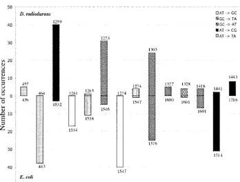
above, the spontaneous spectrum ofrpoBmutations in hotspots involve different sites, as shown in Figure 3 (see also Table 2 and Garibyanet al. 2003), with one
D. radioduransshows three base substitution hotspots at
positions 1273 and 1303 (G:C→A:T) and 1259 (A:T→ exception. Mutations of G:C→A:T at 1303 inD. radio-duransand the corresponding 1576 inE. coliboth repre-C:G). Together, these three hotspots account for 95
of the 150 base substitutions (63%) found among the sent hotspots. Figure 4 shows the DNA sequence align-ments surrounding each of the hotspot sites. In three spontaneous mutations inrpoB, even though these are
Figure 2.—Deletions in
rpoB resulting in the Rifr phenotype inD. radiodurans. The letters in boldface type indicate a 7-bp sequence re-peat separated by 2 bp. See Table 2 for relative frequen-cies of these deletions.
different rates in two of these examples. In one case detected inD. radioduransthat has not yet been detected inE. coli. InE. coli, in which far more extensive studies (E. coli1547), the sequence identity is more extensive.
have been carried out (JinandGross1988;Severinov
We consider reasons for different hotspots in the
dis-et al. 1993; Garibyan et al. 2003; Wolff et al. 2004),
cussion.
alterations at 27 different residues can result in the Rifr Mutations resulting from mutators and mutagens:We
phenotype, whereas so far 15 such residues have been examined the mutational spectrum in auvrDstrain
(Ta-found inD. radiodurans. ble 2). Here, all the mutations are transitions, with
G:C→A:T mutations predominating over A:T→ G:C mutations, although the spectrum is dominated by a
DISCUSSION single hotspot at position 1435. We have also employed
two mutagens, NTG and 5AZ. It is clear from Table 2 In this work, we have successfully applied a genetic that these agents induce specifically G:C→A:T transi- system developed inE. coli toD. radiodurans, a species tions in the case of NTG and G:C→C:G transversions that separated from the rest of the bacterial family tree in the case of 5AZ inD. radioduransand thus have the early in evolution (Battista and Rainey 2001). Al-same specificity as found inE. coliand other organisms though assays measuring the frequency of forward
muta-(CupplesandMiller1989). tion to rifampicin (TempestandMoseley1982),
strep-Altered amino acids in RNA polymerase  in Rifr
tomycin (Kerszman1975), and trimethoprim (Sweet
mutants:The amino acid residues affected in Rifr mu- andMoseley1974) resistance have been described for tants are strikingly similar inE. coliandD. radiodurans, D. radiodurans, currently no genetic system allows the analysis of mutations generated in wild-type and repair-as detailed in Figure 5. However, only one site hrepair-as been
conserved among most prokaryotes, it should be possi-ble to develop a detailedrpoB/Rifrsystem in an organ-ism without sophisticated genetic tools. Investigators from numerous laboratories have analyzed Rifrmutants from a variety of pathogens, includingMycobacterium tuber-culosis (Telentiet al. 1993;Musser1995),M. smegmatis
(Karunakaran andDavies 2000), M. leprae(Cambauet al. 2002),M. kansasii(Kleinet al. 2001),Bacillus anthracis
(Vogleret al. 2002),B. cereus(Vogleret al. 2002), Rhodo-coccus equi (Fines et al. 2001), Legionella pneumophila
(Neilsenet al. 2000),Neisseria meningitides(Stefanelli
et al. 2001), Streptococcus pyogenes (Aubry-Damon et al. 2002),Staphylococcus aureus(Aubry-Damonet al. 1998), andHelicobacter pylori(Heepet al. 2000). In all of these cases, therpoBmutations affect residues corresponding to those altered inE. coliRifrmutants, although a com-Figure4.—DNA sequences and homologies surrounding plete catalog of possible mutations has not been at-the mutational hotspots inD. radioduransandE. coli. The under- tempted in these studies.
lined base is the hotspot site (see also Table 2 and Figure 3). Table 2 shows the results of sequencing⬎250
muta-tions leading to RifrinD. radioduransderived spontane-ously in wild-type anduvrD(mismatch-repair-deficient) backgrounds, and after treatment with N -methyl-deficient mutants ofD. radiodurans. Therefore, we used
N⬘-nitro-N-nitrosoguanidine and 5-azacytidine. We have therpoB/Rifrsystem recently developed inE. coli(
Gari-defined 33 base change substitutions at 22 different sites
byanet al. 2003) to monitor base substitutions. Because
therpoB-encoded-subunit of RNA polymerase is highly (base pairs). This allows us to monitor the A:T→G:C
change at 3 sites, the G:C→A:T change at 7 sites, the theD. radioduranssite has only two occurrences of A:T→ C:G mutations among 150 base substitutions. Similarly, A:T→T:A change at 5 sites, the A:T→C:G change at
the G:C→A:T mutation at position 1273 inD.
radiodur-5 sites, the G:C→T:A change at 6 sites, and the G:C→
ansis a hotspot, whereas the corresponding change at C:G change at 7 sites.
1546 is not inE. coli, despite sharing the same 6 bp on Analysis of therpoB/Rifrsystem provides the first
de-one side and the same nearest neighbor on the other. tailed description of mutagenesis inD. radioduransand
As part of the same sequence segment, the AT→ G:C lays the groundwork for studies of the mechanisms that
change at position 1547 is the most prominent hotspot this species uses to avoid mutation.D. radioduranshas an
inE. coli (40 occurrences among 298 mutations), but astonishing ability to avoid UV- and
ionizing-radiation-no mutations have been found at the corresponding induced mutagenesis (Sweet and Moseley 1974;
position (1274) inD. radiodurans. Normally, one would
Kerszman1975;TempestandMoseley1982). For
ex-not be able to make conclusions regarding this site in ample, even when cultures are exposed to 700 J/m2UV
D. radiodurans, since without any occurrences recorded light, a dose that introducesⵑ5000 thymine-containing
it might be that the A:T→G:C change there does not pyrimidine dimers per genome in exposed cells and
result in a mutation that yields Rifr cells. However, a that kills 90% of the irradiated population (Moseley
look at Figure 5 shows that the aspartic acid residue 1983), there is no evidence of UV-induced mutagenesis
specified by the codon involving 1547 inE. coliand 1274 in this species (TempestandMoseley1982). We
antici-inD. radioduransis an amino acid (residue 516 inE. coli; pate that the introduction of therpoB/Rifrsystem will
425 in D. radiodurans) at which many different ex-encourage further investigation of mutagenesis and
changes yield Rifr. InE. coli, where many more muta-DNA repair inD. radiodurans.
tions have been generated, all five changes at this site Several aspects of the results found in this study are
have been detected and shown to result in Rifr, whereas worth noting. The spontaneous mutations reveal a
dele-so far four of these five changes have already been found tion hotspot centered near sequence repeats that very
in D. radiodurans. It is highly probable that the re-probably serve as substrates for the type of slipped
mis-maining change (to glycine) will also yield a Rifr cell pairing events that generate deletions in other
organ-and that the zero occurrences of an A:T→G:C change isms (Farabaugh et al. 1978; Albertini et al. 1982).
at 1274 inD. radioduranssimply reflect failure to induce Although deletions inrpoBdo not usually generate
via-the mutation at a detectable rate with via-the current sam-ble Rifrstrains, the in-frame deletion of 9 bp removes
ple size and not a failure of the resulting change to three amino acids that alter the-subunit of RNA
poly-generate Rifr colonies. Position 1259 is a hotspot for merase enough to become resistant to inactivation by
the A:T→ C:G transversion inD. radiodurans, but not rifampicin, but not enough to affect function. The
speci-inE. coli. Although this site is part of a 5-bp homology ficities of NTG and 5AZ are the same inD. radioduransas
and a 12 of 14-bp homology (Figure 4), the correspond-found forE. coliand other organisms (see, for instance,
ing sites do not have identical nearest neighbors on one
Cupples and Miller 1989). What is interesting, and
side. The lack of a nearest neighbor in an otherwise not easy to explain, is the failure to detect mutagenesis
homologous stretch does not prevent both 1303 inD.
with EMS, even though NTG is a potent mutagen for
radioduransand 1576 inE. coli(25 occurrences among
D. radiodurans. Also, neither the cytosine analog zebu- 298 mutations) from being hotspots for the G:C→A:T larine (McCormacket al. 1980) nor the adenine ana- transition. That these two sites are hotspots despite be-log 2-aminopurine causes mutations inD. radiodurans, ing in different organisms is remarkable. It is not clear whereas they do inE. coli(seeCupplesand J. H.Miller whether the other sites that are hotspots in one
organ-1989;Leeet al.2004). Finally, therpoBmutations in the ism but not in the other reflect a requirement for a
D. radiodurans uvrDstrain predominate at a single G:C→ more extensive sequence environment as a determinant A:T site, whereas the major hotspot inE. coli rpoB for of mutation rate or whether mutation rates at all sites mismatch-repair-deficient mutations is an A:T → G:C are really organism specific. Additional experiments with mutation. engineered sequences are required to answer these
ques-There are prominent base substitution hotspots in tions.
rpoBin bothD. radioduransandE. coli (Table 2;Gari- We see no reason why therpoB/Rifrsystem cannot be
byan et al. 2003), although with one exception they applied to other genetically intractable bacterial species. occur at different sites (see Figure 3). What makes this The only requirements for implementing this system remarkable is that the DNA sequences surrounding the are that the investigator knows therpoBgene sequence hotspot sites are very similar, as shown in Figure 4. For and has the ability to isolate Rifr mutants from the instance, the E. coli spontaneous hotspot (31 occur- species of interest. It should now be possible to extend rences among 298 mutations) at position 1714 (A:T→ the detailed analysis of spontaneous mutagenesis to a C:G) has a DNA sequence identical to that of the corre- large number of diverse species and to explore how the spondingD. radioduranssite 1441 for 10 bp on one side specifics of this process compare among prokaryotes
Kerszman, G., 1975 Induction of mutation to streptomycin resis-The authors gratefully acknowledge Nicole C. Shank for
construct-tance inMicrococcus radiodurans.Mutat. Res.28:9–14. ing strain NS3113. J.R.B. is supported by Department of Energy grant
Kim, M., T. HuangandJ. H. Miller, 2003 Competition between DE-FG02-01ER63151. J.H.M. is supported by National Institutes of
MutY and mismatch repair at A • C mispairs in vivo. J. Bacteriol. Health grant ES0110875.
185:4626–4629.
Klein, J. L., T. J. BrownandG. L. French, 2001 Rifampicin resis-tance inMycobacterium kansasiiis associated withrpoBmutation. Antimicrob. Agents Chemother.45:3056–3058.
LITERATURE CITED Lee, G., E. WolffandJ. H. Miller, 2004 Mutagenicity of the
cyti-dine analog zebularine inEscherichia coli. DNA Repair3:155–161. Albertini, A. M., M. Hofer, M. P. CalosandJ. H. Miller, 1982 McCormack, J. J., V. E. Marquez, P. S. Liu, D. T. VisticaandJ. S. On the formation of spontaneous deletions: the importance of Driscoll, 1980 Inhibition of cytidine deaminase by 2-oxopyri-short sequence homologies in the generation of large deletions. midine riboside and related compounds. Biochem. Pharmacol.
Cell29:319–328. 29:830–832.
Anderson, A. W., H. C. Nordon, R. F. Cain, G. ParrishandD. Miller, J. H., 1992 A Short Course in Bacterial Genetics: A Laboratory Duggan, 1956 Studies on a radio-resistant micrococcus. Isola- Manual and Handbook for Escherichia coli and Related Bacteria. Cold tion, morphology, cultural characteristics, and resistance to Spring Harbor Laboratory Press, Cold Spring Harbor, NY. gamma radiation. Food Technol.10:575–578. Moseley, B. E., 1983 Photobiology and radiobiology ofMicrococcus Aubry-Damon, H., C. J. SoussyandP. Courvalin, 1998 Character- (Deinococcus) radioduran.Photochem. Photobiol. Rev.7:223–275. ization of mutations in the rpoB gene that confer rifampicin Moseley, B. E., andA. Mattingly, 1971 Repair of irradiation trans-resistance in Staphylococcus aureus. Antimicrob. Agents Chemo- forming deoxyribonucleic acid in wild type and a radiation-sensi-ther.42:2590–2594. tive mutant ofMicrococcus radiodurans.J. Bacteriol.105:976–983. Aubry-Damon, H., M. Galinmand, G. GerbaudandP. Courvalin, Musser, J. M., 1995 Antimicrobial resistance in mycobacteria:
mo-2002 rpoBmutation conferring rifampicin resistance inStrepto- lecular genetic insights. Clin. Microbiol. Rev.8:496–514.
coccus pyogenes.Antimicrob. Agents Chemother.46:1571–1573. Neilsen, K., P. Hindersson, N. HoibyandJ. M. Bangsborg, 2000
Battista, J. R., 1997 Against all odds: the survival strategies ofDeino- Sequencing of therpoBgene inLeionella pneumophilaand
charac-coccus radiodurans.Annu. Rev. Microbiol.51:203–224. terization of mutations associated with rifampicin resistance in
Battista, J. R., and F. A.Rainey, 2001 TheDeinococcaceae, pp. 395– the legionellaacaeae. Antimicrob. Agents Chemother.44:2679– 403 inBergey’s Manual of Systematic Bacteriology, Vol. 1, edited by 2683.
G. M.Garrity. Springer-Verlag, New York. Ovchinnikov, Y. A., G. S. Monastyrskaya, S. O. Guriev, N. F. Cambau, E., P. Bonnafous, E. Perani, W. Sougakoff, B. Jiet al., Kalinina, E. D. Sverdlovet al., 1983 RNA polymerase rifampi-2002 Molecular detection of rifampicin and ofloxacin resis- cin resistance mutations inEscherichia coli: sequence changes and tance for patients who experience relapse of multibacillary lep- dominance. Mol. Gen. Genet.190:344–348.
Petersen-Mahrt, S. K., R. S.Harrisand M. S.Neuberger, 2002 rosy. Clin. Infect. Dis.34:39–45.
AID mutatesE. coli, suggesting a DNA deamination mechanism Campbell, E. A., N. Korzheva, A. Mustaev, K. Murakami, S. Nair
for antibody diversification. Nature418:99–103.
et al., 2001 Structural mechanism for rifampicin inhibition of
Rangarajan, S., G. Gudmundsson, Z. Qiu, P. L. FosterandM. F. bacterial RNA polymerase. Cell104:901–912.
Goodman, 1997 Esherichia coli DNA polymerase II catalyzes Cupples, C., andJ. H. Miller, 1989 A set oflacZ mutations in
chromosomal and episomal DNA synthesis in vivo. Proc. Natl.
Escherichia coli allows rapid detection of each of the six base
Acad. Sci. USA94:946–951. substitutions. Proc. Natl. Acad. Sci. USA86:5345–5349.
Reynolds, M. G., 2000 Compensatory evolution in rifampicin-resis-Dixon, W. J.,andF. J. Massey, Jr.,1969 Introduction to Statistical
tantEscherichia coli.Genetics156:1471–1481.
Analysis. McGraw-Hill, New York.
Severinov, K., M. Soushko, A. GoldfarbandV. Nikiforow, 1993 Drake, J. W., 1991 A constant rate of spontaneous mutation in
DNA-Rifampicin region revisited. J. Biol. Chem.268:14820–14825. based microbes. Proc. Natl. Acad. Sci. USA88:7160–7164.
Stefanelli, P., C. Fazio, G. La Rosa, C. Marianelli, M. Muscillo Earl, A. M., S. K. Rankin, K. P. Kim, O. N. LamendolaandJ. R.
et al., 2001 Rifampicin-resistant meningococci causing invasive
Battista, 2002 Genetic evidence that theuvsEgene product
disease: detection of point mutations in therpoBgene and
molec-of Deinococcus radiodurans R1 is a UV damage endonuclease.
ular characterization of the strains. J. Antimicrob. Chemother. J. Bacteriol.184:1003–1009.
47:219–222. Farabaugh, P. J., U. Schmeissner, M. HoferandJ. H. Miller, 1978
Sweet, D. M., andB. E. Moseley, 1974 Accurate repair of ultra-Genetic studies of the lac repressor. VII. On the molecular nature
violet-induced damage inMicrococcus radiodurans.Mutat. Res.23:
of spontaneous hotspots in thelaclgene ofEscherichia coli.J. Mol.
311–318. Biol.126:847–863.
Telenti, A., P. Imboden, F. Marchesi, D. Lowrie, S. Coleet al., 1993 Fines, M., S. Pronost, K. Maillard, S. TaoujiandR. Leclercq,
Detection of rifampicin resistance mutations inMycobacterium
2001 Characterization of mutations in therpoBgene associated
tuberculosis.Lancet341:647–650.
with rifampicin resistance inRodococcus equiisolated from foals.
Tempest, P. R., andB. E. Moseley, 1982 Lack of ultraviolet muta-J. Clin. Microbiol.39:2784–2787.
genesis in radiation-resistant bacteria. Mutat. Res.104:275–280. Garibyan, L., T. Huang, M. Kim, E. Wolff, A. Nguyenet al., 2003 Udupa, K. S., P. A. O’Cain,V.Mattimoreand J. R.Battista, 1994 Use of therpoBgene to determine the specificity of base substitu- Novel ionizing radiation-sensitive mutants ofDeinococcus radiodur-tion mutaradiodur-tions on theEscherichia colichromosome. DNA Repair ans.J. Bacteriol.176:7439–7446.
2:593–608. Vogler, A. J., J. D. Busch, S. Percy-Fine, C. Tipton-Hunton, K. L. Heep, M., S. Odenbreit, D. Beck, J. Decker, E. Prohaskaet al., 2000 Smithet al., 2002 Molecular analysis of rifampicin resistance Mutations at four distinct regions of therpoBgene can reduce inBacullus anthracisandBacillus cereus.Antimicrob. Agents Che-the susceptibility ofHelicobacter pylori to rifamycin. Antimicrob. mother.46:511–513.
Agents Chemother.44:1713–1715. Wolff, E., M. Kim, K. Hu, H. YangandJ. H. Miller, 2004 Polymer-Jin, D. J., andC. A. Gross, 1988 Mapping and sequencing of muta- ases leave fingerprints: analysis of the mutational spectrum in tions in the Escherichia coli rpoBgene that lead to rifampicin Escherichia coli rpoBto assess the role of Pol IV in spontaneous resistance. J. Mol. Biol.202:45–58. mutation. J. Bacteriol. (in press).
Karunakaran, P., andJ. Davies, 2000 Genetic antagonism and