DOI: 10.1534/genetics.106.063115
SWAN-1, a
Caenorhabditis elegans
WD Repeat Protein of the AN11 Family,
Is a Negative Regulator of Rac GTPase Function
Yieyie Yang,*
,1Jiamiao Lu,* Joel Rovnak,
†Sandra L. Quackenbush
†and Erik A. Lundquist*
,2*Department of Molecular Biosciences, University of Kansas, Lawrence, Kansas 66045 and†Department of Microbiology, Immunology and Pathology, College of Veterinary Medicine and Biomedical Sciences, Colorado State University, Fort Collins, Colorado 80523-1619
Manuscript received July 7, 2006 Accepted for publication September 3, 2006
ABSTRACT
Rac GTPases are key regulators of cell shape and cytoskeletal organization. While some regulators of Rac activity are known, such as GTPase-activating proteins (GAPs) that repress Rac activity, other Rac regulators remain to be identified. The novelCaenorhabditis elegansWD-repeat protein SWAN-1 was identified in a yeast two-hybrid screen with the LIM domains of the Rac effector UNC-115/abLIM. SWAN-1 was found to also associate physically with Rac GTPases. The swan-1(ok267) loss-of-function mutation suppressed defects caused by the hypomorphicced-10(n1993)allele and enhanced ectopic lamellipodia and filopodia formation induced by constitutively active Rac inC. elegansneurons. Furthermore, SWAN-1(1) transgenic expression suppressed the effects of overactive Rac, including ectopic lamellipodia and filopodia formation inC. elegans neurons, ectopic lamellipodia formation in cultured mammalian fibroblasts, and cell polarity and actin cytoskeleton defects in yeast. These studies indicate that SWAN-1 is an inhibitor of Rac GTPase function in cellular morphogenesis and cytoskeletal organization. While broadly conserved across species, SWAN-1 family members show no sequence similarity to previously known Rac inhibitors.
C
ELL and growth cone migration and the establish-ment and maintenance of cell shape are tightly regulated processes controlled by extracellular cues and intracellular physiology (Tessier-Lavigneand Goodman 1996; Yu and Bargmann 2001; Dickson 2002; Raftopoulou and Hall 2004; Vicente-Manzanareset al.2005). Rac GTPases of the Rho subfamily are key regulators of cell shape and act in part by controlling the structure and dynamics of the actin cytoskeleton (Hall 1998). Classically, Rac GTPases were identified by their similarity to Rho and were found to induce the formation of veil-like lamellipodial plasma membrane extensions in serum-starved fibroblasts (Hall 1998; Otieno et al. 2005). InCaenorhabditis elegans neurons, Rac activity promotes the formation of both lamellipo-dia and filopolamellipo-dia, thin finger-like plasma membrane extensions (Struckhoffand Lundquist2003). Loss-of-function studies in Drosophila andC. elegans demon-strate that Rac GTPases are required for a wide variety of morphogenetic events, including axon pathfinding and cell migration (Dickson2001; Lundquist2003). The GTPase regulatory cycle involves active, GTP-bound Racs that hydrolyze GTP to GDP and thus self-inactivate, as the GDP-bound form is inactive. Guanine nucleotide
exchange factors (GEFs) mediate the exchange of GDP for GTP and thus favor the GTP-bound active state of Racs. GTPase-activating proteins (GAPs) stimulate the GTPase activity of Racs and thus favor the inactive state (Van Aelst and D’Souza-Schorey 1997). Another class of Rho GTPase negative regulators are the gua-nine nucleotide dissociation inhibitors (GDIs), which inhibit Rho GTPase association with the plasma mem-brane where they are active (VanAelstand D’Souza -Schorey 1997; Michaelsonet al.2001).
In C. elegans, three Rac GTPases (CED-10, MIG-2, and RAC-2) have compensatory, redundant roles in axon pathfinding and in the migration of neurons, embryonic cells during gastrulation, and vulval cells (Lundquistet al.2001; Kishoreand Sundaram2002; Sotoet al.2002). The actin-binding protein UNC-115/ abLIM, which acts with Racs in axon pathfinding, is required for Rac-induced lamellipodia and filopodia in neurons and UNC-115 itself induces the formation of lamellipodia and filopodia inC. elegansneurons and in serum-starved mammalian fibroblasts (Lundquistet al. 1998; Struckhoff and Lundquist 2003; Yang and Lundquist 2005). UNC-115 might be a downstream cytoskeletal effector of Rac GTPases and might control growth cone lamellipodia and filopodia formation in response to Rac signaling during axon pathfinding. The molecular linkage between Racs and UNC-115 in these events is unclear, and molecules that interact physically with UNC-115 remain to be identified. Fur-thermore, while some Rac inhibitors are known, it will
1Present address:Dana-Farber Cancer Institute, 44 Binney St., Smith 870,
Boston, MA 02115.
2Corresponding author:Department of Molecular Biosciences, University
of Kansas, 1200 Sunnyside Ave., 5049 Haworth Hall, Lawrence, KS 66045. E-mail: [email protected]
be important to identify all of the molecules that reg-ulate Rac activity to precisely control distinct morpho-genetic events.
To identify molecules that act with Rac and UNC-115 in axon pathfinding and cell migration, the LIM do-mains of UNC-115 were used as bait in a yeast two-hybrid screen of aC. eleganscDNA library. This screen identi-fied a novel, conserved molecule called SWAN-1 (s
even-WD-repeat protein of theAN11 family-1), which consists of at least seven WD repeats and a conserved C-terminal tail. In two-hybrid and pull-down assays, SWAN-1 was found to interact both with the UNC-115 LIM domains and with the Racs, suggesting that SWAN-1 is a molec-ular linker between UNC-115 and the Racs. An array of functional tests inC. elegans, mammalian fibroblasts, and yeast indicate that SWAN-1 represses Rac activity. SWAN-1 displays no similarity to other known Rac nega-tive regulators (e.g., GAPs), indicating that SWAN-1 represents a previously unidentified class of negative regulators of Rac GTPases.
MATERIALS AND METHODS
C. elegans culture and genetics: C. elegans were cultured using standard techniques (Epsteinand Shakes1995). All experiments were performed at 20°. Theswan-1(ok267) muta-tion was isolated by theC. elegansGene Knockout Consortium (kindly provided by G. Molder and B. Barstead) and was outcrossed to wild type three times before phenotypic analysis. The homozygosity ofswan-1(ok267)in all outcrosses and single and double mutants was confirmed by single-animal poly-merase chain reaction (PCR) using primers that amplify a band specific to theok267deletion. Germline transformation ofC. eleganswas performed by standard techniques involving injection of a DNA mixture into the syncytial germline of C. eleganshermaphrodites (Epsteinand Shakes1995). RNA-mediated gene knockdown (RNAi) was performed by feeding nematode strainsEscherichia coli expressing double-stranded RNA complementary to eitherswan-1orswan-2(Timmonset al. 2001; Kamathand Ahringer2003).
Numbers of cell corpses in the region of the pharynx at the mid-threefold stage of embryogenesis (750 min postfertiliza-tion) were counted using differential interference contrast microscopy. Gonadal distal tip cell migration defects were scored as the percentage of gonad arms that showed defective morphology as previously described (Lundquistet al.2001). In Table 1, strains harboring a full-lengthswan-1Tgfptransgene were scored. As a control, nontransgene-bearing siblings from the same brood were also scored and presented in Table 1.
Significance of differences of means in Table 1 were cal-culated using the t-test, and significance of differences of proportions in Table 1 and in Figures 4, 5, 7, and 8 were cal-culated using both thet-test and Fisher’s exact analysis.
Molecular biology:All fragments containing coding region that were generated by PCR were sequenced to ensure that no mutations were introduced during the procedure. The sequences of all PCR primers used in this work are available upon request.
Microscopy:See the figure legends for details about each micrograph. Unless otherwise noted, all images were captured on a Leica DMRE microscope with a 403Planapo objective and 103magnifier using a Hamamatsu Orca C4742-94 camera and Openlab software. Images were processed in Photoshop.
UNC-115 LIM domain two-hybrid screen: Vectors for the two-hybrid system were kindly provided by S. Elledge. The coding region for the UNC-115 LIM domains was amplified by PCR and cloned in frame to the GAL4 DNA-binding domain in the vector pAS1-CYH (the ‘‘bait’’ construct). The yeast strain Y190 was transformed with the bait construct by standard techniques, and autoactivation of the bait construct was tested by growth on 25 mm3-aminotriazole (3-AT) and X-Gal, which assess expression ofHIS5andLacZreporter gene expression in Y190. The bait construct showed no autoactivation: it did not allow Y190 yeast to grow on 3-AT (noHIS5expression), nor did blue colonies result when transgenic yeast were grown on X-Gal (noLacZexpression).
Y190-bait transgenic yeast were transformed with aC. elegans random-primed cDNA library cloned into the pACT vector that harbors the GAL4 activation domain (the library was kindly provided by R. Barstead). Yeast were plated at high density on medium containing 25 mm3-AT and X-Gal, and colonies that were blue (indicating HIS5 expression and LacZexpression) were selected for further analysis. From an estimated 2 million cDNAs screened, 2 cDNAs representing theswan-1locus were recovered. These 2 cDNAs did not re-sult in autoactivation when present in Y190 without the bait plasmid, indicating that both plasmids are needed for activation.
The coding regions of the threeC. elegans racgenesced-10, mig-2, andrac-2and the non-LIM-domain-containing portion ofunc-115were generated by PCR and fused in frame to the GAL4 DNA-binding domain in the pAS1-CYH plasmid. None of these constructs resulted in autoactivation. These con-structs were then used in direct two-hybrid tests with the pACTTSWAN-1 plasmid.
Co-immunoprecipitation experiments:HeLa cells and HEK293 cells were grown to80% confluency and were transfected using Fugene6 (Roche, Indianapolis). The full-lengthunc-115 coding region was generated by PCR and cloned in frame to enhanced green fluorescent protein (EGFP) in the pEGFP1 vector (Clontech, Mountain View, CA). The swan-1 coding region was cloned in frame to both the Myc epitope (MYCT SWAN-1) and the 3xFLAG epitope (FLAGTSWAN-1) in the pCMV-Myc and pCMV-3xFLAG vectors, respectively (Clon-tech). MYCTRAC-2 and FLAGTRAC-2 plasmids were similarly constructed. In most cases, 16mg of plasmid were used in single transfections and 8 mg of each plasmid were used in double transfections in 75-cm2flasks. For RAC-2 plasmids, 8mg were used because higher concentrations led to cell inviability. After 48 hr of growth after transfection, cells were washed with ice-cold PBS and 1 ml of ice-cold lysis buffer [150 mmNaCl/ 10 mmTris (pH 7.4)/1 mmEDTA/1 mmEGTA/0.5% NP-40/ 0.2 mmPMSF/1% protease inhibitor cocktail; Sigma, St. Louis] was added. Cells were scraped from the bottom of the flasks, incubated for 1 hr at 4° with shaking, and centrifuged for 15 min at 16,0003gin a microcentrifuge at 4°. Supernatants were precleared by incubating with 80ml of pre-equilibrated protein G Sepharose bead slurry (Amersham Pharmacia Biotech, Piscataway, NJ) for 2 hr at 4°.
Laboratories, Gaithersburg, MD) with chemiluminescent auto-radiography (Pierce, Rockford, IL).
Figure 1, C and D, shows co-immunoprecipitation of UNC-115 and SWAN-1 and RAC-2 and SWAN-1. For each co-immunoprecipitation experiment, the Western blot was also probed with the antibody used for immunoprecipitation to ensure that the immunoprecipitation procedure was robust (Figure 1).
Generation of an UNC-115 antiserum:The coding region for UNC-115 residues 154–527 was inserted into the pQE9 vector (QIAGEN, Valencia, CA) in frame with the RGS-6-histidine epitope, and fusion protein was produced by bac-terial expression. SDS–PAGE gel fragments containing the fusion protein were used as an antigen to immunize two rab-bits (Caltag Laboratories, Healdsburg, CA). On Western blots, serum from the immunized rabbits but not preimmune serum recognized a bacterially expressed glutathione-S-transferase (GST) fusion with the same portion of UNC-115 (data not shown) (residues 154–527, produced by inserting theunc-115 fragment into the pGEX4T vector; Amersham Pharmacia Biotech). UNC-115-specific antibodies were purified from the antiserum by their affinity for the GSTTUNC-115 fusion pro-tein using glutathione Sepharose beads (Amersham Pharmacia Biotech). This affinity-purified polyclonal antibody recog-nized a single band of70 kDa in Western blots of lysates of C. elegans (the estimated size of full-length UNC-115 poly-peptide is 72 kDa) (data not shown).
swan-1expression analysis:The upstream region ofswan-1 excluding the initiator ATG codon (3412–7549 relative to cosmid F53C11) was amplified, inserted into the pPD95.77 GFP expression vector (kindly provided by A. Fire), and used to drive the expression of GFP in transgenic animals. A full-length fusion of the swan-1 coding region to gfp was con-structed by amplifying theswan-1upstream region and entire coding region (3412–9941 relative to cosmid F53C11) from genomic DNA and fusing this fragment in frame togfpsuch that the transgene was predicted to encode a full-length SWAN-1 protein with GFP at the C terminus. RNAi ofswan-1 resulted in greatly reduced GFP expression from this trans-gene, suggesting that the transgene encodes a SWAN-1TGFP molecule (data not shown).swan-1expression constructs were used at 5 ng/ml in transformation experiments.
SWAN-1 transgenic assays in C. elegans: For the loss-of-functionswan-1(ok267)experiments, previously described con-stitutively activerac(G12V)transgenes were used (Struckhoff and Lundquist 2003). PDE morphological defects were scored as previously described (Struckhoffand Lundquist 2003): for each genotype, the percentages of PDE neurons exhibiting ectopic neurites and ectopic lamellipodia and filo-podia were determined. After construction of each transgene in theswan-1(ok267)background, the transgene was crossed away fromswan-1(ok267)and axon defects were scored again to ensure that the transgene caused defects at percentages sim-ilar to those before the experiment began. This ensured that the transgenes had not been altered during the course of the experiment and the phenotypic change was due to the pres-ence ofswan-1(ok267).
For theswan-1(1)expression experiments, new transgenes were constructed that contained a plasmid harboring the swan-1(1)genomic region (bases 3412–9972 inclusive of cosmid F53C11), arac(G12V)plasmid, or both. In all cases anosm-6Tgfp plasmid was included as a neuronal marker to visualize the PDE neuron. When constructing transgenic strains,rac(G12V) plasmids were used at 1 ng/ml and other plasmids were used at 5 ng/ml. Multiple transgenic lines were generated for each experiment to ensure uniformity of results, and results from each line were accumulated to derive a percentage of PDE axon defects. In allC. elegansexperiments, at least 100 animals
were scored for each genotype. Significance of differences in proportions was calculated using thet-test and Fisher’s exact analysis.
SWAN-1 transgenic assays in fibroblasts:Mammalian NIH 3T3 fibroblasts were grown and transformed as previously described (Yangand Lundquist2005). Briefly, cells grown in six-well plates to50% confluency on polylysine-coated cover-slips were transfected by addition of a mixture of the transgene DNA and the Fugene reagent. Cells were transfected with combinations of 1mg each of the MYCTSWR-1 construct, a MYRTUNC-115 construct, and a CFPTRAC-1(Q61L) con-struct, which encodes an activated version of human Rac1 fused to cyan fluorescent protein (CFP) (kindly provided by T. Meyer). Cells were cotransfected with egfp to identify transfected cells, and cells transfected with egfpalone were used as a control. After transfection, cells were allowed to grow for 6 hr and deprived of serum (serum-containing medium was replaced by DMEM). Cells were grown for another 12 hr and fixed in 3.7% paraformaldehyde. The fixed cells were treated with 1mg/ml of rhodamine-labeled phalloidin to visual-ize the actin cytoskeleton. Cells were mounted in 50% glycerin in PBS for epifluorescence microscopy. For each experiment, at least three transfections were performed and .100 cells were scored to ensure consistency of results.
SWAN-1 transgenic assays in yeast:All yeast were grown at 30°. For growth curve analysis, Y190 yeast strains harboring different plasmid constructs were grown in selective medium and diluted to0.1 OD600units in YPD medium. Growth rates were monitored by sampling the OD600culture densities each hour for 8 hr (see Figure 8). Each strain was tested at least twice, and standard deviations were indicated by error bars in Figure 8. To visualize the yeast actin cytoskeleton with rhodamine-labeled phalloidin, cells were grown in selective medium, diluted to 0.1 OD600units in YPD, and grown to midlog phase (0.3–0.7 OD600) (cells harboring the CED-10 plasmid were assayed after 8 hr). Cells were fixed for 1 hr in 20% formal-dehyde in 0.5mpotassium phosphate pH 6.5 and overnight in 4% formaldehyde in 100 mm potassium phosphate pH 6.5. Cells were washed in PBS and permeabilized in 0.2% Triton X-100 in PBS for 15 min, washed three times in PBS, incubated with rhodamine–phalloidin (1 mg/ml) and 49 ,6-diamidino-2-phenylindole dihydrochloride (DAPI) (1mg/ml) in PBS for 1 hr in the dark, washed three times in PBS, and mounted for fluorescence microscopy in PBS with 90% glycerol and 1 mg/ml 1,4-diazabicyclo[2.2.2]octane (DABCO) antifade reagent.
RESULTS
SWAN-1 interacts physically with the UNC-115 LIM domains and with Racs:UNC-115 is a cytoskeletal linker molecule composed of a C-terminal actin-binding villin headpiece domain (VHD) and three N-terminal LIM domains (Figure 1A) (Lundquist et al. 1998). To identify molecules that bind to the UNC-115 LIM do-mains, a yeast two-hybrid screen using the three UNC-115 LIM domains as bait was conducted (Figure 1A). From an estimated 2 millionC. eleganscDNAs screened, two cDNAs were isolated that encode a novel protein that here is named SWAN-1. The presence ofswan-1 two-hybrid fusions with the UNC-115 LIM domain fusion specifically activated both HIS5 expression (data not shown) and lacZ expression (Figure 1B) in the two-hybrid system (see materials and methods). Both
frame, suggesting that only the full-length SWAN-1 protein is active in the two-hybrid assay. SWAN-1 interacted specifically with the UNC-115 LIM domains, but not with the remainder of the UNC-115 molecule in the two-hybrid system (data not shown).
Because UNC-115 acts genetically downstream of Rac signaling (Struckhoffand Lundquist2003), it
was determined if SWAN-1 could also interact with the threeC. elegansRac molecules in the two-hybrid system. SWAN-1 and each of the threeC. elegansRacs, CED-10, MIG-2, and RAC-2, interacted when directly tested in the two-hybrid system (Figure 1B). SWAN-1 interacted with both wild-type RAC-2 (Figure 1B) and constitutively active, GTPase-dead RAC-2(G12V) (data not shown).
The LIM domains of UNC-115 did not interact with any of the three Racs in the two-hybrid system, nor did any other region of UNC-115 (data not shown).
To confirm the physical interactions detected in the two-hybrid system, co-immunoprecipitation of SWAN-1 with UNC-115 and RAC-2 was assayed in lysates of HeLa cells in which tagged versions of the molecules were expressed (Figure 1, C and D). EGFP-tagged full-length UNC-115 co-immunoprecipitated with Myc-tagged SWAN-1 (Figure 1C). Experiments with MYCTRAC-2 and FLAGTSWAN-1 were consistent with co-immuno-precipitation of the molecules, although MYCTRAC-2 and FLAGTSWAN-1 bands were not completely sepa-rated from the light and heavy chains of the antibodies used for immunoprecipitation on Western blots (Figure 1D). These experiments were repeated with similar re-sults in HeLa cells and in HEK293 cells (data not shown). SWAN-1 is a member of a novel, conserved family of seven-WD-repeat proteins:The complete sequences of the twoswan-1cDNAs isolated in the two-hybrid screen were determined, as were the sequences of the in-dependently derived cDNAs yk343g2 and yk326a5 (provided by Y. Kohara) (Figure 2, A and B). The splic-ing pattern and codsplic-ing potential for each of the four cDNAs were identical. Reverse-transcription PCR was conducted on the 59end of theswan-1transcript using a 59 primer complementary to the trans-spliced leader SL1, which revealed that SL1 was spliced onto the 59-end transcript after position 7464 relative to cosmid F53C11 (Figure 2, A and B). cDNA sequences indicated that theswan-1transcript was polyadenylated after position 10306 relative to cosmid F53C11 (Figure 2, A and B).
The swan-1 cDNAs can encode a 388-residue poly-peptide that is predicted to contain at least seven WD-repeat elements (Figure 3, A and B) (Neeret al.1994; Smithet al.1999). WD repeats are conserved structural elements that form a ‘‘b-propeller’’ structure, with each blade of the propeller corresponding to one WD repeat (Sondek et al. 1996). WD repeats have no known enzymatic activity. Rather, they are thought to form a scaffold for protein–protein interaction. Many proteins with seven WD repeats have been identified, includ-ing the b-subunit of G proteins (Smith et al. 1999). However, SWAN-1 is a member of a novel, conserved family of WD-repeat proteins (Figure 3C). The SWAN-1 family is ancient, as obvious SWAN-1 counterparts are found in insects, mammals, protozoa, yeast, and plants (Figure 3C). SWAN-1-family members contain at least seven regions similar to the WD repeat, with a potential eighth region near the C terminus. SWAN-1-family pro-teins also contain a conserved C-terminal tail that is not found in Gb or other WD-repeat proteins (Figure 3C). Generally, the first two predicted WD repeats show more variability between species than do the others.
Two SWAN-1 family members in higher plants have known functions. AN11 from petunia is a cytoplasmic molecule involved in Myc and Myb transcription factor
activation and anthocyanin pigment biosynthesis in re-sponse to light (deVettenet al.1997). In Arabidopsis, the SWAN-1-like molecule TTG1 is involved in the Myb-dependent specification of trichome cells on leaves (Walkeret al.1999). However, the Arabidopsis genome also encodes another SWAN-1 family member, AtAN11 (Figure 3C), which is more similar to SWAN-1 than is TTG1. The role of AtAN11 is not known, nor are the roles of SWAN-1 family members in other organisms.
Theswan-1gene corresponds to the gene F53C11.8 in theC. elegansgenome. Immediately downstream of the
swan-1gene is the coding region for a gene that encodes another SWAN-1 family member here named SWAN-2 (F53C11.7 inC. elegansgenome nomenclature) (Figure 3A). The first exon in aswan-2cDNA is 114 nucleotides downstream of the site ofswan-1polyadenylation (data not shown). SWAN-2 is 45% identical at the amino acid level to SWAN-1 (Figure 3C). RNA-mediated interfer-ence (RNAi) ofswan-2inswan-1(ok267)had no effect on viability, fertility, or gross morphological and behavioral phenotype. Furthermore, neither RNAi ofswan-2nor a
swan-2deletion alleleok964had an effect on activated CED-10 and RAC-2 in PDE development (data not shown).
swan-1(ok267)suppressesced-10(n1993):To determine the role of SWAN-1 inC. elegans, a deletion allele of the gene, called ok267, was obtained from the C. ele-gans Gene Knockout Consortium (kindly provided by G. Molder and B. Barstead). The swan-1 region from
ok267mutants was amplified by PCR and sequenced to determine the breakpoints of the deletion. The 1189-bp
ok267deletion encompassed bases 7846–9034 relative to cosmid F53C11 (Figure 2, A and B). To ensure that the wild-type swan-1 locus had not been duplicated in the
ok267 deletion strain, PCR was performed on swan-1(ok267)genomic DNA using primers that were predicted to amplify a fragment in wild type but not inswan-1(ok267)
(one primer was complementary to a region removed by
ok267). These primers amplified a fragment from wild type but not fromswan-1(ok267)(data not shown).
The ok267 deletion removed the coding region for 223-amino-acid residues of the 388-residue SWAN-1 polypeptide, including all of WD repeats two to six and part of seven (Figure 3, A and B). swan-1(ok267) ani-mals were viable and fertile and displayed no apparent phenotype, including gross morphological or behav-ioral defects. RNAi directed againstswan-1had no de-tectable phenotype. Furthermore, noswan-1transcript was detected by RT–PCR inswan-1(ok267)mutants (data not shown).
Because SWAN-1 interacted physically with UNC-115 and the Racs, double mutants of swan-1(ok267) were built with therac pathway mutantsced-10(Lundquist
and enhanced axon defects caused by activated Rac molecules (see below). No effects were observed in
swan-1(ok267) double mutants with unc-73 or unc-115
(data not shown).
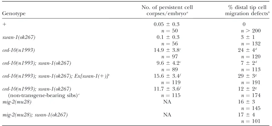
ced-10 Racmutations cause defects in phagocytosis of cells undergoing programmed cell death (Reddienand Horvitz 2000) and defects in the migration of the gonadal distal tip cells (Lundquistet al.2001), result-ing in malformed gonads. swan-1(ok267) significantly
suppressed the cell corpse engulfment defects and the gonadal distal tip cell (DTC) migration defects of ced-10(n1993) mutants (Table 1). ced-10(n1993) mutants alone displayed 14.9 6 3.8 persistent cell corpses in the pharyngeal region of late threefold-stage embryos, compared to 9.664.2 inswan-1(ok267); ced-10(n1993)
double mutants (P , 0.001). Furthermore, 24% of
ced-10(n1993) mutants displayed misshapen gonads due to DTC migration errors, whereas swan-1(ok267);
ced-10(n1993) double mutants displayed 7% (P ,
0.001). swan-1(ok267) alone had little effect on cell corpse engulfment or DTC migration on its own (Table 1). ced-10(n1993) is a hypomorphic allele that retains some ced-10 activity (Reddien and Horvitz 2000; Shakiret al.2006). Thatswan-1(ok267)suppressed the defects caused byced-10(n1993)suggests that the normal role of SWAN-1 might be to repress CED-10 Rac activity [i.e., in the absence of SWAN-1, the remaining activity of
ced-10(n1993) is increased]. While swan-1 slightly sup-pressed the hypomorphicced-10(n1993)allele, it did not suppress the maternal-effect lethality or gonad defects of theced-10(n3417) null allele nor did it suppress the le-thality or axon pathfinding defects associated with the ced-10(n1993); mig-2(mu28)double mutant (data not shown). To confirm that the suppression ofced-10(n1993)was due to theswan-1(ok267)deletion, a full-lengthswan-1T
gfptransgene (predicted to encode a full-length SWAN-1T GFP fusion protein; seematerials and methods) was analyzed for its ability to rescue suppression by
swan-1(ok267). Theswan-1Tgfptransgene rescued the effects
of swan-1(ok267) suppression in ced-10(n1993); swan-1(ok267) mutants (Table 1): transgene-bearing ced-10; swan-1animals displayed 15.663.4 persistent cell corp-ses and siblings that did not inherit the transgene displayed 11.76 3.6 persistent cell corpses (P, 0.001). Furthermore, suppression of DTC migration defects was rescued (29%vs.12%, respectively;P,0.001) (Table 1). Mutations in themig-2 Racgene also cause DTC mi-gration defects (Lundquistet al.2001).swan-1(ok267) did not suppress the DTC migration defects of mig-2(mu28)(Table 1). As opposed toced-10(n1993), which retains some CED-10 function, mig-2(mu28) is a null allele that likely causes complete loss ofmig-2function. This might explain whyswan-1(ok267)did not suppress
mig-2(mu28). Indeed, evidence is presented below that SWAN-1 can regulate MIG-2.
swan-1(ok267) enhances constitutively active Rac in neurons:To further test the idea that SWAN-1 is a nega-tive Rac regulator, the effect ofswan-1loss of function
TABLE 1
swan-1loss of function suppressesced-10(n1993)but notmig-2(mu28)
Genotype
No. of persistent cell corpses/embryoa
% distal tip cell migration defectsb
1 0.0560.3 0
n¼50 n.200
swan-1(ok267) 0.160.3 361
n¼56 n¼132
ced-10(n1993) 14.963.8c 2464d
n¼97 n¼120
ced-10(n1993); swan-1(ok267) 9.664.2c 762d
n¼89 n¼113
ced-10(n1993); swan-1(ok267); Ex[swan-1(1)]e 15.663.4f 2963g
n¼119 n¼191
ced-10(n1993); swan-1(ok267) (non-transgene-bearing sibs)e
11.763.6f 1262g
n¼115 n¼174
mig-2(mu28) NA 1663
n¼145
mig-2(mu28); swan-1(ok267) NA 1764
n¼101
aPersistent corpses in the region of the pharynx were scored in threefold larvae prior to hatching (750 min
postfertilization) (6standard deviation).
bIn young adult hermaphrodites, distal tip cell migration was scored as defective if the gonad arm failed to
execute its complete migration or if the gonad arm was misguided (6standard error of the proportion).
cDistributions are significantly different (t-test,P,0.001).
dProportions are significantly different (t-test and Fisher’s exact analysis,P,0.001).
eEx[swan-1(1)]is a transgenic array harboring wild-typeswan-1. Non-transgene-bearing sibs are the siblings of
transgenic animals from the same brood that did not inherit the transgene.
fDistributions are significantly different (t-test,P,0.001).
gProportions are significantly different (t-test and Fisher’s exact analysis,P,0.001).
on Rac activity in development of the PDE neurons was studied. The PDEs are a pair of ciliated sensory neurons that reside in the posterior-lateral region ofC. elegansin the postdeirid ganglion (Whiteet al.1986; Shakiret al. 2006). A single ciliated dendrite extends dorsally from the cell body, and a single axon extends ventrally to the ventral nerve cord, where it branches and runs anteri-orly and posterianteri-orly (Figure 4A).
Previous studies indicated that constitutively active Rac molecules, produced by the canonical glycine 12 to valine (G12V) mutation, induce ectopic neurite forma-tion and ectopic lamellipodia and filopodia formaforma-tion in the PDE neuron in vivo (Figure 4B) (Struckhoff and Lundquist2003).swan-1(ok267)enhanced the ef-fects of transgenicced-10(G12V)andrac-2(G12V)(Figure 4C). For example, rac-2(G12V) caused 65% ectopic
axon formation and 8% ectopic lamellipod and filopod formation, whereasswan-1(ok267); rac-2(G12V)animals displayed 100 and 74% of the respective defects. While all effects ofswan-1(ok267) andswan-1(RNAi)on CED-10(G12V) and RAC-2(G12V) were significant (P,0.001), the effects of swan-1 on rac-2(G12V) were more pro-nounced than those onced-10(G12V). In addition to in-creasing the penetrance ofrac-2(G12V)andced-10(G12V)
defects, swan-1(ok267) also increased the severity of defects. For example,ced-10(G12V)PDEs generally dis-played one to two ectopic axons, whereasswan-1(ok267)l ced-10(G12V) PDEs often displayed three or more ec-topic axons. Occasionally, so many ecec-topic axons were present that it became impossible to distinguish which axon was the normal PDE axon. RNAi ofswan-1gave similar but weaker results (Figure 4C), indicating that the effects were due to disruption ofswan-1function. These results demonstrate thatswan-1(ok267)enhances the effects ofced-10(G12V)andrac-2(G12V).
The mig-2(rh17) mutant harbors an activating G16 mutation (Zipkin et al.1997). Neithermig-2(rh17) nor the effects of transgenic expression of MIG-2(G16V) were significantly affected byswan-1(ok267)(Figure 4C). SWAN-1(1) transgenic expression represses consti-tutively active Rac: If SWAN-1 is a negative Rac regu-lator, overactivity of wild-type SWAN-1 might repress Rac activity. Transgenic expression of wild-typeswan-1 sup-pressed the effects of ced-10(G12V) and rac-2(G12V)
(Figure 4D; seematerials and methods). For exam-ple,rac-2(G12V) trangenes averaged 65% ectopic PDE axons and 6% ectopic lamellipodia and filopodia, whereas rac-2(G12V) transgenes that also contained wild-type swan-1 DNA averaged 16 and 0% defects, respectively. While all effects of SWAN-1(1) expression on CED-10(G12V) and RAC-2(G12V) were significant (P , 0.001), suppression of ced-10(G12V) was weaker than that ofrac-2(G12V). swan-1 coexpression had no detectable effect onmig-2(G16V)(Figure 4D).
swan-1(ok267) enhances the neuronal effects of dominant Rac alleles: In contrast to ced-10(n1993)
and mig-2(mu28), the mutations ced-10(n3246), ced-10(n1993lq20), mig-2(rh17), and mig-2(lq13) all cause axon defects on their own, indicating that they are not simple loss-of-function mutations (Shakir et al. 2006).mig-2(rh17)affects the glycine 17 residue of the MIG-2 protein (Zipkin et al.1997) and is likely to re-sult in constitutively active, GTPase-dead MIG-2 [the equivalent mutation was included in themig-2(G16V)
transgenes]. It is unknown whetherced-10(n3246), ced-10(n1993lq20), ormig-2(lq13)are activating or dominant-negative mutations.
swan-1(ok267) enhanced the axon defects caused by each of these dominantRac alleles.ced-10(n3246), ced-10(n1993lq20), mig-2(rh17), and mig-2(lq13) caused a variable PDE axon pathfinding phenotype that was cat-egorized as mild (the axon reaches the ventral nerve cord near the normal position but is misguided) or
severe (the axon fails to reach the ventral nerve cord near the normal position) (Figure 5, A and B). The percentages of combined mild and severe axon defects of swan-1(ok267) double mutants with ced-10(n3246),
mig-2(lq13), and mig-2(rh17) were increased, and the severity of all four increased (P,0.001; Figure 5C). For example,ced-10(n3246)alone showed 8% axon defects, most of which were of the mild class, andswan-1(ok267); ced-10(n3246)showed 18% defects with 8% of the severe class.
swan-1is expressed in many cell types including neu-rons: To further understand the role ofswan-1 inRac
signaling, the temporal and spatial pattern of swan-1
expression during development was analyzed. Twoswan-1
expression transgenes were constructed: a transcrip-tional reporter consisting of theswan-1promoter driv-ing green fluorescent protein (gfp) expression and a translation reporter of the full-length swan-1 region fused in frame to gfp such that a full-length SWAN-1 molecule fused to GFP at the C terminus is produced (SWAN-1TGFP) (seematerials and methods). Full-length swan-1Tgfp expression was first detected at
100 min postfertilization when embryonic transcrip-tion begins. Expression was observed in most cells (Figure 6, A–C), including neuroblasts and neurons, and per-sisted throughout development into adulthood. The only cells that did not showswan-1Tgfpexpression were
the intestinal cells and their precursors (Figure 6, A–C). SWAN-1TGFP accumulated predominantly in the cyto-plasm of all cell types analyzed. The transcriptional reporter transgene displayed a temporal and spatial expression pattern identical to that of the full-length fusion transgene (data not shown).
SWAN-1 represses human Rac1 in 3T3 fibroblasts: The above data indicate that SWAN-1 represses Rac activity inC. elegans neuronal morphogenesis. CED-10 Rac and RAC-2 Rac are very similar to human Rac1 (83% identical at the amino acid level) (Lundquist et al. 2001). Expression of constitutively active Rac1 (the glu-tamine to leucine mutation at position 61, Q61L) in serum-starved 3T3 fibroblasts led to lamellipodial mem-brane protrusions along cell edges (Figure 7A). We found that 62% of 3T3 cells expressing Rac1(Q61L) exhibited lamellipodial plasma membrane extensions (Figure 7, A and B) not seen inegfpcontrol-transfected cells. Expression of myc-tagged SWAN-1 alone had little effect on cell morphology (Figure 7B). Fewer cells transfected with both Rac1(Q61L) and SWAN-1 dis-played lamellipodial structures (26% compared to 62% for Rac1 alone; P , 0.001) (Figure 7B), suggesting a partial suppression of Rac1(Q61L) activity by SWAN-1.
c-Src targeted UNC-115 to the plasma membrane and caused overactivation of UNC-115, resulting in ectopic lamellipodia and filopodia formation in serum-starved fibroblasts (Figure 7C). In 24% of MYRT UNC-115-expressing cells, lamellipodial and filopodial mem-brane protrusions were observed (Figure 7, C and D). Cotransfection of SWAN-1 with MYRTUNC-115 re-sulted in 15% of cells exhibiting protrusive morphology, a difference that was not statistically significant (P ¼ 0.1076). While these data indicate that SWAN-1 might slightly repress UNC-115, the effect is not as pro-nounced as the effect of SWAN-1 on Rac1.
SWAN-1 represses Rac activity in yeast:In the course of the directed two-hybrid experiments using SWAN-1 and the Racs, it was noted that yeast transformed with CED-10 and MIG-2 (but not RAC-2) were slow-growing and formed small and irregular-shaped colonies. The doubling times of strains harboring CED-10 and MIG-2 grown in liquid culture were increased compared to that of the parent strain Y190 (Figure 8A). CED-10 cells had a doubling time of.10 hr and MIG-2 cells had a doubling time of6 hr, compared to2.5 hr for Y190. Doubling time compared to that of Y190 was not significantly affected by RAC-2 or SWAN-1 expression (data not
Figure6.—swan-1Tgfpis expressed in all cells except the intestine. Micrographs of an embryo, 270 min after fertilization, harboring the full-length swan-1Tgfp fusion transgene are shown (seematerials and meth-ods). An HCX Planapo 633objective (1.3 numeri-cal aperture) and a 103
magnifier were used. The dashed line surrounds the intestinal precursor cells. Bar in A, 10 mm. (A) A fluorescence micrograph; (B) a differential interfer-ence contrast micrograph; (C) a merged image.
shown). When cell morphology was analyzed, fewer of the CED-10 and MIG-2 cells were in the process of budding during the log phase of growth compared to the parent Y190 strain (Figure 8B). For example, 85% of Y190 cells were in the process of budding whereas 29% of CED-10 and 45% of MIG-2 cells were budding.
SWAN-1 and RAC-2 expression had little effect on percentage of cells undergoing budding (Figure 8B).
CED-10- and MIG-2-expressing cells displayed abnor-mal morphology, including greatly increased cell size and irregular cell shape (18/21 of CED-10-expressing cells and 24/30 MIG-2-expressing cells). Phalloidin staining revealed that the actin cytoskeleton was disor-ganized in these cells. In normal budding cells, the actin cytoskeleton is dramatically polarized; the newly form-ing bud displays actin patches on the cell surface as a result of endocytosis involved in bud growth; and long actin filaments extend from the mother cell to the bud (Adamsand Pringle1984). The actin cytoskeleton of the host strain Y190 displayed this morphology charac-teristic of asymmetric growth and budding (Figure 8C). In CED-10 and MIG-2 cells undergoing budding, actin patches were often observed both in the bud and in the mother cell (Figure 8, D and E), indicating that cell polarity and asymmetric growth had been disrupted. Furthermore, the long actin filaments were often missing (Figure 8, D and E). When the long filaments were present, they were often disorganized with respect to the bud–mother cell axis (Figure 8D). In the large, irregular, nonbudding cells, actin patches were ob-served uniformly on the cell surface (Figure 8G), indi-cating that these cells were undergoing nonpolarized growth without budding. While most RAC-2-expressing cells had normal morphology, some with abnormal polarized growth and actin cytoskeleton disorganization were observed at a low frequency (Figure 8F), which apparently did not significantly interfere with the growth dynamics of RAC-2-expressing yeast. SWAN-1-expressing cells showed no apparent morphology and cytoskeletal defects (data not shown).
The observed defects in budding, polarized growth, and actin disorganization mirrored those seen in se-lected mutants of the six yeast Rho GTPases (Rho1–5p
and Cdc42p) (Dong et al. 2003). Several yeast Rho proteins are required to assemble the long actin fila-ments, which when absent lead to polarity defects and actin patch disorganization; and Cdc42p is required for
the proper orientation of the long actin filaments. While yeast have no true Rac-family GTPases, transgenic expression of CED-10 Rac, RAC-2 Rac, and MIG-2 Rac in yeast might ectopically interfere with the normal roles
of the yeast Rho GTPases, resulting in the cytoskeletal and cell polarity defects described above.
The growth defects caused by CED-10, RAC-2, and MIG-2 expression in yeast were reduced when the yeast were cotransformed with SWAN-1. Doubling time de-creased in the SWAN-1 cotransformed strains (CED-10-expressing cells went from .10 to 4.5 hr, and MIG-2 cells went from 6 to 3.5 hr), and the percentages of budding cells increased (Figure 8A): 29% of CED-10-expressing cells were budding compared to 64% of CED-10 SWAN-1-expressing cells (P,0.001). While the SWAN-1 cotransformed cells still showed some cytoskel-etal disorganization, it was not as severe as that seen in CED-10 and MIG-2 alone, as indicated by increased percentages of budding cells. These data indicate that SWAN-1 repressed CED-10 and MIG-2 activity in yeast cells.
DISCUSSION
Previous studies indicate that the UNC-115/abLIM actin-binding protein controls lamellipodia and filopo-dia formation in response to Rac GTPase signaling (Struckhoffand Lundquist2003; Yangand Lundquist 2005), but the details of this pathway, including other molecules involved, remain unclear. A two-hybrid screen with the UNC-115 LIM domains identified the novel SWAN-1 protein, theC. elegansmember of a novel seven- or eight-WD-repeat protein family conserved in yeast, protozoa, plants, and animals. Subsequent di-rected two-hybrid tests and co-immunoprecipitation ex-periments revealed that SWAN-1 interacted both with the UNC-115 LIM domains and with each of the three
C. elegansRac-family GTPases CED-10, RAC-2, and MIG-2. SWAN-1 might represent a molecular link between Rac GTPases and UNC-115/abLIM. Functional assays inC. elegansneurons, cultured mammalian 3T3 fibro-blasts, and yeast indicate that SWAN-1 is a new class of negative regulator of Rac GTPases.
Expression of activated Rac inC. elegansneurons leads to the formation of ectopic lamellipodia and filopodia that is partially dependent on UNC-115 (Struckhoff and Lundquist 2003). Loss of swan-1 enhanced the effects of CED-10 and RAC-2 activity, and transgenic expression of wild-type swan-1 repressed CED-10 and RAC-2 activity in neurons. In these transgenic assays, no effect of SWAN-1 on MIG-2 activity was detected, despite the fact that SWAN-1 and MIG-2 interacted strongly in the two-hybrid system. swan-1 loss of function did enhance the effects of dominantmig-2alleles on axon pathfinding, suggesting that SWAN-1 also regulates MIG-2. SWAN-1 also repressed MIG-2 activity in yeast, consistent with the idea that SWAN-1 regulates all three Racs, CED-10, RAC-2, and MIG-2. Expression ofC. elegans
SWAN-1 also suppressed the lamellipodia formed in 3T3 fibroblasts in response to human Rac1 harboring the activating Q61L mutation, suggesting that the Rac–
SWAN-1 interaction has been conserved in the evolu-tionary divergence of humans andC. elegans.
swan-1mutation also suppressed the cell corpse en-gulfment defects and gonadal distal tip cell migration defects of the hypomorphic ced-10(n1993) allele, in-dicating that SWAN-1 represses CED-10 activity in mul-tiple developmental processes.swan-1did not suppress the distal tip cell migration defects of mig-2(mu28), which is a predicted null allele ofmig-2. ced-10(n1993)
retains some ced-10 activity, and this residual activity might be enhanced by loss ofswan-1. No residual MIG-2 activity is predicted to be present in the nullmig-2(mu28)
mutant.
These experiments in C. elegans, mammalian fibro-blasts, and yeast support the idea that SWAN-1 inhibits Rac activity.swan-1mutants have no detectable pheno-type on their own, and the effects ofswan-1inracmutant strains are generally weak albeit consistent across many different assays. Generally, sensitized backgrounds were required to see an effect ofswan-1(e.g., hypomorphic and dominant mutations in rac genes and transgenic expression assays). SWAN-1 inhibited human Rac1 and to a lesser extent MYRTUNC-115 in fibroblasts. Possibly SWAN-1 is a direct inhibitor of UNC-115. However, no effect of swan-1 on myrTunc-115 activity in C. elegans
neurons was detected, and SWAN-1 inhibited Rac ac-tivity in the yeast Saccharomyces cerevisiae, which has no molecule similar to UNC-115 encoded in its genome (Goffeauet al.1996). These data indicate that SWAN-1 is a direct inhibitor of Rac GTPase activity. The in-hibition of MYRTUNC-115 by SWAN-1 in fibroblasts might be an indirect effect of Rac1 inhibition (i.e., MYRTUNC-115 might still require Rac activity for full effect). However, it is possible that SWAN-1 is a direct inhibitor of both Rac and MYRTUNC-115.
The effects of SWAN-1 on Rac activity are weak. The related SWAN-2 molecule showed no detectable func-tional overlap or similarity to SWAN-1, suggesting that the two molecules have distinct functions. SWAN-1 might be a fine-scale modulator that is involved in precisely tuning the amount of Rac activity. Modest changes in the levels of Rac activity can have distinct physiological outcomes. For example, inC. elegans, re-ducing the copy number of rac genes can change an attractive semaphorin signal into a repulsive signal (Dalpeet al.2004). In cultured mammalian fibroblasts, reduction of Rac1 activity by as little as 30% leads to a switch from random peripheral lamellipodia protru-sion to directed protruprotru-sion and directed cell migration (Pankov et al. 2005). rac genes display redundant functions in C. elegans and Drosophila whereby pro-gressive loss of rac gene activity leads to progressively more severe axon defects (Lundquistet al. 2001; Ng
et al. 2002). While there is clear overlap in rac gene function, it is also possible that different levels of rac
factor’’ ensuring that background or transient Rac activation is suppressed so that only strong, persistent Rac activity leads to directed protrusion.swan-1(ok267)
has no detectable phenotype on its own, but it is possi-ble that a subtle phenotype was not revealed in our endpoint phenotypic analysis (e.g., the growth cones ofswan-1animals might behave differently from those of wild type, but they might eventually reach their targets).
GTPase-activating proteins and guanine nucleotide dissociation inhibitors are a well-characterized family of Rac negative regulators. GAPs inhibit small GTPases by activating the GTPase activity of the molecules, favor-ing the inactive, GDP-bound state, and GDIs prevent GTPase interaction with the plasma membrane (Van Aelstand D’Souza-Schorey1997; Michaelson et al. 2001). SWAN-1 does not resemble any known GAPs or GDIs, and, as expected, SWAN-1 does not act as a GAP on human Rac1 (I. Blasutigand A. Pawson, personal communication). SWAN-1 interacted physically with both wild-type and G12V-mutant-activated Rac mole-cules, andswan-1interacted genetically with a variety of
racdominant mutations, including the activating G12V and Q61L mutations and the dominant mutations
ced-10(n3246),ced-10(n1993lq20), andmig-2(lq13), which affect Rac by unknown mechanisms. SWAN-1 might bind to Racs and lock them in an inactive conforma-tion, SWAN-1 might block sites of interaction with Rac effectors regardless of the GTP-bound state of Rac, or SWAN-1 might regulate Rac interaction with the plasma membrane.
UNC-115/abLIM and Racs act in the same pathway to promote protrusive activity. SWAN-1 was isolated by its ability to interact with UNC-115 LIM domains, and SWAN-1 is a negative regulator of Rac GTPases in the pathway. UNC-115/abLIM might act as a scaffold for a complex that includes both activators and inhibitors of protrusive activity. SWAN-1 might be recruited with the Rac effector UNC-115/abLIM to Racs to precisely control levels of Rac activity or to prevent spurious, transient Rac activity so that directed protrusion can occur.
We thank E. Struckhoff for technical assistance; R. Barstead, G. Molder, and theC. elegansGene Knockout Consortium forC. elegans strains; R. Barstead for providing the two-hybrid cDNA library; T. Meyer for providing the Rac1(Q61L) clone; K. Neufeld and T. C. Gamblin for reagents and technical assistance; and the Caenorhabditis Genetics Center, sponsored by the National Center for Research Resources, for providingC. elegansstrains. This work was supported by National Institutes of Health (NIH) grant NS40945 and National Science Foundation grant IBN93192 to E.A.L. and NIH grant P20 RR016475 from the INBRE Program of the National Center for Research Resources.
LITERATURE CITED
Adams, A. E., and J. R. Pringle, 1984 Relationship of actin and tu-bulin distribution to bud growth in wild-type and
morphoge-netic-mutant Saccharomyces cerevisiae. J. Cell Biol. 98: 934– 945.
Dalpe, G., L. W. Zhang, H. Zhengand J. G. Culotti, 2004 Con-version of cell movement responses to Semaphorin-1 and Plexin-1 from attraction to repulsion by lowered levels of specific RAC GTPases in C. elegans. Development131:2073–2088.
deVetten, N., F. Quattrocchio, J. Moland R. Koes, 1997 The an11locus controlling flower pigmentation in petunia encodes a novel WD-repeat protein conserved in yeast, plants and ani-mals. Genes Dev.11:1422–1434.
Dickson, B. J., 2001 Rho GTPases in growth cone guidance. Curr. Opin. Neurobiol.11:103–110.
Dickson, B. J., 2002 Molecular mechanisms of axon guidance. Science298:1959–1964.
Dong, Y., D. Pruyneand A. Bretscher, 2003 Formin-dependent actin assembly is regulated by distinct modes of Rho signaling in yeast. J. Cell Biol.161:1081–1092.
Epstein, H. F., and D. C. Shakes, 1995 Caenorhabditis elegans: Modern Biological Analysis of an Organism.Academic Press, New York. Goffeau, A., B. G. Barrell, H. Bussey, R. W. Davis, B. Dujonet al.,
1996 Life with 6000 genes. Science274:546, 563–567. Hall, A., 1998 Rho GTPases and the actin cytoskeleton. Science
279:509–514.
Kamath, R. S., and J. Ahringer, 2003 Genome-wide RNAi screen-ing in Caenorhabditis elegans. Methods30:313–321.
Kishore, R. S., and M. V. Sundaram, 2002 ced-10 Rac and mig-2 function redundantly and act with unc-73 trio to control the orientation of vulval cell divisions and migrations in Caenor-habditis elegans. Dev. Biol.241:339–348.
Lundquist, E. A., 2003 Rac proteins and the control of axon devel-opment. Curr. Opin. Neurobiol.13:384–390.
Lundquist, E. A., R. K. Herman, J. E. Shawand C. I. Bargmann, 1998 UNC-115, a conserved protein with predicted LIM and actin-binding domains, mediates axon guidance in C. elegans. Neuron21:385–392.
Lundquist, E. A., P. W. Reddien, E. Hartwieg, H. R. Horvitzand C. I. Bargmann, 2001 Three C. elegans Rac proteins and several alternative Rac regulators control axon guidance, cell migration and apoptotic cell phagocytosis. Development 128:
4475–4488.
Michaelson, D., J. Silletti, G. Murphy, P. D’Eustachio, M. Rush et al., 2001 Differential localization of Rho GTPases in live cells: regulation by hypervariable regions and RhoGDI binding. J. Cell Biol.152:111–126.
Neer, E. J., C. J. Schmidt, R. Nambudripad and T. F. Smith, 1994 The ancient regulatory-protein family of WD-repeat pro-teins. Nature371:297–300.
Ng, J., T. Nardine, M. Harms, J. Tzu, A. Goldsteinet al., 2002 Rac GTPases control axon growth, guidance and branching. Nature
416:442–447.
Otieno, C. J., J. Bastiaansen, A. M. Ramosand M. F. Rothschild, 2005 Mapping and association studies of diabetes related genes in the pig. Anim. Genet.36:36–42.
Pankov, R., Y. Endo, S. Even-Ram, M. Araki, K. Clarket al., 2005 A Rac switch regulates random versus directionally persistent cell migration. J. Cell Biol.170:793–802.
Raftopoulou, M., and A. Hall, 2004 Cell migration: Rho GTPases lead the way. Dev. Biol.265:23–32.
Reddien, P. W., and H. R. Horvitz, 2000 2/CrkII and CED-10/Rac control phagocytosis and cell migration in Caenorhabdi-tis elegans. Nat. Cell Biol.2:131–136.
Shakir, M. A., J. S. Gilland E. A. Lundquist, 2006 Interactions of UNC-34 Enabled with Rac GTPases and the NIK kinase MIG-15 in Caenorhabditis elegansaxon pathfinding and neuronal migration. Genetics172:893–913.
Smith, T. F., C. Gaitatzes, K. Saxenaand E. J. Neer, 1999 The WD repeat: a common architecture for diverse functions. Trends Biochem. Sci.24:181–185.
Sondek, J., A. Bohm, D. G. Lambright, H. E. Hammand P. B. Sigler, 1996 Crystal structure of a G-protein beta gamma dimer at 2.1A resolution. Nature379:369–374.
Soto, M. C., H. Qadota, K. Kasuya, M. Inoue, D. Tsuboiet al., 2002 The GEX-2 and GEX-3 proteins are required for tissue morphogenesis and cell migrations in C. elegans. Genes Dev.
Steven, R., T. J. Kubiseski, H. Zheng, S. Kulkarni, J. Mancillas et al., 1998 UNC-73 activates the Rac GTPase and is required for cell and growth cone migrations inC. elegans.Cell92:785–795. Struckhoff, E. C., and E. A. Lundquist, 2003 The actin-binding protein UNC-115 is an effector of Rac signaling during axon pathfinding in C. elegans. Development130:693–704. Tessier-Lavigne, M., and C. S. Goodman, 1996 The molecular
biology of axon guidance. Science274:1123–1133.
Timmons, L., D. L. Courtand A. Fire, 2001 Ingestion of bacterially expressed dsRNAs can produce specific and potent genetic inter-ference in Caenorhabditis elegans. Gene263:103–112. VanAelst, L., and C. D’Souza-Schorey, 1997 Rho GTPases and
signaling networks. Genes Dev.11:2295–2322.
Vicente-Manzanares, M., D. J. Webband A. R. Horwitz, 2005 Cell migration at a glance. J. Cell Sci.118:4917–4919.
Walker, A. R., P. A. Davison, A. C. Bolognesi-Winfield, C. M. James, N. Srinivasanet al., 1999 The TRANSPARENT TESTA
GLABRA1 locus, which regulates trichome differentiation and anthocyanin biosynthesis in Arabidopsis, encodes a WD40 repeat protein. Plant Cell11:1337–1350.
White, J. G., E. Southgate, J. N. Thomson and S. Brenner, 1986 The structure of the nervous system of the nematode Cae-norhabditis elegans.Philos. Trans. R. Soc. Lond.314:1–340. Yang, Y., and E. A. Lundquist, 2005 The actin-binding protein
UNC-115/abLIM controls formation of lamellipodia and filopo-dia and neuronal morphogenesis in Caenorhabditis elegans. Mol. Cell. Biol.25:5158–5170.
Yu, T. W., and C. I. Bargmann, 2001 Dynamic regulation of axon guidance. Nat. Neurosci.4(Suppl.): 1169–1176.
Zipkin, I. D., R. M. Kindtand C. J. Kenyon, 1997 Role of a new Rho family member in cell migration and axon guidance in C. elegans. Cell90:883–894.