Open Access
Review
Principles for characterizing the potential human health effects
from exposure to nanomaterials: elements of a screening strategy
Günter Oberdörster
1, Andrew Maynard
2, Ken Donaldson
3,
Vincent Castranova
4, Julie Fitzpatrick*
5, Kevin Ausman
6, Janet Carter
7,
Barbara Karn
8,9, Wolfgang Kreyling
10, David Lai
11, Stephen Olin
5,
Nancy Monteiro-Riviere
12, David Warheit
13, Hong Yang
14and A report from
the ILSI Research Foundation/Risk Science Institute Nanomaterial Toxicity
Screening Working Group
Address: 1Department of Environmental Medicine, University of Rochester, 601 Elmwood Avenue, P.O. Box EHSC, Rochester, NY 14642, USA, 2Project on Emerging Nanotechnologies, Woodrow Wilson International Center for Scholars, 1300 Pennsylvania Avenue, N.W., Washington, DC
20004-3027, USA, 3MRC/University of Edinburgh Centre for Inflammation Research, ELEGI Colt Laboratory Queen's Medical Research Institute,
47 Little France Crescent, Edinburgh EH16 4TJ, UK, 4Pathology and Physiology Research Branch, Health Effects Laboratory Division, National
Institute for Occupational Safety and Health, 1095 Willowdale Road, Morgantown, WV 26505, USA, 5Risk Science Institute, ILSI Research
Foundation, International Life Sciences Institute, One Thomas Circle, N.W., Suite 900, Washington, DC 20005-5802, USA, 6Center for Biological
and Environmental Nanotechnology, MS-63, P.O. Box 1892, Rice University, Houston, TX 77251-1892, USA, 7Respiratory/Inhalation Toxicology,
Central Product Safety, Procter & Gamble Company, PO Box 538707, Cincinnati, OH 45253-8707, USA, 8Office of Research and Development,
United States Environmental Protection Agency, Ariel Rios Building, Mail Code: 8722F, 1200 Pennsylvania Avenue, N.W., Washington, DC 20460, USA, 9Project on Emerging Nanotechnologies, Woodrow Wilson International Center for Scholars, 1300 Pennsylvania Avenue, N.W., Washington,
DC 20004-3027, USA, 10Institute for Inhalation Biology & Focus Network: Aerosols and Health, GSF National Research Centre for Environment
and Health, Ingolstadter Landstrasse 1, 85764 Neuherberg, Munich, Germany, 11Risk Assessment Division, Office of Pollution Prevention &
Toxics, United States Environmental Protection Agency, 7403M, 1200 Pennsylvania Avenue, N.W., Washington, DC 20460, USA, 12Center for
Chemical Toxicology and Research Pharmacokinetics, College of Veterinary Medicine, North Carolina State University, 4700 Hillsborough Street, Raleigh, NC 27606, USA, 13DuPont Haskell Laboratory for Health and Environmental Sciences, P.O. Box 50, 1090 Elkton Road, Newark, DE
19714-0050, USA and 14Department of Chemical Engineering, University of Rochester, Gavett Hall 253, Rochester, NY 14627, USA
Email: Günter Oberdörster - [email protected]; Andrew Maynard - [email protected]; Ken Donaldson - [email protected]; Vincent Castranova - [email protected]; Julie Fitzpatrick* - [email protected]; Kevin Ausman - [email protected]; Janet Carter - [email protected]; Barbara Karn - [email protected]; Wolfgang Kreyling - [email protected]; David Lai - [email protected]; Stephen Olin - [email protected]; Nancy
Monteiro-Riviere - [email protected]; David Warheit - [email protected]; Hong Yang - [email protected]; A report from the ILSI Research Foundation/Risk Science Institute Nanomaterial Toxicity Screening Working Group - [email protected]
* Corresponding author
Abstract
The rapid proliferation of many different engineered nanomaterials (defined as materials designed and produced to have structural features with at least one dimension of 100 nanometers or less) presents a dilemma to regulators regarding hazard identification. The International Life Sciences Institute Research Foundation/Risk Science Institute convened an expert working group to develop a screening strategy for the hazard identification of engineered nanomaterials. The working group report presents the elements of a screening strategy rather than a detailed testing protocol. Based on an evaluation of the limited data currently available, the report presents a broad data gathering strategy applicable to this early stage in the development of a risk assessment process for Published: 06 October 2005
Particle and Fibre Toxicology 2005, 2:8 doi:10.1186/1743-8977-2-8
Received: 03 October 2005 Accepted: 06 October 2005
This article is available from: http://www.particleandfibretoxicology.com/content/2/1/8
© 2005 Oberdörster et al; licensee BioMed Central Ltd.
Particle and Fibre Toxicology 2005, 2:8 http://www.particleandfibretoxicology.com/content/2/1/8
Page 2 of 35
(page number not for citation purposes) nanomaterials. Oral, dermal, inhalation, and injection routes of exposure are included recognizing
that, depending on use patterns, exposure to nanomaterials may occur by any of these routes. The three key elements of the toxicity screening strategy are: Physicochemical Characteristics, In Vitro Assays (cellular and non-cellular), and In Vivo Assays.
There is a strong likelihood that biological activity of nanoparticles will depend on physicochemical parameters not routinely considered in toxicity screening studies. Physicochemical properties that may be important in understanding the toxic effects of test materials include particle size and size distribution, agglomeration state, shape, crystal structure, chemical composition, surface area, surface chemistry, surface charge, and porosity.
In vitro techniques allow specific biological and mechanistic pathways to be isolated and tested under controlled conditions, in ways that are not feasible in in vivo tests. Tests are suggested for portal-of-entry toxicity for lungs, skin, and the mucosal membranes, and target organ toxicity for endothelium, blood, spleen, liver, nervous system, heart, and kidney. Non-cellular assessment of nanoparticle durability, protein interactions, complement activation, and pro-oxidant activity is also considered.
Tier 1 in vivo assays are proposed for pulmonary, oral, skin and injection exposures, and Tier 2 evaluations for pulmonary exposures are also proposed. Tier 1 evaluations include markers of inflammation, oxidant stress, and cell proliferation in portal-of-entry and selected remote organs and tissues. Tier 2 evaluations for pulmonary exposures could include deposition, translocation, and toxicokinetics and biopersistence studies; effects of multiple exposures; potential effects on the reproductive system, placenta, and fetus; alternative animal models; and mechanistic studies.
1.0 Introduction
The rapid proliferation of many different engineered nanomaterials presents a dilemma to regulators regarding hazard identification. The screening strategy developed by the International Life Sciences Institute Research Founda-tion/Risk Science Institute (ILSI RF/RSI) Nanomaterial Toxicity Screening Working Group is an effort to make a significant contribution to the initial hazard identification process for nanomaterial risk assessment.
Engineered nanomaterials are commonly defined as materials designed and produced to have structural fea-tures with at least one dimension of 100 nanometers or less. Such materials typically possess nanostructure-dependent properties (e.g., chemical, mechanical, electri-cal, optielectri-cal, magnetic, biological), which make them desirable for commercial or medical applications. How-ever, these same properties potentially may lead to nanos-tructure-dependent biological activity that differs from and is not directly predicted by the bulk properties of the constituent chemicals and compounds. This report out-lines the elements of a toxicological screening strategy for nanomaterials as the first step – i.e., hazard identification – in the risk assessment process. Both in vitro and in vivo
methodologies were considered in the development of the screening strategy.
Engineered nanomaterials encompass many forms and are derived from numerous bulk substances. Nanoparti-cles form a basis for many engineered nanomaterials, and
are currently being produced in a wide variety of types for a variety of applications; fullerenes (C60 or Bucky Balls),
carbon nanotubes (CNT), metal and metal oxide particles, polymer nanoparticles and quantum dots are among the most common.
Engineered nanomaterials are presenting new opportuni-ties to increase the performance of traditional products, and to develop unique new products. "The ability to cre-ate unusual nanostructures such as bundles, sheets, and tubes holds promise for new and powerful drug delivery systems, electronic circuits, catalysts, and light-harvesting materials." [1]
Many current efforts are predominantly focused on using relatively simple nanostructured materials such as metal oxide nanoparticles and carbon nanotubes in applica-tions such as high performance materials, energy storage and conversion, self-cleaning surface coatings and stain-resistant textiles. Research into more complex nanomate-rials is anticipated to lead to applications such as cellular-level medical diagnostics and treatment and advanced electronics. However, as nanotechnology blurs tradition-ally rigid boundaries between scientific disciplines, a rapid growth in unanticipated applications is to be expected over the next years and decades.
particularly as indicators suggest traditional screening approaches may not be responsive to the nanostructure-related biological activity of these materials.
Several national and international organizations are cur-rently developing standard definitions for comment terms in nanomaterial science including the International Asso-ciation of Nanotechnology's Nomenclature and Termi-nology Subcommittee and the American National Standards Institute Nanotechnology Standards Panel (ANSI-NSP). The following key definitions are used throughout this document.
1. Nanoparticle
A particle with at least one dimension smaller than 100 nm including engineered nanoparticles, ambient ultrafine particles (UFPs) and biological nanoparticles.
2. Engineered/Manufactured Nanoparticle
A particle engineered or manufactured by humans on the nanoscale with specific physicochemical composition and structure to exploit properties and functions associated with its dimensions. Engineered nanoparticles include particles with a homogeneous composition and structure, compositionally and structurally heterogeneous particles (for instance, particles with core-shell structures) and multi-functional nanoparticles (for instance, 'smart' nan-oparticles being developed for medical diagnostics and treatment).
3. Nanomaterial
A material having a physicochemical structure on a scale greater than typically atomic/ molecular dimensions but less than 100 nm (nanostructure), which exhibits physi-cal, chemical and/or biological characteristics associated with its nanostructure.
4. Nanostructured Particle
A particle with a physicochemical structure on a scale greater than atomic/molecular dimensions but less than 100 nm, which exhibits physical, chemical and/or biolog-ical characteristics associated with its nanostructure. A nanostructured particle may be much larger than 100 nm. For example, agglomerates of TiO2 nanoparticles that are
significantly larger than 100 nm in diameter may have a biological activity determined by their nanoscale sub-structure. Other examples include zeolites, meso-porous materials and multifunctional particulate probes.
5. Agglomerate/Aggregate
The terms "agglomerate" and "aggregate" are used differ-ently and even interchangeably in different fields. In the context of this report, the term "agglomerate" is used exclusively to describe a collection of particles that are held together by both weak and strong forces, including
van der Waals and electrostatic forces, and sintered bonds. In this document, the term is used interchangeably with 'aggregate'. However, the importance of understanding how the binding forces of an agglomerate affect the dis-persibility of the component particles under different con-ditions – in essence how easily the agglomerate de-agglomerates – is noted.
6. Nanoporous Material
A material with particles that are larger than 100 nm may have significant structuring on the nanometer size scale, thereby providing properties based upon this smaller structuring that may be toxicologically relevant (e.g., dra-matically increased surface area as compared to the bulk). Nanoporous materials, such as zeolites, are a significant class of materials which have porosity in the sub-100 nm size range but whose primary particles may be large.
2.0 Objectives and Scope
The objective of the ILSI RSI Nanomaterial Toxicity Screening Working Group, which was convened in Febru-ary 2005, was to identify the key elements of a toxicity screening strategy for engineered nanomaterials. The group considered potential effects of exposure to nano-materials by inhalation, dermal, oral, and injection routes; discussed how mechanisms of nanoparticle toxic-ity may differ from those exhibited by larger particles of the same chemical; and identified significant data needs for designing a robust screening strategy.
The elements of a screening strategy for nanomaterials presented by the Nanomaterial Toxicity Screening Work-ing Group include an evaluation of the physicochemical characteristics and dose metrics; acellular assays; in vitro
assays for lung, skin, and mucosal membranes; and in vivo
assays for lung, skin, oral, and injection exposures.
This project was funded by the U.S. Environmental Pro-tection Agency Office of Pollution Prevention and Toxics through a cooperative agreement with the ILSI Research Foundation/Risk Science Institute. It was an outgrowth of another project under the same cooperative agreement that proposed strategies for short-term toxicity testing of fibers [2]. Among the principal conclusions of the latter project were the importance of the physicochemical char-acterization of the fibers, the value of subchronic (1–3 month) rat inhalation exposure studies, and the typically key role in fiber toxicity of biopersistence of inhaled fibers in the lung and of chronic inflammation leading to cell proliferation and interstitial fibrosis.
3.0 Literature Survey
Particle and Fibre Toxicology 2005, 2:8 http://www.particleandfibretoxicology.com/content/2/1/8
Page 4 of 35
(page number not for citation purposes)
are developed and brought into commercial use. To date, few nanotoxicology studies have addressed the effects of nanomaterials in a variety of organisms and environ-ments. However, the existing research raises some con-cerns about the safety of nanomaterials and has led to increased interest in studying the toxicity of nanomateri-als for use in risk assessment and protection of human health and the environment. A new field of nanotoxicol-ogy has been developed to investigate the possibility of harmful effects due to exposure to nanomaterials [3]. Nanotoxicology also encompasses the proper characteri-zation of nanomaterials used in toxicity studies. Charac-terization has been important in differentiating between naturally occurring forms of nanomaterials, nano-scale byproducts of natural or chemical processes, and manu-factured (engineered) nanomaterials. Because of the wide differences in properties among nanomaterials, each of these types of nanoparticles can elicit its own unique bio-logical or ecobio-logical responses. As a result, different types of nanomaterials must be categorized, characterized, and studied separately, although certain concepts of nanotox-icology based on the small size, likely apply to all nanomaterials.
As materials reach the nanoscale, they often no longer dis-play the same reactivity as the bulk compound. For exam-ple, even a traditionally inert bulk compound, such as gold, may elicit a biological response when it is intro-duced as a nanomaterial [4]. New approaches for testing and new ways of thinking about current materials are nec-essary to provide safe workplaces, products, and environ-ments as the manufacturing of nanomaterials and products increases and, as a result, exposure to nanomate-rials increases. The diverse routes of exposure, including inhalation, dermal uptake, ingestion, and injection, can present unique toxicological outcomes that vary with the physicochemical properties of the nanoparticles in question.
The earliest studies investigating the toxicity of nanoparti-cles focused on atmospheric exposure of humans and environmentally relevant species to heterogeneous mix-tures of environmentally produced ultrafine particulate matter (having a diameter <100 nm). These studies exam-ined pulmonary toxicity associated with particulate mat-ter deposition in the respiratory tract of target organisms [5-15]. Epidemiological assessments of the effects of urban air pollution exposure focusing on particulate mat-ter produced as a byproduct of combustion events, such as automobile exhaust and other sources of urban air pollu-tion, showed a link in test populations between morbidity and mortality and the amount of particulate matter [16-19]. Some researchers have found an increased risk of childhood and adult asthma correlated to environmental exposure to ultrafine particulate matter in urban air
[20-22]. However, other research does not indicate the same correlation [23-25].
Laboratory-based studies have investigated the effects of a large range of ultrafine materials through in vivo exposures using various animal models as well as cell-culture-based
in vitro experiments. To date, animal studies routinely show an increase in pulmonary inflammation, oxidative stress, and distal organ involvement upon respiratory exposure to inhaled or implanted ultrafine particulate matter [7,11,26-30]. Tissue and cell culture analysis have also supported the physiological response seen in whole animal models and yielded data pointing to an increased incidence of oxidative stress, inflammatory cytokine pro-duction, and apoptosis in response to exposure to ultrafine particles [31-37]. These studies have also yielded information on gene expression and cell signaling path-ways that are activated in response to exposure to a variety of ultrafine particle species ranging from carbon-based combustion products to transition metals. Polytetrafluor-oethylene fumes in indoor air pollution are nano-sized particles, highly toxic to rats [38]. They elicit a severe inflammatory response at low inhaled particle mass con-centrations, suggestive of an oxidative injury [39-41].
In contrast to the heterogeneous ultrafine materials pro-duced incidentally by combustion or friction, manufac-tured nanomaterials can be synthesized in highly homogenous forms of desired sizes and shapes (e.g., spheres, fibers, tubes, rings, planes). Limited research on manufactured nanomaterials has investigated the interre-lationship between the size, shape, and dose of a material and its biological effects, and whether a unique toxicolog-ical profile may be observed for these different properties within biological models.
Typically, the biological activity of particles increases as the particle size decreases. Smaller particles occupy less volume, resulting in a larger number of particles with a greater surface area per unit mass and increased potential for biological interaction [42-46]. Recent studies have begun to categorize the biological response elicited by var-ious nanomaterials both in the ecosystem and in mam-malian systems. Although most current research has focused on the effect of nanomaterials in mammalian sys-tems, some recent studies have shown the potential of nanomaterials to elicit a phytotoxic response in the eco-system. In the case of alumina nanoparticles, one of the US market leaders for nano-sized materials, 99.6% pure nanoparticles with an average particle size of 13 nm were shown to cause root growth inhibition in five plant spe-cies [46].
materials are associated with fibrotic lung responses and result in inflammation and an increased risk of carcino-genesis. Single-walled carbon nanotubes (SWCNT) have been shown to inhibit the proliferation of kidney cells in cell culture by inducing cell apoptosis and decreasing cel-lular adhesive ability. In addition, they cause inflamma-tion in the lung upon instillainflamma-tion [26,33,47-49]. Multi-walled carbon nanotubes (MWCNT) are persistent in the deep lung after inhalation and, once there, are able to induce both inflammatory and fibrotic reactions [47].
Dermal exposure to MWCNT has been modeled through cell culture and points to the nanoparticles' ability to localize within and initiate an irritation response in target epithelial cells [50]. Proteomic analysis conducted in human epidermal keratinocytes exposed to MWCNT showed both increased and decreased expression of many proteins relative to controls. These protein alterations sug-gested dysregulation of intermediate filament expression, cell cycle inhibition, altered vesicular trafficking/exocyto-sis and membrane scaffold protein down-regulation [50,51]. In addition, gene expression profiling was con-ducted on human epidermal keratinocytes exposed to SWCNT that showed a similar profile to alpha-quartz or silica. Also, genes not previously associated with these particulates before from structural protein and cytokine families were significantly expressed [52]. Dosing kerati-nocytes and bronchial epithelial cells in vitro with SWCNT has been shown to result in increases in markers of oxida-tive stress [50,53,54].
Charge properties and the ability of carbon nanoparticles to affect the integrity of the blood-brain barrier as well as exhibit chemical effects within the brain have also been studied. Nanoparticles can overcome this physical and electrostatic barrier to the brain. In addition, high concen-trations of anionic nanoparticles and cationic nanoparti-cles are capable of disrupting the integrity of the blood-brain barrier. The blood-brain uptake rates of anionic nanoparti-cles at lower concentrations were greater than those of neutral or cationic formulations at the same concentra-tions. This work suggests that neutral nanoparticles and low concentration anionic nanoparticles can serve as car-rier molecules providing chemicals direct access to the brain and that cationic nanoparticles have an immediate toxic effect at the blood-brain barrier [55,56].
Tests with uncoated, water soluble, colloidal C60 fuller-enes have shown that redox-active, lipophilic carbon nan-oparticles are capable of producing oxidative damage in the brains of aquatic species [55]. The bactericidal poten-tial of C60 fullerenes was also observed in these experi-ments. This property of fullerenes has possible ecological ramifications and is being explored as a potential source of new antimicrobial agents [57-59].
Oxidative stress as a common mechanism for cell damage induced by nano- and ultrafine particles is well docu-mented; fullerenes are model compounds for producing superoxide. A wide range of nanomaterial species have been shown to create reactive oxygen species both in vivo
and in vitro. Species which have been shown to induce free radical damage include the C60 fullerenes, quantum dots,
and carbon nanotubes [30,60-66]. Nanoparticles of vari-ous sizes and chemical compositions are able to preferen-tially localize in mitochondria where they induce major structural damage and can contribute to oxidative stress [65].
Quantum dots (QDs) such as CdSe QDs have been intro-duced as new fluorophores for use in bioimaging. When conjugated with antibodies, they are used for immunos-taining due to their bright, photostable fluorescence.
To date, there is not sufficient analysis of the toxicity of quantum dots in the literature, but some current studies point to issues of concern when these nanomaterials are introduced into biological systems. Recently published research indicates that there is a range of concentrations where quantum dots used in bioimaging have the poten-tial to decrease cell viability, or even cause cell death, thus suggesting that further toxicological evaluation is urgently needed [67,68]. While it is well known that bulk cad-mium selenide (CdSe) is cytotoxic, it has been suggested that CdSe quantum dots are cytocompatible, and safe for use in whole animal studies. This postulate is based in part on the use of protecting groups around the CdSe core of the quantum dot. These coatings have been shown to be protective, but their long-term stability has not been evaluated thoroughly. Recent studies exploring the cyto-toxicity of CdSe-core quantum dots in primary hepato-cytes as a liver model found that these quantum dots were acutely toxic under certain conditions. The cytotoxicity correlates with the liberation of free Cd2+ ions due to
dete-rioration of the CdSe lattice. These data suggest that quan-tum dots can be rendered nontoxic initially for in vivo use when appropriately coated. However, the research also highlights the need to further explore the long-term stabil-ity of the coatings used, both in vivo and exposed to envi-ronmental conditions [69].
Particle and Fibre Toxicology 2005, 2:8 http://www.particleandfibretoxicology.com/content/2/1/8
Page 6 of 35
(page number not for citation purposes)
4.0 Elements of a screening strategy for
nanomaterials
While the nanostructure-dependent properties of many engineered nanomaterials may place them in the category of potential hazards, the direct risk they present to human health will depend on the probability of exposures occur-ring, and the extent to which materials entering the body exhibit behavior associated with their nanostructure. Fig-ure 1[70]; Biokinetics of Nano-sized Particles; While many uptake and translocation routes have been demon-strated, others still are hypothetical and need to be inves-tigated. Largely unknown are translocation rates as well as
accumulation and retention in critical target sites and their underlying mechanisms. These as well as potential adverse effects will be largely dependent on physicochem-ical characteristics of the surface and core of nano-sized particles. Both qualitative and quantitative changes in nano-sized particles biokinetics in a disease or compro-mised organism need also to be considered.
In many cases, nanostructured materials will be compo-nents of large-scale products such as nano-composites, surface coatings and electronic circuits, and the potential for direct exposure will be negligible. However, if
Biokinetics of Nano-sized Particles
Figure 1
Biokinetics of Nano-sized Particles. While many uptake and translocation routes have been demonstrated, others still are hypothetical and need to be investigated. Largely unknown are translocation rates as well as accumulation and retention in crit-ical target sites and their underlying mechanisms. These as well as potential adverse effects will be largely dependent on physi-cochemical characteristics of the surface and core of sized particles. Both qualitative and quantitative changes in nano-sized particles' biokinetics in a diseased or compromised organism need also to be considered. Reproduced with permission from Environmental Health Perspectives.
Exposure Media
Uptake Pathways
Translocation
and Distribution
Excretory Pathways
Air, water, clothes
Air
Food, water
Skin
Respiratory tract
GI tract
Blood
Liver
Lymph
Bone marrow
Kidney
Spleen
Urine
Feces
Deposition
n eu
ro ns
neuron s
Ingestion
Inhalation
Other sites
(e.g. muscle, placenta)
(platelets, monocytes, endothelial cells)
Breast milk
Heart
Sweat/exfoliation
Drug delivery
Injection
Confirmed routesPotential routes
Lymph
tracheo-nasal bronchial alveolar
CNS
PNS
nanostructured materials may enter the body, toxicity screening strategies are required to ascertain the potential risk they present.
Nanoparticles are an obvious form of engineered nano-material presenting a significant exposure potential, because they can be readily deposited in the lungs or on the skin, and potentially translocate within the body. However, agglomerates of nanoparticles from a few hun-dred nanometers to a few micrometers in diameter may also be inhaled, ingested or deposited on the skin, and may have the potential to express toxicity associated with their nanostructure. Similarly, it is conceivable that nanostructured particles of a few micrometers in diameter and below (such as fragments of a nano-composite or a nanostructured surface coating) may exhibit nanostruc-ture-dependent biological properties. In each of these cases, exposure potential exists for materials in air and in liquid suspensions or slurries.
In this section, three key aspects of toxicity screening strat-egies are addressed: characterization of nanomaterials, in vitro screening strategies and in vivo screening strategies (covering inhalation, dermal, ingestion, and injection exposure routes). Screening strategies are developed around nanoparticles, but are relevant to all engineered nanomaterials that are capable of entering the body through inhalation, ingestion, dermal penetration, or injection and expressing biological activity which is asso-ciated with their nanostructure.
4.1 Physicochemical Characterization
4.1.1 Introduction
Unlike gases, liquids and many solid materials, the desir-able properties of engineered nanomaterials closely depend on size, shape and structure (both physically and chemically) at the nanoscale. Similarly, there is a strong likelihood that biological activity will depend on physic-ochemical parameters not usually considered in toxicity screening studies. Although quantitative toxicity studies on engineered nanomaterials are still relatively sparse, published data on fullerenes, single walled carbon nano-tubes, nanoscale metal oxides such as TiO2 and nanome-ter-diameter low solubility particles, support the need to carefully consider how nanomaterials are characterized when evaluating potential biological activity [62,71-75]. Respirable fibers present perhaps the closest analogy to a material that is not fully characterized by mass and chem-ical composition alone. However, the diversity and com-plexity of nanomaterials suggests that the level of characterization appropriate to toxicity screening tests will be commensurately more sophisticated.
Until the mechanistic associations between nanomaterial characteristics and toxicity are more fully understood, it
will be necessary to ensure that all nanomaterial charac-teristics that are potentially significant are measured or can be derived in toxicity screening tests. In particular, in as far as it is possible; it is desirable to collect sufficient information to allow retrospective interpretation of toxic-ity data in the light of new findings. In this context, iden-tifying a set of characterization criteria for nanomaterial toxicity screening presents a significant challenge. Clearly, the ideal of characterizing every possible aspect of a test material, while laudable, is impractical. In this document, we have therefore focused on the context under which characterization takes place and the minimum set of char-acterization parameters we consider essential within that context. Essential parameters have been supplemented with those considered desirable and those considered of interest but optional within a screening study. The two overarching characterization contexts discussed are human exposure studies and in vitro/in vivo studies. In the case of the latter, we consider material characterization after administration, characterization at the point of administration and characterization of the bulk material as produced or supplied. The relative importance of char-acterizing dose against different physical metrics during inhalation exposures is also discussed. Recommendations are subsequently made on physicochemical characteriza-tions for nanomaterial toxicity screening tests and charac-terization methods capable of providing the recommended information.
4.1.2 Framework for Material Characterization
Material characterization for toxicity screening studies is most appropriately considered in the context of the stud-ies being undertaken. Requirements for in vitro and in vivo
screening studies will differ according to the material delivery route or method. Additionally, understanding human exposures in the context of developing appropri-ate screening studies will present a further set of character-ization requirements. Four screening study contexts are proposed, and characterization recommendations are developed within these contexts:
• Human exposure characterization
• Characterization of material following administration
• Characterization of administered material
• Characterization of as-produced or supplied material
Human Exposure Characterization
Particle and Fibre Toxicology 2005, 2:8 http://www.particleandfibretoxicology.com/content/2/1/8
Page 8 of 35
(page number not for citation purposes)
relatively few environments where exposures are known to occur. However, if commercialization of products using nanomaterials develops as anticipated, the potential for exposure is likely to increase dramatically over the coming decade. Therefore, estimates of future use and potential human exposures should be considered in the develop-ment of toxicity screening.
Nanomaterial Characterization after Administration
Characterizing delivered nanomaterial after administra-tion in a test system or model provides the highest quality of data on dose and material properties that are related to observed responses, but this is limited by current method-ological capabilities. Characterization after administra-tion is particularly advantageous where the possibility of physicochemical changes in the material before and after administration exists. Examples of potential changes include aggregation state, physisorption or chemisorption of biomolecules and biochemically-induced changes in surface chemistry. In addition, possible physicochemical changes as a result of nanomaterial interactions with the surrounding biological systems such as rapid dissolution of water- or lipid soluble fractions of the nanomaterial need to be carefully considered. While characterization after administration is considered an ideal to work towards, it is recognized that in many cases, characteriza-tion at the point of administracharacteriza-tion will be a more realistic and feasible option. It is also recognized that in many cases, characterization at the point of administration will be essential for the intercomparison of studies, irrespec-tive of whether characterization after administration is carried out.
Characterization of Administered Material
Characterization of administered material in toxicity screening studies is fundamental. This approach addresses potential physicochemical changes between the bulk material and the administered material (such as agglom-eration state) and allows more robust causal associations between the material and observed responses to be devel-oped. However, given the strong sensitivity of many nano-material properties to their local environment, it should be noted that biologically relevant changes in the physic-ochemical nature of a nanomaterial between administra-tion and deposiadministra-tion may have a significant impact on observed responses in some instances.
Characterization of As-Produced or Supplied Material
Characterization of nanomaterials as-produced or as-sup-plied represents the most direct approach to obtaining physicochemical information and may provide useful baseline data on the material under test. Most engineered nanomaterials have a functionality based on their physic-ochemistry. It is therefore likely that information of rele-vance to toxicity screening studies will be available from
suppliers or producers in many cases. However, due to the current lack of accepted nanomaterial characterization standards, it is strongly recommended that wherever pos-sible, independent characterization of test nanomaterials be conducted.
Characterization of supplied nanomaterial may not appropriately represent physicochemical properties of the material during or following administration. For this rea-son exclusive reliance on this approach is discouraged, and is only recommended where characterization of material during or after administration is clearly not feasible.
4.1.3 Key Characteristics
Previous studies of asbestos and other fibers have shown that the dimension, durability and dose (the three D's) of fibrous particles are key parameters with respect to their pathogenicity. In general, fibers with a smaller diameter will penetrate deeper in the lungs. Long fibers (longer than the diameter of alveolar macrophages) stimulate macrophages to release inflammatory mediators and will only be cleared slowly. In addition to fiber length, chem-ical factors play an important role in fiber durability and biopersistence; fibers with high alkali or alkali earth oxide contents and low contents of Al2O3, Fe2O3, TiO2 tend to have low durability and hence low biopersistence [76]. On the other hand, studies of mineral particles have dem-onstrated that the toxic and carcinogenic effects are, in some cases, related to the surface area of inhaled particles and their surface activity [77,78]. Particle surface charac-teristics are considered to be key factors in the generation of free radicals and reactive oxygen species formation and in the development of fibrosis and cancer by quartz (crys-tallized silica) [77].
The unusual properties of nanomaterials are predomi-nantly associated with their nanometer-scale structure, size and structure-dependent electronic configurations and an extremely large surface-to-volume ratio relative to bulk materials. Particles in the nanosize range can deposit in all regions of the respiratory tract including the distal lungs. Due to their small size, nanoparticles may pass into cells directly through the cell membrane or penetrate between or through cells and translocate to other parts of the body. Limited data have suggested possible transloca-tion of inhaled nanoparticles to the nervous system and other organs/tissues [79-81].
molecules are found at the surface compared to those inside. Thus, nanoparticles have a much larger surface area per unit mass compared with larger particles. The increase in the surface-to-volume ratio results in the increase of the particle surface energy which may render them more biologically reactive.
Nano-scale materials are known to have various shapes and structures such as spheres, needles, tubes, plates, etc. Nanoporous materials are materials with defined pore-sizes in the nanometer range. The effects of the shape on the toxicity of nanomaterials are unknown. The shape of nanomaterials may have effects on the kinetics of deposi-tion and absorpdeposi-tion in the body. The results of a recent in vitro cytotoxicity study appear to suggest that single-wall nanotubes are more toxic than multi-wall nanotubes [82].
Chemical composition is another important parameter for the characterization of nanomaterials, which comprise nearly all substance classes, e.g., metal/metal oxides, com-pounds, polymers as well as biomolecules. Some nano-materials can also be a combination of the above components in core-shell or other complex structures. Dependent on the particle surface chemistry, reactive groups on a particle surface will certainly modify the bio-logical effects. Under ambient conditions, some nanopar-ticles can form aggregates or agglomerates. These agglomerates have various forms, from dendritic structure to chain or spherical structures. To maintain the character-istics of nanoparticles, they are often stabilized with coat-ings or derivative surface to prevent agglomeration. The properties of nanoparticles can be significantly altered by surface modification and the distribution of nanoparticles in the body strongly depends upon the surface character-istics. Changes of surface properties by coating of nano-particles to prevent aggregation or agglomeration with different types and concentrations of surfactants have been shown to change their body distribution and the effects on the biological systems significantly [83,84].
Therefore, it is recommended that the following physico-chemical properties of the test materials should be characterized:
• Size distribution
• Agglomeration state
• Shape
• Crystal structure
• Chemical composition – including spatially averaged (bulk) and spatially resolved heterogeneous composition
• Surface area
• Surface chemistry
• Surface charge
• Porosity
4.1.4 Dose Metrics
In any toxicity screening study, careful consideration should be given to the metric used to quantify dose. Although response may be associated with a wide range of physicochemical characteristics, measuring dose against a physical metric of mass, surface area or particle number
for a well-characterized material will enable quantitative interpretation of data. Appropriate selection of the dose metric will depend on the hypothesized parameter most closely associated with anticipated response or the metric which may be most accurately measured. It is strongly rec-ommended that in all cases, sufficient information is col-lected to enable dose against all three primary physical metrics to be derived. This may be achieved where the rela-tionships between nanomaterial mass, surface area and particle number concentration are known, or where meas-urements of particle size distribution are made that enable derivation of all three dose metrics. Where nanomaterials are administered in a liquid medium, such as in the tech-nique of intratracheal instillation or pharyngeal aspira-tion, the nature and amount of material within the suspension should be fully characterized before delivery in terms of number, surface area and mass concentration. Inhalation studies present additional challenges of meas-uring dose over time, and require both on-line (time resolved) and off-line analysis.
Off-line mass concentration measurements using filter-based methods offer continuity with standard inhalation studies and are recommended as an essential component of inhalation nanomaterial screening tests. Likewise, on-line mass concentration measurements are recommended as an essential component of inhalation studies. Gravi-metric and/or chemical analysis of filter samples will pro-vide the most accurate characterization of exposure in many cases when compared to off-line surface area and number concentration analyses. With appropriate addi-tional information, such measurements may be used to calculate aerosol surface area or number concentration. However, the diameter cubed relationship between parti-cle size and mass can lead to large errors when transform-ing from mass to number concentration if the size distribution is broad or there are small numbers of exces-sively large particles present. On-line mass-concentration measurements using instruments such as the Tapered Ele-ment Oscillating Microbalance (TEOM®) potentially offer
Particle and Fibre Toxicology 2005, 2:8 http://www.particleandfibretoxicology.com/content/2/1/8
Page 10 of 35
(page number not for citation purposes)
susceptible to errors where the sampled aerosol contains volatile components. On-line photometric mass concentration methods are generally good for monitoring the temporal stability of aerosol and providing a real-time indication of mass exposure, although they are relatively insensitive to particles smaller than approximately 0.5 µm in diameter [86]. However, in general more appropriate methods should be used for providing real-time measure-ments of number and surface-area exposure [85,87-89].
Aerosol size distribution measurements enable reasona-bly good calculation of exposure against all three physical metrics, if parameters such as particle shape and density are known. Off-line size distribution measurement meth-ods such as Transmission Electron Microscopy (TEM) analysis offer detailed information on this distribution but are extremely time consuming, and frequently limited by the collection techniques and, in the case of TEM anal-ysis, inference of 3-dimensional structure from 2-dimen-sional images. On-line size measurement techniques such as Differential Mobility Analysis [90] are capable of meas-uring aerosol size distribution with a time resolution of tens of seconds. Aerosol number concentration between given particle diameters is easily derived from aerosol size distribution measurements, although interpretation of such data in terms of mass or surface-area dose requires additional information on particle characteristics such as shape and density. It is recommended that for each nano-particle type, size distribution measurement techniques be validated against TEM analysis.
Off-line surface area characterization is possible using isothermal gas-adsorption, although techniques suited to filter samples need to be employed. There is also some possibility that the surface area of the collected material will differ from that of the airborne material due to com-paction and surface occlusion. However, the extent to which this may occur is not well understood. Published studies have shown a good correlation between off-line surface area measurement and biological response [91], suggesting that errors associated with collection and sub-sequent analysis can be small. This holds particularly for insoluble particles; ideally surface area measurements would be required on the insoluble core of a nanomate-rial after its water-and/or lipid soluble compounds have been dissolved from the particle surface. Aerosol diffusion charging has been shown to provide a measure of surface area on-line where the charging rate is low [89], and a small number of aerosol diffusion chargers are commer-cially available. These devices have been shown to meas-ure aerosol surface area well for particles smaller than 100 nm in diameter [87]. At larger diameters, measured sur-face area progressively underestimates aerosol sursur-face area. In particular, the surface area of porous particle structures as well as that of highly aggregated particles will
generally not be determined. Data have been published on a particular aerosol diffusion charger indicating that it provides a measure of aerosol surface area dose in the lungs, as opposed to aerosol surface area exposure [92]. While on-line aerosol surface area measurements are desirable during inhalation exposure studies, uncertain-ties associated with current techniques suggest caution when interpreting such measurements.
Number concentration may be measured on-line with relative ease using instruments such as Condensation Par-ticle Counters [88]. Although it is not clear how biologi-cally relevant number concentration is as a dose metric, the ease with which such measurements are made and their value in tracking temporal changes in exposure lead to their being recommended as essential in inhalation studies.
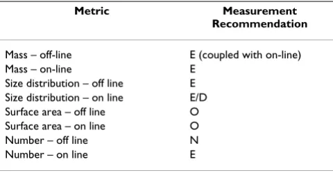
Table 1 summarizes recommendations for measuring exposure during inhalation studies.
4.1.5 Characterization Prioritization
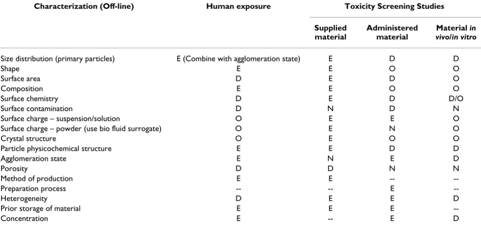
In developing recommendations on material characteriza-tions for nanomaterial toxicity screening studies, three specific factors have been taken into consideration: the context within which a material is being evaluated, the importance of measuring a specific parameter within that context, and the feasibility of measuring the parameter within a specific context. Recommendations on off-line material characterizations for nanomaterial toxicity screening studies are presented in Table 2.
Table 1: Recommendations for measuring exposure during inhalation studies
Metric Measurement
Recommendation
Mass – off-line E (coupled with on-line) Mass – on-line E
Size distribution – off line E Size distribution – on line E/D Surface area – off line O Surface area – on line O Number – off line N Number – on line E
E: These measurements are considered to be essential. D: These measurements are considered to provide valuable information, but are not recommended as essential due to constraints associated with complexity, cost and availability.
O: These measurements are considered to provide valuable but non-essential exposure information.
In addition, recommendations have been made on recording information on nanomaterial production, prep-aration, storage, heterogeneity, and agglomeration state. To enable retrospective interpretation of toxicity data and replication of tests, it is strongly recommended that all information on the production and processing of nano-materials be recorded. Fully documenting storage time and conditions (including temperature, humidity, expo-sure to light and atmosphere composition) is essential, as physicochemical changes may take place over time. If pos-sible, the physicochemical stability of samples over time should be demonstrated. Where a test material is a heter-ogeneous mixture of different components, information is required on the relative abundance of the different com-ponents, and whether associations in the bulk material are maintained in the administered material, or whether different components are preferentially administered with specific delivery mechanisms.
The agglomeration state of a nanomaterial during and fol-lowing administration may have a significant impact on its biological activity. Agglomeration state at different structure scales should be characterized, including pri-mary (pripri-mary particles), secondary (pripri-mary particle agglomerates and self-assembled structures) and tertiary
(assemblies of secondary structures) scales. Ideally, agglomeration state in the biological environment follow-ing administration should be evaluated. If possible, some insight into the binding forces within agglomerates (e.g. relatively weak van der Waals forces or relatively strong sintered bonds) should be obtained. Material agglomeration or de-agglomeration in different liquid media should also be investigated where possible.
Characterization of material as administered is recom-mended as the highest priority, supplemented by charac-terization after in vitro or in vivo administration where possible, and followed in order of preference by character-ization of the material as produced or supplied. Recom-mended characterizations in Table 2 reflect both this hierarchy and the feasibility of making measurements within the respective contexts.
4.1.6 Analysis Methods
Many analytical techniques, both established and devel-opmental, are available for characterizing the nanomate-rial properties listed in Table 2. Table 3 lists some of the more widely available techniques and relates them to the nanomaterial characteristics of interest to toxicity screen-ing studies. Techniques have been categorized with
Table 2: Recommendations on material characterization
Characterization (Off-line) Human exposure Toxicity Screening Studies
Supplied material
Administered material
Material in vivo/in vitro
Size distribution (primary particles) E (Combine with agglomeration state) E D D
Shape E E O O
Surface area D E D O
Composition E E O O
Surface chemistry D E D D/O Surface contamination D N D N Surface charge – suspension/solution O E E O Surface charge – powder (use bio fluid surrogate) O E N O Crystal structure O E O O Particle physicochemical structure E E D D Agglomeration state E N E D
Porosity D D N N
Method of production E E -- --Preparation process -- -- E
--Heterogeneity D E E D
Prior storage of material E E E
--Concentration E -- E D
E: These characterizations are considered to be essential.
D: These characterizations are considered to provide valuable information, but are not recommended as essential due to constraints associated with complexity, cost and availability.
Parti
cle
and
F
ibre
T
oxicol
ogy
200
5,
2
:8
http
://www.particlea
ndfibre
to
xi
colog
y.com/c
onten
t/2/1/8
Pag
e 12 of
3
5
(page nu
mber not
for
cit
a
tion pur
poses)
Table 3: Applicability of a range of analytical techniques to providing specific physicochemical information on engineered nanomaterials, in the context of toxicity screening studies
Analytical technique
Transmission Electron Microscopy
(TEM)
Scanning Electron Microscopy
(SEM)
X-Ray Diffraction
(XRD)
X-ray Photon Spectroscopy
(XPS)
Auger Spectroscopy
(AES)
Secondary Ion Mass Spectrometry
(SIMS)
Scanning Probe Microscopy
Dynamic Light Scattering
(DLS) Zeta potential
Size Exclusion Chromatography
Analytical Ultracentrifugation
Differential Mobility Analysis (DMA)
Isothermal Adsorption (e.g. BET)
Spectroscopic techniques (UV vis, IR, Raman, NMR)
Elemental analysis (eg ICP-MS/AA
etc)
Physicochemical Characteristic
Size distribution (primary particles)
▲ ● ● ● ● ● ● ●
Shape ▲ ● ●
Surface area ● ●
Composition ● ● ● ▲ ● ▲
Surface chemistry
● ● ●
Surface contamination
● ●
Surface charge – suspension/ solution
▲
Surface charge – powder (use bio fluid surrogate)
▲
Crystal structure
● ▲
Particle physicochemic al structure
▲ ●
Agglomeration state
▲ ● ● ●
Porosity ● ●
Heterogeneity ▲ ●
Other applicable techniques are available that have not been listed. ▲Highly applicable
● Capable of providing information in some cases
Capable of providing information in some cases, with validation from more accurate/applicable techniques
Capable of providing qualitative or semi-quantitative information
9 9
9
9 9 9 9
9 9 9
9
9 9 9 9
9
respect to their applicability to specific material character-istics. In general, the table is self-explanatory, and further information on each technique can be obtained from a wide range of sources. A number of techniques are only suitable for materials in certain forms, or specific classes of materials. For instance, while Transmission Electron Microscopy is capable of providing a wealth of informa-tion on nanoparticles and is considered a gold standard for evaluating particle size distribution and shape, dry (or in the case of cryo-TEM, frozen liquid-encapsulated) well-dispersed samples that are sufficiently robust to withstand high vacuums are required. Similarly, techniques such as Infrared (IR) spectroscopy are particularly sensitive to sur-face organic compounds, but are less useful for quantify-ing inorganic surface chemistry. In a number of cases, a complex technique such as TEM can be used to validate a characterization method that is more practical to use on a routine basis.
Given the wide range of analytical techniques available in many disciplines associated with nanotechnology, multi-disciplinary collaborations with research and analysis groups offering state of the art nanomaterial characteriza-tion capabilities are strongly recommended when carrying out nanomaterial toxicity screening studies.
4.1.7 Research Gaps
1. The development of viable in vivo nanomaterial (including nanoparticles) detection techniques.
2. The development and production of inexpensive real-time monitoring instruments and methods for aerosol mass concentration (low concentrations, nanoscale parti-cles), surface area concentration and size distribution.
3. The development of standardized, well characterized nanomaterial samples.
4. The development of radio-labeled nanomaterial sam-ples, and samples that can be tracked and detected through neutron-activation.
5. The development of more advanced surface chemistry characterization techniques, in particular techniques capable of detecting and speciating biological molecules on the surface of nanoparticles and nanomaterials.
6. The development of electron microscopy techniques for biologically-relevant nanoscale analysis.
4.1.8 Recommendations
1. All nanomaterial physicochemical characteristics that are potentially significant should be measured or be deriv-able in toxicity screening tests.
2. Characterization of nanomaterial as administered is strongly recommended, supplemented by characteriza-tion following administracharacteriza-tion where it is technically feasi-ble and practicafeasi-ble. Characterization of the bulk material as-produced or supplied to the exclusion of the above is not recommended, except where more appropriate meas-urements are not feasible.
3. It is recommended that independent characterizations of nanomaterials (beyond information provided by pro-ducers and suppliers) are carried out where possible.
4. It is recommended that the following physicochemical properties of nanomaterials should be characterized in the context of toxicity screening tests: Particle size distri-bution, agglomeration state, particle shape, crystal struc-ture, chemical composition (bulk and spatial), surface area, surface chemistry, surface charge, and porosity.
5. It is recommended that in all cases, sufficient informa-tion be collected to enable derivainforma-tion of the delivered dose against all three primary physical metrics (number, surface area and mass concentration).
6. Off-line mass concentration measurements using filter-based methods are recommended as an essential compo-nent of inhalation nanomaterial screening tests. In addi-tion, off-line measurement of aerosol size distribution is recommended.
7. On-line mass concentration and number measure-ments are recommended as an essential component of inhalation studies.
8. Multidisciplinary collaborations between research and analysis groups offering state of the art nanomaterial char-acterization capabilities are strongly recommended.
9. It is recommended that information on nanomaterial production, preparation, storage, heterogeneity and agglomeration state be recorded for all nanomaterial tox-icity screening studies.
Particle and Fibre Toxicology 2005, 2:8 http://www.particleandfibretoxicology.com/content/2/1/8
Page 14 of 35
(page number not for citation purposes)
4.2 In vitro Testing Methods
4.2.1 Introduction
Before considering the application of specific in vitro test-ing methods to the assessment of the toxicity of nanoma-terials, there are several generic issues that should be noted.
1) Advantages and disadvantages In general in vitro tech-niques are seen as an important adjunct to in vivo studies. These studies allow specific biological pathways to be tested under controlled conditions, as well as isolation of pathways that is not feasible in vivo; e.g., it is difficult to discriminate in vivo whether complement activation has a role in any pro-inflammatory effects of particles. The com-plement system can be isolated in vitro, and its potential role investigated. There are, of course, well-documented problems with in vitro approaches, including lack of vali-dation against in vivo adverse effects, dosimetry mismatch, over-simplicity, non-involvement of the complete inflam-matory response, etc.
2) Control particles It is important, in view of the above, that adequate positive and negative control particles are included in all experiments. This at least allows the test particle to be bench-marked against particles of known toxicity. These can include standard crystalline silica (quartz; e.g, Min-U-Sil or DQ12) as a known cytotoxic particle and fine TiO2 as an inert particle.
3) Expression of dose Toxicity and other responses should be expressed in relation to a range of dose metrics depend-ing on the material and the dose metric data that are avail-able (see Section 4.1).
4) Adsorption of proteins by nanoparticles The large sur-face area of nanoparticles means that they are capable of adsorbing proteins. Nanoparticles of various types have been reported to adsorb key proteins such as albumin [93], fibronectin and TGF-β [94]. This may confound end-points that rely on the measurement of a protein as the protein may be produced but may also remove from the supernatant onto the nanoparticle surface by adsorption, providing a false-negative.
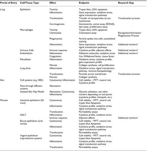
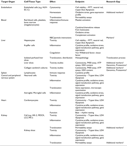
The in vitro tests that are presented will be divided into portal of entry toxicity and target organ toxicity. The potential target cells and associated appropriate end-points will be described. Finally, research gaps and recom-mendations will be identified.
4.2.2 Portals of Entry 4.2.2.1 Lungs
The lungs represent a potential target for any airborne par-ticles, and many in vitro models for the lung exist. Particles deposit on the airway or alveolar epithelium and
encoun-ter mucus or epithelial lining fluid. They may then inencoun-teract with macrophages, which may result in their clearance, or they may enter the interstitium where they may make con-tact with fibroblasts and endothelial cells or cells of the immune system.
The Epithelium
The epithelium is the first barrier that confronts particles that deposit in either the conducting airways or the alveo-lar region. Therefore, both bronchial and alveoalveo-lar epithe-lial cells should be considered as target cells for in vitro
studies. Endpoints for detecting nanoparticle effects could include toxicity measurements, such as LDH release, for necrosis or various cytokine expression (IL-8, MCP-1 etc), [91,95] and activation of inflammation-related transcrip-tion factors such as NF-κB and AP-1[96,97]. Oxidative and nitrosative stress are dominant mechanistic hypothe-ses for cell damage and activation caused by pathogenic particles. These can be monitored by measuring oxidative stress using dichlorofluorescein [98] or oxidized glutath-ione as endpoints [99] and nitrosated proteins as a meas-ure of active nitrogen species [100]. Responses to particle-induced oxidative/nitrosative stress can include up-regu-lation of anti-oxidant genes [101] such as superoxide dis-mutase and glutathione peroxidase, and so these can also be measured. Proliferative effects of nanoparticles can be assessed using a variety of assays including bromo-deoxy-uridine incorporation [102].
If cancer is an endpoint that is under consideration, then direct measures of genotoxicity can be quantified by methods that include COMET assay and 8-hydroxy-deox-yguanosine measurement [103,104]. The translocation of nanoparticles across the epithelium could be an impor-tant discriminator of harmfulness and, although there are few publications specifically addressing transfer of parti-cles across the epithelium in vitro, these should be devel-oped and could contribute to understanding the factors that regulate translocation.
Macrophages
oxide (NO) may also be produced, in response to particles [106] and in the presence of superoxide radical peroxyni-trite, a highly toxic species, can be produced [107]. If the OB or NO production is exaggerated, there could be 'bystander' injury to epithelial cells whilst diminished OB/NO production could mean impaired microbicidal activity that allows infection. Another key macrophage function reported to be impaired by nanoparticles is phagocytosis, [108] and so the effect of test nanoparticles on this function could be considered. The cytoskeleton is key to normal cell functioning and could be targeted by nanoparticles and so could be investigated.
Endothelial cells
Although these are found in the lungs, they are considered a part of the cardiovascular system and are dealt with below.
Fibroblasts
Fibroblasts are found in the interstitium and are liable to be affected by any particle that gains access to this site. At least two important modes of response could be activated by nanoparticle/fibroblast interactions and both modes constitute relevant endpoints for in vitro testing: 1) Pro-inflammatory effects, measured by cytokine/chemokine gene expression (TNFα; etc); or 2) fibrogenic responses activated either by direct stimulation of fibroblast growth or extra-cellular matrix secretion by the nanoparticle, or by autocrine stimulation following nanoparticle-stimu-lated release from the fibroblasts of growth factors such as transforming growth factor beta and platelet-derived growth factor.
The Immune System
Immunopathological effects could be envisaged if parti-cles interact with lymphocytes, or as a consequence of their predilection for entering the interstitium, they mod-ulate dendritic cell function. The effects of nanoparticles on immunological functions including antigen presenta-tion by macrophages and dendritic cells and the subse-quent effects on immune responses in vitro are relevant endpoints and appropriate tests should be designed.
Co-Cultures
In addition to monocultures of lung cells, co-cultures such as epithelial cells/macrophages or epithelial cells/ endothelial cells may more closely represent the in vivo sit-uation, and so such studies are encouraged.
Lung Slices
Methodology to culture whole lung tissue slices is availa-ble, such that multiple pulmonary cell types can be exposed in vitro in the same configuration as they occur in vivo.
Cell Lines vs. Freshly-Derived Cells
If possible, freshly-derived primary cells should be used. Where cell lines are used, these should preferably not be cancer cells. Where cancer cells are used, the endpoint response under study should be carefully compared to non-cancer cells to ensure that, for that endpoint, the fact that the cell is a cancer cell does not greatly modify the response compared to a non-cancer cell.
Whole Heart-Lung Preparation
The Langendorff heart-lung preparation may provide the opportunity to study the behavior of nanoparticles under highly controlled conditions. In this model the exsan-guinated heart and lungs are maintained by perfusion and so transport between the lungs and the vascular space can be studied in the absence of blood [109].
4.2.2.2 Skin
Skin or the integument is the largest organ of the body and is unique because it is a potential route for exposure to nanoparticles during their manufacture and also provides an environment within the avascular epidermis where particles could potentially lodge and not be susceptible to removal by phagocytosis [110]. What are the toxicological consequences of "dirty" nanoparticles (catalyst residue) becoming lodged in the epidermis? In fact, it is this rela-tive biological isolation in the lipid domains of the epi-dermis that has allowed for the delivery of drugs to the skin using lipid nanoparticles and liposomes. Larger par-ticles of zinc and titanium oxide used in topical skin-care products have been shown to be able to penetrate the stra-tum corneum barrier of rabbit skin with highest absorp-tion occurring from water and oily vehicles [111]. This could also apply to manufactured nanoparticles. Can nan-oparticles gain access to the epidermis after topical expo-sure, the first step in a toxicological reaction? Exposure to metallic nanoparticles, whose physical properties would allow them to catalyze a number of biomolecular interac-tions, potentially could produce adverse toxicological effects. More information is required regarding the effi-ciency of decontamination of nanoparticles from skin since solubilization and dilution, the two hallmarks of post-exposure decontamination, might be less efficacious for these solid structures.
Particle and Fibre Toxicology 2005, 2:8 http://www.particleandfibretoxicology.com/content/2/1/8
Page 16 of 35
(page number not for citation purposes)
become sequestered within the epidermis to increase their exposure time to viable epidermal keratinocytes.
Nanomaterials are difficult to obtain in large quantities; therefore, it is best to conduct in vitro tests to estimate in vivo starting doses for toxicity testing [112]. At least three or four concentrations with controls should be used in all
in vitro systems. These data would provide a preliminary, but relevant, assessment of both systemic exposure after topical administration as well as cutaneous hazard after both topical and systemic exposure, two essential compo-nents of any risk assessment.
Cell Culture
Human epidermal keratinocyte (HEK) monolayers can be affected by nanoparticle interactions. It has already been shown that changes in biomarkers of viability and toxicity can occur with exposure to multi-wall carbon nanotubes [50]. Cytotoxicity endpoints should be evaluated: 1) cell viability-metabolic markers such as mitochondrial reduc-tion of tetrazolium salts into insoluble dye (MTT), 2) decreased cell viability-membrane markers like neutral red uptake into cell lysosomes, trypan blue exclusion and cell attachment/cell detachment, and 3) pro-inflamma-tory cytokine affects measured by TNFα, IL-8, IL-6, IL-10, or IL-1β. Genomics and proteomics assays could be used to explore the mechanism behind the toxicity. However, caution must be taken when using carbon black or any other material as a control because complications may occur. Carbon can adsorb the viability dyes, such as neu-tral red, and interfere with the absorption spectra. False positives will occur. The type of carbon black used is extremely important. For instance, ultrafine carbon black has been utilized in inhalation studies but dosing in cell culture gives different results, especially when conducting viability and cytokine assays.
Three dimensional skin cell cultures are also available commercially. They have shown to be able to predict irri-tation but may significantly overestimate absorption or penetration [113-116]. Assays listed above can be used but may not be applicable with nanomaterials due to adsorption.
Flow-through Diffusion Cell Studies
Diffusion cell system consists of flow-through diffusion blocks each containing multiple Teflon cells perfused by a constant temperature circulator through a Silastic oxygen-ator, an automatic fraction collector, and a desiccant. Cir-cular fresh skin from pigs (pig skin mimics human skin and eliminates the extreme variability seen with random source human skin) or humans are placed epidermal – side up in Teflon flow-through diffusion cell. Compound containing nanoparticles is dosed on the epidermal side whilst the dermal side in each cell is bathed with receptor
fluid at a set flow rate. The perfusate is collected at defined intervals up to 24 hrs and nanoparticles flux in the per-fusate can be assessed by radioactivity counting, fluores-cence, or UV detection. The skin surface can then be swabbed to remove non-absorbed surface particles and then tape stripped to remove a stratum corneum sample to assess nanoparticle penetration into this outermost epi-dermal layer. Serial sectioning of the skin can also be car-ried out [117,118].
Isolated Perfused Porcine Skin Flap (IPPSF)
The isolated perfused porcine skin flap (IPPSF) would be an ideal model to study the absorption and toxicity of nanomaterials. The IPPSF has an intact functional micro-circulation, a viable epidermis and dermis and can be well controlled. A single-pedicle, axial pattern tubed skin flap is obtained from the abdomen of pigs following surgical creation of the flap perfused primarily by the caudal superficial epigastric artery and its associated paired venae commitantes. The IPPSF is transferred to the perfusion apparatus that is a custom designed temperature and humidity-regulated chamber. Nanomaterials can be topi-cally dosed to the skin surface and perfusate samples col-lected over an eight hour period and assessed for nanoparticle flux [119-121].
Other acute toxicity in vitro assays are available but are used to test corrosives (rat transcutaneous electrical resist-ance (TER), commercially available EPISKIN, Epiderm and Corrositex) and irritation (EPISKIN, and Epiderm). However, the major traditional endpoint for skin toxicity is using the cell viability assay MTT reduction that has been shown to be unpredictable with nanomaterials due to marker interactions with nanoparticles.
4.2.2.3 Mucosa
Mucosa is the moist tissue that lines particular organs and body cavities throughout the body, including the nasal cavity, oral cavity, lungs, vagina and gastrointestinal tract. Potentially one of the most important portals of entry for nanoparticle exposure (excluding the nasal cavity and lung, which has been detailed above) is the gastrointesti-nal tract. Either accidental or intentiogastrointesti-nal exposure via oral administration to the GI tract can lead to significant expo-sures. Efficient uptake of nanoparticles via the GI tract has been well documented in oral feeding studies and gavage studies using particles ranging from 10 nm to 500 nm [122-124]. In these studies nanoparticles translocated through the mucosal lining and epithelial barrier of the intestine and were associated with the GALT (gastroinstet-inal associated lymphatic tissue) and circulatory system within as little as 60 minutes time [125].