Electronic Thesis and Dissertation Repository
8-12-2014 12:00 AM
Evolutionary and in silico analysis of the antiviral TRIM22 gene
Evolutionary and in silico analysis of the antiviral TRIM22 gene
Jenna KellyThe University of Western Ontario
Supervisor Stephen Barr
The University of Western Ontario
Graduate Program in Microbiology and Immunology
A thesis submitted in partial fulfillment of the requirements for the degree in Doctor of Philosophy
© Jenna Kelly 2014
Follow this and additional works at: https://ir.lib.uwo.ca/etd Part of the Immunity Commons
Recommended Citation Recommended Citation
Kelly, Jenna, "Evolutionary and in silico analysis of the antiviral TRIM22 gene" (2014). Electronic Thesis and Dissertation Repository. 2327.
https://ir.lib.uwo.ca/etd/2327
This Dissertation/Thesis is brought to you for free and open access by Scholarship@Western. It has been accepted for inclusion in Electronic Thesis and Dissertation Repository by an authorized administrator of
EVOLUTIONARY AND IN SILICO ANALYSIS OF THE ANTIVIRAL TRIM22 GENE
(Thesis format: Integrated Article)
by
Jenna N. Kelly
Graduate Program in Microbiology and Immunology
A thesis submitted in partial fulfillment of the requirements for the degree of
Doctor of Philosophy
The School of Graduate and Postdoctoral Studies The University of Western Ontario
London, Ontario, Canada
ii
Abstract
Tripartite motif protein 22 (TRIM22) is an evolutionarily ancient interferon-induced protein that been shown to potently inhibit human immunodeficiency virus (HIV), hepatitis B virus (HBV), and influenza A virus (IAV) replication. Altered TRIM22 expression levels have also been linked to autoimmune disease, cancer, and cellular proliferation. Despite its important role in a number of biological processes, the factors that influence TRIM22 expression and/or antiviral activity remain largely unknown. To identify key functional sites in TRIM22, we performed extensive evolutionary and in silico analyses on the TRIM22 coding region. These tools allowed us to pinpoint multiple
sites in TRIM22 that have evolved under positive selection during mammalian evolution, including one site that coincides with the location of a common non-synonymous SNP (nsSNP) in the human TRIM22 gene (TRIM22 rs1063303:G>C). We found that the
frequency of TRIM22 rs1063303:G>C varied considerably among different ethnic
populations and African (AFR), American (AMR), and European (EUR) populations contained an excess of intermediate frequency TRIM22 rs1063303:G>C alleles when
compared to a neutral model of evolution. The latter is typically indicative of balancing selection, a non-neutral selective process that maintains polymorphism in a population. Interestingly, we also found that the TRIM22 nsSNP rs1063303:G>C had an inverse
impact on TRIM22 function. TRIM22 rs1063303:G>C increased TRIM22 expression
levels, but decreased its anti-HIV activity and altered its subcellular localization pattern. In addition to these studies, we used a variety of in silico methods to prioritize and
delineate other functional sites in TRIM22. We showed that the majority of positively selected sites in the C-terminal B30.2 domain of TRIM22 are located in one of four surface-exposed variable loops that are critical for the anti-HIV effects of the closely-related TRIM5α protein. Moreover, we used six different in silico nsSNP prediction
programs to screen all of the nsSNPs in the TRIM22 gene and identified 14 high-risk
nsSNPs that are predicted to be highly deleterious to TRIM22 function. Finally, to examine the TRIM22 nsSNP rs1063303:G>C in a more isolated population, we
genotyped this nsSNP in two Inuit populations (Canadian and Greenlandic Inuit). We found that the TRIM22 rs1063303:C allele is inordinately prevalent in the Inuit compared
iii
intermediate frequency TRIM22 rs1063303:G>C alleles compared to a neutral model of
evolution, indicating that site TRIM22 rs1063303:G>C has not evolved under balancing
selection in the Inuit. Lastly, we found an interesting association between the TRIM22
rs1063303:C allele and serum levels of triglycerides (TG) and high-density lipoprotein (HDL). Taken together, the results presented here identify a number of pertinent sites in the TRIM22 protein that likely influence its biological and/or antiviral functions.
Keywords
iv
Co-Authorship Statement
Chapter 2 of this thesis was published in Human Mutation 35: 1072-1081, June 2014 doi: 10.1002/humu.22595. I was involved in performing all of the experiments with the exception of those described in Fig 2.4d, which were primarily performed by M. W. Woods.
v
Acknowledgements
I am fortunate to have many remarkable and inspiring people in my life who have made the completion of this dissertation possible. I would like to thank my supervisor, Dr. Stephen Barr, for his guidance and support throughout my PhD studies. His positive attitude and encouragement helped me overcome many of the challenging situations I encountered during my research. I am also thankful to him for giving me the freedom to explore my own ideas and forge my own path as a researcher. I would like to thank my advisory committee members, Dr. Greg Dekaban and Dr. Joe Mymryk, for their many insightful comments and suggestions. Their guidance, support, and ‘open-door policy’ have been invaluable through both successes and difficulties. I would also like to thank Dr. Robert Hegele for allowing me to work in his lab and for sharing his genotyping expertise with me. His knowledge, guidance, and commitment to the highest research standards inspired and motivated me.
vi
Table of Contents
Abstract ... ii
Acknowledgements ... v
Table of Contents ... vi
List of Tables ... xi
List of Figures ... xiii
List of Abbreviations...xiv
1 Introduction ... 1
1.1 Host restriction factors ... 5
1.1.1 Inhibition of HIV replication by host restriction factors ... 6
1.1.2 Innate immune regulation by host restriction factors ... 11
1.2 The TRIM family ... 11
1.2.1 Evolution and classification ... 11
1.2.3 The SPRY and PRY/SPRY (B30.2) domains ... 22
1.1.3 Antiviral activity ... 23
1.2 The TRIM22 protein ... 24
1.2.1 Origins and evolution ... 25
1.3 Rationale and Experimental Approach ... 37
1.4 References ... 39
Chapter 2 ... 57
2 Ancient and recent adaptive evolution in the antiviral TRIM22 gene: identification of a single nucleotide polymorphism that impacts TRIM22 function ... 57
2.1 Introduction ... 58
vii
2.2.1 Sequence analysis ... 59
2.2.2 Cells, plasmids, and transfections ... 60
2.2.3 RNA isolation and real-time PCR... 61
2.2.4 Western Blotting ... 62
2.2.5 Confocal immunofluorescence microscopy ... 62
2.2.6 Neutrality tests ... 63
2.2.7 Statistical analysis ... 63
2.3 Results 63 2.3.1 Positive selection in multiple TRIM22 domains among mammals ... 63
2.3.2 Genetic Variation in the Human TRIM22 Gene ... 73
2.4 Discussion ... 81
2.5 References ... 87
Chapter 3 ... 95
3 In silico analysis of functional single nucleotide polymorphisms in the human TRIM22 gene ... 95
3.1 Introduction ... 96
3.2 Materials and methods ... 97
3.2.1 Retrieval of nsSNP data ... 97
3.2.2 In silico nsSNP analysis ... 98
3.2.3 Phylogenetic analysis ... 98
3.2.4 Comparative molecular modeling ... 98
3.2.5 Prediction of post-translational modification sites ... 99
3.2.6 Protein stability analysis ... 99
3.3 Results and Discussion ... 100
3.3.1 SNP dataset ... 100
viii
3.3.3 Conservation Profile of High-Risk nsSNPs ... 105
3.3.4 Comparative Modeling of High-Risk nsSNPs ... 110
3.3.5 Prediction of Post-Translational Modification Sites in TRIM22 ... 113
3.4 Conclusions ... 121
3.5 References ... 125
Chapter 4 ... 134
4.1 Introduction ... 135
4.2 Materials and methods ... 137
4.2.1 Study subjects ... 137
4.2.2 Clinical characteristics and biochemical analyses ... 138
4.2.3 Genotype analyses ... 138
4.2.4 Neutrality tests ... 138
4.2.5 Statistical analyses ... 138
4.3 Results 139 4.3.1 Baseline phenotypic characteristics of study subjects ... 139
4.3.2 TRIM22 rs1063303:G>C genotype and allele frequencies ... 139
4.3.3 TRIM22 rs1063303:G>C is not evolving under balancing selection in the Canadian or Greenlandic Inuit ... 142
4.3.4 Association between TRIM22 rs1063303:G>C and plasma lipoproteins in the Canadian Inuit ... 142
4.5 References ... 150
Chapter 5 ... 157
5 Discussion ... 157
5.1 Summary of results ... 157
ix
5.1.2 In silico analysis of functional single nucleotide polymorphisms in the
human TRIM22 gene ... 158
5.1.3 The TRIM22 nsSNP rs1063303:G>C is not evolving under balancing selection in the Inuit and is associated with low serum TG and high serum HDL levels in the Canadian Inuit... 159
5.2 Multiple sites in TRIM22 have evolved under positive and/or balancing selection ... 160
5.2.1 Positive selection ... 160
5.2.2 Balancing selection ... 163
5.3 TRIM22 contains 14 high-risk deleterious nsSNPs and numerous putative PTM sites ... 165
5.3.1 High-risk deleterious nsSNPs ... 165
5.3.2 Putative PTM sites ... 167
5.4 The TRIM22 nsSNP rs1063303:G>C influences diverse TRIM22-mediated biological functions ... 170
5.4.1 The TRIM22 nsSNP rs1063303:G>C increases TRIM22 expression levels 170 5.3.2 The TRIM22 nsSNP rs1063303:G>C decreases TRIM22-mediated antiviral activity against HIV-1 ... 171
5.3.3 The TRIM22 nsSNP rs1063303:G>C is associated with low serum TG and high serum HDL levels in the Canadian Inuit ... 173
5.5 Concluding remarks ... 173
5.5 References ... 175
Appendix A ... 182
A.1 Overview of serum lipoproteins... 182
A.1.1 Introduction to serum lipoproteins ... 182
A.2 Serum lipoproteins during the APR ... 189
A.2.1 Overview of the APR ... 189
x
xi
List of Tables
Table 1.1: Characteristics of well-studied host restriction factors. ... 12
Table 1.2: Factors that alter TRIM22 protein expression levels. ... 30
Table 2.1: Mammalian TRIM22 coding sequences used for Selecton analysis. ... 64
Table 2.2: Models of evolution applied to TRIM22 coding sequences. ... 69
Table 2.3: Positive selection in TRIM22 protein domains. ... 72
Table 2.4: Results of Tajima’s D and Fu’s FS neutrality tests. ... 78
Table 3.1 In silico prediction results for nsSNPs in TRIM22... 101
Table 3.2 Summary of in silico prediction results for all nsSNPs in TRIM22. ... 104
Table 3.3: TRIM22 nsSNPs predicted to be functionally significant by four or more SNP prediction algorithms. ... 106
Table 3.4 Conservation profile of amino acids in TRIM22 that coincide with the location of high-risk nsSNPs. ... 109
Table 3.5: RMSD (Å) and TM-score for the 9 high-risk nsSNPs in the B30.2 domain of TRIM22... 114
Table 3.6 Putative ubiquitylation and sumoylation sites in the TRIM22 protein. ... 116
Table 3.7 Putative phosphorylation sites in the TRIM22 protein. ... 119
Table 3.8: I-Mutant results for selected nsSNPs in the TRIM22 protein. ... 122
Table 4.1 Baseline phenotypic characteristics (mean ± SD) of Canadian Inuit, Greenland Inuit, and European Caucasian populations. ... 140
xii
Table 4.3: Results of Tajima’s D and Fu’s Fs neutrality tests. ... 143
Table 4.4: Summary of ANOVA results showing determinants of serum lipoproteins in the Canadian Inuit, Greenlandic Inuit, and European Caucasian populations. ... 145 Table 4.5: Significant associations between the TRIM22 rs1063303:G>C genotype and
xiii
List of Figures
Figure 1.1: ISG induction by type I IFN signaling. ... 4
Figure 1.2: Stages of HIV replication targeted by host restriction factors. ... 8
Figure 1.3: Subgroup classification of the TRIM family. ... 16
Figure 1.4: The RING E3 ligase-mediated ubiquitylation pathway. ... 20
Figure 1.5: TRIM22 evolution, genomic organization, and protein structure. ... 27
Figure 2.1: Mapping positively selected sites in the TRIM22 protein. ... 68
Figure 2.2: Selecton analysis of mammalian TRIM22 coding sequences. ... 71
Figure 2.3: Genetic variation at amino acid site 242 in the TRIM22 protein. ... 76
Figure 2.4: nsSNP rs1063303:G>C alters TRIM22 expression and antiviral activity. .... 80
Figure 3.1: ConSurf analysis of amino acid residues in the TRIM22 protein. ... 108
Figure 3.2: Structural models for wild type TRIM22 and high-risk nsSNPs in the B30.2 domain... 112
Figure 3.3: Putative sumo-interacting motifs (SIM) in TRIM22. ... 118
Figure 3.4 Putative functional sites in the TRIM22 protein. ... 124
Figure A.1 General structure of serum lipoprotein. ... 185
xiv
List of Abbreviations
AIC Akaike Information Content AP-1 Activator protein 1
APOBEC3G Apolipoprotein B mRNA-editing enzyme catalytic polypeptide 3G APR Acute phase response
ARF ADP ribosylation factor-like domain BB B-box domain
BR Bromodomain
BST-2 Bone marrow stromal antigen 2 CB Cajal bodies
CC Coiled-coil domain
CD4 Cluster of differentiation 4
COS C-terminal subgroup one signature domain CP Core promoter
Cys Cysteine residue
E1 Ubiquitin activating enzyme E2 Ubiquitin conjugating enzyme E3 Ubiquitin ligating enzyme
xv
FIL Filamin-type immunoglobulin domain FN3 Fibronectin type 3 domain
Fv1 Friend virus susceptibility 1 locus G1 Group 1 TRIM
G2 Group 2 TRIM
GAS Gamma interferon activation site HBV Hepatitis B virus
HCV Hepatitis C virus
HDL High density lipoprotein
HECT Homologous to the E6-AP carboxyl terminus HIV Human immunodeficiency virus
HIV-1 Human immunodeficiency virus type 1 HIV-2 Human immunodeficiency virus type 2 HOM Hominoids
huTRIM5α Human TRIM5α IFN Interferon
IFNAR Interferon alpha/beta receptor IL-1 Interleukin 1
xvi
IRF-7 Interferon regulatory factor 7 IRF-8 Interferon regulatory factor 8 ISG Interferon stimulated gene ISG15 Interferon stimulated gene 15 ISGF3 Interferon stimulated gene factor 3 ISRE Interferon stimulated response element
JAK/STAT Janus kinases/signal transducers and activators of transcription LDL Low density lipoprotein
LPS Lipopolysaccharide LTA Lipoteichoic acid LTR Long-terminal repeat Lys Lysine residue
MATH Merpin and tumor-necrosis factor receptor-associated factor homology domain
MDM Monocyte derived macrophages NHL NHL repeats
N-MLV N-tropic murine leukemia virus NB Nuclear bodies
NF-κB Nuclear factor-kappa B
xvii
NLS Nuclear localization signal NP Nucleoprotein
nsSNP Non-synonymous single nucleotide polymorphism (SNP) NWM New world monkey
OWM Old world monkey
PAMP Pathogen-associated molecular pattern PHD Plant homeodomain
PIC Preintegration complex
PML Promyelocytic leukemia protein (also called TRIM19)
PRR Pattern recognition receptor PTM Post-translational modification
qPCR Quantitative polymerase chain reaction RBCC RING, BB1 and/or BB2, and CC domains rhTRIM5α Rhesus macaque TRIM5α
RING Really interesting new gene domain
RLR Retinoic acid inducible gene (RIG-I)-like receptor
SAMHD1 Sterile alpha motif and histidine/aspartic acid domain-containing protein
xviii
SIV Simian immunodeficiency virus SLE Systemic lupus erythematosus SNP Single nucleotide polymorphism SPRY SplA/ryanodine receptor domain SUMO Small ubiquitin-like modifier
TAK1 Transforming growth factor beta-activated kinase 1 TG Triglyceride
TLR Toll-like receptor TM Transmembrane domain TNFα Tumor necrosis factor alpha TRIM Tripartite motif protein TRIM22 Tripartite motif protein 22 TRIM5α Tripartite motif protein 5 alpha
UBE2D Ubiquitin conjugating enzyme 2/E2 class D UBE2E Ubiquitin conjugating enzyme 2/E2 class E v1-v4 Variable loops 1-4
Chapter 1
1
Introduction
The immune system has evolved over millennia to provide effective defense measures against invading pathogens and promote host survival. The vertebrate immune system can be divided into innate and adaptive arms, which together comprise both germline-encoded and acquired immune responses. The innate immune system is evolutionarily more ancient than the adaptive immune system and constitutes the first line of defense against infectious agents. It is also essential for initiating subsequent adaptive immune responses, which unlike innate immune responses, are characterized by specificity and the generation of immunological memory 1. Innate immunity is activated upon pathogen
recognition by a limited repertoire of germline-encoded pattern recognition receptors (PRR). PRRs can be grouped into several distinct families: 1) the membrane bound toll-like receptors (TLR), 2) the cytosolic retinoic acid inducible gene (RIG-I)-toll-like receptors (RLR), 3) the nucleotide binding oligomerization domain (NOD)-like receptors (NLR), and 4) DNA sensors 2,3. PRRs recognize conserved products of microbial metabolism called pathogen-associated molecular patterns (PAMP). PAMPs, which are not usually present in host cells, bind to specific PRRs to activate innate immunity. For example, specific TLR family members recognize genomic material or replication intermediates from viruses (i.e. TLRs 3, 7, 8, and 9), while other TLRs recognize diverse microbial products from parasites, fungi, and/or bacteria 4–8.
secreted cytokines that are essential for host antiviral defense and immune activation. Type I IFNs are produced in direct response to viral infection and comprise multiple subtypes of IFN-α plus IFN-β and several additional IFNs (δ, ε, κ, τ, ω) with less-definitive roles in antiviral immunity. The other two major classes of IFNs, type II and type III, each contain only one subtype. IFN-γ (type II IFN) is generally produced by activated T lymphocytes or natural killer cells in response to cytokine secretion from infected cells or macrophages. Similar to type I IFNs, IFN-λ (type III IFN) is typically produced in direct response to viruses; however type III IFN binds to different cognate receptors than type I IFNs. Type III IFN receptors are found mainly on epithelial cells, whereas type I IFN receptors are present on all cell types 10–13.
Figure 1.1: ISG induction by type I IFN signaling.
Figure 1.1
1.1 Host restriction factors
Host restriction factors are IFN-induced proteins that inhibit specific steps in the viral replication lifecycle. Researchers first coined the term ‘restriction factor’ in the 1970’s following the characterization of the mouse friend virus susceptibility 1 (Fv1) locus,
which conferred resistance to murine retroviruses 16. Since this discovery, restriction factors have been identified in many vertebrate species, and today the term is used to describe host proteins that inhibit the replication of any animal virus 17,18. Several major characteristics distinguish restriction factors from other host proteins. Typically, host restriction factors: 1) have antiviral activity as their main biological function, 2) are induced by IFN signaling and/or viral infection, 3) contain evolutionary signatures of positive selection, and 4) are antagonized by a viral protein 18,19. Characteristics for a number of well-known host restriction factors, including the apolipoprotein B mRNA-editing enzyme catalytic polypeptide 3G (APOBEC3G), tripartite motif protein 5 alpha (TRIM5α), bone marrow stromal antigen 2 (BST-2 or tetherin), and sterile alpha motif and histidine/aspartic acid domain-containing protein (SAMHD1), are summarized in Table 1.1. at the end of this section.
Type I IFNs have long been recognized as potent inhibitors of human immunodeficiency virus (HIV) replication 20. As such, HIV has been used for many years as a system to identify and characterize the IFN-induced host restriction factors responsible for this inhibition. Type I IFNs block both early and late stages of the HIV lifecycle 21–25. Early
Nef, Vpu, and Vpr, which are only required for viral replication in the presence of host restriction factors 26. Whereas HIV type 1 (HIV-1) encodes only the latter accessory proteins, a number of strains of simian immunodeficiency virus (SIV) and HIV type 2 (HIV-2) also encode an additional accessory protein called Vpx 27. Stages of HIV-1 replication (and/or SIV or HIV-2 replication) targeted by the APOBEC3G, TRIM5α, BST-2/tetherin, and SAMHD1 host restriction factors plus their viral antagonists (if known) are shown in Fig 1.2 and outlined in further detail in the text below.
1.1.1
Inhibition of HIV replication by host restriction factors
Multiple host restriction factors that inhibit HIV replication have been identified within the last decade, some of which have been studied extensively. One such protein is the host restriction factor APOBEC3G, which was first identified as a HIV-1 restriction factor in 2002 28. APOBEC3G is targeted by the HIV-1 Vif accessory protein; however
when Vif is not present, APOBEC3G is packaged into assembling HIV-1 virions and released into the cytoplasm of newly infected target cells. APOBEC3G is a cytidine deaminase enzyme or an enzyme that converts deoxycytidine into deoxyuridine in a nucleic acid sequence. When APOBEC3G is present in newly infected target cells, it converts deoxycytidine to deoxyuridine in the nascent viral single-stranded negative-sense cDNA. This results in deoxyguanine to deoxyadenine hypermutation and loss of genetic integrity in the HIV-1 plus-strand sequence and inhibits HIV-1 replication at the level of reverse transcription (Fig 1.2) 29–32. When Vif is present in cells, APOBEC3G becomes polyubiquitylated and is targeted for proteasomal degradation. Vif facilitates this process by binding directly to both APOBEC3G and the Cul5 E3 ligase ubiquitin complex. Interestingly, Vif antagonism is not always fully efficient and hypermutated HIV-1 sequences are readily recovered from HIV-1 infected individuals. Moreover, hypermutation frequency has been shown to correlate inversely with plasma viremia in several sizable HIV-1 cohorts 33–37.
Figure 1.2: Stages of HIV replication targeted by host restriction factors.
Figure 1.2
Figure 1.2: Stages of HIV replication targeted by host restriction factors.
from a host species other than its own. For example, the human TRIM5α (huTRIM5α) protein strongly inhibits N-tropic murine leukemia virus (N-MLV), but has very weak activity against HIV-1. Similarly, rhesus monkey TRIM5α (rhTRIM5α) does not inhibit SIVmac (SIV strain isolated from rhesus macaques that were experimentally inoculated
with SIV from asymptomatic sooty mangabey monkeys [SIVsmm]38). The majority of
studies have shown that rhTRIM5α inhibits HIV-1 by targeting the assembled HIV-1 capsid protein found on the mature viral core. Capsid is the main structural protein of HIV-1 and forms the inner viral protein shell that surrounds the HIV-1 genome and its core proteins in mature virions 39. To inhibit HIV-1 replication, rhTRIM5α binds to the incoming mature viral core and facilitates its premature disassembly. This leads to the inhibition of reverse transcription and subsequent HIV-1 replication (Fig 1.2) 40–42. Of
interest, rhTRIM5α, which localizes in subcellular structures called cytoplasmic bodies, has been shown to mediate its own polyubiquitylation and proteasomal degradation during the HIV-1 restriction process. Self-ubiquitylation of rhTRIM5α may destabilize the HIV-1 capsid lattice and induce its disassembly. Notably, other studies have shown that proteasome inhibitors do prevent inhibition of reverse transcription by rhTRIM5α, but do not disrupt overall rhTRIM5α-mediated HIV-1 restriction 43–46. Thus, it has also been proposed that rhTRIM5α may inhibit HIV-1 replication in two or more redundant ways, such as by blocking the nuclear translocation of the viral preintegration complex (PIC). The viral PIC includes the viral cDNA plus both viral and host proteins and its nuclear translocation is required for proper integration of HIV-1 into the host genome 47. To date, there have been no HIV-1 antagonists to rhTRIM5α identified.
for BST-2/tetherin configuration have been proposed, experimental evidence strongly supports a parallel-membrane-spanning model. This models postulates that one end of two BST-2/tetherin monomers (either the N-terminus or C-terminus) is anchored to the cell membrane and the other end of both monomers is anchored to the viral membrane. BST-2/tetherin is downregulated from the cell surface when Vpu is present and likely degraded via the proteasomal and/or lysosomal pathway. However, some studies have shown that Vpu may simply sequester BST-2/tetherin in the cytoplasm. These methods are unlikely to be mutually exclusive and may work together to counteract the BST-2/tetherin protein. The mechanism of Vpu antagonism may also depend on the cellular context of HIV-1 restriction 19,57.
SAMHD1, the most recently identified host restriction factor, restricts SIV/HIV-2 viral replication in non-dividing cell types, including CD4+ T cells, dendritic cells, and
1.1.2
Innate immune regulation by host restriction factors
In addition to their direct antiviral effects, a growing number of host restriction factors indirectly influence the antiviral response by regulating important innate immune signaling pathways. For example, TRIM5α was recently shown to function as a PRR for the HIV-1 capsid lattice. Upon capsid recognition, TRIM5α activates the transforming growth factor beta-activated kinase 1 (TAK1), AP-1 and NF-κB signaling, and the transcription of proinflammatory cytokines 64. This signaling is required for
TRIM5α-mediated retroviral restriction. Thus, in addition to its direct antiviral effects, TRIM5α also inhibits HIV-1 replication by activating innate immune signaling pathways. BST-2/tetherin and SAMHD1 have also recently been implicated in the activation of innate immune and proinflammatory signaling pathways 65,66.
1.2 The TRIM family
The tripartite motif or TRIM family is a large group of evolutionarily ancient proteins that are involved in multiple biological processes and have been linked to a number of different human diseases. Many TRIM proteins are upregulated by type I and type II IFNs and some, such as the host restriction factor TRIM5α, have antiviral properties. TRIM proteins are particularly adept at targeting retroviruses and to date, ~20 TRIM proteins have been shown to inhibit retrovirus replication 67–70. Recent studies have also
identified several TRIM proteins that regulate key proteins involved in innate immune signaling, including IRF-3, IRF-8, RIG-I, and NF-κB 71,72. In addition to their role in
antiviral immunity, TRIM proteins have also been implicated in numerous Mendelian inherited disorders and autoimmune diseases, cell cycle progression, development, and several types of cancer 73–76.
1.2.1
Evolution and classification
Table 1.1
Table 1.1: Characteristics of well-studied host restriction factors.
Restriction factor IFN
induced? Viral target Stage of life cycle inhibited Viral antagonists
1 Positive
selection?
Fv1 (mouse) No Retroviruses Capsid uncoating 77 None known Yes 78
MxA Yes Orthomyxoviruses
Paramyxoviruses Hepadnaviruses Rhabdoviruses Alphaviruses Bunyaviruses Togaviruses Picornaviruses
Nucleocapsid transport or another early life cycle
step 79 None known Yes
80
MxB Yes Retroviruses Nuclear import or integration 81 None known ND
IFITM1, IFITM2,
IFITM3 Yes Orthomyxoviruses Flaviviruses
Coronavirus Rhabdoviruses Alphaviruses Bunyaviruses Filoviruses
Endosomal fusion or uncoating 82,83 None known ND
TRIM5α Yes Retroviruses
Hepadnaviruses Capsid uncoating
84 None known Yes 85
APOBEC3G No Retroviruses
Hepadnaviruses Adenoviruses Paramyxoviruses Retrotransposons
Reverse transcription 31,33,35,77,86–90 Vif (HIV-1)
Bet (PFV)
glycol-Gag (MLV)
Yes 91
SAMHD1 Yes Retroviruses
Herpesviruses Reverse transcription
58,60,92,93 Vpx (HIV-2)
Vpr (SIV) Yes
Poxviruses
BST-2/Tetherin Yes Retroviruses
Flaviviruses Herpesviruses Rhabdoviruses Paramyxoviruses Arenaviruses Filoviruses
Budding 48,51,53,54,95–98 Vpu (HIV-1)
Nef (SIV) Env (HIV-2) VP40 (Ebola) K5 (KSHV) NA (IAV) gM (Herpes)
Yes 99
PKR Yes Retroviruses
Orthomyxoviruses Filoviruses Paramyxoviruses Bunyaviruses Herpesviruses Poxviruses
Viral protein translation 100–102 NS1 (IAV, IBV)
NSs (RVFV) VP35 (Ebola)
Yes 103
HERC5 Yes Retroviruses
Orthomyxoviruses Nuclear export, assembly
104–106 None known Yes 106
1 Viral antagonists: this is not an exhaustive list, particularly for PKR. Please refer to 107 for a more comprehensive list of PKR antagonists.
Drosophila melanogaster and Caenorhabditis elegans genomes contain 7 and 18 TRIM
genes, respectively. In stark contrast, the vertebrate Mus musculus genome contains 64 TRIM genes and 100 TRIM genes have now been identified in the human genome.
Notably, the TRIM family may still be expanding in humans. One study recently identified 11 TRIM genes that are specific to humans and African apes and another 7 TRIM genes that are found only in humans. According to this study, the novel TRIM
genes were acquired through multiple segmental duplication events, the majority of which originated from a single chromosomal locus. One Han Chinese woman with 12 extra copies of these TRIM genes was also identified, documenting TRIM copy number
variation in humans for the first time 108.
Figure 1.3: Subgroup classification of the TRIM family.
Figure 1.3
1.2.2
The RBCC motif
Members of the TRIM family are defined by their N-terminal TRIM or RBCC motif, which is comprised of a ‘Really Interesting New Gene’ (RING) domain, one or two BB domains, and a predicted coiled-coil (CC) region. The entire RBCC motif is present in most TRIM proteins; however, in TRIM proteins that lack one of these domains, the other domains are conserved in both order and spacing 68,112. The C-terminal domain of TRIM family members is variable and contains any of 10 distinct motifs alone or in combination. The majority of TRIM proteins have a C-terminal SPRY or PRY/SPRY domain (also called a B30.2 domain). Other possible C-terminal domains include ARF (ADP ribosylation factor-like), the COS (C-terminal subgroup one signature) box, PHD (plant homeodomain), MATH (merpin and tumor-necrosis factor receptor-associated factor homology), FN3 (fibronectin type 3), and/or FIL (filamin-type immunoglobulin) (Fig 1.3) 112,113.
(homologous to the E6-AP carboxyl terminus). Unlike HECT-containing E3 ligase proteins, RING E3 ligases bind directly to both the E2 conjugating enzyme and the target protein and this increased proximity facilitates the direct transfer of ubiquitin from the E2 enzyme to the target protein 23,29. TRIM proteins represent the largest group of RING-containing E3 ubiquitin ligases and multiple TRIM family members were recently shown to interact with the UBE2D and UBE2E (ubiquitin conjugating enzymes 2/E2 class D and class E) classes of E2 conjugating enzymes; however, other combinations of TRIM-E2 proteins have also been identified 64,113,120,121,126.
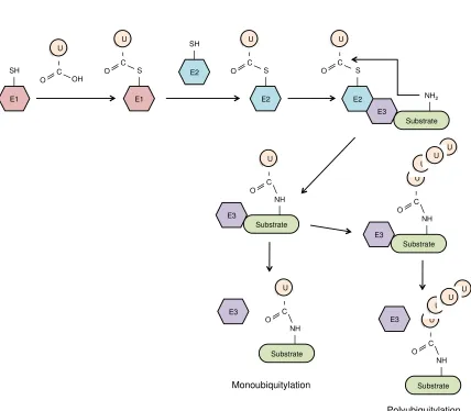
Substrate proteins can be modified by a single ubiquitin moiety (monoubiquitylation) or can be tagged with a chain of ubiquitin (polyubiquitylation). Ubiquitin chains are linked together via the formation of an isopeptide bond between the C-terminal glycine residue of one ubiquitin and one of seven internal lysine residues of a second ubiquitin moiety. Notably, ubiquitin chains linked through distinct Lys residues have different cellular functions. Lys48 ubiquitin chains mark proteins for degradation by the 26S proteasome, whereas Lys63, other Lys-based ubiquitin chains, and monoubiquitylation often serve non-proteolytic functions. Depending on the substrate proteins and enzymes involved, these modifications can serve as signals for diverse cellular processes, including DNA repair, transcription, signal transduction, and/or intracellular trafficking 127. Thus far, most well-characterized TRIM proteins have been shown to facilitate either Lys48 or Lys63 polyubiquitylation. Many TRIM proteins can also undergo self-ubiquitylation, including TRIM5α (both huTRIM5α and rhTRIM5α proteins), which induces its own proteasomal degradation and rapid turnover. Several groups have proposed that self-ubiquitylation of rhTRIM5α indirectly leads to the proteasomal degradation of HIV-1 virions. While the precise mechanism of TRIM5α-mediated restriction is still not fully understood, some studies have shown that HIV-1 restriction is dependent on TRIM5α’s E3 ubiquitin ligase activity 43,45,46,128–131.
Figure 1.4: The RING E3 ligase-mediated ubiquitylation pathway.
Figure 1.4
E1 SH U C OH O E1 S U CO E2
SH E2 S U C O Substrate E3 E2 S U C O NH2 Substrate E3 NH U C O Substrate E3 NH U C O U U U Monoubiquitylation Polyubiquitylation Substrate NH U C O U U U Substrate NH U C O E3 E3
proteins are activated and conjugated to target proteins using a distinct set of enzymes; however, the overall process is analogous to ubiquitylation 135. Notably, unlike other SUMO E3 ligase proteins, the SUMO E3 ligase activity of TRIM proteins is dependent on both RING and BB domains 132. Some TRIM family members have been shown to undergo SUMO modification themselves (self-sumoylation) and several TRIM proteins contain SUMO-interacting motifs (SIMs). SIMs bind to other proteins that have been modified with SUMO. Interestingly, two SIMs in huTRIM5α and rhTRIM5α play an important role in N-MLV and HIV-1 restriction 136–139.
Following the RING finger domain, TRIM proteins contain one or two BB domains. Similar to the RING domain, BB domains are zinc binding motifs with a number of conserved cysteine and histidine residues; however, unlike the RING domain, BBs are found exclusively in the TRIM family. There are two types of BBs, BB1 and BB2, that share similar but distinct consensus motifs. When both BB are present in TRIMs, BB1 always precedes BB2. If only one BB is present it is always BB2 68,113,120. Structural
studies of several human TRIM proteins have revealed that TRIM BBs have a similar ternary structure as the RING finger domain 140–142. Studies on the BB2 domains of human TRIM63 (MuRF1) and TRIM5α identified two unusual conserved clusters of hydrophobic residues on the surface of the domain. Two hydrophobic clusters that are flanked by a number of charged residues were identified on the surface of the BB2 domain of TRIM5α. A number of these residues (e.g. W115, L116, and R119) were required for major TRIM5α functions, including TRIM5α turnover, higher order self-association, formation of cytoplasmic bodies, HIV-1 capsid binding, and/or HIV-1 restriction 143. TRIM63 had a similar cluster of solvent-exposed hydrophobic residues located at the surface of its BB2 domain, which form a dimer interface and mediate TRIM63 self-association 142.
higher-molecular-weight complexes that among other functions, define specific subcellular structures. For example, the CC domain of TRIM19 (also referred to as promyelocytic leukemia protein [PML]) is essential for the proper assembly and maintenance of macromolecular called nuclear bodies (NB). Moreover, the CC domain of TRIM5α facilitates TRIM5α trimerization. Mutations in CC that disrupt TRIM5α trimerization impair TRIM5α-mediated HIV-1 restriction 144–147. Interestingly, it has been proposed that the pleiotropic effects of TRIM proteins may be due to the ability of the CC to facilitate diverse homomeric and heteromeric interactions 120,148.
1.2.3
The SPRY and PRY/SPRY (B30.2) domains
The SPRY and PRY/SPRY (B30.2) domains are the most common C-termini found in TRIM family proteins. The evolutionarily ancient SPRY domain (~140 amino acids) is present alone or fused to a related domain called the PRY domain (~60 amino acids), which always precedes the SPRY domain. Unlike the SPRY-only domain, the fused PRY/SPRY domain is only found in vertebrates and PRY/SPRY-containing proteins (including TRIMs) have expanded rapidly during vertebrate evolution. The reasons for this expansion are still unclear; however, it has been proposed that the PRY/SPRY domain has been selected and maintained in vertebrates as a component of immune defense 68,149. Indeed, the PRY/SPRY domain in TRIM proteins is often critical for TRIM-mediated virus inhibition. For example, rhTRIM5α interacts with the HIV-1 capsid protein via its PRY/SPRY domain and both PRY and SPRY portions of the domain are necessary for this interaction. Furthermore, the PRY/SPRY domain in the TRIM25 protein, which activates the RIG-I signaling cascade, is both necessary and sufficient for its interaction with RIG-I. These PRY/SPRY-mediated protein-protein interactions are required for both rhTRIM5α and TRIM25 to execute their antiviral functions 40,122,150–153.
families that contain proteins involved in ubiquitylation processes. As mentioned in the previous section, the TRIM family also contains a sizeable number of proteins that are involved in ubiquitylation. Thus, although SPRY and PRY/SPRY-containing proteins have diverse biological functions, it has been proposed in the literature that they may function primarily as target substrate recognition modules for E3 ubiquitin ligases 154,155. This is consistent with experimental evidence for some members of the TRIM family, such as TRIM27, which interacts with the NOD2 (nucleotide-binding oligomerization domain-containing protein 2) protein through its PRY/SPRY domain and subsequently facilitates NOD2 polyubiquitylation and degradation using its RING domain 156. However, in the majority of cases, the mechanistic details and/or interacting substrate proteins required for TRIM-mediated biological activities are unknown.
Several SPRY and PRY/SPRY domain structures have now been resolved, including a limited number of structures in complex with their binding partners. These studies have revealed that the SPRY and PRY/SPRY domain structures are extremely versatile and can interact with diverse ligands 157,158. The core fold of the PRY/SPRY domain is a bent β-sandwich comprised of two antiparallel β sheets. The majority of conserved residues are located in the hydrophobic core between the antiparallel β sheets, whereas loops of variable length and sequence protrude from the core β-sandwich. In many PRY/SPRY-containing proteins, these variable loops form protein binding surfaces that determine substrate binding specificity 154,155,159–162. For example, four variable (v) loops in the recently solved structure of the rhTRIM5α PRY/SPRY domain comprise the HIV-1 capsid binding site. The binding surface is dominated by one variable loop (v1) that is highly flexible and interacts weakly with multiple capsid epitopes. Interestingly, the authors of this study suggests that capsid recognition by rhTRIM5α may function in a similar manner as IgM-mediated antigen recognition 163.
1.1.3
Antiviral activity
replication of diverse retroviruses, including HIV-1 and N-MLV. A comprehensive screen for antiretroviral activity of 55 human and mouse TRIMs revealed that 19 additional TRIM proteins inhibit the entry or release of HIV-1, MLV, and/or avian leucosis virus. Interestingly, unlike TRIM5α, most of the additional TRIM proteins inhibited late stages of the viral life cycle 69. Multiple TRIM proteins have also been shown to have antiviral activity against hepatitis B virus (HBV). These TRIM proteins have been shown to significantly reduce the HBV transcription in HepG2 cells 164,165. Some TRIM proteins, such as TRIM19/PML, have been implicated in the inhibition of additional viruses, including herpes simplex virus, human cytomegalovirus, vesicular stomatitis virus, and influenza A virus (IAV) 166. Recently, TRIM56 was shown to inhibit the replication of bovine viral diarrhea virus in vitro and mouse-specific TRIM79α was
identified as a potent inhibitor of tick-borne encephalitis virus 167,168. In addition,
TRIM21 was previously shown to act as an intracellular IgG receptor that neutralizes antibody-coated virus in the cytoplasm by targeting it for degradation by the 26S proteasome 169. These studies suggest that TRIM family proteins restrict evolutionarily diverse viruses and target a variety of stages in the viral replication life cycle.
1.2 The TRIM22 protein
1.2.1
Origins and evolution
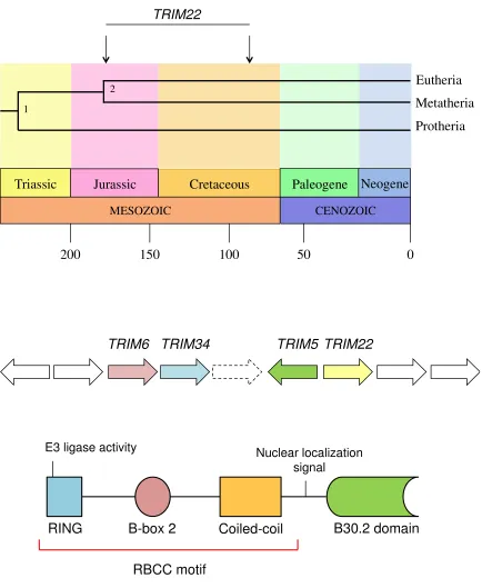
Human TRIM22 is located on chromosome 11 within a cluster of closely-related TRIM
genes that also includes TRIM5, TRIM6, and TRIM34 171,172. The origins of the entire TRIM5/6/22/34 gene cluster can be traced back to the Cretaceous period, or more
specifically, to approximately 90-180 million years ago (Fig 1.5). Studies have shown that TRIM5/6/22/34 is absent in Metatherian (marsupial) mammals (e.g. opossum and
chicken), but present in all major Eutherian (placental) groups (e.g. cow, dog, and human) 172. As such, the TRIM5/6/22/34 gene cluster must have emerged after the
divergence of Metatherian and Eutherian mammals, but before the separation of major Eutherian groups. Of interest, several groups have proposed that TRIM5/6/22/34 likely
arose through tandem gene duplication, as TRIM5, TRIM6, TRIM22, and TRIM34 are
close human paralogs, and because major gene re-arrangements have been observed in this chromosomal region 108,172,173. Gene duplication plays an important role in evolution
and a number of TRIM genes have been shown to undergo gene duplication in both
primates and teleost fish 108,174.
Within the TRIM5/6/22/34 gene cluster, TRIM22 and TRIM5 have an interesting and
dynamic evolutionary relationship. In some Eutherian groups, such as cow, there are multiple copies of the TRIM5 gene but no TRIM22 gene. However, in other Eutherian
groups, such as dog, the TRIM22 gene is present but the TRIM5 gene is not 172. In
addition, TRIM22 and TRIM5 have evolved in a mutually exclusive manner, whereby
positive selection has acted on either TRIM22 or TRIM5 (but not both) in different
primate lineages. This striking anti-correlative pattern of evolution is thought to occur due to tight genetic linkage between the two genes 172. Both TRIM22 and TRIM5 are
classified as Subgroup G (also Group 2) TRIM genes according to the most recent
phylogenetic-based classification system (TRIM6 and TRIM34 are also Subgroup G,
Figure 1.5: TRIM22 evolution, genomic organization, and protein structure.
An approximate timeline of Metatherian and Eutherian mammalian evolution is shown on the top panel. The estimated time (~90-180 million years ago) that TRIM22 (and the
rest of the TRIM5/6/22/34 gene cluster) emerged in Eutherian mammals is indicated by
two arrows. The middle panel shows the genomic organization of the TRIM5/6/22/34
Figure 1.5
MESOZOIC
Triassic Jurassic Cretaceous
CENOZOIC
Neogene Paleogene
Protheria Metatheria Eutheria
200 150 100 50 0
2 1
TRIM22
TRIM22 TRIM5
TRIM34 TRIM6
RBCC motif
RING B-box 2 Coiled-coil B30.2 domain
E3 ligase activity Nuclear localization
signal
1.2.2
Protein structure
Similar to other Subgroup G (and Group 2) members, the TRIM22 protein is comprised of an N-terminal RBCC motif that includes RING, BB2, and CC domains and a C-terminal B30.2 domain (Figs. 1.3 and 1.5). The RING domain of TRIM22 has been shown to possess both E3 ubiquitin and E3 SUMO ligase activity 132,175. TRIM22’s E3 ubiquitin ligase activity is dependent on two catalytic cysteine (Cys) residues (i.e. Cys15 and Cys18) in its RING domain. These Cys residues help stabilize the zinc finger motif and facilitate the transfer of ubiquitin to the appropriate substrate protein 113,117. TRIM22 also mediates its own ubiquitylation and 26S proteasomal degradation when combined with the E2 conjugating enzyme UbcH5B (also referred to as UBC2D2) 175. TRIM22
inhibits viral replication by both E3 ubiquitin ligase dependent and independent mechanisms. For example, TRIM22’s E3 ubiquitin ligase activity is required for TRIM22-mediated inhibition of HBV, IAV, and EMCV 164,176,177. However, HIV-1
inhibition occurs in both the presence and absence of TRIM22’s E3 ubiquitin ligase activity 178,179.
proteins. While TRIM22 has been shown to undergo trimerization, the biological and antiviral significance of TRIM22 trimerization is unknown 112.
The TRIM22 protein contains a C-terminal B30.2 (PRY/SPRY) domain. Although the structure of this domain has not been resolved for TRIM22, several domain-deletion studies have shown that it is integral for many TRIM22 functions. For example, one study showed that B30.2 mutants of TRIM22 were no longer able to inhibit HBV replication and localized exclusively to the cytoplasm in HepG2 cells 164. Several additional studies have also reported the TRIM22’s B30.2 domain is necessary for its nuclear localization and the formation of nuclear bodies (NB), including a study that showed that amino acids 491-494 are critical for nuclear localization and amino acids 493-494 are critical for NB formation 184,185. TRIM22’s B30.2 domain may also be
required for EMCV restriction, self-ubiquitylation, and/or the ubiquitylation of other substrate proteins. One study that investigated TRIM22-mediated EMCV restriction showed that a C-terminal TRIM22 mutant lacking both CC and B30.2 domains was unable to inhibit EMCV replication or facilitate ubiquitylation of target proteins even though its RING domain was still intact and bound to the ubiquitylated E2 enzyme 176. An N-terminal TRIM22 mutant lacking both RING and BB2 domains lost the ability to undergo self-ubiquitylation, suggesting that the B30.2 domain of TRIM22 may be ubiquitylated by its own RING finger domain 176. While the role of TRIM22’s B30.2 domain in HIV-1 restriction is still unclear, rhTRIM5α’s B30.2 domain is required for HIV-1 restriction 146. Moreover, several hyper-variable regions (v1-v4) in the B30.2 domain of rhTRIM5α form the binding surface for HIV-1 capsid and confer virus specificity for TRIM5α-mediated restriction of retroviruses 186. The B30.2 domain of TRIM22 also contains these hyper-variable regions (v1-v4); however, it is unknown whether they play a role in its antiviral activity or specificity.
1.2.3
Induction and expression
Table 1.2
Table 1.2: Factors that alter TRIM22 protein expression levels.
Change Cell type and/or Tissue 1
Cytokines
IFN-α Increase Primary MDM; CEM, Jurkat, THP-1, H9, HepG2, U937 U-937-4, Daudi,
and HeLa cells
IFN-β Increase HOS, Daudi, and HeLa cells
IFN- Increase HeLa, HepG2, and MCF7 cells
IL-1-β Increase Coronary artery endothelium
IL-2 Increase CD4+, CD8+, NK cells
IL-15 Increase CD4+, CD8+, NK cells
Progesterone Increase ABC28 and T47D cells
TNF-α Increase Coronary artery endothelium
Antigens/Infection
EBV infection Increase BL41-EBV cells
EBV LMP-1 Increase DG75 cells
HBV infection Increase Liver tissue
HCV infection Increase Liver tissue
HIV-1 infection Increase Immature DCs, Primary MDMs
HIV-1 Tat Increase Immature DCs
HPV infection Decrease Human keratinocytes
KSHV infection Increase KSHV lesion
LPS Increase Primary MDMs
Rubella infection Increase ECV304 cells
Activation/Differentiation/Cell cycle
1α,25-dihydroxyvitamine D33 Increase Primary MDMs
Anti-CD2 Increase Primary T cells
Anti-CD2/CD28 Decrease Primary T cells
Anti-CD2/CD28/CD3 Decrease CD4+, CD8+, NK cells
All-trans retinoic acid Increase Primary MDMs; HL60 and NB4 cells
p53 Increase K562 and U-937-4 cells
p73 Increase U-937-4 cells
Pioglitazone Increase Primary MDMs
UV-irradiation Increase MCF-7 cells
Disease
Systemic Lupus Erythematosus Increase CD4+ T cells from SLE patient
Wilms Tumor Decrease Primary Wilms Tumor
Neuroblastoma Decrease Primary Neuroblastoma
Breast Cancer Decrease Primary Breast cancer and 10 Breast cancer cell lines
1 Please refer to 170for comprehensive list of all references
164,172,178,179,187–193. TRIM22 expression is also induced by several viral antigens (e.g.
Epstein-Barr virus [EBV], HIV-1, HBV, Kaposi’s sarcoma-associated herpes virus [KSHV], and Rubella), cytokines, and hormones. Studies have shown that TRIM22 contains two IFN-stimulated response elements (ISRE-1, ISRE-2) and one IFN-γ activation site (GAS) in its 5’ promoter region. Notably, ISRE-1 and GAS are not required for IFN-γ induction of TRIM22. Instead, induction of TRIM22 by IFN-γ requires ISRE-2 plus six upstream nucleotides (referred to as the 5’ extended ISRE or eISRE) 190. This induction is dependent on the chromatin remodeling enzyme brahma-related gene 1, which recruits IFN regulatory factor 1 (IRF-1) to the eISRE, and histone deacetylase activity, which prevents the proteasomal degradation of IRF-1 192,194. JAK, phosphatidylcholine-phospholipase C, and protein kinase C are also required for induction of TRIM22 by IFN-γ. p300 enhances IFN-γ induced expression of TRIM22 and is also required for the recruitment of RNA polymerase II to the 5’ TRIM22 promoter. IRF-1 binding to eISRE appears to also be required for IFN-α induced and basal TRIM22 expression 195.
1.2.4
Sub-cellular localization
Several determinants of TRIM22 sub-cellular localization have been identified. The TRIM22 protein contains a bipartite nuclear localization signal (NLS) in the Spacer 2 domain (SP2), which was previously shown to be necessary, but not sufficient, for nuclear localization 197. Although the B30.2 domain does not contain a known NLS, multiple groups have shown that it is required for nuclear localization 164,184,196,197. One group in particular showed that the amino acids 491-494 are essential for TRIM22 nuclear localization and that the amino acids 493-494 are critical for the formation of TRIM22 NBs in MCF7 cells 197. Another group reported that the amino acids 395, 396, and 400 are required for the cytoplasmic localization of TRIM22 in COS-7 (African green monkey) cells 196. However, this group did not investigate how these amino acids influence TRIM22 localization in human cells. Notably, some studies that observed cytoplasmic TRIM22 localization used a shorter form of TRIM22 (442 instead of 498 amino acids) that is translated from a 1329 mRNA coding sequence. As such, in these studies the lack of amino acids 491-494 likely contribute to the cytoplasmic localization of TRIM22.
1.2.5
Antiviral function
Human TRIM22 was first identified in 1995 during a search for IFN-induced genes in Daudi cells. Following sequence analysis, which revealed that TRIM22 was highly homologous to the mouse Rpt-1 gene, exogenous TRIM22 expression was shown to
downregulate transcription from the HIV-1 LTR 187,200. Although this experiment was
performed using a luciferase reporter gene instead of the entire HIV-1 genome, it was first to report an antiviral (and anti-HIV-1) function for TRIM22. In 2006, another independent study showed that TRIM22 was highly up-regulated in primary monocyte-derived macrophages (MDM) in response to HIV-1 infection, IFN-α treatment, or stimulation with lipopolysaccharide (LPS). In addition, they showed that exogenous TRIM22 expression inhibited HIV-1 infection by 50-90% in 293T cells and primary MDMs 189. In 2008, Barr and colleagues showed that TRIM22 was a key mediator of type I IFN-induced inhibition of HIV-1 replication 178. Two different methods of HIV-1 inhibition were observed. In HOS and HeLa cell lines, TRIM22 expression inhibited HIV-1 particle production by preventing the trafficking of the Gag polyprotein to the plasma membrane. This effect was dependent on the E3 ligase activity of TRIM22. Because TRIM22 was also shown to interact with Gag it was thought that TRIM22-mediated post-translational modification of Gag may be responsible for altered Gag trafficking. However, to date, TRIM22 has never been shown to modify Gag post-translationally. Unlike in HOS and HeLa cells, in U2OS and 143B cells, TRIM22 inhibited the accumulation of intracellular Gag protein. Although the mechanism of restriction was not identified in these cell lines, several potential explanations were suggested, including inhibition of LTR-driven transcription or degradation of the Gag RNA and/or polyprotein 178. Notably, these experiments provided the first mechanistic data for restriction of HIV-1 replication by TRIM22.
It has since been confirmed that TRIM22 can restrict HIV-1 transcription 179. In 2011,
non-permissive and non-permissive clones. LTR transcription levels in non-non-permissive clones were decreased 7-10 fold compared to permissive clones; however, non-permissive transcription levels were increased when shRNA was used to knockdown TRIM22. Moreover, when TRIM22 was expressed in permissive clones transcription levels decreased to those observed in non-permissive clones. Further examination of these clones revealed that TRIM22 inhibited basal LTR-driven HIV-1 transcription and that these effects were independent of NF-κB binding sites in the LTR, Tat-mediated LTR transactivation, and TRIM22 E3 ligase activity 179.
In addition to these in vitro studies, there is also in vivo evidence to support a role for
TRIM22 as an anti-HIV-1 effector. A 2011 study demonstrated that increased TRIM22 expression was associated with significantly lower viral loads and significantly higher CD4+ T-cell counts in HIV-1 positive individuals in the primary phase of infection 201.
Recently, a follow-up study showed that TRIM22 expression was also associated with significantly lower viral loads in HIV-1 positive individuals in the chronic phase of infection 202. These data suggest that TRIM22 expression contributes to HIV-1 disease progression in infected individuals.
replication. HBV CP inhibition was dependent on TRIM22’s B30.2 domain and its E3 ligase activity, even though a ubiquitylation target was not identified 164. Interestingly, TRIM22 expression is also significantly upregulated during clearance of HBV and hepatitis C virus (HCV) in chimpanzees 203,204. In human HCV infection, TRIM22 is significantly upregulated in the cirrhotic liver of HCV positive individuals and individuals with mild chronic HCV with no fibrosis 205.
1.2.6
Other functions
Several reports have implicated TRIM22 in other biological processes, including cell cycle regulation and cell proliferation/differentiation. One study identified a functional p53 response element in intron 1 of the TRIM22 gene and demonstrated that upon p53
binding this element activates TRIM22 expression. Moreover, the same study showed that overexpression of TRIM22 in U937 cells led to decreased clonogenic growth and that endogenous TRIM22 was upregulated during induced differentiation in NB4 cells
188. A later study investigating TRIM22 expression during hematopoietic differentiation
showed that TRIM22 is highly expressed in CD34+ bone marrow progenitor cells, but declines in mature populations. Notably, although TRIM22 expression was inversely correlated with differentiation in both lineages, its expression pattern differed during erythroid versus granulocytic differentiation. Decreased TRIM22 expression was more pronounced and lasting during erythroid differentiation and undetectable in nucleated erythroid populations 193.
TRIM22 has also been linked to other human diseases, including certain cancers and autoimmune diseases. Two studies have shown that decreased TRIM22 expression is associated with increased progression, relapse, and mortality in cases of Wilms tumor
206,207. In addition, a recent study reported that TRIM22 expression is significantly
p53-inducible in breast cancer cells, suggesting that a defect in p53-mediated TRIM22 regulation was responsible for TRIM22 downregulation in breast cancer 208. Two gene profiling studies have also implicated TRIM22 in the pathogenesis of systemic lupus erythematosus (SLE). The first study showed that TRIM22 was overexpressed in the CD4+ T cells of individuals with active SLE compared to individuals with non-active SLE. The second study also reported that TRIM22 was upregulated in SLE-positive CD4+ T cells and that, along with a number of other IFN-induced genes, TRIM22 was significantly hypomethylated in SLE cells compared to healthy controls 209,210.
1.3 Rationale and Experimental Approach
Genetic variation in immune genes plays an integral role in host susceptibility to and progression of infection and disease. Much of this variation is due to single nucleotide polymorphisms (SNPs), which are defined as single base changes in a DNA sequence. While many SNPs are phenotypically neutral, non-synonymous or amino acid altering SNPs (nsSNP) often have deleterious effects on protein structure and/or function 211–218. Previous studies have shown that nsSNPs do not appear randomly in the genome, but emerge based on genomic location and selective pressures. Since innate immune genes are located at the interface of the microbial environment, they tend to be exposed to a wide range of selective pressures. As such, innate immune genes often contain genetic signatures of positive (directional) and/or balancing selection, whereas most other host genes are dominated by negative (purifying) selection 219–221.
follows that a number of studies on host restriction factors have used an evolutionary approach to identify essential residues in these proteins 62,85,91,99,106,222. In addition, other studies have used in silico nsSNP prediction programs to identify functionally and/or
clinically relevant polymorphisms in innate immune genes 19–24. Further, in silico
methods have also been used to identify important functional regions in these genes (e.g. post-translational modifications) and support evolutionary analyses 229–232. One of the major advantages of performing in silico analyses is that it allows for the systematic
analysis and prioritization of specific functional sites. This is becoming increasingly important given the tremendous number of SNPs in the human genome and the vast amount of genetic data that is generated on a daily basis. Collectively, evolutionary and
in silico analyses provide valuable insight into protein function and are powerful tools
that can help identify key structural and/or functional sites in a protein.
Given the important role played by TRIM22 in multiple biological processes, including the host antiviral response, and the paucity of information about key functional sites in the TRIM22 protein, we conducted an extensive evolutionary and in silico analysis to
identify critical amino acid residues that mediate TRIM22 function. This approach has previously been used to pinpoint specific amino acid residues that are essential for the activities of other host restriction factors, including the APOBEC3G, TRIM5α, BST-2/tetherin, and SAMHD1 proteins. We hypothesized that evolutionary forces have been acting on TRIM22 and have selected for specific amino acids that impact TRIM22
function. To address this hypothesis, we characterized the evolutionary forces acting on
TRIM22 and used a number of in silico methods to delineate and prioritize potential
functional sites in human TRIM22 that may be relevant to its overall antiviral and/or biological functions. We then investigated how various TRIM22 nsSNPs may affect
1.4 References
1. Hoebe, K., Janssen, E. & Beutler, B. The interface between innate and adaptive immunity. Nat. Immunol.5, 971–4 (2004).
2. Akira, S., Uematsu, S. & Takeuchi, O. Pathogen recognition and innate immunity.
Cell124, 783–801 (2006).
3. Brennan, K. & Bowie, A. G. Activation of host pattern recognition receptors by viruses. Curr. Opin. Microbiol.13, 503–7 (2010).
4. Medzhitov, R. & Janeway, C. A. Innate immunity: the virtues of a nonclonal system of recognition. Cell91, 295–8 (1997).
5. Janeway, C. A. & Medzhitov, R. Innate immune recognition. Annu. Rev. Immunol. 20, 197–216 (2002).
6. Yoneyama, M. et al. The RNA helicase RIG-I has an essential function in
double-stranded RNA-induced innate antiviral responses. Nat. Immunol.5, 730–7 (2004).
7. Thompson, M. R., Kaminski, J. J., Kurt-Jones, E. A. & Fitzgerald, K. A. Pattern recognition receptors and the innate immune response to viral infection. Viruses3,
920–40 (2011).
8. Lester, S. N. & Li, K. Toll-Like Receptors in Antiviral Innate Immunity. J. Mol. Biol.426, 1246–1264 (2014).
9. Kawai, T. & Akira, S. Antiviral signaling through pattern recognition receptors. J. Biochem.141, 137–45 (2007).
10. Levy, D. E., Marié, I. J. & Durbin, J. E. Induction and function of type I and III interferon in response to viral infection. Curr. Opin. Virol.1, 476–86 (2011).
11. Platanias, L. C. Mechanisms of type-I- and type-II-interferon-mediated signalling.
Nat. Rev. Immunol.5, 375–86 (2005).
12. Boehm, U., Klamp, T., Groot, M. & Howard, J. C. Cellular responses to interferon-gamma. Annu. Rev. Immunol.15, 749–95 (1997).
13. Randall, R. E. & Goodbourn, S. Interferons and viruses: an interplay between induction, signalling, antiviral responses and virus countermeasures. J. Gen. Virol. 89, 1–47 (2008).
14. Stetson, D. B. & Medzhitov, R. Type I interferons in host defense. Immunity 25,
15. Schneider, W. M., Chevillotte, M. D. & Rice, C. M. Interferon-Stimulated Genes: A Complex Web of Host Defenses. Annu. Rev. Immunol. 32, 140220103029009
(2013).
16. Pincus, T., Hartley, J. W. & Rowe, W. P. A major genetic locus affecting resistance to infection with murine leukemia viruses. Tissue culture studies of naturally occurring viruses. J. Exp. Med.133, 1219–33 (1971).
17. Liu, L. et al. A whole genome screen for HIV restriction factors. Retrovirology8,
94 (2011).
18. Duggal, N. K. & Emerman, M. Evolutionary conflicts between viruses and restriction factors shape immunity. Nat. Rev. Immunol.12, 687–95 (2012).
19. Zheng, Y.-H., Jeang, K.-T. & Tokunaga, K. Host restriction factors in retroviral infection: promises in virus-host interaction. Retrovirology9, 112 (2012).
20. Meylan, P. R., Guatelli, J. C., Munis, J. R., Richman, D. D. & Kornbluth, R. S. Mechanisms for the inhibition of HIV replication by interferonsalpha, beta, and -gamma in primary human macrophages. Virology193, 138–48 (1993).
21. Poli, G., Orenstein, J. M., Kinter, A., Folks, T. M. & Fauci, A. S. Interferon-alpha but not AZT suppresses HIV expression in chronically infected cell lines. Science 244, 575–7 (1989).
22. Agy, M. B., Acker, R. L., Sherbert, C. H. & Katze, M. G. Interferon treatment inhibits virus replication in HIV-1- and SIV-infected CD4+ T-cell lines by distinct mechanisms: evidence for decreased stability and aberrant processing of HIV-1 proteins. Virology214, 379–86 (1995).
23. Fernie, B. F., Poli, G. & Fauci, A. S. Alpha interferon suppresses virion but not soluble human immunodeficiency virus antigen production in chronically infected T-lymphocytic cells. J. Virol.65, 3968–71 (1991).
24. Coccia, E. M., Krust, B. & Hovanessian, A. G. Specific inhibition of viral protein synthesis in HIV-infected cells in response to interferon treatment. J. Biol. Chem. 269, 23087–94 (1994).
25. Shirazi, Y. & Pitha, P. M. Alpha interferon inhibits early stages of the human immunodeficiency virus type 1 replication cycle. J. Virol.66, 1321–8 (1992).
26. Malim, M. H. & Emerman, M. HIV-1 accessory proteins--ensuring viral survival in a hostile environment. Cell Host Microbe3, 388–98 (2008).