! " #
$#$ !% & &$
' ( ))) *
Finding Tumor Volume Using Segmentation from MR image
Prof. Samir Kumar Bandyopadhyay
Professor,
Dept of Computer Science & Engineering, University of Calcutta, 92 A.P.C. Road,
Kolkata , India [email protected]
Abstract: An accurate segmentation is critical, especially when the tumor morphological changes remain subtle, irregular and difficult to assess by clinical examination. Brain tissue and tumor segmentation in MR images has been an active area of research today. The region growing segmentation is used to segment the brain tumors due to its wide range of applications and automatic features. After taking the image of the tumorous brain there is a need to process it. The image clearly shows the place of the tumorous portion of the brain. The image does not give the information about the numerical parameters such as area and volume of the tumorous portion of the brain. After segmentation the desired tumor area is selected from the segmented image. An approach to segmentation is proposed that removes the adverse effect on the boundary, which is unwanted especially from the point of view of volume rendering. This approach gives more accurate boundary detection and holes filling after segmentation. Volume of the tumor is calculated for the decision to be taken by the doctors.
Keywords: Edge Detection, Brain Tumor Segmentation, Magnetic resonance imaging and Region Growing Segmentation
I. INTRODUCTION
Image processing is a rapidly growing area of computer science. Its growth has been based by technological advances in digital imaging, computer processors and mass storage devices. Fields which traditionally used analog imaging are now switching to digital systems, for their flexibility and affordability. Important examples are medicine, and video production, photography, remote sensing, and security monitoring. These and other sources produce huge volumes of digital image data every day, more than could ever be examined manually. Digital image processing is concerned primarily with extracting useful information from images. Ideally, this is done by computers, with little or no human intervention.
MRI is often the medical imaging method of choice when soft tissue delineation is necessary. This is especially true for any attempt to segment brain tissues, normal or abnormal. Image segmentation is a tool that has been applied to medical imaging modalities to differentiate tissue types for purposes of volume measurement and visualization.
Image processing algorithms may be placed at three levels. At the lowest level are those techniques which deal directly with the raw, possibly noisy pixel values, with denoising and edge detection being good examples. In the middle are algorithms which utilise low level results for further means, such as segmentation and edge linking. At the highest level tumor is isolated from the region for further processing.
Automated detection of the abnormalities in medical images is an important and sometimes necessary procedure in medical diagnostics, planning, and treatment. While
detection of abnormalities such as tumors is possible by experts, manual segmentation is usually tedious and time consuming, and subject to error. There are many methods that find a tumor in MR images semi-automatically. In such methods, human intervention is required, which again makes the process time consuming and expensive. The critical problem is finding the tumor location automatically and later finding its boundary precisely. An important factor in detecting tumor from healthy tissues is the difference in intensity level. However, relying only on the intensity level is usually not enough. The spatial information available in clusters of pixels that form a tumor should also be used in the detection process [1-2].
Tumor pixels are usually geometrically connected, but tumor edges are of lower intensity than its center. To identify the boundary of the tumor in the Region of Interest (ROI), we use region growing to detect tumor pixels. For this purpose, we need to find the threshold between the tumor and its surrounding. In this paper next section discussed about currents methods and subsequent section proposes a method to find out tumor by segmentation. The method shows good results and can be applicable to other areas of medical science.
II. REVIEW WORKS
extract important features from the image data, from which a description, interpretation, or understanding of the scene can be provided by the machine. The segmentation of brain tumor from magnetic resonance images is an important but time-consuming task performed by medical experts The digital image processing community has developed several segmentation methods. and necrotic tissues. These types of algorithms are used for dividing the brain images into three categories (a) Pixel based (b) Region or Texture Based (c) Structural based.
Thresholding method is frequently used for image segmentation. This is simple and effective segmentation method for images with different intensities. [6] The technique basically attempts for finding a threshold value, which enables the classification of pixels into different categories. A major weakness of this segmentation mode is that: it generates only two classes. Therefore, this method fails to deal with multichannel images.
Besides, it also ignores the spatial characteristics due to which an image becomes noise sensitive and undergoes intensity in-homogeneity problem, which are expected to be found in MRI. Both these features create the possibility for corrupting the histogram of the image. For overcoming these problems various versions of thresholding technique have been introduced that segments medical images by using the information based on local intensities and connectivity [7]. Though this is a simple technique, still there are some factors that can complicate the thresholding operation, for example, non-stationary and correlated noise, ambient illumination, busyness of gray levels within the object and its background, inadequate contrast, and object size not commensurate with the scene. [8].
Researchers introduced a new image thresholding method based on the divergence function. In this method, the objective function is constructed using the divergence function between the classes, the object and the background [9]. The required threshold is found where this divergence function shows a global minimum.
Due to high reliability and accurate measurement of the dimensions and location of tumor [10], MRI is frequently used for observing brain pathologies. Previously, region growing and shape based methods were heavily relied upon for observing the brain pathologies.
[11] Proposed a Bayes-based region growing algorithm that estimates parameters by studying characteristics in local regions and constructs the Bayes factor as a classifying criterion. The technique is not fully automatic, i.e. it requires user interaction for the selection of a seed and secondly the method fails in producing acceptable results in a natural
image. It only works in homogeneous areas. Since this technique is noise sensitive, therefore, the extracted regions might have holes or even some discontinuities [1, 7].
Shape based method provides an alternative approach for the segmentation of brain tumor. But the degree of freedom for application of this method is limited too. The algorithm demands an initial contour plan for extracting the region of interest. Therefore, like region growing approach, this method is also semiautomatic. Both of these methods are error sensitive because, an improper or false description of initial plan and wrong selection of the seed image will lead to disastrous results. Statistical methods and fuzzy logic approaches seems to be reliable and are the best candidates for the replacement of the above mentioned techniques.
Supervised and un-supervised methods for image processing are frequently applied [3, 12-14]. An Attempt has been made to segment the volume as a whole using KNN and both hard and fuzzy c-means clustering [13]. Results showed, however, that there appears to be enough data non-uniformity between slices to prevent satisfactory results. Supervised classification enables us to have sufficient known pixels to generate representative parameters for each class of interest. In an un-supervised classification pre hand knowledge of classes is not required. It usually uses clustering algorithm for classifying an image data.
III. PROPOSED METHOD
Segmentation of images holds an important position in the area of image processing. It becomes more important while typically dealing with medical images where pre-surgery and post-pre-surgery decisions are required for the purpose of initiating and speeding up the recovery process [5]. Computer aided detection of abnormal growth of tissues is primarily motivated by the necessity of achieving maximum possible accuracy.
Manual segmentation of these abnormal tissues cannot be compared with modern day’s high speed computing (CSF) and sometimes pathological tissues like tumor etc.
After the brain image was processed by the Homogeneity nature of brain we obtain an image that is homogenous and have pixels in gray scale of sixteen level scales. This image forms the input for the next level of processing for detection of the edge, i.e. brain boundary of both sides. In this paper we initially find out the edge using a window of size by end pixels of both horizontal and vertical boundaries of the input image
Output: Horizontal Edge (horM), Vertical Edge (verM), Edge Map (resM)
Begin:
Step1. Open MR Image
Step2. Loop row=0 to MRI.Height Loop col=0 to MRI.Width
val= MRI.Pixel[row,col] AND 000000ff k= col+1
Loop k to MRI.Width
kval = MRI.Pixel[row,k] AND 000000ff If kval <> val
Break EndIf k=k+1 End Loop
horM.Pixel[row,k-1]=ff000000 col=k
End Loop row=row+1 End Loop
Step3. Loop col=0 to MRI.Width Loop row=0 to MRI.Height
val= MRI.Pixel[row,col] AND 000000ff k= row+1
Loop k to MRI.Height
kval = MRI.Pixel[k,col] AND 000000ff If kval <> val
Break EndIf k=k+1 End Loop
verM.Pixel[k-1,col]=ff000000 row=k
End Loop col=col+1 End Loop
Step4. Loop row=0 to MRI.Height Loop col=0 to MRI.Width
Bval=horM.Pixel[row,col] AND 000000ff Cval=verM.Pixel[row,col] AND 000000ff If Bval = 255 AND Cval = 255
resM.Pixel[row,col]= ffffffff Else
resM.Pixel[row,col]= ff000000 EndIf
col=col+1 End Loop row=row+1 End Loop
Step5. End
To further evaluate the performance of our method, we have considered noisy images. Gaussian white noise with mean zero and variance 0.02 has been added to the images. The noisy image and enhanced image are shown in figure 1 and
figure 2.
Figure 1 Noisy Image
Figure 2 Enhanced Image
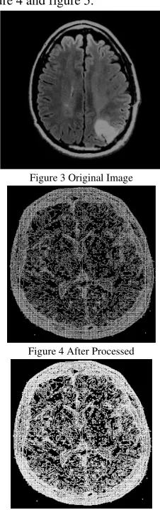
The tumor is in a cluttered area and it is hard to see its boundary. Force is able to detect the tumor area, as one of the centroids moves inside the tumor. It is difficult to determine the tumor area more precisely. These are shown in figure 3, figure 4 and figure 5.
Figure 3 Original Image
Figure 4 After Processed
We start by locating all the edge paths that originates from the first row of the image. The algorithm travels each individual path and find out the changed value of pixel and stores them in an one dimensional list. This list is plotted on another image if the edge path is circular or end on the last row of the image or the vertical line representing the left boundary of the brain. After all pixel paths on the first row are traversed the algorithm repeats similar scanning and traversal of all pixel paths from the row that is indicated by dividing the image vertically into sixteen segments. The first row of each segment is used for locating pixel paths for traversal. All complete paths are then plotted on another image thus providing the anatomical regions of the brain. Algorithm to detect the Segmentation regions of Brain: Step !:Scan the image from the Left side of the image to locate the leftmost pixel of the brain region. Draw a vertical line along this pixel from top to bottom representing the Left baseline or boundary.
Step 2:Scan the image from the right side of the image to locate the rightmost pixel of the brain region.
Step 3: Draw a vertical line along this pixel from top to bottom.
Step 4: Partition the obtained rectangle horizontally into sixteen segments and start with the first row of the first segment.
Step 5: Scan the enclosed rectangle from the right side to left, from the first row of the segment.
Step6 :Obtain a pixel that is black indicating an edge path, traverse the pixel path by considering all the surrounding pixels in a clockwise priority and consider the pixel with the highest priority.
Step7: The pixels that surrounded the edge pixel, but are of lower priority are stored in a Backtrack Stack to be used only if the traversal process reaches a dead end.
If a dead end is reached, pop out from the Backtrack stack a lesser priority pixel and continue with the traversal process. Step 8: Traversal continues to the next pixel till it reaches the left baseline or the bottom of the image or the start position is reached.
Step 9: If the traversal is terminated, the plotting list is erased and continues from Step7
Else plot pixels from the Plotting List.
Step 10: Continue to Step6 till all black pixels, indicating an edge path, is traversed.
Step 11: Move to the first row of the next segment and continue from Step6 to Step11.
Step 12: If all rows are considered then stop.
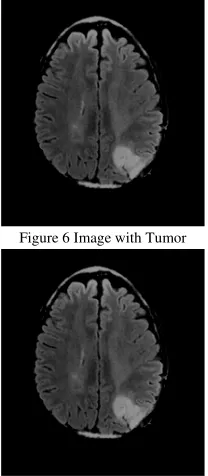
Figure 6 Image with Tumor
Figure 7 Centroids on original image After higher intensity levels
Figure 8 Segmentation of noisy image
Figure 9 Extraction of tumor
The above figures from figure 6-figure 9 were calculated and isolated brain tumor. The first stage is to determine the edge input image labels and the number of pixels in each label. The second stage is to determine the segmented output requested region to get total number of pixels accessed. From the whole slices we can calculate the number of pixels into the 3D volume. Segmented areas are automatically calculated and multiplied by the MRI slice thickness plus the inter slice gap to calculate a per- slice tumor volume. The total tumor volume is obtained by summing the tumor bearing slices [18-19].
In this paper, a fully automated tumor detection method based on Force algorithm is proposed. “Force” is an unsupervised data clustering method which has been modified here to be used as a method to find the region that contains tumor in brain MRI. The proposed method has three steps of preprocessing, coarse tumor detection using Force, and fine tumor detection. The performance of this method has been evaluated; and it is shown that it is robust and able to find the tumor boundaries in MR images with different intensity, dynamic range, noise, tumor size and shape. The proposed clustering algorithm can be employed in 3D space with no change. Therefore, the entire tumor detection approach is extendable to 3D. The clustering method can also be applied to other medical images, e.g., brain or liver MRI.
V. REFERENCES
[1] Bezdek, J.C., Hall, L.O., Clarke, L.P.: Review of MR image segmentation techniques using pattern recognition. Medical physics 20(4), 1033–1048 (1993) [2] Held, K., Rota, K.E., Krause, B.J., Wells III, W.M.,
Kikinis, R., Muller-Gartner, H.W.:Markov random field segmentation of brain MR images. IEEE Trans. Med. Imaging, 878– 886 (1997)
[3] Clarke, L., et al.: MRI segmentation: Methods and applications. Magnetic Resonance Imaging 13(3), 343– 368 (1995)
[4] Cuadra, M.B., Gomez, J., Hagmann, P., Pollo, C., Villemure, J.G., Dawant, B.M., Thiran, J.: Atlas-based segmentation of pathological brains using a model of tumor growth. In: Medical Image Comp.& Computer-Assisted Intervention MICCAI (2002), pp. 380– 387.Springer, Heidelberg (2002)
[5] Clark, M.C., Hall, L.O., Goldgof, D.B., Velthuizen, R., Murtagh, F.R., Silbiger, M.S.:Automatic tumor-segmentation using knowledge-based techniques. IEEE Trans. on Medical Imaging 17, 187–201 (1998) [6] Kaus, M.R., Warfield, S.K., Nabavi, A., Chatzidakis,
E., Black, P.M., Jolesz, F.A.,Kikinis, R.: Segmentation of meningiomas and low grade gliomas in MRI. In: Taylor, C.,Colchester, A. (eds.) MICCAI 1999. LNCS, vol. 1679, pp. 1–10. Springer, Heidelberg (1999) [7] Prastawa, M., Bullitt, E., Ho, S., Gerig, G.: A Brain
Tumor Segmentation FrameworkBased on Outlier Detection. Medical Image Analysis 8, 275–283 (2004) [8] M. Mancas, B. Gosselin, B. Macq, 2005, "Segmentation
Using a Region GrowingThresholding", Proc. of the Electronic Imaging Conference of the International Society for Optical Imaging (SPIE/EI 2005), San Jose (California, USA).Dong-yong Dai; Condon, B.; Hadley, D.; Rampling, R.; Teasdale, G.; "Intracranial
deformation caused by brain tumors: assessment of 3-D surface by magnetic resonance
[9] imaging"IEEE Transactions on Medical Imaging Volume 12, Issue 4, Dec. 1993 Page(s):693 – 702 [10] Matthew C. Clark “Segmenting MRI Volumes of the
Brain With Knowledge- Based Clustering” MS Thesis, Department of Computer Science and Engineering, University of South Florida, 1994
[11] Dzung L. Pham, Chenyang Xu, Jerry L. Prince;"A Survey of Current Methods in Medical Medical Image Segmentation" Technical Report JHU / ECE 99-01, Department ofElectrical and Computer Engineering. The Johns Hopkins University, Baltimore MD 21218, 1998.
[12] M. Sezgin, B. Sankur " Survey over image thresholding techniques and quantitativeperformance evaluation" J. Electron. Imaging 13 (1) (2004) 146-165.
[13] Chowdhury, M.H.; Little, W.D,;"Image thresholding techniques" IEEE Pacific RimConference on Communications, Computers, and Signal Processing, 1995. Proceedings.17-19 May 1995 Page(s):585 – 589 [14] Zhou, J.; Chan, K.L.; Chong, V.F.H.; Krishnan, S.M
“Extraction of Brain Tumor from MR Images Using One-Class Support Vector Machine” 27th Annual International Conference of the Engineering in Medicine and Biology Society, 2005. IEEE-EMBS 2005,Page(s):6411 – 6414
[15] Pan, Zhigeng; Lu, Jianfeng;;"A Bayes-Based Region-Growing Algorithm for MedicalImage Segmentation" Computing in Science & Engineering, Volume 9, Issue 4, July-Aug.2007 Page(s):32 – 38
[16] J. C. Bezdek, L. O. Hall, L. P. Clarke "Review of MR image segmentation techniquesusing pattern recognition. " Medical Physics vol. 20, no. 4, pp. 1033 (1993).
[17] Velthuizen RP, Clarke LP, Phuphanich S, Hall LO, Bensaid AM, Arrington JA, Greenberg HM and Silbiger ML. "Unsupervised Tumor Volume Measurement Using Magnetic Resonance Brain Images," Journal of Magnetic Resonance Imaging , Vol. 5, No. 5, pp. 594-605, 1995.
[18] M. Sato, S. Lakare, M. Van and A. Kaufaman, “A gradient magnitude based region growing algorithm for accurate segmentation”, Proceeding of international conference onimage processing, vol. 3, pp. 448-451, 2000.