1556-6811/10/$12.00 doi:10.1128/CVI.00408-09
Copyright © 2010, American Society for Microbiology. All Rights Reserved.
Optimization and Application of a Multiplex Bead-Based Assay To
Quantify Serotype-Specific IgG against
Streptococcus pneumoniae
Polysaccharides: Response to the Booster Vaccine after
Immunization with the Pneumococcal 7-Valent
Conjugate Vaccine
䌤
Karin E. M. Elberse, Irina Tcherniaeva, Guy A. M. Berbers, and Leo M. Schouls*
Laboratory for Infectious Diseases and Perinatal Screening, National Institute for Public Health and the Environment, Antonie van Leeuwenhoeklaan 9, Bilthoven 3721 MA, Netherlands
Received 12 October 2009/Returned for modification 12 November 2009/Accepted 27 January 2010
We describe the optimization and application of a multiplex bead-based assay (Luminex) to quantify antibodies against polysaccharides of 13 pneumococcal serotypes. In the optimized multiplex immunoassay (MIA), intravenous immune globulin was introduced as an in-house reference serum, and nonspecific reacting antibodies were adsorbed with the commercial product pneumococcal C polysaccharides Multi. The antibody concentrations were assessed in 188 serum samples obtained pre- and post-booster vaccination at 11 months after administration of a primary series of the pneumococcal seven-valent conjugate vaccine (PCV-7) at 2, 3, and 4 months of age. The results of the MIA were compared with those of the ELISA for the serotypes included in the seven-valent conjugated polysaccharide vaccine and for a non-vaccine serotype, serotype 6A. The geometric mean concentrations of the antibodies determined by MIA were slightly higher than those deter-mined by ELISA. The correlations between the assays were good, withR2
values ranging from 0.84 to 0.91 for all serotypes except serotype 19F, for whichR2
was 0.70. The concentrations of antibody against serotype 6A increased after the administration of PCV-7 due to cross-reactivity with serotype 6B. The differences between the results obtained by ELISA and MIA suggest that the internationally established protective threshold of 0.35
g/ml should be reevaluated for use in the MIA and may need to be amended separately for each serotype.
In 2006, the pneumococcal 7-valent conjugate vaccine (PCV-7; Prevenar; Wyeth Vaccines, Pearl River, NY) was in-troduced into the National Immunization Program (NIP) in the Netherlands. The vaccine is administered to children at the ages of 2, 3, and 4 months and a booster is given at 11 months of age. During the prevaccination era, the seven serotypes covered by the vaccine accounted for approximately 60% of the cases of invasive pneumococcal disease among children 0 to 4 years in age in the Netherlands (14). Recently, new pneu-mococcal conjugate vaccines that protect against more sero-types than PCV-7 have been developed, including a 10-valent vaccine (PCV-10; Synflorix; GSK, Middlesex, United King-dom) and a 13-valent vaccine (PCV-13; Wyeth Vaccines) It is anticipated that one of these vaccines will replace the current PCV-7.
Efficacy and immunogenicity studies with PCV-7 demon-strated that it has 97% efficacy against invasive disease (3). A Finnish trial evaluated the efficacy of the vaccine against otitis media and found 57% efficacy against the vaccine serotypes (9). Most immunogenicity studies were performed by enzyme-linked immunosorbent assay (ELISA) (3, 4, 6), the “gold stan-dard” for quantifying the concentrations of antibodies to
pneu-mococcal serotype-specific polysaccharides. In 2000, guidelines for the pneumococcal ELISA were described in an interna-tional standard protocol, referred to as the WHO protocol (http://www.vaccine.uab.edu). This protocol was initially devel-oped for evaluation of the immunogenicities of pneumococcal vaccines. On the basis of a meta-analysis of the results of the various vaccine trials (3, 8, 12, 15, 19), WHO recommends the use of an antibody concentration of 0.35g/ml as a correlate of protection if it is assessed by ELISA (29). This recommended protective concentration is identical for all serotypes.
The standard reference serum used in the ELISA is lot 89S serum. This comprises a pool of serum samples from 17 adults immunized with the 23-valent pneumococcal polysaccharide vaccine (23). Quantification of the serotype-specific IgG con-centrations present in lot 89S was performed by an antibody-capture reference ELISA (24, 25). This serum can be obtained through the FDA; however, the supply of this serum is finite, and the current stock is running low. A replacement for the current reference serum, 007sp, is currently being produced and characterized (10).
The polysaccharides used in the ELISA also contain cell wall polysaccharides (CWPSs) that are covalently bound to the serotype-specific capsular polysaccharide by peptidoglycan (5, 28). Antibodies reacting to CWPSs are present in serum sam-ples, but these antibodies do not offer functional protection against the pneumococcus and may cause a nonspecific signal in the assay (18, 30). To remove CWPS antibodies that may react with CWPS in the ELISA, CWPS is added as a sorbent
* Corresponding author. Mailing address: Laboratory for Infectious Diseases and Perinatal Screening, National Institute for Public Health and the Environment, Antonie van Leeuwenhoeklaan 9, Bilthoven 3721 MA, Netherlands. Phone: 31 30 2742121. Fax: 31 30 2744418. E-mail: [email protected].
䌤Published ahead of print on 3 February 2010.
674
on August 17, 2020 by guest
http://cvi.asm.org/
bead-based assay that uses the Luminex technology to quantify antibodies against 13 pneumococcal polysaccharides simulta-neously. The results obtained by this multiplex immunoassay (MIA) were compared with those obtained by ELISA for eight serotypes. In the assay, we used an in-house reference serum, intravenous immune globulin (IVIG). Furthermore, we used the newly available CWPS Multi to adsorb the non-type-spe-cific anti-CWPS antibodies. For evaluation of the performance of the MIA developed, sera from children taken pre- and post-booster vaccination at 11 and 12 months of age were used.
MATERIALS AND METHODS
Serum samples obtained pre- and post-booster immunization with PCV-7.
The antibody concentrations in serum samples (n⫽188) obtained pre-booster vaccination (n⫽93) and post-booster vaccination (n⫽95) at 11 and 12 months from 95 children vaccinated by use of the primary vaccination scheme at 2, 3, and 4 months of age were assessed by ELISA and MIA during a study that was conducted to monitor the effect of the changes to the pertussis vaccine in the Dutch NIP (study ISRCTN97785537) (1). All children in the study were immu-nized four times with the combination product, which consisted of the diphthe-ria-attenuated pertussis-tetanus vaccine, inactivated polio vaccine, and Hae-mophilus influenzaetype b vaccine (Pediacel; Sanofi-Pasteur, Lyon, France) and PCV-7 (Wyeth Vaccines) in 2007.
Calibration sera.The calibration panel used for the pneumococcal ELISA consisted of 12 serum samples and was supplied by the National Institute for Biological Standards and Control (NIBSC; Hertfordshire, United Kingdom). The concentrations of IgG antibodies against the seven serotypes included in PCV-7 for this panel were assessed by MIA and ELISA and were compared with the IgG concentrations published elsewhere (http://www.vaccine.uab.edu/qc3 .pdf).
Coupling of polysaccharides to beads.The coupling of the polysaccharides to carboxylated microspheres was performed as described previously (11, 16, 20). Briefly, purified capsular polysaccharides were conjugated to poly-L-lysine. The conjugates were coupled to carboxylated beads (Bio-Rad Laboratories, Hercu-les, CA). All capsular polysaccharides except polysaccharide 6A were obtained from the American Type Culture Collection (ATCC; Manassas, VA); polysac-charide 6A was kindly provided by Wyeth Vaccines.
The same procedure was also used to create CWPS-specific beads by using the CWPS Multi preparation (Statens Serum Institute, Copenhagen, Denmark). Of the CWPS Multi polysaccharides, 2.5 mg was used for the coupling of the polysaccharide to poly-L-lysine. The coupling of the conjugate to the beads was performed by use of a 1.5-h incubation.
MIA.MIA was performed as described previously (16, 20) but with minor modifications. Sera were diluted and incubated for 1 h or overnight in adsorbent buffer containing 15g/ml CWPS Multi and 5% antibody-depleted human se-rum (ADHS; Valley Biomedical, Winchester, VA) in phosphate-buffered saline (pH 7.2). A 10% (wt/vol) solution of IVIG (lyophilized IVIG; Sanquin, Amster-dam, Netherlands) was used as an in-house reference serum. IVIG contains purified IgG from a pool of at least 1,000 plasma samples obtained from blood donors from the Dutch population. The donors were not immunized with a pneumococcal vaccine. A total of 3,000 beads of each serotype-specific bead set were used per well. Analysis of the beads was performed on a BioPlex 100 apparatus (Bio-Rad) and by use of the BioPlex software package (version 4.1.1; Bio-Rad).
ELISA. ELISA was performed according to the WHO guidelines for the ELISA for the quantitation of serotype-specific IgG (www.vaccine.uab.edu
/WHO2.pdf). The concentrations of antibodies against serotype 4, 6A, 6B, 9V, 14, 18C, 19F, and 23F polysaccharides were assessed.
Cross-reactivity.Sera that reacted with both serotype 6A- and serotype 6B-specific beads were used for assessment of possible cross-reactivity between serotypes 6A and 6B. In the first step, 500l of serum (diluted 1,000, 2,000, or 5,000 times) was incubated with 1.25⫻105
CWPS Multi-coupled Luminex beads overnight at room temperature. Subsequently, the mixture was centrifuged at 14,000⫻gto remove the CWPS antibodies bound to the CWPS-coupled beads, the supernatant was mixed with 1.25⫻105beads coupled with either serotype 6A or serotype 6B polysaccharides, and the mixture was incubated for 2 h at room temperature. The mixtures were centrifuged at 14,000⫻g, and the super-natant was used in the MIA to determine the amount of antibodies that was removed by the absorption procedure.
Statistical methods.Antibody concentrations were calculated by interpolation from a five-parameter logistic standard curve (13, 22), in which the median fluorescent intensity (MFI) was plotted against the IgG concentration of the reference serum (BioPlex software; Bio-Rad). The geometric mean concentra-tions (GMCs) were calculated, and linear regression was used to correlate the concentrations from the MIA with the concentrations from the ELISA; the result was expressed as the square of the correlation coefficient (R2
) and the slope. The slope equals the change in theyaxis divided by the change in thexaxis of the trend line. The concentrations of the calibration sera obtained by the ELISA could be evaluated by using the WHO guidelines (www.vaccine.uab.edu).
RESULTS
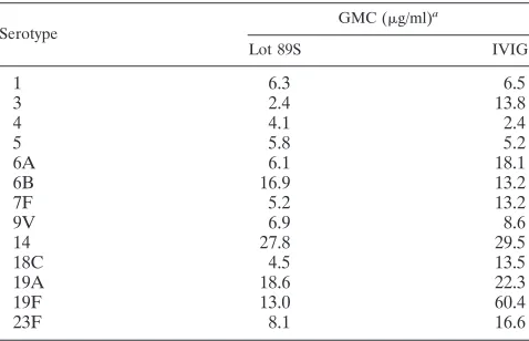
Evaluation of IVIG as an in-house reference serum.In this study, IVIG was introduced as an in-house reference serum for the MIA. IVIG is a pool of purified IgG from plasma obtained from approximately 1,000 healthy blood donors. The serotype-specific IgG concentrations in IVIG were determined by using the lot 89S sera as a reference in the MIA (Table 1). The undiluted IVIG contained a 10% (wt/vol) concentration of IgG, which is approximately 10 times greater than the nor-mal IgG concentration in serum. As a result, the concentra-tions of IgG antibodies against various pneumococcal poly-saccharides were also higher than those in healthy adult serum.
The serotype-specific concentrations in the sera from chil-dren pre- and post-booster vaccination were assessed by using both lot 89S and IVIG as reference sera in the MIA. The use of IVIG yielded the same antibody concentrations as those obtained by the use of lot 89S, as indicated by the almost perfect correlation between those concentrations (R2ⱖ0.98).
9V 6.9 8.6
14 27.8 29.5
18C 4.5 13.5
19A 18.6 22.3
19F 13.0 60.4
23F 8.1 16.6
a
Lot 89S was used as the reference.
on August 17, 2020 by guest
http://cvi.asm.org/
Furthermore, the curves of both reference serum pools were virtually identical (Fig. 1).
Evaluation of adsorbent buffer. Recently, CWPS Multi, which carries both types of CWPSs, has become commercially available, and we investigated whether this reagent could be used to replace adsorption with CWPS plus capsular polysac-charide 22F. To assess the degree to which CWPS Multi re-moved the anti-CWPS antibodies in serum, six serum samples with high CWPS concentrations were adsorbed with CWPS Multi and tested by a Luminex assay with CWPS Multi-cou-pled beads. The CWPS Multi adsorption yielded a reduction in the CWPS Multi signal of 96% or more (data not shown).
The effect of CWPS Multi adsorption was compared with the effect of adsorption with CWPS plus capsular polysaccha-ride 22F on all pre- and post-booster vaccination serum sam-ples in the MIA for all 13 serotypes (Fig. 2). The correlation between the results of the two adsorption methods wasⱖ0.8 for the vaccine serotypes. For all serotypes except serotypes 19A and 19F, higher concentrations were obtained over the high concentration ranges, showing that the dynamic range of the MIA increased by the use of CWPS Multi as the sorbent. In contrast, serotype 19A yielded overall lower concentrations in the MIA and the serotype 19F concentrations were lower over the high concentration range. The results obtained by the use of CWPS Multi and CWPS plus capsular polysaccharide 22F adsorptions in the MIA were compared with those ob-tained by their use in the ELISA for eight serotypes. Linear regression, used to correlate the results of MIA and those of the ELISA, yielded similar or higherR2values for CWPS Multi
adsorption, and the slopes improved when CWPS Multi was used. The only exception was for serotype 19F, for which both the correlation and the slope of the trend line improved by the use of CWPS plus capsular polysaccharide 22F adsorption. As the results for virtually all serotypes were better when CWPS Multi was used and the use of CWPS Multi would enable the inclusion of serotype 22F in the MIA in the future, CWPS Multi was chosen as the sorbent to be used in the optimized MIA.
In the previously described protocol for the pneumococcal MIA (11, 16), newborn bovine serum (NBBS) was used as the blocking reagent in the buffers. However, the results of another polysaccharide Luminex assay, which combined
polysaccha-rides fromHaemophilus influenzaetype b and meningococcal serogroups A, C, Y, and W-135, correlated better with those of the ELISA when ADHS was used (7). Therefore, we evaluated the use of 5% ADHS as a blocking reagent and compared the results obtained with 5% ADHS with those obtained by the use of 5% NBBS. The results obtained for serotypes 6B, 9V, 14, and 18C with either blocking agent were quite similar. How-ever, for serotypes 4, 19F, and 23F, the addition of 5% ADHS considerably increased the dynamic range of the assay (data not shown). The use of 50% ADHS did not yield results better than those obtained by the use of 5% ADHS (data not shown), and therefore, 5% ADHS was used in this study.
Comparison of MIA and ELISA. To evaluate the perfor-mance of the MIA, the correlation of the results of MIA with those of the gold standard assay, the WHO ELISA, was de-termined. The correlation between the results of MIA and those of the ELISA for the calibration panel of 12 serum samples was high, withR2values ranging from 0.75 to 0.92, on
the basis of the published ELISA results for all 12 serum samples (Table 2). Comparison of the results of the MIA with those of the WHO ELISA performed in-house resulted in lower correlations. However, on the basis of the results for the 12 serum samples only, theR2value was still fairly good (Table
2). By using the WHO guidelines, which state that the results for 75% of the serum samples should be within an error range of 40%, all serotypes met the WHO criterion when they were
FIG. 1. Similarities of the curves obtained by MIA with serial di-lutions of the lot 89S and IVIG reference sera for serotypes 6A, 6B, and 14. The median fluorescent intensities are plotted against the IgG concentration. Similar curves were obtained for the other serotypes.
FIG. 2. Correlation between IgG concentrations obtained by sero-type 14- and serosero-type 23F-specific MIAs by using adsorption with CWPS Multi (xaxis) and CWPS plus serotype 22F capsular polysac-charide (yaxis). The dotted lines indicate the perfect correlation. The IgG concentrations are log transformed.
on August 17, 2020 by guest
http://cvi.asm.org/
tested by our ELISA. However, when this criterion was applied to the MIA, only two of the seven serotypes met this error range. For two serotypes, 4 of the 12 serum samples did not have results within the error range, and the result for one of
assays. The assessment of the error range for these sera ob-tained by MIA is therefore only exploratory and not restrictive. In the future, error ranges could be defined for the standard-ization of MIAs.
The correlation between the results of MIA and those of the ELISA was also determined by using pre- and post-booster vaccination samples from immunized children after they re-ceived a primary series of three immunizations with PCV-7
23F 0.91 0.90 0.94
a
n⫽12 serum samples for the calibration panel. b
In-house WHO ELISA. c
Published results of the WHO ELISA (http://www.vaccine.uab.edu/qc3.pdf).
FIG. 3. High degree of correlations between the IgG concentrations obtained by ELISA and MIA by using infant sera obtained pre- and post-booster vaccination with PCV-7. The dotted lines indicate the perfect correlation. The IgG concentrations are log transformed.
on August 17, 2020 by guest
http://cvi.asm.org/
(Fig. 3). The correlations varied from 0.69 (serotype 19F) to 0.91 (serotype 4). The mean of the R2 values for all eight
serotypes was 0.85, demonstrating that MIA yields antibody concentrations that are comparable to those obtained by ELISA. For serotypes 4, 6B, 9V, 18C, 19F, and 23F, the anti-body concentrations measured by MIA were higher than those obtained by ELISA, particularly over the high concentration range. For serotype 14, almost identical concentrations were obtained by the two assays. MIA yielded antibody concentra-tions in the low concentration range lower than those obtained by ELISA only for serotype 6A.
On average, the concentrations measured by MIA were higher than those measured by ELISA, and the results also suggested that the dynamic range of the MIA is larger than that of the ELISA. The latter was corroborated when the results for dilution series of the lot 89S reference serum ana-lyzed by ELISA and MIA were compared (Fig. 4). As an example, the concentrations of antibody against serotype 14 could be determined by ELISA over a range of 0.1 to 30 ng/ml. In contrast, MIA enabled measurements over a 0.1- to 90-ng/ml range. Thus, the range over which anti-serotype 14 an-tibody concentrations can be accurately measured by MIA is approximately 3-fold greater than that for the ELISA. The dynamic range of the MIA for the other serotypes was also larger, ranging from 3-fold to up to 50-fold.
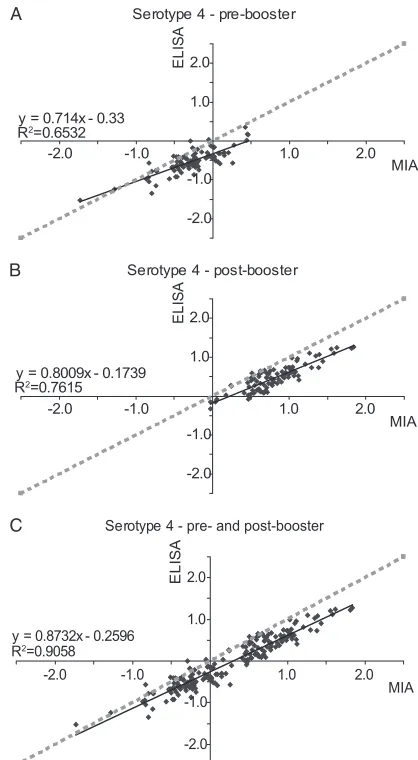
LowR2
values due to small concentration ranges.The rel-atively poor correlation between the results obtained by MIA and ELISA for serotype 19F prompted further analysis. That analysis showed that the distribution of the concentrations had a considerable impact on the correlation between the results of MIA and those of the ELISA. As an example, theR2value was
calculated separately for the anti-serotype 4 pre- and post-booster vaccination concentrations. TheR2 values were only
0.65 for the pre-booster vaccination concentrations and 0.76 for the post-booster vaccination concentrations. However, if the pre- and post-booster vaccination concentrations were combined, theR2value increased to 0.91 (Fig. 5). This
illus-trates thatR2may be inaccurate if only a small range of
con-centrations is used. TheR2values for the pre-booster
vaccina-tion, the post-booster vaccinavaccina-tion, and the combined comparisons
are given in Table 3. Measurement of theR2values over a small
concentration range yielded lowerR2values for all serotypes.
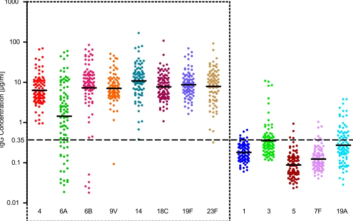
Response to booster vaccine after immunization with PCV-7.
The concentrations of antibodies to 13 serotypes in the pre-and post-booster vaccination sera were assessed by the MIA. For the serotypes present in PCV-7, virtually all serum samples had antibody concentrations above the protective concentra-tion of 0.35g/ml after the booster vaccination (Fig. 6). Only five serum samples had low concentrations of antibodies to serotype 6B, a single sample had low concentrations of anti-bodies to serotype 9V, and another sample had low concen-trations of antibodies to serotype 23F. Similar results were obtained by the ELISA (1). The GMCs for the non-vaccine serotypes were well below the level of protection, with the exception of the GMC for serotype 6A.
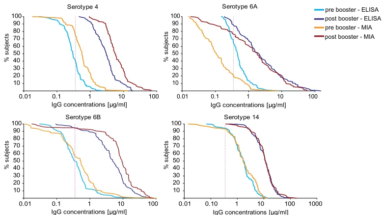
The percentage of children with concentrations above the protective level can be derived from the relative cumulative distribution curves (RCDCs) (Fig. 7). The concentrations
mea-FIG. 4. Visualization of the larger dynamic range of the serotype 14-specific MIA compared to that of the serotype 14-specific ELISA. Serial dilutions of the lot 89S reference serum were made and tested in both assays. The MFI (leftyaxis) and optical density (OD; righty
axis) are plotted against the IgG concentration. By the MIA, the dynamic range was 0.1 to 90 ng/ml, whereas the ELISA allowed as-sessment of the IgG concentration only over a range of 0.1 to 30 ng/ml.
FIG. 5. Influence of the IgG concentration range on the correla-tions between the results of the MIA and ELISA. The IgG concentra-tions were obtained for serotype 4 pre-booster vaccination (A), post-booster vaccination (B), and pre- and post-post-booster vaccination combined (C). The IgG concentrations are log transformed.
on August 17, 2020 by guest
http://cvi.asm.org/
sured by MIA were generally higher, and therefore, as deter-mined by MIA, a higher percentage of children had antibody concentrations above the protective concentration even before the booster vaccination. The graphs also illustrate the larger dynamic range of the MIA compared with that of the ELISA, and this was particularly obvious for serotype 6A. The RCDC for serotype 6A showed that, according to the MIA, 20% and 77% of the children had concentrations above the protected concentration pre- and post-booster vaccination, respectively. In contrast, the ELISA results suggested that 67% of the chil-dren had protective antibody concentrations pre-booster vac-cination and that 89% reached this level after the booster vaccination.
Cross-reactivity of serotypes 6A and 6B.Although PCV-7 does not contain the serotype 6A capsular polysaccharide, the serum samples from the vaccinated children contained a
con-6A, we were able to fully adsorb the serotype 6A signal, but only 40 to 60% of the serotype 6B signal could be removed.
DISCUSSION
In this report, we describe the optimization and application of an MIA for the simultaneous assessment of the concentra-tions of antibodies directed against 13 different pneumococcal capsular polysaccharides in infant sera in a single assay. This MIA will enable us to perform large immunosurveillance stud-ies that require the assessment of the concentrations of anti-bodies against a large number of different antigens simulta-neously. Such studies would be extremely time-consuming and expensive if they were performed by ELISA and therefore would be virtually impossible to perform by ELISA. To assess the performance of the assay, we compared the concentrations of antibodies against the seven serotypes included in PCV-7 and serotype 6A, as measured by the MIA and the WHO ELISA with serum samples from infants pre- and post-booster vaccination with PCV-7. The correlation between the results obtained by MIA and ELISA was good, with virtually all R2
values exceeding 0.85. A relatively low correlation was found
9V 0.3 0.9 0.52 2.3 7.1 0.69 0.83
14 1.6 1.6 0.85 10.4 10.8 0.70 0.87
18C 0.2 0.7 0.60 1.9 7.7 0.75 0.87
19F 0.9 2.7 0.51 4.5 8.7 0.60 0.69
23F 0.2 0.6 0.64 3.1 7.9 0.73 0.88
FIG. 6. IgG concentrations and GMCs for 13 different serotypes obtained by MIA of sera taken from infants 1 month post-booster vaccination with PCV-7. Each colored dot represents an individual serum sample. GMCs are indicted by the black dashes in the colored dots. The horizontal dashed line indicates the protective concentration of 0.35g/ml. The serotypes that are depicted within the box with a dashed border are the serotypes used to compare MIA and ELISA in this study.
on August 17, 2020 by guest
http://cvi.asm.org/
for serotype 19F. However, the range of concentrations of IgG antibodies against this serotype was narrow, and we demon-strated that the use of data with a small range may result in low correlation values. The dynamic range of the MIA appeared to be at least 3-fold larger than that of the WHO ELISA, which we demonstrated by comparing the results for serial dilutions of lot 89S obtained by the MIA and the ELISA. For most serotypes, the GMCs obtained by MIA were higher than those obtained by ELISA; the exceptions were the GMCs for sero-types 14 and 6A. For serotype 14, the GMCs obtained by MIA and ELISA were virtually identical, whereas the MIA for se-rotype 6A yielded lower concentrations in the low concentra-tion range. As a result of the higher average GMCs obtained by the MIA, the number of serum samples with concentrations exceeding the proposed concentration of 0.35g/ml required for protection was higher by the MIA than by the ELISA. The protective concentration of 0.35 g/ml is based on a meta-analysis of studies that used ELISA. The actual correlate of protection is difficult to assess, as this would require monitor-ing of invasive pneumococcal disease in children with various antibody levels. Such experiments are impossible to perform. The value of 0.35g/ml was set as a cutoff value for noninfe-riority but is often used as a surrogate level of protection.
Because of the differences between MIA and ELISA, we be-lieve that the protective concentration of 0.35g/ml should be amended and that there should be a distinct value for each serotype for MIA and for ELISA as well. Although we used a 13-plex pneumococcal MIA, we were able to truly evaluate the performance of the assay for only eight of these serotypes in this study, because we used serum samples from children im-munized with PCV-7. In order to assess the performance of the MIA for the other five serotypes, serum samples from infants immunized with these capsular polysaccharides (e.g., a 13-valent PCV) are required. No such sera were available to us during our study.
The results of previously described pneumococcal multiplex assays were also shown to correlate well with those of the ELISA (2, 16, 20). In the study whose results are presented here, we introduced an alternative and readily available refer-ence serum sample and the use of CWPS Multi as the sorbent. A replacement for lot 89S, 007sp, has been prepared, but this serum sample was not available to us (10). The introduction of IVIG as a new in-house reference serum sample has the main advantage that it is available in large quantities, and therefore, the same reference serum sample can be used in a number of large-scale studies. The calculated concentrations of antibodies in the samples from the immunized infants were the same whether IVIG or lot 89S was used as the reference serum sample. The curves obtained by the use of serial dilutions of both reference serum samples were nearly identical, even though the nature of the individuals who donated blood for those serum pools was quite different, that is, unimmunized versus immunized individuals. Once it is calibrated, a single batch can be used for a large number of studies. The 3-g batch that we purchased for this purpose is sufficient for analysis of approximately 4⫻106serum samples. Due to the variety of
antibodies present in IVIG, this serum sample pool can also be used as a reference for other serological assays targeting
anti-FIG. 7. Relative cumulative distribution curves of the pre- and post-booster vaccination IgG concentrations for four serotypes obtained by MIA and ELISA. The vertical dotted line indicates the protective concentration of 0.35g/ml. The percentage of subjects (yaxis) is plotted against the IgG concentration (xaxis). The graph for serotype 4 is illustrative for the other serotypes, for which the results are not depicted here.
TABLE 4. Absorption of serotype 6A and 6B cross-reactive antibodies in serum samples from four children
after booster vaccination with PCV-7
MIA serotype
Adsorbent serotype
% inhibition
Child 1 Child 2 Child 3 Child 4 Lot 89S (n⫽11)
6A 6A 86 93 91 87 95
6A 6B 98 99 99 98 44
6B 6A 64 62 61 42 9
6B 6B 95 98 98 94 68
on August 17, 2020 by guest
http://cvi.asm.org/
dynamic range of the MIA. CWPS Multi will most likely re-move anti-CWPS2 antibodies more efficiently than the capsule polysaccharide preparation obtained from serotype 22F iso-lates, because in the latter preparation, CWPS2 makes up only approximately 5% (wt/wt) of the total polysaccharide compo-sition (28). In addition, the use of CWPS Multi enables the future expansion of the MIA to include serotype 22F as well. The PCV-7 vaccination induced an serotype 6A anti-body response. This suggests cross-reactivity between serotype 6B and the related serotype 6A antigen, a phenomenon de-scribed earlier (2, 11, 16, 17). The absorption experiments in the study presented here conclusively show that vaccination with the serotype 6B antigen evokes antibodies against one or more epitopes carried by both the serotype 6B and serotype 6A capsular polysaccharides and against serotype 6B-specific epitopes. In similar experiments, Findlow et al. (11) obtained somewhat different results. Unlike the results obtained in our experiments, they could not completely remove antibodies re-acting with serotype 6B-specific beads by serotype 6B absorp-tion. In addition, the antibodies reacting with serotype 6A-specific beads were only partially removed by absorption with serotype 6B. Although absorption with serotype 6A completely removed the anti-serotype 6A antibodies, the anti-serotype 6B antibodies were apparently left unaffected, a result which dif-fers from our results. The experiments described by Findlow et al. were performed with reference serum lot 89S. Lot 89S is a pool of sera from adults who were vaccinated with the 23-valent polysaccharide vaccine, which does not contain the se-rotype 6A polysaccharide. It is likely that these adults have developed antibody responses against serotype 6A both through natural exposure to this serotype and by cross-reactiv-ity with the serotype 6B polysaccharide which is present in the 23-valent vaccine. In our study, we used individual serum sam-ples from PCV-7-vaccinated young children, who are less likely to have been naturally exposed to pneumococci of serotype 6A. In addition, we removed the cross-reacting antibodies with beads to prevent possible interference by the low-avidity anti-bodies that are released from the sorbent during the incuba-tion steps in the MIA. These factors may explain the differ-ences between the results of our absorption experiments and those found in other studies. Irrespective of the reasons for the induction of anti-serotype 6A and anti-serotype 6B antibodies, it is clear that the concentrations of antibodies against sero-types 6A and 6B should be carefully interpreted.
In conclusion, we optimized and evaluated the performance of an MIA for quantification of the levels of antibodies di-rected against 13 different pneumococcal polysaccharides. We have introduced a new in-house reference serum sample and a new adsorption buffer, and we showed that the results of the
ACKNOWLEDGMENTS
We thank Helen Findlow (HPA, Manchester, United Kingdom) for transfer of the pneumococcal Luminex knowledge. We thank the chil-dren and parents who participated in this study, as well as the clinical and laboratory staff.
REFERENCES
1.Berbers, G. A., and N. Jones.2008. Serological surveillance of the effect of the changes to pertussis vaccines in the NIP from 2004 till 2008. Switch from whole cell to acellular vaccine in children of 1 year of age (ISRCTN97785537). RIVM report. National Institute for Public Health and the Environment, Bilthoven, Netherlands.
2.Biagini, R. E., S. A. Schlottmann, D. L. Sammons, J. P. Smith, J. C. Snawder, C. A. Striley, B. A. MacKenzie, and D. N. Weissman.2003. Method for simultaneous measurement of antibodies to 23 pneumococcal capsular polysaccharides. Clin. Diagn. Lab. Immunol.10:744–750.
3.Black, S., H. Shinefield, B. Fireman, E. Lewis, P. Ray, J. R. Hansen, L. Elvin, K. M. Ensor, J. Hackell, G. Siber, F. Malinoski, D. Madore, I. Chang, R. Kohberger, W. Watson, R. Austrian, and K. Edwards.2000. Efficacy, safety and immunogenicity of heptavalent pneumococcal conjugate vaccine in chil-dren. Northern California Kaiser Permanente Vaccine Study Center Group. Pediatr. Infect. Dis. J.19:187–195.
4.Capeding, M. Z., T. Puumalainen, C. P. Gepanayao, H. Kayhty, M. G. Lucero, and H. Nohynek.2003. Safety and immunogenicity of three doses of an eleven-valent diphtheria toxoid and tetanus protein-conjugated pneumo-coccal vaccine in Filipino infants. BMC Infect. Dis.3:17.
5.Concepcion, N. F., and C. E. Frasch.2001. Pneumococcal type 22F polysac-charide absorption improves the specificity of a pneumococcal-polysaccha-ride enzyme-linked immunosorbent assay. Clin. Diagn. Lab. Immunol.
8:266–272.
6.Dagan, R.2002. Immunisation with a pneumococcal 7-valent conjugate vac-cine. Int. J. Clin. Pract.56:287–291.
7.de Voer, R. M., R. M. Schepp, F. G. Versteegh, F. R. van der Klis, and G. A. Berbers.2009. Simultaneous detection of Haemophilus influenzae type b polysaccharide-specific antibodies and Neisseria meningitidis serogroup A, C, Y, and W-135 polysaccharide-specific antibodies in a fluorescent-bead-based multiplex immunoassay. Clin. Vaccine Immunol.16:433–436. 8.Ekstrom, N., M. Vakevainen, J. Verho, T. Kilpi, and H. Kayhty.2007.
Functional antibodies elicited by two heptavalent pneumococcal conjugate vaccines in the Finnish Otitis Media Vaccine Trial. Infect. Immun.75:1794– 1800.
9.Eskola, J., T. Kilpi, A. Palmu, J. Jokinen, J. Haapakoski, E. Herva, A. Takala, H. Kayhty, P. Karma, R. Kohberger, G. Siber, and P. H. Makela.
2001. Efficacy of a pneumococcal conjugate vaccine against acute otitis media. N. Engl. J. Med.344:403–409.
10.Feavers, I., I. Knezevic, M. Powell, E. Griffiths, and WHO Consultation on Serological Criteria for Evaluation and Licensing of New Pneumococcal Vaccines.2009. Challenges in the evaluation and licensing of new pneumo-coccal vaccines, 7–8 July 2008, Ottawa, Canada. Vaccine27:3681–3688. 11.Findlow, H., G. Laher, P. Balmer, C. Broughton, E. D. Carrol, and R.
Borrow.2008. Competitive inhibition flow analysis assay for the non-culture detection of pneumococcal serotype capsular polysaccharide. Clin. Vaccine Immunol.16:222–229.
12.Goldblatt, D., J. Southern, L. Ashton, P. Richmond, P. Burbidge, J. Tasevska, A. Crowley-Luke, N. Andrews, R. Morris, R. Borrow, K. Cartwright, and E. Miller.2006. Immunogenicity and boosting after a reduced number of doses of a pneumococcal conjugate vaccine in infants and toddlers. Pediatr. Infect. Dis. J.25:312–319.
13.Gottschalk, P. G., and J. R. Dunn.2005. The five-parameter logistic: a characterization and comparison with the four-parameter logistic. Anal. Bio-chem.343:54–65.
14.Jansen, A. G., G. D. Rodenburg, S. C. de Greeff, E. Hak, R. H. Veenhoven, L. Spanjaard, L. M. Schouls, E. A. Sanders, and A. van der Ende.2009. Invasive pneumococcal disease in the Netherlands: Syndromes, outcome and potential vaccine benefits. Vaccine27:2394–2401.
on August 17, 2020 by guest
http://cvi.asm.org/
15.Klugman, K. P., S. A. Madhi, R. E. Huebner, R. Kohberger, N. Mbelle, and N. Pierce.2003. A trial of a 9-valent pneumococcal conjugate vaccine in children with and those without HIV infection. N. Engl. J. Med.349:1341– 1348.
16.Lal, G., P. Balmer, E. Stanford, S. Martin, R. Warrington, and R. Borrow.
2005. Development and validation of a nonaplex assay for the simultaneous quantitation of antibodies to nine Streptococcus pneumoniae serotypes. J. Immunol. Methods296:135–147.
17.Lee, H., M. H. Nahm, R. Burton, and K. H. Kim.2009. Immune response in infants to the heptavalent pneumococcal conjugate vaccine against vaccine-related serotypes 6A and 19A. Clin. Vaccine Immunol.16:376–381. 18.Nielsen, S. V., U. B. Sorensen, and J. Henrichsen.1993. Antibodies against
pneumococcal C-polysaccharide are not protective. Microb. Pathog.14:299– 305.
19.O’Brien, K. L., L. H. Moulton, R. Reid, R. Weatherholtz, J. Oski, L. Brown, G. Kumar, A. Parkinson, D. Hu, J. Hackell, I. Chang, R. Kohberger, G. Siber, and M. Santosham.2003. Efficacy and safety of seven-valent conju-gate pneumococcal vaccine in American Indian children: group randomised trial. Lancet362:355–361.
20.Pickering, J. W., T. B. Martins, R. W. Greer, M. C. Schroder, M. E. Astill, C. M. Litwin, S. W. Hildreth, and H. R. Hill.2002. A multiplexed fluorescent microsphere immunoassay for antibodies to pneumococcal capsular polysac-charides. Am. J. Clin. Pathol.117:589–596.
21.Plikaytis, B. D., D. Goldblatt, C. E. Frasch, C. Blondeau, M. J. Bybel, G. S. Giebink, I. Jonsdottir, H. Kayhty, H. B. Konradsen, D. V. Madore, M. H. Nahm, C. A. Schulman, P. F. Holder, T. Lezhava, C. M. Elie, and G. M. Carlone.2000. An analytical model applied to a multicenter pneumococcal enzyme-linked immunosorbent assay study. J. Clin. Microbiol.38:2043–2050. 22.Prentice, R. L.1976. A generalization of the probit and logit methods for
dose response curves. Biometrics32:761–768.
23.Quataert, S., D. Martin, P. Anderson, G. S. Giebink, J. Henrichsen, M.
Leinonen, D. M. Granoff, H. Russell, G. Siber, H. Faden, D. Barnes, and D. V. Madore. 2001. A multi-laboratory evaluation of an enzyme-linked immunoassay quantitating human antibodies to Streptococcus pneumoniae polysaccharides. Immunol. Invest.30:191–207.
24.Quataert, S. A., C. S. Kirch, L. J. Wiedl, D. C. Phipps, S. Strohmeyer, C. O. Cimino, J. Skuse, and D. V. Madore.1995. Assignment of weight-based antibody units to a human antipneumococcal standard reference serum, lot 89-S. Clin. Diagn. Lab. Immunol.2:590–597.
25.Quataert, S. A., K. Rittenhouse-Olson, C. S. Kirch, B. Hu, S. Secor, N. Strong, and D. V. Madore.2004. Assignment of weight-based antibody units for 13 serotypes to a human antipneumococcal standard reference serum, lot 89-S (f). Clin. Diagn. Lab. Immunol.11:1064–1069.
26.Romero-Steiner, S., J. Fernandez, C. Biltoft, M. E. Wohl, J. Sanchez, J. Feris, S. Balter, O. S. Levine, and G. M. Carlone.2001. Functional antibody activity elicited by fractional doses of Haemophilus influenzae type b con-jugate vaccine (polyribosylribitol phosphate-tetanus toxoid concon-jugate). Clin. Vaccine Immunol.8:1115–1119.
27.Skovsted, I. C., M. B. Kerrn, J. Sonne-Hansen, L. E. Sauer, A. K. Nielsen, H. B. Konradsen, B. O. Petersen, N. T. Nyberg, and J. O. Duus.2007. Purification and structure characterization of the active component in the pneumococcal 22F polysaccharide capsule used for adsorption in pneumo-coccal enzyme-linked immunosorbent assays. Vaccine25:6490–6500. 28.Sorensen, U. B., and J. Henrichsen.1984. C-polysaccharide in a
pneumo-coccal vaccine. Acta Pathol. Microbiol. Immunol. Scand. C92:351–356. 29.World Health Organization.2005. Recommendations for the production
and control of pneumococcal conjugate vaccines. World Health Organ. Tech. Rep. Ser.927:64–98.
30.Yu, X., Y. Sun, C. Frasch, N. Concepcion, and M. H. Nahm.1999. Pneumo-coccal capsular polysaccharide preparations may contain non-C-polysaccha-ride contaminants that are immunogenic. Clin. Diagn. Lab. Immunol.6:519– 524.