The power of ion mobility-mass spectrometry for structural characterisation
and the study of conformational dynamics
Francesco Lanucara,†1,2 Stephen W. Holman,† 1,2 Christopher Gray†2 and Claire E. Eyers*1,2 1Protein Function Group, Department of Biochemistry, Institute of Integrative Biology, University of
Liverpool, Crown Street, Liverpool L69 7ZB
2Michael Barber Centre for Mass Spectrometry, Manchester Institute of Biotechnology, School of Chemistry, University of Manchester, 131 Princess Street, Manchester M1 7DN
†These authors contributed equally
Corresponding author: Claire E. Eyers
Protein Function Group, Institute of Integrative Biology, University of Liverpool, Crown Street, Liverpool L69 7ZB
Email: [email protected]
Abstract
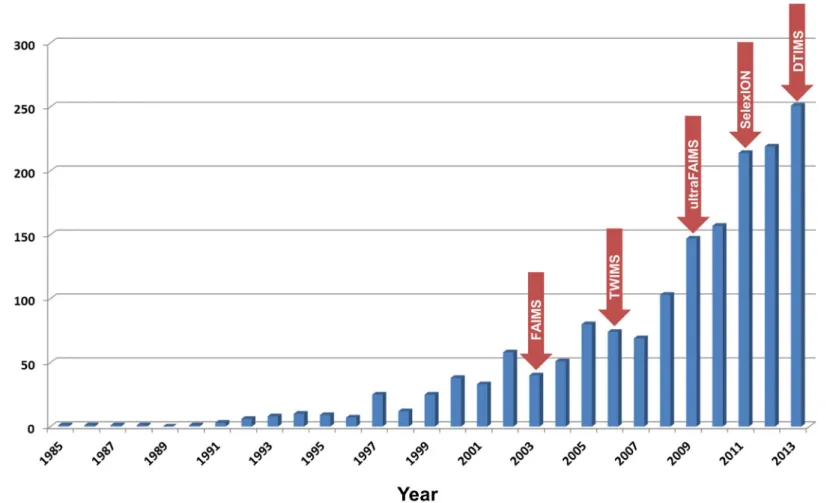
Ion mobility spectrometry (IMS) is an analytical technique that separates gas-phase ions based on their size and shape, analogous to electrophoresis in the condensed phase. The technique has long been used for the detection of illegal or dangerous substances e.g. explosives at border crossings, and to garner evidence for the illegal use of chemical agents.1 Mass spectrometry (MS) is also used in the analysis of gas-phase ions, permitting determination of their mass-to-charge (m/z) ratios. Therefore, the two techniques can be used to ascertain complementary information about analytes. The coupling of the two strategies, though first described in 1962,2 has only recently become relatively commonplace, primarily due to commercialisation of the necessary instrumentation. The last ten or so years has seen an explosion in research using ion mobility-mass spectrometry (IM-MS), as demonstrated by a rapid increase in the annual number of peer-reviewed publications (Figure 1), where the benefits of combining both analytical strategies have been exploited.
Instrumentation for ion mobility spectrometry
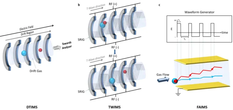
Primarily, three IMS techniques are employed in IM-MS: drift time ion mobility spectrometry (DTIMS), travelling wave ion mobility spectrometry (TWIMS) and field asymmetric ion mobility spectrometry (FAIMS) (Figure 2, Table 1); these are described in detail below.
Drift time ion mobility spectrometry (DTIMS)
DTIMS is the oldest and conceptually simplest form of ion mobility (Figure 2a). Ions are introduced into a drift tube. The application of a static uniform electric field (5-100 V) then propels these ions in the direction of the applied field. The tube is filled with a drift gas, typically helium. The time taken for an ion to drift through the tube is related to its rotationally averaged cross sectional area, i.e. the area covered by a particle, or more simply its collision cross section (CCS), Ω. Compact structures travel faster than more elongated (extended) ions due to fewer interactions with the gas. The recorded drift time of an ion allows calculation of its Ω, according to the Mason-Schamp equation3:
Ω=
3
ze
16
N
(
2
π
μk
BT
)
1
2
1
K
0where K0 is the reduced mobility (measured mobility at standard temperature and pressure), z is the
charge state of the ion, e is the elementary charge, N is the number density of the drift gas, µ is the reduced mass of the ion-neutral pair, kB is the Boltzmann constant and T is the gas temperature. The
proportional relationship between Ω and K0 is only true at or below the “low field limit”, where the
V•cm2).4 Determination of the CCS (which gives an indication of an ion’s size and shape) provides structural information that is characteristic for each compound, and can be compared to data acquired using other structural techniques such as X-ray crystallography and Nuclear Magnetic Resonance (NMR) spectroscopy. The level and quality of structural information obtainable using IMS compared to traditional techniques will be discussed below. DTIMS is advantageous due to its relatively high resolving power (R >100 Ω / Ω) compared to other IM devices, meaning that an ion with Ω of 1000 Å2 can theoretically beseparated from an ion with Ω +/- 10 Å2. However, DTIMS can suffer from low duty cycle i.e. percentage of ions detected relative to those generated by the ionisation source, on devices where the ions are pulsed into the drift cell, or where entry/exit of ions into the drift cell occurs through extremely small apertures. However, limitations in duty cycle have been overcome in newer generation DTIMS instruments by incorporation of some form of ion-trapping funnel prior to the drift cell, which can accumulate ions whilst a previous pulse is being mobility separated.5, 6
Travelling wave ion mobility spectrometry (TWIMS)
A TWIMS device comprises a series of ring electrodes called a stacked ring ion guide (SRIG), to which a travelling voltage wave is applied.7 Radio-frequency (RF) voltages of opposite phases are applied to adjacent electrodes (Figure 2b). These voltages radially confine the ions, whilst application of a transient direct current (DC) voltage to each electrode in succession from one end of the device to the other propels the ions axially. The DC voltage pulse applied to each electrode in turn creates the ‘travelling wave’ upon which ions ‘surf’ and traverse the mobility cell. By altering the speed and magnitude of the travelling voltage wave, mobility separation of ions can be achieved. Higher mobility ions are ‘carried’ by the wave, whilst ions of lower mobility ‘roll over’ the wave, thus taking longer to move through the mobility cell. Complex mixture separation can be achieved by sending several travelling waves through the device in quick succession. The TWIMS device is operated below the low field limit and, following calibration, determination of CCS is therefore possible.8-11 Calibration of the drift time through the TWIMS cell under defined conditions (gas type/pressure, travelling wave speed/height etc.) is necessary as the direct relationship between Ω and K0 is no
Field asymmetric ion mobility spectrometry (FAIMS)
FAIMS devices are constructed of two electrodes, across which an electric field is established (Figure 2c).12 Ions are introduced into the device perpendicular to the electric field and collinearly to the drift gas. Two voltages are used to achieve separation: the dispersion voltage (DV) and the compensation voltage (CV). The DV is an alternating asymmetric waveform, where the value of the positive voltage is greater than that of the negative. However, the negative voltage is applied for a longer time, resulting in an equal voltage•time product for each part of the waveform (Figure 2c). This waveform exploits the fact that under high electric field conditions (>5000 Vs-1), as generated by the positive voltage period, the mobility of ions can be different to that under the low electric field conditions (<200 Vs-1) provided by the negative voltage period. This electric field-dependent mobility means that ions drift radially as they traverse the electrodes (Figure 2c). Ultimately, dispersion of the ions in this manner would lead to contact with the electrodes and neutralisation. Therefore, to refocus ions through the electrodes, a CV is applied. For a given CV, only ions of specific mobility will be repelled from the electrodes, whilst other ions will continue along their path and be neutralised. Manipulation of the CV can therefore be used to selectively permit ions of interest through the electrodes whilst removing ions of differing mobility. The FAIMS device thus operates as a mobility filter and typically finds application as an orthogonal separation technique to liquid chromatography (LC), separating ions based on different physico-chemical properties, prior to MS analysis, enabling increased selectivity and peak capacity.13 The major limitation of FAIMS is that CCS cannot be determined, as its operating electric field strength causes it to exceed the low field limit (Table 1); this prohibits the correlation of drift time with structural features as the mobility of an analyte is not directly related to its structure, explaining why FAIMS devices are primarily used to filter out unwanted interferences and/or to separate analytes of interest. Although the resolving power of FAIMS separation is traditionally poor (R < 20), the preferential inclusion of light gases in which the ions are more mobile (i.e. He and/or H2) as opposed to N2 in the carrier gas, has been demonstrated to significantly increase resolution, with resolving powers of up to 500 being reported.14-16
popularity as FAIMS devices.17, 19 Newer generation DMA instrumentation with higher resolving power (R > 50) may yet encourage growth in this area.20
While the IMS devices discussed will all separate ions based on differences in their mobility through a buffer gas, only the time-based mobility devices (DTIMS, TWIMS) can be used to determine information about cross-sectional area, either with or without the requirement for drift time calibration if there is a requirement to define CCS in Å2. In contrast, FAIMS (and DMA) devices function preferentially as filtration devices, much like quadrupoles can be used in MS analysis. However, whether the utility of IMS separation is for analyte filtration and/or differentiation, or more targeted structural analysis, the key strength comes in combining this analytical feature with mass spectrometry, due to the complementary information garnered. Examples of how IMS devices can be used either to improve the MS-based analysis of complex mixtures, or in the case of DTIMS/TWIMS, facilitate enhanced structural analysis and the study of conformational dynamics are described below.
Enhancing the MS analysis of mixtures with IMS
The coupling of IMS to MS can significantly improve experimental analysis as defined by a range of criteria often used to benchmark such methods: selectivity, speed and limit of detection. The improvement gained for each is considered below.
Selectivity
adding volatile (often chiral) dopants, such as 2-butanol.23, 24 Detailed studies have shown that the change in selectivity induced by such dopants is obtained through differential formation of ion– molecule clusters.25, 26 Addition of shift reagents, as utilised in NMR spectroscopy, is another strategy that can improve the separation of ions of similar mobility in their native forms.27-29 Selective complex formation of only one of the compounds with the shift reagent changes their mobility and facilitates separation. One particularly effective use of this approach was reported by Howdle et al., who used a pharmaceutical excipient, polyethylene glycol (PEG), already present in the sample as the shift reagent.27 Inclusion of PEG changed the mobility of the target drug, lamivudine, so that it occupied an interference-free region of the spectrum and could be analysed in isolation.
The separation capabilities of IMS have also demonstrated particular utility in MS analyses where chromatographic separation either has not or cannot be employed, providing a crucial additional analytical dimension. For example, MS imaging experiments, where analytes are ionised directly from a complex matrix such as a tissue section, are notably enhanced by incorporation of IMS. Stauber and co-workers used TWIMS in a matrix-assisted laser desorption/ionisation (MALDI)-MS imaging experiment facilitating separation of ions of the same m/z value (i.e. isobaric).30,31 While analysis of these ions in the absence of IMS produced a composite image of both species, meaning that distinct localisation could not be ascertained, incorporation of IMS enabled acquisition of separate spatial distributions for each individual compound. A similar study recorded the spatial distribution of the cytotoxic agent vinblastine following administration to rats. Although this analysis initially suffered from matrix interference at the same m/z value as the target compound, the additional selectivity afforded by TWIMS enabled vinblastine distribution to be accurately determined.32
Another area where the additional separation provided by IMS has proven beneficial is non-targeted discovery proteomics. Shliaha and co-workers analysed a tryptic digest of an E. coli whole cell lysate and demonstrated a ~60% increase in the number of identifications at both the peptide and protein levels using LC-TWIMS-MS compared to LC-MS alone.33 A similar study comparing the same analytical strategies reported an even greater increase in the number of identifications, with both peptide and protein assignments rising due to the complementary separation mechanism and additional peak capacity afforded by the TWIMS device.34
Speed
timescale, faster than chromatography (typically on the scale of seconds), and could potentially be used to increase the speed of analysis whilst retaining the necessary separation. For example, Parson and co-workers separated isomeric glucuronide metabolites of propranolol over 15 times faster using FAIMS than had been achieved using high-performance liquid chromatography (HPLC).35 Many other examples exist of IMS being employed to improve the rate of pharmaceutical discovery and/or screening by obviating (or significantly reducing) the need for chromatographic separation prior to MS. For example, application of TWIMS significantly increased the rate at which biological samples could be screened for the presence of natural products as potential lead therapeutics.36 TWIMS has also been used in metabonomics to provide additional online separation, facilitating a reduction in the upstream LC method duration and thus improving throughput.37
Limit of detection
The limit of detection by MS is much lower than most other analytical techniques, with LC-MS shown to routinely detect low attomole amounts of analyte even in complex mixtures.38, 39 The addition of IMS to the analytical workflow can further lower limits of detection by removing background interference. The group of Thibault demonstrated a greater than 10-fold improvement in the detection limits of peptides in complex mixtures when FAIMS was incorporated into their LC-MS platform.40 The improved limits of detection and increased power of separation doubled the number of peptide40 and phosphopeptide41 identifications from unpurified cellular extracts. Likewise, Ibrahim and co-workers used a set of model isomeric modified peptides to demonstrate that DTIMS can overcome the necessity to fragment peptides for discovering the site of covalent modification, using instead the mobility of each isomer for characterisation.42 By incorporating DTIMS for ions of known behaviour, determined using authentic standards, these researchers circumvented the need for tandem MS and reported an order of magnitude improvement in detection limits.
samples taken from athletes, reducing the detection limits in some cases to sub-picogram per mL (pgmL-1) quantities.45 Improvements in detectability opens up the future possibility of identifying a wide range of substances in biological fluids at significantly lower levels (and for longer periods post-dosage), with important implications for monitoring healthcare and the screening of drugs of abuse.
IM for enhanced structural analysis of (bio)molecules
While all modes of ion mobility spectrometry will separate analytes based on their conformation, application of DTIMS or TWIMS can further be used to determine CCS. Unlike other biophysical techniques used to characterise analyte structure, IM-MS can be used to ascertain structural information using only small amounts of sample (nanogram quantities). Moreover, as the ions of interest can be selectively isolated from complex samples, material of much lower purity can be used than is typically needed to structurally characterise compounds using X-ray crystallography or NMR spectroscopy (Table 2).46 Furthermore, by using IM-MS to determine changes in mobility and thus conformation and CCS, properties like conformational dynamics,47-52 folding and unfolding intermediates,52-54 ligand-induced conformational changes,55, 56 aggregation intermediates57, 58 and quaternary structures (topology) can be determined.59-60
Determination of ion collision cross sections
Analyte structure is determined from experimentally derived rotationally averaged temperature-dependent CCS values, which reflect the conformation and shape adopted by the ions in the gas phase under defined experimental conditions.61 Experimental CCS can be compared with theoretical CCS values generated by molecular dynamic (MD) simulations, often using X-ray and NMR structures as input for the calculations.62-65 Computational algorithms have been developed for this purpose, although choosing the right algorithm is extremely important for correct interpretation of the experimental IM-MS data. When dealing with large proteins and protein complexes, the complexity brought about by the range of molecular shapes and dimensions that arise pose serious limitations to the applicability of such algorithms, either due to their inability to deal with particular kinds of intra- and intermolecular interactions, or to the extremely demanding computational costs.
arising due to scattering between the ions and the drift gas. Due to its inability to factor for multiple collisions, this method usually underestimates CCS for ions > 2 kDa and is therefore primarily used for predicting CCS of small molecules. The EHSS method takes account of the scattering and the collisions with the drift gas but ignores long-range interactions between gas and ions. In contrast, the TM is generally considered more reliable, particularly for large biomolecules, as it takes into account long-range interactions between the drift gas and the analyte ion, as well as collision effects.69 However, a major limitation of the TM is the time required to perform calculations, particularly for very large molecules; for such analytes, the EHSS method is therefore often a good compromise.
Recently, the projection superposition approximation (PSA) method was introduced by Bleiholder and co-workers.70-73 Like PA, this algorithm computes molecular shapes as a projection approximation, but with an extra feature of superposition of atomic potentials and inclusion of a shape factor. This approach is of particular value considering that long-range, attractive van der Waals interactions between ions and buffer gas are proportional to the analyte's molecular weight and the atom density, and are therefore a function of the molecular shape. An algorithm that is capable of taking into account these size and shape effects, concomitantly lowering computational demands, is therefore particularly beneficial. The PSA method was shown to outperform EHSS following tests on three sets of molecules chosen to represent different sizes and shapes, providing results in very good agreement with TM, but with a 100-1000 fold reduction in computational time.70
IM-MS as a stand-alone technique for structural studies
greater number of protonation sites, and has an immediate effect on their ionic mobilities.74 Similarly, ligand binding or protein post-translational modification often induce conformational changes that can be evaluated using IM-MS without necessarily having to rely on the availability of calculated CCS values (i.e. prior detailed structural determination), and such measurements have also been used to explore temperature-dependent conformational dynamics.75 Indeed, one might argue that IM-MS is a unique analytical tool for (bio)molecules unsuited to conventional approaches. Importantly, unlike many other biophysical techniques that provide an averaged structure, IMS can be used to interrogate dynamic heterogeneity, placing IM-MS in a privileged position over both crystallography and NMR spectroscopy (Table 2). The ability to monitor dynamics allows snapshots of short-lived folding intermediates and conformational transitions to be obtained.47, 48 Identification of transient conformations is becoming ever apparent as the techniques develop: recent work from Shvartsburg and colleagues has demonstrated the additional utility of different drift gases, notably hydrogen-rich gas mixtures, for enhancing the separation of dynamic protein conformers by FAIMS.76 The five-fold improvement in resolution achieved under these conditions enabled distinction of ~15 conformations of ubiquitin, where previously only a handful of conformers had been distinguishable. This strategy will undoubtedly prove invaluable for future investigation into protein conformation. For biopolymers like proteins and protein complexes, investigation of both conformation and dynamics furthers our understanding of the intramolecular and intermolecular interactions that control the folding and the conformational landscapes that biomolecules can adopt in vacuo. A comparison of the results obtained in the gas-phase with data obtained for the same system in solution can improve our understanding of the role played by solvent molecules, counter-ions and other chemical entities in driving the formation of thermodynamically stable structures (see below). The additional dimension of separation offered by IM in IM-MS also means that the resolution of isobaric molecular species is feasible. This has enormous potential in the case of polymeric systems where subunits can generate isobaric multimeric assemblies that would not be distinguishable by virtue of their m/z value alone. As illustrated by examples below, IM-MS has been successfully used to explore oligomerisation processes of polypeptides such as -amyloid, whose aggregation is linked to the aetiology of neurodegenerative diseases.77, 78
Small molecules (<500 Da)
spectroscopy. LC-MS/MS, although having a limit of detection suitable for metabolite analysis, will often not be able to discriminate between isomeric species even after multiple rounds (n) of fragmentation (MSn).65 This is especially true for aromatic hydroxylated metabolites. Dear and co-workers overcame this limitation by applying TWIMS-MS and MD simulations to the analysis of ondansetron and its metabolites, thereby allowing the 6-, 7- and 8-(OH) regioisomers to be discerned, even though they generated identical product ion mass spectra (Figure 3).64 Regioisomers of glucuronidated metabolites have also been distinguished and identified using TWIMS-MS.79 Determining the CCS of product ions generated by collisional dissociation, in addition to the precursor ion from which they are derived, may also be useful since MD simulations are more accurate for molecules with fewer degrees of freedom.80 Such studies may also allow stereochemical information to be harnessed that would otherwise be difficult to determine from the precursor ion alone. For example, Both et al. were able to use drift time measurements to differentiate the stereochemistry of monosaccharide product ions generated after collision-induced dissociation (CID) of glycopeptides and free di-, tri- and pentasaccharides, suggesting that IM-MSn may have significant utility for glycan sequencing.81 Clemmer and colleagues have also used a novel IMS-ion trap hybrid instrument to determine the ion mobility distributions (and thus conformation) of select precursor ions by monitoring for specific CID products,82 a strategy which could feasibly be extended to other types of product ions, such as those arising following electron transfer dissociation. IM-MS has also been shown to be capable of resolving diastereoisomers indistinguishable by HPLC, MS and NMR spectroscopy, and that were unsuitable for X-ray diffraction due to the formation of polycrystalline solids.80
Peptides
by the adduction of metal ions (cationisation) to peptides have also been studied by IM-MS, revealing new information with respect to the CCS of cationised versus protonated peptide ions. Also, and arguably potentially of greater interest, analogous studies have determined how conformation changes induced by metal ion adduction can influence protein–ligand interactions.89-92 IM-MS has also proven extremely useful in advancing our understanding of gas-phase peptide dissociation.93 Studies on product ion conformations after CID of peptides have revealed that both linear and cyclic product ions are formed regardless of the energy deposited.94 Subsequent ring opening of the resultant macrocycles at different positions leads to scrambling of the original linear peptide sequence, complicating sequence determination by tandem MS.95, 96 Furthermore, conformations adopted by gas-phase radical cationic products can now be more readily investigated with the development of a new commercial IM-MS instrument capable of performing electron-transfer dissociation (ETD).97
Large biomolecules
Early IM-MS studies of biologically relevant polymers focused on drift time measurements of isolated small proteins (enabling CCS calculation), and were mainly used to improve understanding of the relationship between solution-phase and gas-phase conformations. Such structures had already been explored by measuring charge state distribution by electrospray ionisation (ESI)-MS74 as lower charge states are generally believed to be more representative of the folded, compact conformations adopted by a protein in solution. However, care should be taken in assuming that a reduced charge state always corresponds to a native structure (see below). Indeed, depending on the contribution of hydrophobic, ionic and hydrogen bond interactions, the three dimensional structure might evolve in very specific ways upon desolvation. Therefore any general relationship between low charge state and compactness is hard to define a priori.74
Proteins and protein complexes under native conditions
on the corresponding gas-phase ions. IM-MS analyses of ions of reduced charge states are therefore optimal, as they are more likely to represent the biologically relevant, native folded species. However, protein complexes containing a central cavity have been observed to collapse during IM-MS, yielding ions of low charge state with more compact structures, and thus smaller CCS values, than that of their native-like conformation.103
A key concept in the IM-MS analysis of proteins and protein complexes from native conditions is the ability to transfer intact species from solution into the gas phase without inducing major conformational changes upon the loss of solvating molecules, as occurs during ESI.100 In order for a structural analysis to be meaningful, it is important to ensure that the structure being analysed resembles that of its native, functional state. Water molecules play a major role in controlling the stability of protein (and protein complex) conformations in solution, mainly by inducing hydrophobic regions to orient themselves towards the core of the structure and thus minimise contact with the aqueous solvent.104 It has therefore been suggested that removal of these solvent molecules might induce major conformational changes leading to non-native (‘inside-out’) structures.69, 105 A significant amount of effort has been devoted to understanding the relationship between solution and gas-phase conformation both theoretically and experimentally, and much of the currently accepted theory in this area has been generated by IM-MS measurements.106-109
formation of otherwise disfavoured salt bridges.115 Such studies have advanced our understanding of the mechanisms of ESI and how this process can contribute to preserving or disrupting the original solution-phase structure.113
While IM-MS determined CCS values for lower charge states are generally in good agreement with data calculated for the same species based on X-ray crystallography or NMR spectroscopy data, gas-phase CCS values are typically slightly lower than those calculated for solid-gas-phase crystal structures.69 As recently reported for a collection of theoretical and experimental CCS values of proteins of different molecular weight, the discrepancy between the two sets of data tend to increase with the size of the protein.69 Considering the fraction of crystal volume occupied by solvent molecules, this variation can be explained by invoking a partial collapse of the gas-phase protein ions upon desolvation.116 DMA-MS studies have revealed a difference as large as 40% between the gas-phase CCS and the X-ray structure of the GroEL tetradecamer, suggesting substantial compaction of the native structure in vacuo.117 However, despite this noticeable difference, the authors were still able to demonstrate that the tetradecameric complex maintains its native topology upon desolvation.
It is clear that the relationship between gas-phase and solution- and solid-phase conformations is not easily addressed, mainly because the changes which an ion undergoes upon transfer to the gas phase are largely dependent on the relative roles played by electrostatic and hydrophobic interactions in dictating the native structure. Nonetheless, IM-MS measurements of CCS of biologically interesting species, such as proteins and protein assemblies, are invaluable as they readily enable investigation of conformation dynamics and folding/unfolding equilibria that are not easily accessible by solid-phase or solution-phase strategies. IM-MS therefore plays an important role in unravelling structural features. Arecent example includes a 3D ion trap-
IM-quadrupole-time of flight (Q-ToF)-MS-based study to monitor the effect of charge reduction on the conformation of gas-phase cytochrome c ions.119 Protein ions of high charge state were selected to undergo proton transfer reactions in the ion trap and the resulting CCS values compared to those of the same charge states extracted from the solution by ESI. The two values were similar, suggesting that initially unfolded, highly charged species can be induced to refold to their original compact states through charge reduction. These elegant experiments, which could only be facilitated by coupling an ion trap with IM-MS, permitted researchers to follow conformational transitions and demonstrate how coulombic-induced unfolding might be reversed. Analogous experiments followed the temporal evolution of ESI generated conformations of several charge states of cytochrome c over a timescale ranging from 10 ms to 10 s, demonstrating that initially folded conformations can relax into new minima on the conformational potential energy surface by temporarily populating unfolded intermediates.120 A similar study on the evolution of the native and a partially unfolded state of ubiquitin using DTIMS was also recently reported by Wyttenbach and Bowers.121 The compact state was found to survive for up to 100 ms in vacuo, whereas the ‘A state’, extracted from an acidic solution expected to induce partial unfolding, showed a faster (≤ 100 ms) decay to conformational intermediates, which then further refolded to new compact species. Electron paramagnetic resonance (EPR) spectroscopy in combination with TWIMS-MS has also revealed information about the conformational dynamics and molecular recognition processes which contribute to the functioning of the NarJ chaperone protein when bound to its substrate NarG.122 ESI-IM-MS measurements of the unbound and bound chaperone under native solution conditions clearly demonstrated that substrate binding was dependent on recognition and selection of a single conformer.
hydrophobic proteins from stabilising detergent micelles during ESI, although this process needs to be carefully controlled.123, 124 As an alternative to detergents as solubilising agents that may unduly influence protein structure, Leney et al. recently described an amphipathic polymer for solubilising two bacterial, β-barrel-functional outer membrane proteins in a detergent-free solution prior to IM-MS analysis.125 Once in the gas phase, the proteins were released from their complexes with the polymer by voltage-induced activation in the source and transfer regions of the mass spectrometer, and their conformational properties studied by TWIMS. Both proteins retained their native structures based on comparison with theoretically predicted CCS, highlighting the potential of IM-MS for the examination of membrane proteins. Such strategies will undoubtedly be of great importance considering the interest that these, and other, membrane proteins have garnered as therapeutic targets.
The analysis of pathways leading to the formation of non-covalent, insoluble protein aggregates, such as amyloid fibrils, which are associated in vivo with the onset of pathological states including Parkinson’s and Alzheimer’s disease, has been revolutionised by IM-MS.126 Of particular relevance are contributions by the Bowers' group, including a fundamental study characterising the mechanism of amyloid-β protein oligomerisation and generation of toxic amyloid fibrils,77 and investigations into the effects of spermine binding on -synuclein protein conformation leading to protein compaction, a likely precursor for -synuclein aggregation and the onset of Parkinson’s disease.78 Measurement of drift times (and calculation of experimental CCS values) for different protein assemblies and prediction of model structures for the amyloid-aggregates using the PA model allowed specific pathways to be defined for the observed self-assembling processes, showing the potential of IM-MS as a complementary, and yet independent, structural tool for the investigation of these challenging systems. In addition to permitting structural characterisation of the intermediate, oligomeric species in aggregate formation,57, 77, 127 these studies employed IM to deconvolute isobaric peaks in the mass spectrum, originating from combinations of different numbers and charge states of homo-oligomers that coalesce to the same m/z value.128 Analogous studies using DTIMS129, 130 or TWIMS131 have also been used to investigate the binding mode of inhibitors (small molecules, peptide mimetics and peptides) of formation of toxic β-amyloid aggregates, providing evidence for the mechanism of action of effective therapeutic agents for the potential treatment of Alzheimer’s disease.
solvent molecules. Indeed, this was the first IM-MS study to succeed in showing that a large multimeric protein assembly (relative molecular mass of 90 kDa) could be transferred from aqueous solution to the gas phase whilst maintaining its quaternary topology.60 The researchers made use of TWIMS coupled to a modified ToF mass spectrometer to measure the CCS of four charge states (19+ -22+) of the 11-mer complex, demonstrating that the lowest charge state exhibited the largest CCS, with a value in close agreement to that estimated for a ring structure determined by X-ray crystallography. Further details of the identity of the conformers underlying this ionic population were provided by thorough molecular dynamic simulations using the EHSS method, showing how the experimentally determined CCS for the 19+ charge state comprised the native ring structure as well as partial ring and buckled ring conformers. IM-MS studies also revealed how binding of a 53 base pair strand of RNA to the TRAP complex had a stabilising effect on the gas-phase conformation. Another fertile research area is virus structure and assembly, with the Heck group reporting IM-MS analysis of hepatitis B virus (HBV) capsids as large as 4 MDa.59 These virus particles are known to form two icosahedral capsids of different sizes depending on the number of dimers: T3 with 90 or T4 with 120 dimers. TWIMS-MS analysis identified two distinct conformations for each of the virus capsids, generating CCS values that were found to be in close agreement with the radii obtained for the same structures analysed by electron microscopy.
The study of membrane-embedded molecular machines represents a significant challenge from a structural determination point of view and has been revolutionised by IM-MS in conjunction with other MS-based techniques. Other biophysical techniques have been able to analyse subunits of these large protein machineries, generating a wealth of informative data but failing to report any structural characterisation of intact complexes. The first report on the gas-phase structure of an intact adenosine triphosphatase (ATPase)/synthase machinery appeared in 2011 using an IM-MS instrument specifically modified for transmission of high mass complexes.132, 133 The recorded mass spectra revealed the subunit stoichiometries for the two complexes, and IM studies of the transmembrane proteins within the complex demonstrated conformational heterogeneity of subunit I (in ICL12), but not subunit CL12 (as determined by a broader ATD of ICL12) (Figure 5). It was only by using this IM-MS-derived structural data that a mechanism of function of this proton channel could be proposed.133
Other large molecules
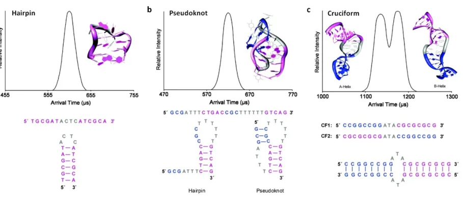
The gas-phase conformations adopted by different DNA secondary structures, including hairpins, pseudoknots and cruciforms (Holliday junction) have been studied using both IM-MS and molecular dynamics (Figure 6). At lower charge density, all these secondary structures are stable over the typical millisecond timescale of IM analysis. Where both pseudoknots and hairpins can be formed, pseudoknots are found to be favoured due to the formation of extra Watson–Crick pairs between the initial secondary structure formed (usually a hairpin) and another single-stranded region. At higher charge density these structures become elongated as Watson–Crick pairs are broken. Interestingly, the cruciform that was extensively studied formed a B-helix in the solution phase. However, this same analyte was found to exist as both a B-helix and an A-helix when desolvated, demonstrating that IM-MS can be used as tool to study structures adopted in a solvent-free environment.136 G-quadruplex formation, of particular importance in understanding the mechanism of cellular ageing and transformation, has also been monitored by IM-MS. In solution, these structures are known to form stacked planar rings where the plane is formed from four guanosine residues stabilised by atypical Hoogsteen hydrogen bonds and cations between the planes.140 These structures are retained upon vaporisation as confirmed by IM-MS. Analysis of tandem repeats of the telomeric region, where G-quadruplexes are often observed, allowed distinction and determination of G-quadruplex isomers.141 Using IM-MS, the intermediates in the assembly of these G-quadruplexes could be characterised and it was only by using such a strategy that it could be demonstrated that the final kinetically stable tetramers form an equilibrium state of DNA monomers, dimers and trimers, upon addition of ammonium cations.138
Synthetic molecules including metal-ring host-guest complexes, rotaxanes, catenanes, macrocyclic porphyrin-like systems and synthetic baskets like resorcinarenes represent an exciting branch of chemistry due to their potential applications in biosensing, drug delivery, catalysis, and material engineering. In many cases, the interaction between host and guest is controlled by fine structural differences, such as cis-trans isomerism. In-depth characterisation is therefore challenging, especially when classical biophysical methodologies are not applicable. The coupling of IM to MS offers a unique strategy to address the structural features of these systems with evident advantages over other techniques, combining the possibility of resolving different chemical entities by virtue of their m/z values before measuring their gas-phase conformations, without the need for highly pure, concentrated sample solutions and offering high speed of analysis. Recent studies have highlighted the importance of IM-MS to resolve very similar structures: cis-trans cyclometallated cages143 and rigid coordination-based rectangular, triangular and prismatic platinum-complexes144 were separated using DTIMS-MS; regio- and stereo-isomeric pairs of multi-ruthenated porphyrins145 and entire libraries of supramolecular assemblies using TWIMS.146 In many instances, prediction of candidate structures by molecular modelling was also used to help corroborate these results.
Conclusions and future perspectives
Traditionally, MS, which operates on the microsecond timescale, has been combined with chromatographic separation (either liquid or gas) operating in the region of seconds to minutes, for the analysis of mixtures. IM separation typically operates on the millisecond timescale, and consequently can be nested between chromatographic separation and MS analysis. Thus, the benefits of IMS for improved analysis of mixtures can be leveraged without detriment to the already established complementarity of chromatography and MS. We anticipate that in the future, (GC/LC)-IM-MS will become standard for optimal analytical capability in many situations. However, significant improvements in IM resolution will be required for it to ‘outperform’ recent developments in ultra-high performance LC, and its benefits for high-throughput analyte separation lie in its capability of separating ions by different physico-chemical properties.
A great strength of IM-MS is the ability to characterise and compare dynamic changes in analyte structure, which has previously been extremely difficult for most biological samples. At present IM-MS studies often require ‘validation’ by other strategies, in part due to natural scepticism towards ‘new’ (or rather unfamiliar) techniques. However, the field is now approaching sufficient maturity that it should be possible for IM-MS generated structural information, acquired under carefully controlled conditions, to stand alone on its own merits. Indeed, structures inferred by X-ray crystallography, should ideally be used to complement, rather than purely validate the gas-phase conformational information generated by IM-MS. IMS is limited in that it is not capable of providing detailed atomic-level structural information. However, this is counterbalanced by its utility in studying conformational dynamics of rapidly evolving systems.
Table 1 Comparison of the three main types of IMS: drift tube ion mobility separation (DTIMS), travelling wave ion mobility spectrometry (TWIMS) and field asymmetric ion mobility spectrometry (FAIMS). CCS: collisional cross-section; CV: compensation voltage; FWHM: full width at half maximum
DTIMS TWIMS FAIMS
Advantages
Rotationally averaged collisional cross section
(CCS; Ω) i.e. ‘shape’ can be measured (Å2) Rotationally averaged CCS can be determined
Can be used to separate species of very similar mobility i.e. has high resolving power
(>100 as defined by Ω/ΔΩ measured at full width at half maximum (FWHM))155
High resolving power (≤~100 as defined by Ω/ ΔΩ at FWHM)14
Relatively straightforward to transfer the ion mobility device between different mass
spectrometers Can be used to mobility separate product
ions generated either by collision-induced dissociation or electron-transfer dissociation
Disadvantages CCS determination requires calibration of the
drift time through the TWIMS cell, ideally using a calibrant of similar physical and
chemical properties
CCS cannot be determined
Relatively low resolving power (≤ ~45 as defined by Ω/ΔΩ at FWHM)150
The geometric configuration of current commercial DTIMS-MS instruments means that it can only be used to separate analytes
immediately post-ionisation
The geometric configuration of a FAIMS-MS instrument means that it can only be used to
separate analytes immediately post-ionisation
Gating-type instruments are susceptible to ion losses when transferred from atmospheric pressure during ionisation to the
Ion heating can occur as ions are injected into the TWIMS cell which may affect gas-phase
conformation. Unless carefully controlled,
The percentage of ions detected relative to those generated following ionisation (i.e. the
reduced pressure required for analysis the process of measurement may therefore perturb analyte structure
under conditions where the compensation voltage (CV) is ramped (CV scanning mode),
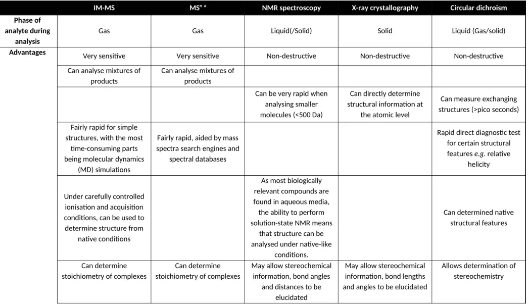
Table 2 Some advantages and disadvantages of commonly applied analytical techniques for determining structural information about analytes156, 157
IM-MS MSn a NMR spectroscopy X-ray crystallography Circular dichroism
Phase of analyte during
analysis
Gas Gas Liquid(/Solid) Solid Liquid (Gas/solid)
Advantages Very sensitive Very sensitive Non-destructive Non-destructive Non-destructive
Can analyse mixtures of products
Can analyse mixtures of products
Can be very rapid when analysing smaller molecules (<500 Da)
Can directly determine structural information at
the atomic level
Can measure exchanging structures (>pico seconds)
Fairly rapid for simple structures, with the most
time-consuming parts being molecular dynamics
(MD) simulations
Fairly rapid, aided by mass spectra search engines and
spectral databases
Rapid direct diagnostic test for certain structural features e.g. relative
helicity
Under carefully controlled ionisation and acquisition conditions, can be used to determine structure from
native conditions
As most biologically relevant compounds are found in aqueous media, the ability to perform solution-state NMR means
that structure can be analysed under native-like
conditions.
Can determined native structural features
Can determine stoichiometry of complexes
Can determine stoichiometry of complexes
May allow stereochemical information, bond angles
and distances to be elucidated
May allow stereochemical information, bond lengths and angles to be elucidated
Measures 3-D structure in dynamic motion
Products of electron-mediated dissociation can
be used to infer 3D-structure
Conventional solution state NMR spectroscopy can measure 3D structure in
dynamic motion
Disadvantages Analyte must be able to be
ionised
Analyte must be able to be ionised
Difficult to analyse mixtures of products. Samples often have to be purified and concentrated,
which may affect the structure of biological
samples
Requires purified and crystallised material. May
be time-consuming or impossible to generate
crystals
Cannot be used for mixtures. Requires relatively concentrated
(~0.5 mgmL-1) purified
samples
Relies on MD simulations to indirectly determine
precise structural information from CCS values. MD simulations become more challenging
as molecules become larger
Difficult to ascertain detailed 3D structure information directly from
mass spectra
Analysis of spectra becomes difficult for larger
molecules
Gives no specific structural information at the atomic
level
Destructive Destructive Analyte may be damaged
by the X-rays
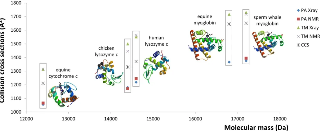
Figure 4 Comparison of experimentally estimated collision cross sections (Å2) (CCS) of five protein standards. CCS values calculated using either
Figure 5 Conformational heterogeneity and dissociation of subunit I from ICL12 implies a mechanism for closing the H+ channel. Arrival time
Figure 6 Polynucleotide structures as determined using a combination of molecular dynamic simulations and experimentally determined arrival time distributions. Depicted (top) are the arrival time distributions (ATDs) and structures as predicted using molecular dynamics (MD), together with the polynucleotide sequence(s) and expected conformations (bottom). a) 5’-TGCGATACTCATCGCA-3’ adopts a hairpin secondary structure where the nucleotide backbone ‘loops’ back to form a series of Watson-Crick pairs. b) 5’-GCGATTTCTGACCGCTTTTTTGTCAG-3’ could adopt one of two potential conformations, forming either a hairpin structure, or a pseudoknot where two loops are formed and held in place by Watson-Crick pairs to the adjacent nucleotide sequence. MD and ion mobility-mass spectrometry (IM-MS) measurements both indicate the presence of a single structure corresponding to the pseudoknot. c) The DNA strands CF1 (5’-CCGGCCGGATACGCGCGCG-3’) and CF2 (5’-CGCGCGCGATACCGGCCGG-3’) together form two IM resolvable structures, that MD shows are cruciform secondary structures adopting both A and B helical conformations. Adapted with permission from 134. Copyright
REFERENCES
1. D'Agostino, P.A. & Chenier, C.L. Desorption electrospray ionization mass spectrometric analysis of organophosphorus chemical warfare agents using ion mobility and tandem mass spectrometry. Rapid Commun. Mass Spectrom. 24, 1617-1624 (2010).
2. McDaniel, E.W., Martin, D.W. & Barnes, W.S. Drift-tube mass spectrometer for studies of low-energy ion-molecule reactions. Rev. Sci. Instrum. 33, 2-7 (1962).
3. Mason, E.A. & Schamp Jr., H.W. Mobility of gaseous ions in weak electric fields. Ann. Phys. 4, 233-270 (1958).
4. Creaser, C.S. et al. Ion mobility spectrometry: a review. Part 1. Structural analysis by mobility measurement. Analyst 129, 984-994 (2004).
5. Wyttenbach, T., Kemper, P.R. & Bowers, M.T. Design of a new electrospray ion mobility mass spectrometer. Int. J. Mass Spectrom. 212, 13-23 (2001).
6. Harvey, S.R., MacPhee, C.E. & Barran, P.E. Ion mobility mass spectrometry for peptide analysis. Methods 54, 454-461 (2011).
7. Giles, K. et al. Applications of a travelling-wave based radio-frequency-only stacked ring ion guide. Rapid Commun. Mass Spectrom. 18, 2401-2414 (2004).
8. Bush, M.F. et al. Collision cross sections of proteins and their complexes: a calibration framework and database for gas-phase structural biology. Anal. Chem. 82, 9557-9565 (2010).
9. Campuzano, I. et al. Structural characterization of drug-like compounds by ion mobility mass spectrometry: Comparison of theoretical and experimentally derived nitrogen collision cross sections. Anal. Chem. 84, 1026-1033 (2012).
10. Bush, M.F., Campuzano, I.D. & Robinson, C.V. Ion mobility mass spectrometry of peptide ions: Effects of drift gas and calibration strategies. Anal. Chem. 84, 7124-7130 (2012).
11. Chawner, R. et al. QconCAT standard for calibration of ion mobility-mass spectrometry systems. J. Proteome Res. 11, 5564-5572 (2012).
12. Purves, R.W. & Guevremont, R. Electrospray ionization high-field asymmetric waveform ion mobility spectrometry-mass spectrometry. Anal. Chem. 71, 2346-2357 (1999).
13. Canterbury, J.D., Yi, X., Hoopman, M.R. & MacCoss, M.J. Assessing the dynamic range and peak capacity of nanoflow LC-FAIMS-MS on an ion trap mass spectrometer for proteomics. Anal. Chem. 80, 6888-6897 (2008).
14. Shvartsburg, A.A., Danielson, W.F. & Smith, R.D. High-resolution differential ion mobility separations using helium-rich gases. Anal. Chem. 82, 2456-2462 (2010).
15. Shvartsburg, A.A. & Smith, R.D. Accelerated high-resolution differential ion mobility separations using hydrogen. Anal. Chem. 83, 9159-9166 (2011).
16. Shvartsburg, A.A. et al. High-definition differential ion mobility spectrometry with resolving power up to 500. J. Am. Soc. Mass Spectrom. 24, 109-114 (2013).
17. Knutson, E.O. & Whitby, K.T. Aerosol classification by electric mobility: Apparatus, theory, and applications. J. Aerosol Sci. 6, 443-451 (1975).
18. Pomareda, V., Lopez-Vidal, S., Calvo, D., Pardo, A. & Marco, S. A novel differential mobility analyzer as a VOC detector and multivariate techniques for identification and quantification. Analyst 138, 3512-3521 (2013).
19. de la Mora, J.F., de Juan, L., Eichler, T. & Rosell, J. Differential mobility analysis of molecular ions and nanometer particles. TRAC - Trend. Anal. Chem. 17, 328-339 (1998).
20. Rus, J. et al. IMS-MS studies based on coupling a differential mobility analyzer (DMA) to commercial API-MS systems. Int. J. Mass Spectrom. 298, 30-40 (2010).
21. Creese, A.J. & Cooper, H.J. Separation and identification of isomeric glycopeptides by high field asymmetric waveform ion mobility spectrometry. Anal. Chem. 84, 2597-2601 (2012). 22. Varesio, E., Le Blanc, J.C.Y. & Hopfgartner, G. Real-time 2D separation by LC x differential ion
23. Howdle, M.D., Eckers, C., Laures, A.M.-F. & Creaser, C.S. The effect of drift gas on the separation of active pharmaceutical ingredients and impurities by ion mobility-mass spectrometry. Int. J. Mass Spectrom. 298, 72-77 (2010).
24. Fernández-Maestre, R., Wu, C. & Hill, J., H. H. Using a buffer gas modifier to change separation selectivity in ion mobility spectrometry. Int. J. Mass Spectrom. 298, 2-9 (2010). 25. Fernández-Maestre, R., Wu, C. & Hill, J., H. H. Buffer gas modifiers effect resolution in ion
mobility spectrometry through selective ion-molecule clustering reactions. Rapid Commun. Mass Spectrom. 26, 2211-2223 (2012).
26. Holness, H.K., Jamal, A., Mebel, A. & Almirall, J.R. Separation mechanism of chiral impurities, ephedrine and pseudoephedrin, found in amphetamine-type substances using achiral modifiers in the gas phase. Anal. Bioanal. Chem. 404, 2407-2416 (2012).
27. Howdle, M.D., Eckers, C., Laures, A.M.-F. & Creaser, C.S. The use of shift reagents in ion mobility-mass spectrometry: Studies on the complexation of an active pharmaceutical ingredient with polyethylene glycol excipients. J. Am. Soc. Mass Spectrom. 20 (2009).
28. Hilderbrand, A.E., Myung, S. & Clemmer, D.E. Exploring crown ethers as shift reagents for ion mobility spectrometry. Anal Chem 78, 6792-6800 (2006).
29. Bohrer, B.C. & Clemmer, D.E. Shift Reagents for Multidimensional Ion Mobility Spectrometry-Mass Spectrometry Analysis of Complex Peptide Mixtures: Evaluation of 18-Crown-6 Ether Complexes. Anal Chem 83, 5377-5385 (2011).
30. Kiss, A. & Heeren, R.M.A. Size, weight and position: ion mobility spectrometry and imaging MS combined. Anal. Bioanal. Chem. 399, 2623-2634 (2011).
31. Stauber, J. et al. On-tissue protein identification and imaging by MALDI-ion mobility mass spectrometry. J. Am. Soc. Mass Spectrom. 21, 338-347 (2010).
32. Trim, P.J. et al. Matrix-assisted laser desorption/ionization-ion mobility separation-mass spectrometry imaging of vinblastine in whole body tissue sections. Anal. Chem. 80, 8628-8634 (2008).
33. Shliaha, P.V., Bond, N.J., Gatto, L. & Lilley, K.S. Effects of travelling wave ion mobility separation on data independent acquisition in proteomics studies. J. Proteome Res. 12, 2323-2339 (2013).
34. Rodriguez-Suarez, E. et al. An ion mobility assisted data independent LC-MS strategy for the analysis of complex biological samples. Curr. Anal. Chem. 9, 199-211 (2013).
35. Parson, W.B. et al. Rapid analysis of isomeric exogenous metabolites by differential mobility spectrometry-mass spectrometry. Rapid Commun. Mass Spectrom. 25, 3382-3386 (2011). 36. Esquenazi, E., Daly, M., Bahrainwala, T., Gerwick, W.H. & Dorrestein, P.C. Ion mobility mass
spectrometry enables the efficient detection and identification of halogenated natural products from cyanobacteria with minimal sample preparation. Bioorgan. Med. Chem. 19, 6639-6644 (2011).
37. Harry, E.L., Weston, D.J., Bristow, A.W.T., Wilson, I.D. & Creaser, C.S. An approach to enhancing coverage of the urinary metabonome using liquid chromatography-ion mobility-mass spectrometry. J. Chromatogr. B. 871, 357-361 (2008).
38. Picotti, P., Bodenmiller, B., Mueller, L.N., Domon, B. & Aebersold, R. Full dynamic range proteome analysis of S. cerevisiae by targeted proteomics. Cell 138, 795-806 (2009).
39. Brownridge, P. et al. Global absolute quantification of a proteome: Challenges in the deployment of a QconCAT strategy. Proteomics 2011, 2957-2970 (2011).
40. Saba, J., Bonneil, E., Pomiès, C., Eng, K. & Thibault, P. Enhanced sensitivity in proteomics experiments using FAIMS coupled with a hybrid linear ion trap/orbitrap mass spectrometer. J. Proteome Res. 8, 3355-3366 (2009).
42. Ibrahim, Y.M., Shvartsburg, A.A., Smith, R.D. & Belov, M.E. Ultrasensitive identification of localization variants of modified peptides using ion mobility spectrometry. Anal. Chem. 83, 5617-5623 (2011).
43. Cuyckens, F. et al. Identifying metabolite ions of peptide drugs in the presence of an in vivo matrix background. Bioanalysis 4, 595-604 (2012).
44. Blackburn, M.A. et al. Identification and subsequent removal of an interference by FAIMS in the bioanalysis of dianicline in animal plasma. Bioanalysis 3, 2119-2127 (2011).
45. Guddat, S., Thevis, M., Kapron, J., Thomas, A. & Schänzer, W. Application of FAIMS to anabolic androgenic steroids in sport drug testing. Drug Testing Anal. 1, 545-553 (2009). 46. Scarff, C.A., Thalassinos, K., Hilton, G.R. & Scrivens, J.H. Travelling wave ion mobility mass
spectrometry studies of protein structure: Biological significance and comparison with X-ray crystallography and nuclear magnetic resonance spectroscopy measurements. Rapid Commun. Mass Spectrom. 22, 3297-3304 (2008).
47. Gidden, J., Bushnell, J.E. & Bowers, M.T. Gas-phase conformations and folding energetics of oligonucleotides: dTG- and dGT-. J. Am. Chem. Soc. 123, 5610-5611 (2001).
48. Gidden, J. & Bowers, M.T. Gas-phase conformational and energetic properties of deprotonated dinucleotides. Eur. Phys. J. D 20, 409-419 (2002).
49. Wyttenbach, T., Grabenauer, M., Thalassinos, K., Scrivens, J.H. & Bowers, M.T. The effect of calcium ions and peptide ligands on the relative stabilities of the calmodulin dumbbell and compact structures. J. Phys. Chem. B 114, 437-447 (2010).
50. Jenner, M. et al. Detection of a protein conformational equilibrium by electrospray ionisation-ion mobility-mass spectrometry. Angew. Chem. Int. Ed. 50, 8291-8294 (2011). 51. Bereszczak, J.Z. et al. Structure, stability and dynamics of norovirus P domain derived protein
complexes studied by native mass spectrometry. J. Struct. Biol. 177, 273-282 (2012).
52. Shi, H., Pierson, N.A., Valentine, S.J. & Clemmer, D.E. Conformation types of ubiquitin [M+8H]8+ ions from water:methanol solutions: Evidence for the N and A states in aqueous solution. J. Phys. Chem. B 116, 3344-3352 (2012).
53. Smith, D.P., Giles, K., Bateman, R.H., Radford, S.E. & Ashcroft, A.E. Monitoring copopulated conformational states during protein folding events using electrospray ionization-ion mobility spectrometry-mass spectrometry. J. Am. Soc. Mass. Spectrom. 18, 2180-2190 (2007).
54. Nabuchi, Y., Hirose, K. & Takayama, M. Ion mobility and collision-induced dissociation analysis of carbonic anhydrase 2. Anal. Chem. 82, 8890-8896 (2010).
55. Hyung, S.-J., Robinson, C.V. & Ruotolo, B.T. Gas-phase unfolding and disassembly reveals stability differences in ligand-bound multiprotein complexes. Chem. Biol. 16, 382-390 (2009). 56. Hopper, J.T.S. & Oldham, N.J. Collision induced unfolding of protein ions in the gas phase studied by ion mobility-mass spectrometry: The effect of ligand binding on conformational stability. J. Am. Soc. Mass Spectrom. 20, 1851-1858 (2009).
57. Smith, D.P., Radford, S.E. & Ashcroft, A.E. Elongated oligomers in 2-microglobulin amyloid assembly revealed by ion mobility spectrometry-mass spectrometry. P. Natl. Acad. Sci. USA
107, 6794-6798 (2010).
58. Beveridge, R., Chappuis, Q., MacPhee, C. & Barran, P. Mass spectrometry methods for intrinsically disordered proteins. Analyst 138, 32-42 (2013).
59. Uetrecht, C. et al. Stability and shape of hepatitis B virus capsids in vacuo. Angew. Chem. Int. Ed. 47, 6247-6251 (2008).
60. Ruotolo, B.T. et al. Evidence for macromolecular protein rings in the absence of bulk water. Science 310, 1658-1661 (2005).
62. Wyttenbach, T., von Helden, G. & Bowers, M.T. Gas-phase conformation of biological molecules: Bradykinin. J. Am. Chem. Soc. 118, 8355-8364 (1996).
63. Baumketner, A. et al. Amyloid β-protein monomer structure: A computationaly and experimental study. Protein Sci. 15, 420-428 (2006).
64. Dear, G.J. et al. Sites of metabolic substitution: Investigating metabolite structures utilising ion mobility and molecular modelling. Rapid Commun. Mass Spectrom. 24, 3157-3162 (2010).
65. Cuyckens, F. et al. Product ion mobility as a promising tool for assignment of positional isomers of drug metabolites. Rapid Commun. Mass Spectrom. 25, 3497-3503 (2011).
66. Wyttenbach, T., von Helden, G., Batka, J.J., Carlat, D. & Bowers, M.T. Effect of the long-range potential on ion mobility measurements. J. Am. Soc. Mass Spectrom. 8, 275-282 (1997). 67. Shvartsburg, A.A. & Jarrold, M.F. An exact hard-spheres scattering model for the mobilities
of polyatomic ions. Chem. Phys. Lett. 261, 86-91 (1996).
68. Shvartsburg, A.A., Schatz, G.C. & Jarrold, M.F. Mobilities of carbon cluster ions: Critical importance of the molecular attractive potential. J. Chem. Phys. 108, 2416-2423 (1998). 69. Jurneczko, E. & Barran, P.E. How useful is ion mobility mass spectrometry for structural
biology? The relationship between protein crystal structures and their collision cross sections in the gas phase. Analyst 136, 20-28 (2011).
70. Bleiholder, C., Wyttenbach, T. & Bowers, M.T. A novel projection approximation algorithm for the fast and accurate computation of molecular collision cross sections (I). Method. Int. J. Mass Spectrom. 308, 1-10 (2011).
71. Bleiholder, C., Contreras, S., Do, T.D. & Bowers, M.T. A novel projection approximation algorithm for the fast and accurate computation of molecular collision cross sections (II). Model parameterization and definitions of empirical shape factors for proteins. Int. J. Mass Spectrom. 345-347, 89-96 (2013).
72. Bleiholder, C., Contreras, S. & Bowers, M.T. A novel projection approximation algorithm for the fast and accurate computation of molecular collision cross section (IV). Application to polypeptides. Int. J. Mass Spectrom. 354-355, 275-280 (2013).
73. Wyttenbach, T., Bleiholder, C. & Bowers, M.T. Factors contributing to the collision cross section of polyatomic ions in the kilodalton to gigadalton range: Application to ion mobility measurements. Anal. Chem. 85, 2191-2199 (2013).
74. Hall, Z. & Robinson, C.V. Do charge state signatures guarantee protein conformations? J. Am. Soc. Mass Spectrom. 23, 1161-1168 (2012).
75. Berezovskaya, Y., Porrini, M. & Barran, P.E. The effect of salt on the conformations of three model proteins is revealed by variable temperature ion mobility mass spectrometry. Int. J. Mass Spectrom. 345, 8-18 (2013).
76. Shvartsburg, A.A. & Smith, R.D. Separation of protein conformers by differential ion mobility in hydrogen-rich gases. Anal. Chem. 85, 6967-6973 (2013).
77. Bernstein, S.L. et al. Amyloid-β protein oligomerization and the importance of tetramers and dodecamers in the aetiology of Alzheimer's disease. Nat. Chem. 1, 326-331 (2009).
78. Grabenauer, M. et al. Spermine binding to Parkinson's protein α-synuclein and its disease-related A30P and A53T mutants. J. Phys. Chem. B 112, 11147-11154 (2008).
79. Shimizu, A., Ohe, T. & Chiba, M. A novel method for the determination of the site of glucuronidation by ion mobility spectrometry-mass spectrometry. Drug Metab. Dispos. 40, 1456-1459 (2012).
80. Baker, E.S. et al. Diastereomer assignment of an olefin-linked bis-paracyclophane by ion mobility mass spectrometry. J. Am. Chem. Soc. 126, 6255-6257 (2004).
81. Both, P. et al. Discrimination of epimeric glycans and glycopeptides using ion-mobility mass spectrometry and its potential for carbohydrate sequencing. Nat. Chem. 6, 65-74 (2014). 82. Lee, S. et al. Extracted fragment ion mobility distributions: A new method for complex
83. Florance, H.V. et al. Evidence for alpha-helices in the gas phase: A case study using melittin from honey bee venom. Analyst 136, 3446-3452 (2011).
84. Jarrold, M.F. Helices and sheets in vacuo. Phys. Chem. Chem. Phys. 9, 1659-1671 (2007). 85. Zilch, L.W., Kaleta, D.T., Kohtani, M., Krishnan, R. & Jarrold, M.F. Folding and unfolding of
helix-turn-helix motifs in the gas phase. J. Am. Soc. Mass Spectrom. 18, 1239-1248 (2007). 86. McLean, J.R. et al. Factors that influence helical preferences for singly charged gas-phase
peptide ions: The effects of multiple potential charge-carrying sites. J. Phys. Chem. B 114, 809-816 (2010).
87. Albrieux, F. et al. Conformation of polyalanine and polyglycine dications in the gas phase: Insight from ion mobility spectrometry and replica-exchange molecular dynamics. J. Phys. Chem. A 114, 6888-6896 (2010).
88. Albrieux, F. et al. Structural preferences of gas-phase M2TMP monomers upon sequence variations. J. Phys. Chem. A 115, 4711-4718 (2011).
89. Wu, C., Klasmeier, J. & Hill, H.H. Atmospheric pressure ion mobility spectrometry of protonated and sodiated peptides. Rapid Commun. Mass Spectrom. 13, 1138-1142 (1999). 90. Liu, D.F. et al. Oxytocin-receptor binding: Why divalent metals are essential. J. Am. Chem.
Soc. 127, 2024-2025 (2005).
91. Rožman, M. & Gaskell, S.J. Non-covalent interactions of alkali metal cations with singly charged tryptic peptides. J. Mass Spectrom. 45, 1409-1415 (2010).
92. Chen, L., Gao, Y.Q. & Russell, D.H. How alkali metal ion binding alters the conformation preferences of gramicidin A: A molecular dynamics and ion mobility study. J. Phys. Chem. A
116, 689-696 (2012).
93. Garcia, I.R., Giles, K., Bateman, R.H. & Gaskell, S.J. Studies of peptide a- and b-type fragment ions using stable isotope labeling and integrated ion mobility/tandem mass spectrometry. J. Am. Soc. Mass Spectrom. 19, 1781-1787 (2008).
94. Polfer, N.C., Bohrer, B.C., Plasencia, M.D., Paizs, B. & Clemmer, D.E. On the dynamics of fragment isomerization in collision-induced dissociation of peptides. J. Phys. Chem. A 112, 1286-1293 (2008).
95. Saminathan, I.S. et al. The extent and effects of peptide sequence scrambling via formation of macrocyclic b ions in model proteins. J. Am. Soc. Mass Spectrom. 21, 2085-2094 (2010). 96. Chawner, R., Gaskell, S.J. & Eyers, C.E. Proposal for a common nomenclature for peptide
fragment ions generated following sequence scrambling during collision-induced dissociation. Rapid Commun. Mass Spectrom. 26, 205-206 (2012).
97. Moss, C.L. et al. Assigning structures to gas-phase peptide cations and cation-radicals. An infrared multiphoton dissociation, ion mobility, electron transfer, and computational study of a histidine peptide ion. J. Phys. Chem. B 116, 3445-3456 (2012).
98. van den Heuvel, R.H.H. & Heck, A.J.R. Native protein mass spectrometry: from intact oligomers to functional machineries. Curr. Opin. Chem. Biol. 8, 519-526 (2004).
99. Kaddis, C.S. & Loo, J.A. Native protein MS and ion mobility: Large flying proteins with ESI. Anal. Chem. 79, 1778-1784 (2007).
100. Heck, A.J.R. Native mass spectrometry: A bridge between interactomics and structural biology. Nat. Methods 5, 927-933 (2008).
101. Kondrat, F.D.L., Kowald, G.R., Scarff, C.A., Scrivens, J.H. & Blindauer, C.A. Resolution of a paradox by native mass spectrometry: Facile occupation of all four metal binding sites in the dimeric zinc sensor SmtB. Chem. Commun. 49, 813-815 (2013).
102. Konijnenberg, A., Butterer, A. & Sobott, F. Native ion mobility-mass spectrometry and related methods in structural biology. BBA-Proteins Proteom. 1835, 1239-1256 (2013). 103. Hall, Z., Politis, A., Bush, M.F., Smith, L.J. & Robinson, C.V. Charge-State Dependent