Global Perspectives
Section Editors Johan A. Aarli, MD
Oded Abramsky, MD, PhD, FRCP
Michael J. Potchen, MD Sam D. Kampondeni,
MMed
Khalid Ibrahim, MS Joseph Bonner, PhD Karl B. Seydel, MD Terrie E. Taylor, DO Gretchen L. Birbeck, MD
Correspondence to Professor Birbeck: Gretchen_Birbeck@URMC. Rochester.edu
NEUROINTERP: A METHOD FOR FACILITATING NEUROIMAGING RESEARCH ON CEREBRAL MALARIA
Radiologic data are increasingly important in clinical care guidelines for neurologic disorders and in the conduct of clinical trials assessing novel therapies. The infrastructure and expertise for neuroradiologic evaluations remain scarce in resource-limited set-tings, but where available, MRI and CT capacity can offer new insights into common, globally devas-tating diseases. In vivo data for frequently fatal trop-ical conditions such as cerebral malaria have been largely limited to autopsy studies, which only provide information on nonsurvivors at a single point in time. New imaging facilities in sub-Saharan Africa offer opportunities for expanded research on tropical neurologic disorders.1However, data
man-agement challenges hamper the research utility of radiologic evaluations.
Traditional methods of capturing radiographic data from neuroradiologic evaluations provided for clinical purposes rely on unstructured narrative descriptions. Narratives are unsuitable for research analysis for several reasons: the text data are not structured for statistical analysis; reports are rarely standardized among radiol-ogists or among institutions; determining whether an unmentioned feature is absent or inadvertently disre-garded in the narrative is frequently impossible; search-ing for unambiguous words in text reports to develop a database (e.g., swelling vs edema) is haphazard; and, as with many physician-driven technologies, clinical inter-pretations may have considerable inter- and intrareader variability.
To improve the utility of MRI in clinical research of cerebral malaria, we devised a data entry applica-tion and workflow instrument, NeuroInterp. As an instrument, NeuroInterp prompts radiologists to sys-tematically glean data from their evaluations. As a workflow instrument, NeuroInterp ensures that serial readings are evaluated by multiple readers with dis-cordant data identified and adjudication addressed. This methodologic approach to developing a data management instrument for MRI data could also be
applied to other tropical conditions whose radio-graphic correlates have not been well-characterized.
The NeuroInterp instrument. Development of the NeuroInterp instrument began in 2008, when, as part of a long-standing NIH-funded study of pediat-ric cerebral malaria, brain MRI acquisition became part of the standard evaluation of comatose patients admitted to the pediatric research ward at Queen Elizabeth Central Hospital in Blantyre, Malawi.1
CT and autopsy studies of cerebral malaria sug-gested a number of likely radiographic findings, including acute edema, chronic atrophy, inflamma-tory lesions, and ischemia.2,3To develop appropriate
items for inclusion in the NeuroInterp platform for pediatric cerebral malaria findings, 2 years of narrative-based findings were summarized and dis-tilled into discrete items for structured entry. Two fellowship-trained radiologists, one in neuroradiol-ogy (M.J.P.) and one in MRI (S.D.K.), read all stud-ies. The radiologists reviewed all MRIs from the research ward and collaboratively developed a sys-tematic scoring procedure for the potential cerebral malaria cases that included graded measures of cere-bral edema, periventricular white matter changes, cortical abnormalities, and brainstem changes, among others.4 These included variables denoting
anatomical localization and characterization of spe-cific MRI pulse sequence characteristics, such as presence or absence of T2 abnormalities and/or diffusion-weighted imaging changes. The variables for each feature were created to permit unambiguous and mutually exclusive options (e.g., “Were there T2 signal abnormalities present within the supraten-torial cortex? [yes/no]”). Equivocation options of “not applicable” were also included for most fea-tures. Some features cannot be endorsed with con-fidence. To address the ambivalence, we included a field to capture the reviewer confidence on the pres-ence or abspres-ence of the feature in question (,25%, 25%–50%, 50%–75%, .75%). Fields were also created to capture the change in a clinical feature over the course of serial readings in the same patient. Fields capturing longitudinal data were given the
From Michigan State University (M.J.P., K.I., J.B., K.B.S., T.E.T., G.L.B.); Blantyre Malaria Project (T.E.T., K.B.S.); and Queen Elizabeth Central Hospital (S.D.K.), Blantyre, Malawi.
following answer options: progressed, remained the same, or regressed. Importantly, this was an iterative process whereby new insights into relevant findings led to additional items for inclusion in NeuroInterp and/or further delineation of existing variables until item saturation when no additional new findings were routinely identified.
The NeuroInterp application. To begin the data entry application, we generated metadata to describe each of the fields required by the instrument. The meta-data as a set contain the caption or description, the field or variable name, the data type (number or text), and the answer options (e.g., 1 5 yes; 2 5 no). The data entry forms for NeuroInterp are built on a proprietary research information management system called RIX. This platform generates hyper-text markup language (similar to Web pages) forms based on the metadata with fields required by the instrument. Each data entry form is dedicated to one radiologist’s evaluation of one neuroradiologic inter-pretation for one patient. The metadata can be used to generate forms to capture structured data as guided by the instrument.
The NeuroInterp workflow.To begin the workflow, ra-diologists assemble the files for the study, launch the NeuroInterp form, find or create a patient, review the
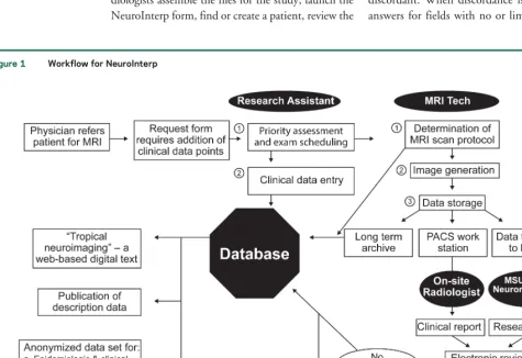
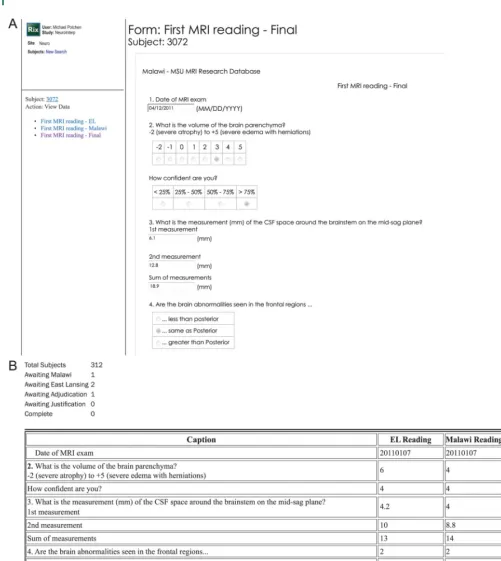
files, and enter their findings as prompted by the form, which follows the instrument paradigm. Peri-odically, an adjudicating radiologist views a report showing which patients have 2 readings and await a discordance review (see figures 1 and 2). From the report, a patient is selected and the adjudicator (in collaboration or solo) makes a judgment for a final answer for each discordant field.
Discordance algorithms for identifying cases for adjudication were developed to compare endorsed values from 2 readers and identify discordant values. When a field has identical values for each field, the value is the automatic final answer. Fields that cap-ture an unambiguous endorsement of the presence or absence of a specific feature allow no discordance between readers. Fields that capture the reader’s confidence in an unambiguous endorsement have greater tolerance for discordance. For these fields, the workflow allows the“worse case” value as the automatic final answer. Fields that capture continu-ous measures such as volume also have a higher tol-erance for discordance. For these fields, the average value between the 2 endorsed answers becomes the automatic final answer unless the values exceed the accepted variability and are therefore noted to be discordant. When discordance is detected between answers for fields with no or limited tolerance, the
Figure 1 Workflow for NeuroInterp
MSU5Michigan State University; PACS5picture archiving and communication system.
586 Neurology 81 August 6, 2013
adjudicating user must choose a final answer upon review of the neuroradiologic images/files/studies. Open-ended text fields require a review of both readers’ evaluations to enter a final adjudicated answer.
NeuroInterp—The final product. The NeuroInterp instrument guides structured evaluation of scaled quantitative assessments of neurologic structures.
disagreement on variables detailing degrees of abnor-mality or involvement.
To date, NeuroInterp has facilitated the systematic study of the radiologic findings of cerebral malaria sur-vivors with neurologic sequelae5as well as a study of
normative brain MRI findings in a representative com-munity-based sample of Malawian children.6
Neuro-Interp offers a feasible approach to the challenges of radiologic data acquisition and management, especially for conditions with little prior descriptive data avail-able. We are presently enhancing NeuroInterp further with instructional images to facilitate reader training and improve interreader reliability. Plans are also under way to transition from propriety software to open source software, which should allow more broad use of this data management tool in cerebral malaria imag-ing research.
AUTHOR CONTRIBUTIONS
Michael J. Potchen: conceptualized the NeuroInterp program and worked on early prototypes, developed the first draft of the manuscript, reviewed and edited subsequent versions, and approved the submitted version. Sam D. Kampondeni: worked on early prototypes of NeuroInterp and provided substantive feedback into early development, provided critical intellectual reviews and editing on early versions of the manuscript, and approved the submitted version. Khalid Ibrahim: technical and logistical support for the full development of NeuroInterp and provided substan-tive feedback into early development, provided critical reviews and edited early versions of the manuscript, and approved the submitted version. Joseph Bonner: technical and logistical support for the full development of NeuroInterp and provided substantive feedback into early develop-ment, reviewed and edited early versions of the manuscript, and approved the submitted version. Karl B. Seydel and Terrie E. Taylor: assisted with regular assessments of the needed data from NeuroInterp as the product was being developed and provided substantive feedback into early devel-opment, reviewed and edited early versions of the manuscript, and approved the submitted version. Gretchen L. Birbeck: conceptualized the NeuroInterp program and worked on early prototypes, developed the first draft of the manuscript, reviewed and edited subsequent versions, and approved the submitted version.
STUDY FUNDING
Early prototypes of NeuroInterp were supported through a CDFP grant from Michigan State University (09-CDFP-1771). The definitive devel-opment of NeuroInterp was supported by award number R21NS069228 from the Fogarty International Center and the National Institute of Neurological Disorders and Stroke. The content is solely the responsibil-ity of the authors and does not necessarily represent the official views of the NIH.
DISCLOSURE
M.J. Potchen has received research funding from the US NIH and the Dana Foundation. S.D. Kampondeni, K. Ibrahim, and J. Bonner have received funding from the US NIH. K.B. Seydel has received research funding from the US NIH and the Dana Foundation. T.E. Taylor has received research funding from the US NIH. G.L. Birbeck has received research funding from the US NIH and the Dana Foundation. Go to Neurology.org for full disclosures.
REFERENCES
1. Potchen M, Kampondeni S, Birbeck G, et al. Magnetic resonance imaging in Malawi: contributions to clinical care, medical education and biomedical research. Malawi Med J 2011;7:60–64.
2. Taylor TE, Fu WJ, Carr RA, et al. Differentiating the pathologies of cerebral malaria by postmortem parasite counts. Nat Med 2004;10:143–145.
3. Potchen MJ, Birbeck GL, Demarco JK, et al. Neuroimaging findings in children with retinopathy-confirmed cerebral malaria. Eur J Radiol 2010;74:262–268.
4. Potchen MJ, Kampondeni SD, Seydel KB, et al. Acute brain MRI findings in 120 Malawian children with cerebral malaria: new insights into an ancient disease. AJNR Am J Neuroradiol 2012;33:1740–1746.
5. Kampondeni S, Birbeck G, Beare N, et al. Findings in brain injured survivors of pediatric cerebral malaria. Presented at the Annual Meeting for the Radiologic Society of North America; 2012; Seattle.
6. Potchen M, Kampondeni S, Mallewa M, et al. Clinically normal is structurally normal: findings from Malawi’s Brain Imaging in Normal Kids (BRINK) Study. Presented at the Annual Meeting for the Radiologic Society of North Amer-ica; 2012; Seattle.
588 Neurology 81 August 6, 2013
DOI 10.1212/WNL.0b013e31829e6ed5
2013;81;585-588
Neurology
Michael J. Potchen, Sam D. Kampondeni, Khalid Ibrahim, et al.
NeuroInterp: A method for facilitating neuroimaging research on cerebral malaria
This information is current as of August 5, 2013
rights reserved. Print ISSN: 0028-3878. Online ISSN: 1526-632X.
1951, it is now a weekly with 48 issues per year. Copyright © 2013 American Academy of Neurology. All ® is the official journal of the American Academy of Neurology. Published continuously since
Services
Updated Information &
http://n.neurology.org/content/81/6/585.full including high resolution figures, can be found at:
References
http://n.neurology.org/content/81/6/585.full#ref-list-1
This article cites 4 articles, 1 of which you can access for free at:
Citations
http://n.neurology.org/content/81/6/585.full##otherarticles This article has been cited by 6 HighWire-hosted articles:
Subspecialty Collections
http://n.neurology.org/cgi/collection/parasitic_infections Parasitic infections
http://n.neurology.org/cgi/collection/mri MRI
http://n.neurology.org/cgi/collection/health_systems Health systems
http://n.neurology.org/cgi/collection/diagnostic_test_assessment_ Diagnostic test assessment
http://n.neurology.org/cgi/collection/all_pediatric All Pediatric
http://n.neurology.org/cgi/collection/all_epidemiology All epidemiology
http://n.neurology.org/cgi/collection/access_to_care Access to care
following collection(s):
This article, along with others on similar topics, appears in the
Permissions & Licensing
http://www.neurology.org/about/about_the_journal#permissions its entirety can be found online at:
Information about reproducing this article in parts (figures,tables) or in
Reprints
http://n.neurology.org/subscribers/advertise
Information about ordering reprints can be found online:
rights reserved. Print ISSN: 0028-3878. Online ISSN: 1526-632X.
1951, it is now a weekly with 48 issues per year. Copyright © 2013 American Academy of Neurology. All ® is the official journal of the American Academy of Neurology. Published continuously since