Available Online at www.ijcsmc.com
International Journal of Computer Science and Mobile Computing
A Monthly Journal of Computer Science and Information Technology
ISSN 2320–088X
IMPACT FACTOR: 6.199IJCSMC, Vol. 8, Issue. 9, September 2019, pg.49 – 60
Brain Computer Interface (BCI)
Abdallah Abdelaziz
Department of Biomedical Engineering, Istanbul University, Istanbul, Turkey Academia.edu: independent.academia.edu/lioabdo
Abstract: With the technological development in the world especially in computers, a computer simulation was observed with almost all things around as in a lot of fields such as engineering, medical, chemical,...etc., so can a computer understand the human's emotions? Emotions are intrinsically connected to the way that people interact with each other. Emotion constitutes a major influence in determining human behaviors. A human being can read the emotional state of another human, and behave in the best way to improve their communication at that moment. This is because emotions can be recognized through words, voice intonation, facial expression, and body language. Can a computer understand the human's brain? Making the computer more empathic to the human is one of the aspects of affective computing. The computer can actually take a look inside the human’s head to observe their mental state. In this study, we will understand how can a computer understand human brain by understanding the electric signals that come from it.
Keywords: BCI, the human brain, electrical signals, Brain-computer interaction, emotion, emotion recognition, valence-arousal model, affective computing.
Introduction
BCI has many applications, especially for disabled persons. It reads the waves produced by the brain and translates these signals into actions and commands that can control the computer(s). A brain-computer interface (BCI) can enable such physically challenged people to achieve greater independence by making technology accessible. BCI technology provides an alternative communication channel between the human brain (that does not depend on the brain's normal output channels of the peripheral nerves and muscles) and a computer. The three most commonly discussed diseases/ injuries cited in the BCI literature as being a case of a locked-in syndrome are ALS, high spinal cord injury and brain stem stroke.
- Patients suffering from ALS can undergo severe physical impairment due to the degeneration of nerve cells that control the voluntary muscles. In the later.
- Spinal cord injury (SCI) can result in damage to myelinated fiber tracts or the nerve roots that carry the signals to and from the brain. Incomplete SCI, most of the motor functions and sensation below the neurological level are affected or completely lost. SCI has a global annual incidence of 15-40 cases per million population and less than 5% of people suffering from SCI recover locomotion.
- Brain stem stroke can be fatal, as the brain stem controls many of the basic and fundamental activities for life, such as breathing, heart rate, blood pressure, swallowing and eye movement People with severe brain stem stroke may also enter into a locked-in state and lose motor functions. BCI (i.e., electroencephalography [EEG]-based communication pro-duces new channels for controlling devices which would not be possible through the modes of communication that require eye movement or some muscle activity.
The farmer work on BCI was reported in the 1970s (Vidal, 1973). however until the 1990s the BCI technology was on standby. More powerful computers were needed for processing the electroencephalogram (EEG). Nowadays, the (BCI) achieves high accuracies (>90%) in classifying the intention of the user. However, the BCI is low performance compared with other kinds of HMI for example, HMI based on electromyography. eye-tracker, and voice-commanding. Nevertheless, these inter-fares need an open pathway between the user and the machine being controlled (a muscular interaction). a BCI has the unique characteristic of being used by people with severe disabilities, such as tetraplegia. The BCI can be used for different applications (e.g., gaming, Military forces) but generally, the BCI are used for helping people with disabilities. In this way, a BCI is a new tool for the communication of people suffering from partial or total paralysis of the body. According to the World Report on Disability, about 15 out of every 100 people in the world has a disability (more than 780 million persons).This value is increasing over the years. It estimated that between 110 and 190 million (24-4%) have a severe disability. that in quadriplegia, severe depression. or blindness (World Health Organization and The World Bank. 2011). In Argentina. according to the National Census on Disability. 7.1%(2.2 million) suffer from a disability and approximately 1.1 million have a motor disability. Out of this 1-million-plus people, 61.6% (702,096) cannot move their legs and 30%(342.186) cannot move their arms and legs (INDEC. 20183, Pantano. 2005). It follows dart, just in Argentina. more than a million people can benefit from BO technology. More than ma 000 people do not have arm or leg mobility, and possibly, the only way to overcome that 4 by using a BCE Curiously, you do not see these people out in public because they are relegated to staying in the home and, generally. in bed all day Thus. the development of assistive technologies like BCI is indispensable for the many people suffering from severe disabilities. So how does it work or how can we interface computer with human brain?
How does it work
from electrodes placed on the scalp, or in the special cases on the cortex. The resulting traces are known as an electroencephalogram (EEG) and represent so-called brainwaves. After this measurement comes to the stage of processing these signals and classification stage to move to control command in order to control system(machine). The operation of a BCI generally involves several steps, as seen in Fig1.
1. Acquisition to capture the signal from the brain.
2. Preprocessing to prepare the raw data obtained for further steps. 3. Signature/feature extraction.
4. Feature classification.
5. Translation of the classified signature/feature into computer commands. 6. Application interface for the actual application design.
The techniques used for these steps vary according to the application that is to be developed.
Figure 1 stages of BCI
Both the brain and the machine should be able to interact with the other one, in other words, the brain should be healthy and the machine should be intelligent (at least to some degree)
The brain
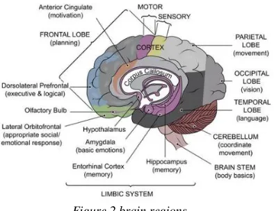
The brain is the central controlling organ of the human being. Various scientific studies have proved that some regions of the brain are involved in thinking of emotions, responding to extreme emotional stimuli, and viewing emotional situations. There are a lot of regions inside the human brain every one with a special function as shown in Fig 2.
Figure 2 brain regions.
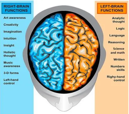
electrodes are placed over the motor cortex, EEG signals associated with motor imagery (MI) can be acquired. can be interpreted as the mental rehearsal of a motor act. Understanding of the structure of the brain is therefore important for informing the placement of electrodes on the scalp of the BCI user. If the optimal recording sites are correctly associated with the command-related EEG activity then the probability of success of the BCI may be increased. This is accomplished by wearing an electrode cap, which helps to obtain the EEG signals from the scalp. This approach is preferred, as it does not require surgical implantation of electrodes inside the brain by invasive means and makes the BCI practically usable and user-friendly. If we would split the brain right down the middle into two symmetrical, or equal parts, we would have a right and left hemisphere as shown in the figure 3.
The right side is Responsible for control of the left side of the body and is the more artistic and creative side of the brain It is responsible for creative awareness also dominant in emotional expression and dominant in the perception of facial expression, body posture, and prosody.
The left side is Responsible for control of the right side of the body and is the more academic and logical side of the brainIt also performs logical tasks such as those found in science and mathematics, dominant in language and important for preprocessing social emotions.
Loops of the brain
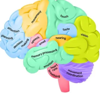
The brain‘s cerebral cortex is the outermost layer that gives the brain its characteristic wrinkly appearance. Traditionally, each of the hemispheres has been divided into four lobes: frontal, parietal, temporal and occipital.
Figure 4 brain’s loops
Although we now know that most brain functions rely on many different regions across the entire brain working in conjunction, it is still true that each lobe carries out the bulk of certain functions.
Figure 5 brain loops and function
Frontal lobe
The frontal lobe is generally where higher executive functions including emotional regulation, planning, reasoning and problem solving occur. This is why in frontotemporal dementia, personality changes are often the first signs of the disease.
Parietal lobe
Temporal lobe
The temporal lobe also contains regions dedicated to processing sensory information, particularly important for hearing, recognizing the language, and forming memories, include:
- Auditory information
The temporal lobe contains the primary auditory cortex, which receives auditory information from the ears and secondary areas, and processes the information so we understand what we’re hearing (e.g. words, laughing, a baby crying).
- Visual processing
Certain areas in the temporal lobe make sense of complex visual information including faces and scenes. - Memory
The medial (closer to the middle of the brain) temporal lobe contains the hippocampus, a region of the brain important for memory, learning and emotions.
Occipital lobe
The occipital lobe is the major visual processing center in the brain.
The primary visual cortex, also known as V1, receives visual information from the eyes. This information is relayed to several secondary visual processing areas which interpret depth, distance, location and the identity of seen objects.
Electroencephalography
Electroencephalography (EEG) is a non-invasive brain-imaging method that records the brain's electrical activity at the surface of the scalp. EEG was first used in 1929 by Hans Berger, who recorded brain activity beneath the closed skull and reported changes during different states. In 1957 Gray Walter was the first person to record the brain with electrodes and showed that brain rhythms changed according to different mental tasks. The brain’s electrical activity is picked up by electrodes attached on the patient’s scalp and amplified on the EEG machine to be viewed as brain waves.
Brain Waves
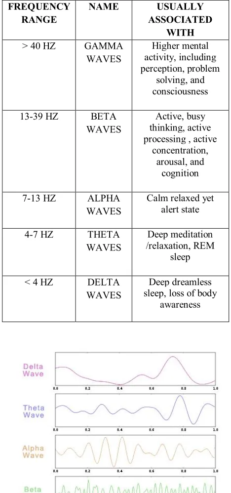
Brain waves are oscillating electrical voltages in the brain measuring just a few millionths of a volt. There are five widely recognized brain waves, and the main frequencies of human EEG waves are listed in Table 1 along with their characteristics.
Brain wave samples for different waveforms are shown in Fig. 6.
Table 1brain waves
Figure 6 brain waves
FREQUENCY RANGE
NAME USUALLY
ASSOCIATED WITH
> 40 HZ GAMMA WAVES
Higher mental activity, including perception, problem
solving, and consciousness
13-39 HZ BETA WAVES
Active, busy thinking, active processing , active
concentration, arousal, and
cognition
7-13 HZ ALPHA WAVES
Calm relaxed yet alert state
4-7 HZ THETA WAVES
Deep meditation /relaxation, REM
sleep
< 4 HZ DELTA WAVES
Deep dreamless sleep, loss of body
Measuring brain activity
The electrodes are located over the scalp. The measure recorded with this technique is named EEG. The electrodes are distributed over the scalp according to the International 10-20 system (Fig. 7).
Figure 7 International 10-20 system
Each site has a letter to identify the lobe and a number to identify the hemisphere location. The letters F, T, C, P and O stand for frontal, temporal, central, parietal, and occipital lobes, respectively. (Note that there exists no central lobe; the ―C‖ letter is used only for identification purposes.) Even numbers (2,4,6,8) refer to electrode positions on the right hemisphere, whereas odd numbers (1,3,5,7) refer to those on the left hemisphere. A ―z‖ (zero) refers to an electrode placed on the midline.
PREPROCESSING: A SIGNAL ENHANCEMENT REQUIREMENT ALONG WITH NOISE REDUCTION
This section describes the signal preprocessing requirement and core methodologies that have been implemented by a various research group. The primary requirement of the preprocessing stage in BCI is to filter out the noise, unwanted signals that are embedded within the EEG. In view of the fact that EEG-based MI classification depends solely on the EEG as the signal of interest, the signals generated by muscle movement (known as electromyogram (EOG), the activity due to movement of the eyes (known as electrooculogram ECG and the electrocardiogram (EGG) should also be considered as unwanted signals or artifacts. The EMC activity is the most common artifact and exists in the frequency range of 20 and 200 Hz whereas movement artifacts appear at frequencies of less than 10 Hz. The EOG exists within the frequency range between 0.1 and 38 Hz, but typically below 20 Hz. The ECG artifacts are related to the field of the heart potentials over the surface of the scalp. These artifacts are located in the lower frequency ranges. There are various techniques, discussed below, to remove artifacts. Most of the BCI groups utilize a band-pass filter as one of the most common preprocessing tools to extract information related to the EEG frequencies of interest. There is a minimum probability of artifacts in the region of 8-12 Hz (lower frequency band) and 16-24 Hz (higher frequency band) because the other frequencies have been removed by way of band-pass filtering. These optimal bands of interest may vary from one subject to another, and hence finding subject-dependent frequency bands is necessary to maximize BCI performance. A notch filter of 50 Hz can be utilized to remove the common electrical power line interference/artifact. A good preprocessing tool can enhance the performance of the complete BCI while using the same feature extraction (FE) and classification processes, so preprocessing the EEG signal is often considered the most important BCI stage. A few of the best-known preprocessing techniques commonly employed in BCI systems are therefore discussed in the following subsections.
1- Referencing Method
of the activity at the reference site and then subtracting this average produces. in principle a de-referenced solution. CAR is mathematically expressed as:
eq1
Where is the potential difference between the electrode and the reference, and n is the total number of
electrodes in the montage. it‘s important to note here that when i=j, the CAR can over fit because the electrode
potential is reduced due to the averaging process. CAR has been widely used in BCI systems where the noise is distributed over EEG montage.
2- Principal Component Analysis [PCA]
Principal Component Analysis (PCA) is a common statistical technique for identifying and re-referencing the data by linear mapping, which transforms a number of possibly correlated variables into a smaller number of uncorrelated variables known as principal components. The primary axis or the first principal component is calculated such that it accounts for the largest amount of variability in the data. Similarly, the subsequent components or axes are calculated and they account for the direction of the remaining variability but in decreasing order of the amount of variability in the data. The subsequent axes thus represent the direction of the next largest variation and so on. Since the transformed data have most of the variation in the first few components, the remaining components can be ignored to reduce the dimensionality but simultaneously not significantly compromise the accuracy of the data representation. Applying PCA to a set of data is a five-step process. The steps involved are the following:
1. The first step is to subtract the mean horn each of the data dimensions. This generates a data set whose mean is zero, i.e., the data matrix is centered.
2. The next step is to calculate the covariance matrix of the centered data.
3. The next task is to calculate the eigenvectors and the eigenvalues of the covariance matrix. An eigenvalue represents the amount of variance within a given component.
4. These eigenvectors are then ordered according to their eigenvalues, highest to lowest. Thus, these are the principal components of the data set. This is necessary to order the components in the order of significance. The final data set will have fewer dimensions than the original if the components with lesser significance are removed.
5. The final step is to form a feature vector by taking the retained eigenvectors. The eigenvectors in the feature vector should be arranged column-wise.
Feature Vector = ( .. ).The Feature Vector is then transposed and the transposed mean is subtracted from the original data the centered data, to obtain the PCA data. Thus, the data in its final form is represented an
PCA_data = Transposed_FeatureVector Transposed XTransposed_OriginalMeanSubtractedData.
Hence, if the data were initially n dimensional, if the first p eigenvectors are chosen, the final data set will consist of only p dimensions.
Amplifier
An amplifier is a device that receives an input signal, guides a power source, and creates an amplified copy of the original signal. The increase in voltage is called gain and is calculated from the ratio of the output and input voltages. Most amplifiers used in recordings of biologic signals are a combination of amplifiers called a differential amplifier.
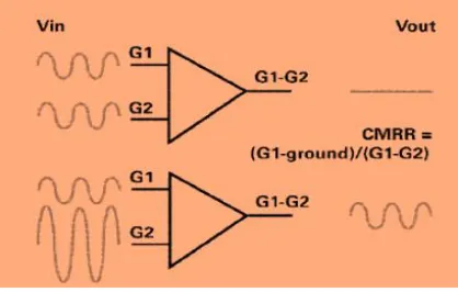
The output of a differential amplifier is the amplified difference between two inputs, called G1 and G2. (‗‗G‘‘ comes from the days when the electrical contacts to tube amplifiers were grids)
Figure 8 Amplifier
The main benefit of the differential amplifier is noise reduction, quantified by the term common mode rejection. Any voltage seen in common between G1 and G2 adds up to zero and thereby cancels out. One can calculate the common mode rejection ratio.
Processing the signals
So the final step is to process the signal to make program in the microcontroller in order to control and converting these signals into action.
BCI functions
Communication and control
Brain-computer interface (BCI) systems build a communication bridge between the human brain and the external world eliminating the need for typical information delivery methods.
User state monitoring
Early BCI applications have targeted disabled users who have mobility or speaking issues. Their aim was to provide an alternative communication channel for those users. But later on, BCI enters the world of healthy people as well. It works as a physiological measuring tool that retrieves and uses information about an individual‘s emotional, cognitive or effectiveness state.
BCI Applications
- Medical Applications
Healthcare field has a variety of applications that could take advantage of brain signals in all associated phases
- Detection and diagnosis
- Rehabilitation and restoration
Mobility rehabilitation is a form of physical rehabilitation used with patients who have mobility issues, to restore their lost functions and regain previous levels of mobility or at least help them adapt to their acquired disabilities. People suffer from serious injuries or events such as strokes may also be able to fully recover.
- Neuroergonomics and smart environment
As previously mentioned, deploying brain signals is not exclusive to the medical field. Smart environments such as smart houses, workplaces or transportations could also exploit brain computer interfaces in offering further safety, luxury and physiological control to humans‘ daily life.
- Educational and self-regulation
Neurofeedback is a promising approach for enhancing brain performance via targeting human brain activity modulation.
And a lot of others applications for BCI.
Advantages and Disadvantages of the Method
EEG has two clear advantages for brain research. The first is characteristic of an electrical recording system—high precision time measurements. Changes in the brain‘s electrical activity occur very quickly, and extremely high time resolution is required to determine the precise moments at which these electrical events take place. Today‘s EEG technology can accurately detect brain activity at a resolution of a single millisecond (and even less). Unlike other electrical recording devices that require inserting electrodes into the brain, EEG electrodes are simply stuck onto the scalp. It is, therefore, a non-invasive procedure that allows researchers clear access to a healthy human brain (which they would not probe inside to explore, of course). In addition, EEG equipment is relatively inexpensive compared with other devices and simple to operate.
The main disadvantage of EEG recording is a poor spatial resolution. Since measurements are taken at the scalp, the received signal is, essentially, the sum of the electric field (in the direction perpendicular to the scalp) that is produced by a large population of neurons. The spatial resolution of a single electrode is in the order of one centimeter of the cortex, which contains hundreds of thousands of neurons. Particularly strong electrical activity can be picked up by several neighboring electrodes. The EEG signal, therefore, is not useful for pinpointing the exact source of the activity, and it does not allow researchers to distinguish between activities originating in different but closely adjacent locations. That said, today there are more advanced techniques available for analyzing EEG which allow for more accurate estimates of the signal source.
Conclusion
Brain signals reflect the handled activities and controlling behavior of the brain or the influence of the received information from other body parts either sensing or internal organs. Brain Computer Interfacing provides a channeling facility between brain and external equipment.
BCI applications have attracted the research community. Several studies have been presented in this paper regarding the growing interest in BCI application fields such as medical, organizational, transportation, games and entertainment, and security and authentication fields. It also demonstrates the various devices used for capturing brain signals.
been widely spread in other application fields due to its advantages over the invasive one. Other challenges and issues posed as a result of utilizing brain signals have also been discussed along with some solutions offered by different algorithms at various BCI processing components.
References
[1]. J. van Erp, F. Lotte, M. Tangermann, Brain-computer interfaces: beyond medical applications Computer, 45 (4) (2012), pp. 26-34.
[2]. C.S. Ang, M. Sakel, M. Pepper, M. Phillips, Use of brain computer interfaces in neurological
rehabilitation.
[3]. Brit J Neurosci Nurs, 7 (3) (2011), pp. 523-528
[4]. Tan H, Kong K, Shee C, Wang C, Guan C, Ang W. Post-acute stroke patients use brain-computer interface to activate electrical stimulation. In: Engineering in Medicine and Biology Society (EMBC), 2010 Annual International Conference of the IEEE. IEEE; 2010. p. 4234–37.
[5]. Q. Wei, Y. Wang, Z. Lu, Channel reduction by cultural-based multi-objective particle swarm optimization based on filter bank in brain-computer interfaces, Unifying Electrical Engineering and Electronics Engineering, Springer (2014).
[6]. EEG research Nidhin Thomas on Apr 20, 2011,Scribd.
[7]. VAIBHAV GANDHI B.ENG,M.ENF,PH.D. BRAIN-COMPUTER INTERFACING FOR ASSISTIVE ROBOTICS 2015.school of science and technology, Middlesex University ,London ,UK.
[8]. Smart Wheelchairs and Brain-computer Interfaces: Mobile Assistive Technologies, Pablo Diez, Academic Press, 2018, 0128128933, 9780128128930.
[9]. Introduction to EEG- and Speech-Based Emotion Recognition,Priyanka A. Abhang ,Bharti W. Gawali ,And Suresh C. Mehrotra ,Department of Computer Science and Information Technology, Dr. Babasaheb Ambedkar Marathwada University, Aurangabad, India.
[10].Review Of Clinical Electroencephalography,G.R.Shamsaei Assistant Professor of Neurology, Jundishapour University of Medical Sciences.
[11].Ismail K, Mansor W, Khuan L, Che Wan Fadzal C. Spectral analysis of eeg signals generated from imagined writing. In: Signal Processing and its Applications (CSPA), 2012 IEEE 8th International Colloquium on. IEEE; 2012. p. 510–13.