1
Mycobacterium abscessus: environmental bacterium turned clinical nightmare
1 2
Rose C. Lopeman1, James Harrison1, Maya Desai2 and Jonathan A. G. Cox1* 3
4
1School of Life and Health Sciences, Aston University, Aston Triangle, Birmingham, UK, B4 7ET 5
2Birmingham Children’s Hospital, Birmingham Women’s and Children’s NHS Foundation Trust, 6
Steelhouse Lane, Birmingham, UK, B4 6NH 7
8
*Author to whom correspondence should be addressed: 9
Dr Jonathan A. G. Cox, School of Life and Health Sciences, Aston University, Aston Triangle, 10
Birmingham, UK, B4 7ET; J.a.g.cox@aston.ac.uk; 0(+44)121 204 5011 11
ORCID ID: 0000-0001-5208-4056 12
13
Key Words:
14
Mycobacterium abscessus; Non-tuberculous mycobacteria; Antimicrobial drug discovery; Cystic
15
fibrosis 16
17
Abstract:
18
Mycobacteria are a large family of over 100 species, most of which do not cause diseases in humans. 19
The majority of the mycobacterial species are referred to as nontuberculous mycobacteria (NTM), 20
meaning they are not the causative agent of tuberculous (TB) or leprosy, i.e. Mycobacterium 21
tuberculous complex and Mycobacterium leprae, respectively. The latter group is undoubtedly the
22
2 most infamous, with TB infecting an estimated 10 million people and causing over 1.2 million deaths 23
in 2017 alone (1). TB and leprosy also differ from NTM in that they are only transmitted from person 24
to person and have no environmental reservoir, whereas NTM infections are commonly acquired from 25
the environment (2) (3). It took until the 1950’s for NTM to be recognised as a potential lung pathogen 26
in people with underlying pulmonary disease and another 3 decades for NTM to be widely recognised 27
by the medical community when NTM, particularly Mycobacterium avium complex (MAC) was 28
recognised as the most common group of opportunistic pathogens in AIDS patients (4). This review 29
focusses on an emerging NTM called Mycobacterium abscessus (M. abs). M. abs is a rapidly growing 30
(RGM) NTM that is responsible for opportunistic pulmonary infections in patients with structural lung 31
disorders such as cystic fibrosis (CF) and bronchiectasis (5), as well as a wide range of skin and soft 32
tissue infections (SSTIs) in humans (6) (7). M. abs is a weakly staining Gram-positive mycobacterium 33
that is neverand is, like other NTM, most often seen in soil and aquatic environments (8). The bacillus-34
shaped bacterium is 1-6µm long and 0.2-0.5µm in diameter, with curved ends and the presence of 35
cord factor, or trehalose 6-6’-dimycolate, a glycolipid found in the cell wall of virulent species of 36
mycobacteria that results in “serpentine cord” cell morphology is sometimes observed(8)(9). On solid 37
growth medium, M. abs can display either a rough (M. abs-R) or smooth (M. abs-S) morphotype, with 38
the rough morphotype displaying a more virulent phenotype than its smooth variant (10). The rough 39
morphotype is characterised by irregular parallel filaments that form ridges across the colony, 40
whereas a smooth morphology is displays a wet, smooth colony with no filaments or ridges (79). This 41
morphology is driven by cell wall glycopeptidolipid (GPL); a loss of GPL results in the reversion from 42
rough to smooth morphotype (80) (81). Moreover, it has been shown using human tissue culture 43
models of infection that M. abs-R is able to persist and multiply within the host macrophage whereas 44
M. abs-S lacks this capacity, hence its role in virulence (82). Like all other mycobacteria, M. abs are
45
aerobic, non-motile and acid-fast organisms with a characteristically thick, lipid-rich cell wall that is 46
hydrophobic. Due to their unusually impermeable, thick cell wall, mycobacteria are notoriously 47
3 became available in 2009, elucidation of the resistance mechanisms of M. abs became an area of focus 49
for scientific research, as the considerable threat it poses to public health became more apparent (12) 50
(13) (14). In this review we will discuss how we came to understand the pathogen, how it is currently 51
treated, as well as a discussion of drug resistance mechanisms and novel treatments currently in 52
development. 53
54
Introduction:
55
M. abs was first isolated in 1952 by Moore and Frerichs from a 63-year-old woman’s knee abscess (6)
56
and since then, our understanding of the pathogen has rapidly and somewhat turbulently expanded. 57
When it was first isolated, it was suggested by the authors that M. abs was an entirely new species of 58
NTM and was given its name due to its ability to produce subcutaneous abscesses. Interestingly, at 59
this point, M. abs was considered to be a pathogen of low virulence due to the perception that it was 60
primarily a pathogen causing cutaneous infections that appeared transient and self-limiting (6). 40 61
years after its discovery, M. abs was first implicated in pulmonary infections after an analysis of 154 62
patients with RGM pulmonary infections revealed that 82% of the isolates were M. abs; the disease 63
was considered to be slowly progressive but virulent nonetheless (15). Since its first identification, M. 64
abs nomenclature and species/subspecies identification have undergone many changes.
65
In 1952 (6), M. abs was believed to be identical to Mycobacterium cholenae, another RGM that infects 66
fish and amphibians, as it presented identical biochemical features (16). Then, in 1972, following an 67
international collaborative study by the International Working Group on Mycobacterial Taxonomy, 68
M.abs was designated subspecies status (16). 20 years later, in 1992, Kusunoki and Ezaki used DNA
69
hybridisation to establish that there is only 35% DNA relatedness between M. chelonae subsp. 70
chelonae and M. chelonae subsp. abscessus. In light of this, M. abs was finally re-elevated to species
71
4 However, in 2004, an unusual mycobacterium was isolated from a patient with hemoptoic pneumonia, 73
and researchers were unable to accurately identify the species using the techniques described above. 74
They developed partial PCR sequencing of the rpoB gene and were able to demonstrate that the 75
isolate shared 96.0% partial rpoB sequence similarity and a 98.0% recA gene sequence similarly with 76
only the M. abs type strain. They had previously proposed that rpoB gene sequence difference of >3% 77
and a recA gene sequence difference of >2% was sufficient to differentiate between different NTM 78
species. Using this new rpoB gene sequencing technique aided with the more traditional biochemical 79
assays and 16S rRNA gene sequencing, the authors were able to produce an accurate phylogenetic 80
tree of various NTM. They concluded that this novel isolate was a new species closely related to and 81
likely recently derived from M. abs. This was subsequently named Mycobacterium massiliense (17). 82
In 2006, rpoB gene sequencing was used on 59 clinical isolates of RGM (18), and they found that 15.3% 83
of these isolates were novel, corresponding to 3 new species of mycobacteria. One of these species, 84
named Mycobacterium bolletii by the authors, was found to share 100% 16S gene similarity and 95.6% 85
rpoB gene sequence similarity with M. abs.
86
In 2011 it was proposed by Leao et. al., (19) that the M. abscessus complex (MABS complex) should 87
be amended to include M. abscessus subsp. abscessus (as before) and to combine the two subspecies 88
to form one single subspecies, M. abscessus subsp. bolletii. Finally, in 2013, whole-genome sequencing 89
(WGS) was used by Bryant et. al., to identify transmission between patients with CF (20). The authors 90
subjected 168 clinical isolates of M. abs to WGS and a phylogenetic tree produced from the isolates 91
showed clearly, for the first time, that M. abscessus subsp. abscessus, M. abscessus subsp. bolletii, and 92
M. abscessus subsp. massiliense are three distinct subspecies belonging to the MABS complex. The
93
idea that MABS is a complex that contains 3 subspecies that are genetically very similar, but 94
phenotypically divergent was given more traction in 2016 when Tortoli et. al., (21) published an 95
amended description of the MABS complex that highlighted the importance of subspecies 96
5 i.e. “genetically close organisms that diverge in phenotype” is appropriate in this case, considering the 98
genetic similarity and the presence of an inducible and functional erm(41) gene conferring macrolide 99
resistance in only M. abscessus subsp. bolletii and M. abscessus subsp. abscessus isolates whereas M. 100
abscessus subsp. massiliense has a non-functional erm(41) gene.
101
102
103
104
Figure 1: Timeline of Mycobacterium abscessus taxonomy from 1950 through to the present day. The
105
first 50 years since its discovery, no congruent terminology was in widespread use to accurately 106
describe and differentiate M. abs from other NTM. In the mid-2000s improved molecular technology 107
resulted in the discovery of the two M. abscessus subspecies; M. abscessus subsp. massiliense and M. 108
abscessus subsp. bolletii in 2004 and 2006, respectively. Then in 2011 it was proposed that M.
109
abscessus subsp. massiliense and M. abscessus subsp. bolletii should be merged into one subspecies,
110
M. abscessus subsp. massiliense. This caused some confusion within the medical community, until in
111
2013, when whole genome sequencing (WGS) showed genetic divisions that clearly identified the 112
6 114
Speciation of the M. abscessus complex:
115
Over the years, many different biochemical and molecular techniques have been employed to identify 116
NTM species. Up until the early 2000’s, the sodium chloride tolerance test was used to identify species 117
of RGM, particularly in distinguishing between M. abs and M. chelonae species, as M. abs is able to 118
grow on Löwenstein-Jensen medium with 5% sodium chloride but M. chelonae is not (23). However 119
several investigators reported that this method is unreliable, likely due vague criteria and the cross-120
over of biochemical features between differing species of RGM (23) (24) (25). The citrate utilization 121
assay perhaps provides more reliability, the premise being that M. abs is unable to use citrate as a 122
carbon source whereas other RGM such as M. chelonae are (26). As is also the case with the sodium 123
chloride test, this assay takes up to 8 weeks to complete and therefore is losing traction in the clinical 124
setting (23). High Performance Liquid Chromatography (HPLC) has also been used to generate mycolic 125
acid patterns and thus distinguish between RGM species, however this technique has limitations as 126
several RGM have similar mycolic acid profiles (27). Despite its widespread use in species 127
identification, 16S rRNA sequencing has been shown to be inadequate for species identification of 128
mycobacteria (17). An assay with superior specificity was needed to differentiate between NTM 129
species and subspecies. 130
131
M. abscessus and Cystic Fibrosis:
132
NTM species are ubiquitous in the environment (unlike M. tuberculosis and M. leprae which require a 133
living host and are transmitted patient to patient or zoonotically), suggesting that NTM exposure is 134
extremely common, whereas NTM disease is still relatively rare. Those with pre-existing lung diseases 135
undoubtedly have some predisposition to NTM infection, leading some to describe a “two-hit” theory 136
of NTM disease acquisition (28). Undoubtedly, the leading population affected by M. abs is the CF 137
7 CF is an autosomal recessive disorder caused by mutations in the CF transmembrane conductance 139
regulator gene (CFTR). Despite being a multi-organ disease, one of the most prominent features in CF 140
is chronic pulmonary infection. The major pathogen associated with lung infection in CF is 141
Pseudomonas aeruginosa, and unfortunately, 80 to 90% of patients with CF die from respiratory
142
failure as a result of chronic bacterial infection (29). Even from infancy, the lungs of CF patients are 143
already commonly colonised with a variety of organisms such as Staphylococcus aureus and 144
Haemophilus influenzae. Before 1990, NTM infection was not often associated with CF. However, since
145
then, reports of M. abs infection (along with other NTM species) have been increasingly common. 146
Several large-scale studies have been performed over the past decade or so, revealing an NTM 147
prevalence in CF patients in some areas as high as 20% (table 1). 148
149
Table 1: Prevalence of non-tuberculous mycobacterial lung disease in cystic fibrosis patients in
150
differing geographical areas between 2004 and 2014. 151
152
Age is a strong correlator of NTM infection in this group, with 40% of CF patients over the age of 40 153
having NTM smear positive results, as opposed to 4-20% in the under 40s population (35). Other risk 154
factors for NTM infection in CF patients appears to be lower body mass index (BMI) values, worse 155
forced expiratory volume (FEV1), current infection with Pseudomonas aeruginosa and 156
Stenotrophomonas maltophilia, experience of pneumothorax requiring chest drain, the use of inhaled
157
Study Location Sample size NTM prevalence in CF
Oliver, KN (2004) (30) USA 750 13% (majority M. avium
complex)
Roux, AL, et. al. (2009) (31) France 1582 6.6% (M. abs most common)
Seddon, P, et. al. (2013) (32) UK 3805 adults 3317 children
5% adults 3.3% children
Adjemian, J, et. al. (2014) (33) USA 18,003 10-20%; depending on area
8 antibiotics and other medical interventions. (36). One study performed in Israel found a significant 158
association between Aspergillus species and NTM species in sputum cultures of CF patients (37). 159
160
M. abscessus infection in non-CF populations
161
It is well documented that a risk factor for NTM pulmonary disease is patients with low body fat. The 162
mechanisms behind this are not well understood, however it is possible that leptin plays a role in NTM 163
predisposition (38). 164
Aside from pulmonary infections, M. abs is also able to produce skin and soft tissue infections (SSTIs) 165
in otherwise healthy hosts. There have been cases of M. abs outbreaks following the use of 166
contaminated needles and other surgical instruments (39) and even, as was the case in a cohort of 167
‘lipotourists’ (i.e., people who travel abroad for cosmetic surgery for fat removal), severe outbreaks 168
following cosmetic surgery (40). Interestingly, M. abs has also been linked to late-onset wound 169
infections following crush trauma sustained by Swedish survivors of the 2004 tsunami that killed over 170
200,000 people and caused serious crush injuries in another >2000(41) 171
M. abs also causes serious disseminated infections following transplantation (42). A single case study
172
involving post-transplant M. abs SSTI resulted in disseminated pulmonary infection and eventually the 173
death of the patient, despite aggressive pre- and peri-operative anti-mycobacterial therapy (43). For 174
this reason, many have recommended that M. abs colonisation should be viewed as a contraindication 175
to lung transplantation. This suggestion, however, has been met with criticism. Some studies have 176
shown that it is possible to perform a lung transplant on patients with M. abs colonisation and that 177
subsequent clearance of infection is possible, albeit with a strong possibility of severe complications 178
(44)(45). Despite this uncertainty surrounding the outcome of lung transplantation in patients 179
colonised with M. abs, it is increasingly clear that effective treatments for M. abs lung infection must 180
be developed, as lung transplantation is a potentially life-saving therapy for end-stage lung disease 181
9 183
Environmental reservoirs and transmission:
184
NTM are ubiquitous in the environment; especially water sources and soil (4). They are prone to 185
biofilm formation and this contributes to their ability to persist in harsh environments(46). NTM can 186
persist in environments that are in close proximity to human populations, particularly human water 187
sources, hospital water supplies (sinks, showerheads), and homes. 188
M. abs, like other NTM, is able to survive in harsh, nutrient-starved environments where other
189
competing microorganisms would not survive, such as in chlorinated water (47). The presence of the 190
lipid-rich cell wall results in a hydrophilic cell surface, which facilitates the formation of biofilms, their 191
slow growth and adherence to surfaces, thus aiding their survival and providing them with a selective 192
advantage (48)(49)(50). Furthermore, many RGM are oligotrophic, requiring low levels of two carbon 193
sources and minimal amounts of metal ions (51), further indicating their hardiness and persistence in 194
harsh environments. The impenetrable nature of the M. abs cell wall in comparison to other non-195
mycobacterial pathogens also contributes to its resistance to many antibiotics and disinfectants (52) 196
(12). The ability of M. abs to survive in the human environment presents a huge problem for human 197
health, with most studies up until this point suggesting that patients with CF predominately acquire 198
NTM infection from the environment(20). This long-held belief was called into question in 2013 when 199
Floto and his team used WGS to show possible patient to patient transmission of M. abs within a CF 200
clinic in the UK (53). 201
In 2009, Feazel et al demonstrated that showerheads provide an enriched environment for NTM 202
biofilm formation; the presence of human pathogens including NTM were >100 fold higher in 203
showerhead biofilms compared to the background water contents(54). A study in Hawaii investigated 204
the prevalence of NTM in household plumbing; areas such as showerheads, sinks, taps, shower drains, 205
and refrigerator water dispensers were sampled. The authors found that 69% of households surveyed 206
10 an outbreak of M. abs skin infections in children who were exposed to the same indoor wading pool 208
(56). This study demonstrates the importance of identifying M. abs environmental reservoirs, 209
reporting M. abs cases and subsequent environmental remediation in order to reduce the risk of 210
infection. 211
The persistence and spread of NTM species within healthcare environments is fast becoming a serious 212
problem and a significant threat to human health (57). It was a long-held belief in the scientific 213
community that NTM is transmitted to humans from the environment, and that patient to patient 214
transmission is unlikely. Resulting in a clinical focus on reducing the risk of environmental transmission 215
using effective sterilising techniques and other hygiene practices. Such as it is, the CF Trust published 216
M. abs infection control recommendations that include general infection control measures such as
217
hand washing and more specific recommendations such as segregation of infected patients from other 218
patients (58). 219
The mode of transmission of pathogenic NTM to humans is still poorly understood, with many studies 220
seeking evidence of human to human transmission using molecular techniques such as WGS. A study 221
undertaken in 2001 sought to address this question; a retrospective analysis of 1062 respiratory 222
specimens taken from 214 patients with CF revealed 5 patients with M. abs lung infection. These 5 223
patients each had isolates with a unique genotype that was not shared with any of the other patients, 224
which led the authors to conclude that patient to patient transmission of M. abs was not occurring 225
within their cohort (59). 226
In 2014, a small-scale study was performed on 27 M. abs isolates from 20 paediatric CF patients (60). 227
The authors used a combination of epidemiology, variable number tandem repeat (VNTR) profiling 228
and WGS to find evidence of cross-infection between paediatric CF patients. They hypothesized that 229
patients with strains that had identical VNTR profiles would have had intense exposure to each other 230
compared with patients with strains that had different VNTR profiles. They found little evidence of 231
11 intensity of exposure. They concluded that cross-infection was uncommon in their cohort, and that 233
transmission is most likely to be from a common environmental source (60). 234
The biggest shift in our understanding of transmission came in 2013 when a major study was published 235
in which WGS was used to identify transmission of M. abs between patients at an adult CF centre in 236
the UK between 2007 and 2011 (20). The authors found a high level of relatedness between isolates 237
of M. abscessus subsp. abscessus, but clusters were clearly segregated from one another, indicating 238
that patients have independently acquired either genetically diverse strains or a dominant circulating 239
clone. In the case of M. abscessus subsp. massiliense, however, the authors found isolates from 240
different individuals with almost identical genomic sequences, strongly indicating transmission 241
between patients. Analysis of the environment revealed no NTM species isolated from the water 242
supply to the clinic, showerheads, dish washers, bronchoscopes or the local River Cam or Papworth 243
Hospital Pond. Further investigation into possible transmission routes revealed patients with isolates 244
from the same genetic relatedness clusters were present in the clinic at the same time as each other, 245
further supporting their hypothesis that M. abscessus subsp. massiliense is likely transmitted from 246
patient to patient rather than independently from the environment. This finding represents a major 247
clinical advance which may require patients infected with M. abs to be segregated from M. abs-naïve 248
patients to prevent onward transmission. 249
Following on from the localised retrospective study published in 2013 (20), a global WGS initiative was 250
launched on 1080 isolates from 517 patients from the UK, USA, Republic of Ireland, mainland Europe 251
and Australia (53). This study found that the majority of isolates were from densely clustered 252
genotypes that were not diverse, suggesting a high level of human-human transmission. Phylogenetic 253
analysis also revealed that there are 3 dominant circulating clones globally, and these clones are 254
associated with higher virulence and poor clinical outcomes. Human-human transmission appears to 255
have facilitated the evolution of M. abs from an environmental pathogen to a transmissible human 256
12 258
Diagnosis and treatment:
259
As M. abs and other NTM species are ubiquitous in the environment, including drinking water supplies, 260
the presence of culture-positive respiratory tract sample for NTM does not always indicate NTM-261
pulmonary disease (NTM-PD). Therefore, patients must also have characteristic symptoms, 262
compatible radiology, and two or more positive sputum samples for the same NTM species, as well as 263
the exclusion of other potential causes of pulmonary disease (61). 264
For clinical laboratory identification of NTM species, the British Thoracic Society (BTS) recommends 265
that isolates be obtained from sputum samples, and if this is not possible (for example in children), 266
bronchoalveolar lavage or transbronchial biopsy samples should be taken when NTM pulmonary 267
disease is suspected (61). NTM infection can be validated in the laboratory, with the use of auramine-268
phenol staining and microscopy, as well as culture on solid and liquid media. 269
All clinical isolates of M. abs undergo susceptibility testing for clarithromycin, cefoxitin and amikacin. 270
They also recommend that other antibiotics such as tigecycline, imipenem, minocycline, moxifloxacin 271
and clofazimine are tested in this manner(61). 272
Treatment
273
When M. abs was first isolated in 1952, it was thought the patient was initially infected with the 274
pathogen at the age of 14 years old. The patient’s condition resolved without intervention and so for 275
some time, treatment wasn’t considered a priority in M. abs infections (6). 276
Of course, today it is well known that treatment for M. abs pulmonary infection is essential to give the 277
patient the best chance of survival. Unfortunately, antimicrobial chemotherapy for M. abs infection is 278
particularly difficult due to its intrinsic and acquired resistance to most of the commonly used 279
antibiotic classes. Further complications in the treatment of M. abs infection is the lack of evidence 280
13 (62). Because chemotherapy-based treatment of M. abs infection is often unsuccessful, the American 282
Thoracic Society advises that certain patients may have the best chance of disease regression with 283
resectional surgery, especially if the patient exhibits a poor response to drug therapy, if macrolide-284
resistance develops, or if the patient is experiencing disease-related complications such as 285
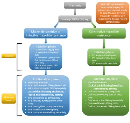
haemoptysis (62). 286
Current treatment guidelines from the BTS (61) recommend that treatment for M. abs pulmonary 287
disease should consist of an initial phase antibiotic regimen that includes intravenous (I.V.) and oral 288
antibiotics, followed by a continuation phase comprising of oral and inhaled antibiotics (Figure 2). 289
Further genetic analysis of clinical isolates can provide information on the erm(41) (inducible 290
macrolide resistance) and/or presence of 23S rRNA point mutation (constitutive macrolide resistance) 291
14 293
Figure 2: Flow chart showing treatment regimen for M. abs-pulmonary disease based on laboratory
294
susceptibility testing results as recommended by the British Thoracic Society. Treatment will differ 295
based on the whether the isolate displays macrolide sensitivity/inducible macrolide resistance or 296
constitutive macrolide resistance. The initial phase of treatment involves three intravenous (I.V.) 297
antibiotics, and for macrolide sensitive/inducible macrolide resistance 1 of 2 oral macrolides, and this 298
phase lasts one month. The continuation phase also depends on laboratory susceptibility testing 299
results and clinicians will typically administer 1-4 oral antibiotics over a period of at least 12 months. 300
It is also important to note that the American Thoracic Society recommends surgical resection of 301
infected area if the patient is not responding to therapy, if macrolide resistance develops, and/or if 302
15 Side effects of M. abs treatment are common and can be severe. A retrospective analysis of 65 304
patients undergoing treatment for M. abs lung disease in South Korea(63) revealed frequent adverse 305
reactions to cefoxitin; 51% of patients developed leukopenia, 6% of patients developed 306
thrombocytopenia, and 15% of patients experienced drug-induced hepatotoxicity. As a result, 307
cefoxitin was discontinued in 60% of patients and side effects resolved. Another common side effect 308
observed was gastrointestinal problems (nausea, anorexia, or diarrhoea), which affected 22% of 309
patients and caused 4 patients (6%) to completely stop antibiotic treatment. A clinical 310
recommendation was made to consider imipenem as an alternative to cefoxitin, however prolonged 311
treatment with imipenem can cause neutropenia. 312
Another study that analysed treatment outcomes in 65 patients with M. abs in North America also 313
found a high prevalence of side effects. IV amikacin (65% of patients) and azithromycin (71% of 314
patients) were the most commonly used antimicrobials in this cohort. They found 74 different side 315
effects reported in 62% of patients, most commonly nausea/vomiting (31%) and skin changes (20%). 316
They attributed many of these side effects to amikacin or tigecycline, and as a result, of those received 317
amikacin or tigecycline therapy, 51% and 36% of patients, respectively, had to adjust or stop 318
medication due to severe side effects such as ototoxicity. Similar to the South Korean study, 4 patients 319
had to totally stop treatment because of their side effects (64). 320
Clarithromycin is one of the most commonly used antibiotics to treat M. abs (35). However, 321
clarithromycin has been associated with hearing loss, with one study citing a 7% hearing loss rate in 322
their patients. This side effect did resolve in all but one patient, but the authors state that the patient 323
had a pre-existing condition that hindered their ability to attribute this hearing loss solely to 324
clarithromycin (65). A case study on an 81-year-old woman, who was being treated with 325
clarithromycin for infective exacerbation of chronic pulmonary obstructive disease (COPD) showed 326
another example of clarithromycin-related permanent hearing loss, despite evidence that 327
16
M. abs is the presence of a functional inducible erm(41) gene that confers macrolide resistance in both
329
M. abscessus subsp. abscessus and M. abscessus subsp. bolletii but not M. abscessus subsp.
330
massiliense.
331
332
Future perspectives for M. abscessus
333 334
The resistance problem: why the drugs don’t work:
335
M. abs is known for its intrinsic resistance to most chemotherapeutic agents, including all the
anti-336
tuberculous drugs used to treat M. tuberculosis infection (68) (69). Furthermore, in vitro drug 337
susceptibility testing on M. abs often proves unhelpful in guiding treatment regimens (70). There are 338
a number of natural resistance mechanisms displayed by M. abs (along with other mycobacteria), 339
including a waxy and impermeable cell wall, drug export systems, antibiotic modifying/inactivating 340
enzymes, and genetic polymorphism of target genes (12). 341
342
343
17
Figure 3: Graphical summary of the resistance mechanisms exhibited by Mycobacterium abscessus
345
(M. abs). There are several mechanisms involving different physiological, enzymatic and genomic 346
processes that contribute to the notoriously drug-resistant profile of M. abs. It is likely that these 347
processes work in synergy to produce a highly resistant pathogen, such as efflux pumps and drug 348
resistance genes. 349
350
The greatest contributing factor to the lack of M. abs sensitivity to many major classes of antibiotic is 351
the mycobacterial cell wall, the role of which has long been studied. The high lipid content and unusual 352
thickness of the mycobacterial cell wall provides an effective barrier for hydrophilic and lipophilic 353
agents (71). In 1990 it was shown that the lack of permeability of the M. chelonae (then grouped 354
together with M. abs) cell wall plays a vital role in making the pathogen resistant to antibiotics (52). 355
The cell wall barrier is also responsible for M.abs’ intrinsic resistance to acids and alkalis (72). The cell 356
wall of mycobacteria also contains porins, it was shown in 1990 that M. chelonae possesses a 59 kDa 357
cell wall protein that allows for the diffusion of small, hydrophilic solutes. This porin, however, is 358
minor, unlike that of E. coli where they are the most abundant cell wall protein, explaining the low 359
permeability to hydrophilic solutes (11). The cell wall cannot explain all of the intrinsic drug resistance 360
seen in M. abs, in fact it is known that the cell wall, particularly the porins, act synergistically with 361
internal systems that are activated by the presence of intracellular antibiotics, and that the low 362
permeability of the mycobacterial cell wall means that the bacteria has time to induce the expression 363
of drug resistance genes (73). 364
As a constituent of the mycobacterial cell wall, active efflux pumps can be described as one of the 365
main causative factors of drug resistance in mycobacteria (12)(74)(75). They primarily act to protect 366
bacteria against toxic compounds and bacterial homeostasis by transporting toxins or metabolites to 367
the extracellular environment (75). M. abs encodes protein members of the major facilitator family 368
18 transporters are found in all forms of life and make use of adenosine triphosphate (ATP) to transport 370
molecules across membranes. The MmpL transporter family is a subclass of a large family of multidrug 371
resistance pumps known as Resistance-Nodulation-Cell-Division (RNCD) permeases. MmpLs export 372
lipid components across the cell envelope of mycobacteria (77). The role of MmpLs in M. abs drug 373
resistance is yet to be fully understood, however there is evidence that MmpL7 in M. tuberculosis 374
confers resistance to isoniazid (78), suggesting that MmpLs may play a major role. 375
Macrolides are one of the mainstays of M. abs treatment (35), yet despite this, M. abs infections tend 376
to respond poorly to macrolide therapy, even when they appear sensitive to clarithromycin in vitro 377
(79). A study performed in 2009 revealed the presence of an inducible erm(41) gene in 7 out of 10 M. 378
abs clinical isolates that confers resistance to macrolides with a minimum inhibitory concentration
379
(MIC) of ≥32 µg/mL. The 3 remaining susceptible isolates had erm(41) gene, however it appeared to 380
be non-functional (79). The erm(41) gene produces a functional 23S rRNA methylase, contributing to 381
macrolide resistance along with point mutations in the rrl encoding 23S rRNA gene (80). Following on 382
from this, it was shown that macrolides may be useful in treating approximately 20% of M. abs 383
infections in the U.S., and that sequencing of the erm(41) gene is a potentially useful tool in predicting 384
macrolide susceptibility (81). It is also noteworthy that M. abscessus subsp. massiliense contains a 385
large 97 base pair deletion in erm(41), rendering it useless and therefore meaning M. abscessus subsp. 386
massiliense retains susceptibility to macrolides, except in the case of rrl mutants (82)(79)(83)(84). M. 387
abs isolates possessing an rrl mutant display constitutive resistance to macrolide antibiotics. This
388
phenomenon is known to be mediated by a mutation in rrl encoding the bacterial 23S rRNA gene, 389
particularly at positions 2058 and 2059, i.e. the drug binding pocket of the gene (85). 390
If macrolide therapy is not advised due to evidence of constitutive resistance, there are of course other 391
chemotherapeutic options available. However, in many of the conserved genes in M. abs that can 392
potentially act as drug targets there is the presence of genetic polymorphisms, which can often confer 393
19 A 1998 study showed revealed an amino acid substitution at position 83 (Ser83Ala) in the quinolone-395
resistance-determining-region (QRDR) in fluoroquinolone-resistant isolates of M. abs (86). This 396
substitution occurs in the region of DNA gyrase subunit GyrA that binds DNA, and as fluoroquinolones 397
bind strongly to the gyrase-DNA complex, and weakly to protein or DNA alone, this mutation results 398
in fluoroquinolone resistance (87). Genetic polymorphisms also occur within the emb operon that 399
codes for several homologous arabinosyl transferases. These are enzymes involved in the 400
polymerisation of arabinogalactan, an essential component of the mycobacterial cell wall and can be 401
inhibited by the tuberculosis drug ethambutol. A 1997 study showed that polymorphisms at position 402
306 in a highly conserved embB gene conferred natural resistance across many species of 403
mycobacteria, including M. abs (88). M. abs has high natural levels of resistance to ethambutol (MIC 404
>64mg/L), and the same study transferred the M. abs emb region to ethambutol-susceptible M. 405
smegmatis resulted in a 500-fold increase in the MIC to ethambutol (88).
406
M. abs also produces a number of target-modifying enzymes. Rifampicin ADP-ribosyl transferase,
407
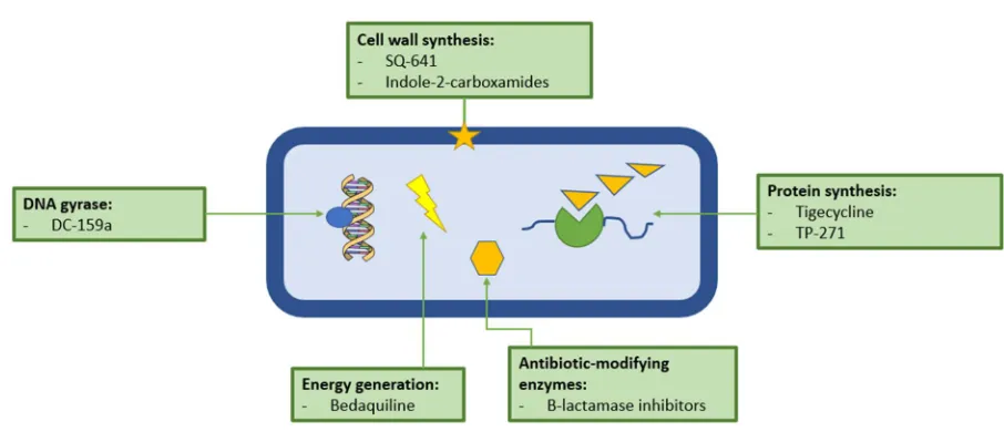
Arr_Mab inactivates rifamycins such as rifampicin. Aminoglycoside 2’-N-acetyltransferase and 408
aminoglycoside phosphotransferases mediate the susceptibility to aminoglycoside antibiotics. M. abs 409
has also been shown to produce an endogenous β-lactamase (BlaMab), that efficiently hydrolyses the 410
β-lactam ring of β-lactam antibiotics, rendering them ineffective (89). 411
Aside from antibiotic-specific internal drug resistance mechanisms, a family of transcriptional 412
regulators, the WhiB family, is excusive to actinomycetes and may be involved in conferring drug 413
resistance in M. abs. Members of this family have been shown to regulate systems of drug resistance 414
in M. tuberculosis, including antibiotic export and activation (90). M. abs has been shown to possess 415
a homologue of the M. tuberculosis WhiB7. When M. abs WhiB7 is deleted, the result is increased 416
sensitivity to clinically relevant antibiotics that target the ribosome, such as clarithromycin, amikacin 417
and tetracycline (91). 418
20
Future treatments:
420
It perhaps goes without saying that there is an urgent, unmet need for safe and effective treatments 421
against M. abs pulmonary disease. There have been instances of successful treatment of M. abs with 422
already available antibiotics. One such case was reported in 2002, where a 63-year-old patient whose 423
infection had not responded to the traditional regimen was prescribed a course of faropenem, a new 424
member of the β-lactam antibiotic class. Treatment was successful and produced no adverse side 425
effects (92). It is not just antimicrobials that have potential in enhancing M. abs treatment. In 2012, 426
Okazaki et. al. reported that the use of clarithromycin, amikacin and imipenem/cilastatin to treat a 427
case of M. abs pulmonary was greatly enhanced with the addition of corticosteroids. The authors 428
recommend that the presence of organising pneumonia (a non-specific inflammatory pulmonary 429
process) or an allergic reaction may have helped to explain the poor response to antibiotic treatment 430
alone in some patients, and that this possibility should be considered when applicable to improve 431
treatment outcomes (93). 432
One of the enzymatic resistance mechanisms employed by M. abs is the production of an endogenous 433
β-lactamase, BlaMab (Figure 3). Cefoxitin and imipenem, both β-lactam antibiotics, are commonly used 434
to treat M. abs. In order to improve the efficacy of these antibiotics, a β-lactamase inhibitor may be 435
administered in conjunction during therapeutic treatment. A 2015 study revealed that avibactam, a 436
β-lactamase inhibitor is able to efficiently inhibit BlaMab (94), and a subsequent 2017 study showed 437
that avibactam improves the efficacy of imipenem against M. abs both in vitro and in macrophage, 438
and zebrafish models of infection (95). 439
Aside from these examples, very few case studies have reported successful treatment with 440
repurposed antibiotics. Therefore, novel drug targets in M. abs must be discovered and elucidated, 441
and novel compounds that safely and effectively inhibit these targets discovered. 442
There are potentially a wide variety of viable drug targets in M. abs (Figure 4) Many of the most 443
21
M. tuberculosis, which a handful of researchers have applied to M. abs and other NTM species.
445
Unfortunately, only a small percentage of the novel drugs which are active against M. tuberculosis, 446
are also active against M. abscessus, further highlighting just how resistant and dangerous this 447
pathogen is proving to be. 448
449
450
Figure 4: Graphical summary of the exploitable drug targets in Mycobacterium abscessus (M. abs).
451
There are several potential target areas in M. abs including physiological, genomic, enzymatic and 452
metabolic processes. Many of the drugs with potential to be used as part of M. abs treatment are 453
old classes of antibiotics that have been repurposed, such as β-lactamase inhibitors, or have been 454
discovered as part of the anti-tuberculous drug discovery pipelines, such as bedaquiline. 455
456
One potential target in M. abs is DNA gyrase, despite the fact that M. abs is naturally resistant to 457
quinolones (96), a novel fluoroquinolone, DC-159a was developed in 2010 as part of the Working 458
Group on TB Drugs, and was found to be active against M. abs with an MIC of 16 µg/mL, which was 4 459
to 8-fold lower than the other already available quinolones tested (97). The authors stressed the 460
importance of in vivo testing of DC-159a, however, no publications attesting to the in vitro activity of 461
22 The mycobacterial cell wall, in all its complexity, can offer an attractive range of potential antibiotic 463
targets. The three distinct layers of the mycobacterial cell wall: core peptidoglycan, arabinogalactan 464
and mycolic acids are each essential to the pathogen and involve a number of exploitable processes 465
(98). A 2010 study subjected several species of NTM to a capuramycin analogue SQ641 (99). 466
Capuramycins are a novel class of nucleoside antibiotics that work by targeting phosphor-N-467
acetylmuramyl-pentapeptide-translocase (translocase-1 or TL-1) which is essential for peptidoglycan 468
synthesis. They found that the drug had an MIC of 0.25-1 µg/mL, as well as finding synergy between 469
SQ641 and rifabutin and streptomycin. This drug has great potential as it is fast-acting and displays a 470
long post-antibiotic effect (100). In 2017 a study was published in which several members of the newly 471
synthesized MmpL3 inhibitors, indole-2-carboxamides, have shown potent activity against M. abs. 472
These inhibitors have been shown to work by inhibiting the transfer of mycolic acids to their cell 473
envelope acceptors in M. abs strains (101). Further work has been done on this class of inhibitors; in 474
2019, Pandya et. al. reported that oral administration of the inhibitors shows a statistically significant 475
reduction in bacterial load in the lungs and spleens of M. abs-infected mice (102). 476
It has been demonstrated that M. abs displays high levels of intrinsic resistance to the tetracycline 477
class of antibiotics via the monooxygenase, MabTetX, a WhiB7-independent pathway (103). This is not 478
the end of the road for this class of antibiotics. Tigecycline, the first developed glycylcycline, a new 479
class of tetracycline antibiotics originally developed for SSTIs, was shown in 2014 to be highly effective 480
in vivo against M. abs pulmonary disease (104). Further work in 2018 revealed that tigecycline is a
481
poor substrate of MabTetX and is incapable of inducing its expression, explaining its high efficacy in 482
comparison with other tetracycline antibiotics (103). Tigecycline is now of the recommended 483
treatment options for M. abs pulmonary disease, and is arguably one of the most effective, with one 484
study citing clinical improvement in >60% patients with M. abs pulmonary disease when tigecycline is 485
employed as part of the multi-drug regimen against M. abs (104). Tigecycline is not the only 486
tetracycline showing activity against M. abs. A 2012 study tested the in vitro activity of a novel 487
23 found all the isolates to have an MIC of ≤1 µg/mL with an average of 0.5 µg/mL, which is decidedly 489
superior than that of the other orally available tetracycline antibiotics such as moxifloxacin and 490
tetracycline (105). 491
Bedaquilin, the latest drug indicated for the treatment of multi-drug resistant TB (MDR-TB) was 492
approved by the FDA in 2011, and it works by targeting the ATP synthase of mycobacteria. Obregon 493
et. al. (106) demonstrated an MICs of 1.0 µg/mL against M. abs reference strain and then in 2017,
494
Vesenbeckh and colleagues pointed to bedaquiline as a potential antimicrobial against M. abs after 495
the drug exhibited MICs of ≤1 µg/mL against 20 M. abs clinical isolates in vivo. (107) 496
497
Summary:
498
M. abs is increasingly being recognised as an important pathogen responsible for a wide range of
499
infections and implicated in severe, and often untreatable pulmonary infections in people with CF and 500
other structural lung disorders. Almost all of the currently available antibiotics are useless against the 501
pathogen, with even official guideline treatment regimens having little to no evidence of in vivo 502
efficacy. With such high treatment failure rates, clinicians are often forced to administer last-resort 503
antibiotics in the hope of a cure. Coupled with increasing prevalence and its already extensively drug 504
resistant profile, it is glaringly obvious that novel, effective and safe treatments are needed. Many of 505
the novel drugs mentioned above are in various phases of clinical trial against M. tuberculosis and 506
there is a significant paucity of data regarding their efficacy against M. abs and other NTM species. 507
Furthermore, there is a startling lack of in vivo efficacy data for any of these drugs, which is particularly 508
worrying considering the inconsistencies between in vitro and in vivo anti-M. abs activity. Whilst TB 509
has many dedicated drug-discovery programmes, NTM has none. A dedicated NTM drug discovery 510
pipeline is essential to ensure the disease burden of NTM does not become overwhelming. 511
24
Author Contributions:
513
R. C. L., J. H., M. D. and J. A. G. C. reviewed the literature, intellectually conceived and wrote the 514
manuscript. 515
516
Funding: This research was funded by Birmingham Women’s and Children’s Hospital Charity Research
517
Foundation (BWCHCRF) (R. C. L. 50% PhD Studentship, match funded by Aston University Prize 518
Scheme) and the Academy of Medical Sciences and Global Challenges Research Fund with a 519
Springboard Grant (SBF003\1088:). 520
521
Acknowledgements: J. A. G. C. is grateful to the Academy of Medical Sciences, Global Challenges
522
Research Fund and Birmingham Women’s and Children’s Hospital Charity Research Foundation 523
(BWCHCRF) for their continued support of the Mycobacterial Research Group at Aston University. 524
525
References:
526
1. Organisation, World Health. Global tuberculosis report. Geneva : s.n., 2018. CC BY-NC-SA 3.0 IGO. 527
2. Human-to-human transmission of Mycobacterium kansasii or victims of a shared source? Ricketts, 528
WM, O'Shaughnessy, TC and van Ingen, J. 2014, European Respiratory Journal, Vol. 44, pp.
1085-529
1087. 530
3. Distinguishing Tuberculosis from Nontuberculosis Mycobacteria Lung Disease, Oregon, USA. 531
Kendall, BA, et al. 3, 2011, Emerging Infectious Diseases, Vol. 17, pp. 506-509.
532
4. Nontuberculous mycobacteria: opportunistic environmental pathogens for predisposed hosts. 533
Cook, J. 1, 2010, British Medical Bulletin, Vol. 96, pp. 45-59.
25 5. An Official ATS/IDSA Statement: Diagnosis, Treatment, and Prevention of Nontuberculous
535
Mycobacterial Diseases. Griffith, DE, et al. 4, 2007, ATS Journals, Vol. 175.
536
6. An Unusual Acid-Fast Infection of the Knee with Subcutaneous, Abscess-Like Lesions of the Gluteal 537
Region. Moore, M and Frerichs, JB. 2, 1953, Journal of Investigative Dermatology, Vol. 20.
538
7. Cutaneous infection with Mycobacterium abscessus. Fitzgerald, DA, et al. 1995, Journal of 539
Dermatology, Vol. 132, pp. 800-804. 540
8. Laboratory Maintenance of Mycobacterium abscessus. Cortes, MAM, Nessar, R and Kumar Singh, 541
A. 2010, Current Protocols in Microbiology, Vol. 18, pp. 10D.1.-10D.1.12.
542
9. Spontaneous reversion of Mycobacterium abscessus from a smooth to a rough morphotype is 543
associated with reduced expression of glycopeptidolipid and reacquisition of an invasive phenotype.
544
Howard, ST, et al. 2006, Microbiology, Vol. 152, pp. 1581-1590.
545
10. Preliminary Characterization of a Mycobacterium abscessus mutant in human and murine models 546
of infection. Byrd, TF and Lyons, CR. 1999, Infection and Immunity, Vol. 67, pp. 4700-4707.
547
11. Mycobacterial cell wall: Structure and role in natural resistance to antibiotics. Jarlier, V and 548
Nikaido. 1994, FEMS Microbiology Letters, Vol. 123, pp. 11-18.
549
12. Mycobacterium abscessus: a new antibiotic nightmare. Nessar, R, et al. 4, 2012, Journal of 550
Antimicrobial Chemotherapy, Vol. 67, pp. 810-818. 551
13. Nontuberculous Mycobacteria in Adult Patients with Cystic Fibrosis. Kilby, JM, et al. 1, 1992, 552
Chest Journal, Vol. 102, pp. 70-75. 553
14. Nontuberculous Mycobacteral Disease in Adult Cystic Fibrosis Patients. Aitken, ML, et al. 4, 1993, 554
26 15. Clinical Features of Pulmonary Disease Caused by Rapidly Growing Mycobacteria: An Analysis of 556
154 Patients. Griffith, DE, Girard, WM and Wallace, RJ. 5, 1993, American Review of Respiratory
557
Disease, Vol. 147. 558
16. A Co-operative Numberical Analysis of Rapidly Growing Mycobacteria. Kubica, GP, et al. 1972, 559
Journal of General Microbiology, Vol. 73, pp. 55-70. 560
17. Amoebal Coculture of "Mycobacterium massiliense" sp. nov. from the Sputum of a Patient with 561
Hemoptoic Pneumonia. Adekambi, T, et al. 12, 2004, Journal of Clinical Microbiology, Vol. 42, pp.
562
5493-5501. 563
18. rpoB gene sequence-based characterization of emerging non-tuberculous mycobacteria with 564
descriptions of Mycobacterium bolletii sp. nov., Mycobacterium phocaicum sp. nov. and
565
Mycobacterium aubagnense sp. nov. Adekambi, T, et al. 2006, International Joural of Systematic and
566
Evolutionary Microbiology, Vol. 56, pp. 133-143. 567
19. Proposal that Mycobacterium massiliense and Mycobacterium bolletii be united and reclassified 568
as Mycobacterium abscessus subsp. bolletii comb. nov., designation of Mycobacterium abscessus
569
subsp. abscessus subsp. nov. and emended description of Mycobacteri. Leao, SC, et al. 2011,
570
International Journal of Systematic and Evolutionary Microbiology, Vol. 61, pp. 2311-2313. 571
20. Whole-genome sequencing to identify transmission of Mycobacterium abscessus between 572
patients with cystic fibrosis: a retrospective cohort study. Bryant, JM, et al. 2013, The Lancet, Vol.
573
381, pp. 1551-1560. 574
21. Emended description of Mycobacterium abscessus, Mycobacterium abscessus subsp. abscessus 575
and Mycobacterium abscessus subsp. bolletii and designation of Mycobacterium abscessus subsp.
576
massiliense comb. nov. Tortoli, E, Kohl, TA and Brown-Elliot, BA. 2016 , International Journal of
577
27 22. Report of the Ad Hoc Committee on Reconciliation of Approaches to Bacterial Systematics. 579
Wayne, LG, et al. 4, 1987, International Journal of Systematic Bacteriology, Vol. 37, pp. 463-464.
580
23. Variables Affecting Results of Sodium Chloride Tolerance Test for Identification of Rapidly 581
Growing Mycobacteria. Conville, P and Witebsky, FG. 6, 1998, Journal of Clinical Microbiology, Vol.
582
36, pp. 1555-1559. 583
24. Deoxyribonucleic Acid Relatedness Study of the Mycobacterium fortuitum-Mycobacterium 584
chelonae Complex. Levy-Frebault, V, et al. 3, 1986, International Journal of Systematic Bacteriology,
585
Vol. 36, pp. 458-460. 586
25. Clinical Significance, Biochemical Features and Susceptibility Patterns of Sporadic Isolates of the 587
Mycobacterium chelonae-Like Organism. Wallace, RJ, et al. 12, 1993, Journal of Clinical
588
Microbiology, Vol. 31. 589
26. Identification of Clinically Significant Mycobacterium fortuitum Complex Isolates. Silcox, VA, 590
Good, RC and Floyd, MM. 6, 1981, Journal of Clinical Microbiology, Vol. 14, pp. 686-691.
591
27. Mycobacterium immunogenum sp. nov., a novel species related to Mycobacterium abscessus and 592
associated with clinical disease, pseudo-outbreaks and contaminated metalworking fluids: an
593
international cooperative study on mycobacterial taxonomy. Wilson, RW, et al. 2001, International
594
Journal of Systematic and Evolutionary Microbiology, Vol. 51, pp. 1751-1764. 595
28. Understanding nontuberculous mycobacterial lung disease: its been a long time coming. Griffith, 596
DE and Aksamit, TR. 2016, F1000 Research, pp. 1-8.
597
29. Lung Infections Assocaited with Cystic Fibrosis. Lyczak, JB, Cannon, CL and Pier, GB. 2, 2002, 598
Clinical Microbiology Reviews, Vol. 15, pp. 194-222. 599
30. The natural history of nontuberculous mycobacteria in patients with cystic fibrosis. Olivier, KN. 600
28 31. Multicenter study of prevalence of nontuberculous mycobacteria in patients with cystic fibrosis in 602
France. Roux, AL, et al. 12, 2009, Journal of Clinical Microbiology, Vol. 47, pp. 4124-4128.
603
32. Prevalence of nontuberculous mycobacteria in cystic fibrosis clinics, United Kingdom, 2009. 604
Seddon, P, et al. 7, 2013, Emerging Infectious Disease, Vol. 19, pp. 1128-1130.
605
33. Nontuberculous Mycobacteria among Patients with Cystic Fibrosis in the United States. Screening 606
Practices and Environmental Risk. Adjemian, J, Olivier, KN and Prevots, DR. 5, 2014, American
607
Journal Critical Care Medicine, Vol. 190, pp. 581-586. 608
34. Nontuberculous mycobacteria in cystic fibrosis associated with allergic bronchopulmonary 609
aspergillosis and steroid therapy. Mussaffi, H, et al. 2005, European Respiratory Journal, Vol. 25, pp.
610
324-328. 611
35. An Official ATS/IDSA Statement: Diagnosis, Treatment, and Prevention of Nontuberculosis 612
Mycobacterial Diseases. Griffith, DE, et al. 4, 2007, American Journal of Respiratory and Critical Care
613
Medicine, Vol. 175, pp. 367-417. 614
36. Epidemiology of nontuberculous myocbacteria (NTM) amongst individuals with cystic fibrosis 615
(CF). Viviani, L, et al. 5, 2016, Journal of Cystic Fibrosis, Vol. 15, pp. 619-623.
616
37. Multicenter Cross-Sectional Study of Nontuberculous Mycobacteria Infections among Cystic 617
Fibrosis Patients, Israel. Levy, I, et al. 3, 2008, Vol. 14, pp. 378-384.
618
38. Leptin modulates the T-cell immune response and reverses starvation-induced 619
immunosuppression. Lord, GM, et al. 6696, 1998, Nature, Vol. 394, pp. 897-901.
620
39. Clinical and taxonomic status of pathogenic nonpigmented or late-pigmenting rapidly growing 621
mycobacteria. Brown-Elliott, BA and Wallace, RJ. 4, 2002, Clinical Microbiology Review, Vol. 15, pp.
622
29 40. Outbreak of Mycobacterium abscessus wound infections among "lipotourists" from the United 624
States who underwent abdominoplasty in the Dominican Republic. Furuya, EY, et al. 8, 2008, Clinical
625
Infectious Diseases, Vol. 46, pp. 1181-1188. 626
41. Late-Onset Posttraumatic Skin and Soft-Tissue Infections Caused by Rapid-Growing Mycobacteria 627
in Tsunami Survivors. Appelgren, P, et al. 2, 2008, Clinical Infectious Diseases, Vol. 47, pp. 11-16.
628
42. Mycobacterium abscessus infections in lung transplants: 15 year experience from a single 629
institution. Osmani, M, et al. 2, 2018, Transplant Infectious Disease, Vol. 20, pp. 1-8.
630
43. Mycobacterium abscessus chest wall and pulmonary infection in a cystic fibrosis lung transplant 631
recipient. Taylor, JL and Palmer, SM. 8, 2006, Journal of Heart and Lung Transplant, Vol. 25, pp.
985-632
988. 633
44. Lung transplant outcomes in cystic fibrosis patients with pre-operative Mycobacterium abscessus 634
respiratory infections. Lobo, LJ, et al. 4, 2013, Clinical Transplantation, Vol. 27, pp. 523-529.
635
45. Lung transplantation in patients with cystic fibrosis and Mycobacterium abscessus infection. 636
Gillijam, M, et al. 4, 2010, Journal of Cystic Fibrosis, Vol. 9, pp. 272-276.
637
46. Microcolony and biofilm formation as a survival strategy for bacteria. Johnson, LR. 1, s.l. : Journal 638
of Theoretical Biology, 2008, Vol. 251, pp. 24-34. 639
47. Nontuberculosis mycobacteria and the lung: from suspicion to treatment. McGrath, EE, et al. 4, 640
2010, Lung, Vol. 188, pp. 269-282. 641
48. Health Impacts of Environmental Mycobacteria. Primm, T, Lucero, CA and JO, Falkinham. 1, 642
2004, Clinical Microbiology Review, Vol. 17, pp. 98-106. 643
49. The Envelope of Mycobacteria. Brennan, P and Nikaido, H. 1995, Annual Review of Biochemistry, 644
30 50. Physiochemical Cell Surface and Adhesive Properties of Coryneform Bacteria Related to the 646
Presence and Chain Length of Mycolic Acids. Bendinger, B, et al. 11, 1993, Applied and
647
Environmental Microbiology, Vol. 59, pp. 3973-3977. 648
51. Impact of human activities on the ecology of nontuberculosis mycobacteria. Falkinham, JO. 6, 649
2010, Future Microbioloy, Vol. 5, pp. 951-960. 650
52. Permeability Barrier to Hydrophilic Solutes in Mycobacterium chelonei. Jarlier, V and Nikaido, H. 651
3, 1990, Journal of Bacteriology, Vol. 172, pp. 1418-1423. 652
53. Whole-Genome Sequencing Reveals Global Spread of Mycobacterium Abscessus Clones Amongst 653
Patients With Cystic Fibrosis. Grogono, D, et al. Washington : American Thoracic Society, 2017. C25
654
Non-Tuberculosis Mycobacteria: From Bench to Clinic. Vol. 195. 655
54. Opportunistic Pathogens Enriched in Showerhead Biofilms. Feazel, LM, et al. 38, 2009, 656
Proceedings of the National Academy of Sciences, Vol. 106, pp. 16393-16399. 657
55. Environmental Nontuberculous Mycobacteria in the Hawaiian Islands. Honda, JR, et al. 10, 2016, 658
PLOS Neglected Tropical Diseases, Vol. 10, pp. 1-17. 659
56. First United States Outbreak of Mycobacterium abscessus Hand and Foot Disease Among 660
Children Associated With a Wading Pool. Carter, KK, et al. 2018, Pediatric Infectious Diseases
661
Society, pp. 1-6. 662
57. General Overview on Nontuberculous Mycobacteria, Biofilms and Human Infection. Faria, S, Joao, 663
I and Jordao, L. 2015, Journal of Pathogens, Vol. 2015, pp. 1-10.
664
58. Group, Cystic Fibrosis Trust Mycobacterium abscessus Infection Control Working. 665
Mycobacterium abscessus: Suggestions for infection prevention and control (Interim guidance -
666
October 2013). s.l. : Cystic Fibrosis Trust, 2013.
31 59. Lack of Transmission of Mycobacterium abscessus among Patients with Cystic Fibrosis Attending 668
a Single Clinic. Bange, FC, et al. 11, 2001, Clinical Infectious Diseases, Vol. 32, pp. 1648-1650.
669
60. Whole-Genome Sequencing and Epidemiological Analysis Do Not Provide Evidence for Cross-670
transmission of Mycobacterium abscessus in a Cohort of Pediatric Cystic Fibrosis Patients. Harris, KA,
671
et al. 7, 2015, Clinical Infectious Diseases: an official publication of the Infectious Diseases Society of
672
America, Vol. 60, pp. 1007-1016. 673
61. British Thoracic Society Guideline for the management of non-tuberculous mycobacterial 674
pulmonary disease (NTM-PD). Haworth, CS, et al. 2017, BMJ Open Respiratory Research, Vol. 4, pp.
675
1-12. 676
62. An Official ATS/IDSA Statement: Diagnosis, Treatment and Prevention of Nontuberculosis 677
Mycobacterial Disease. Griffith, DE, et al. 4, 2007, American Thoracic Society Journals, Vol. 175, pp.
678
367-417. 679
63. Antibiotic Treatment of Mycobacterium abscessus Lung Disease. Jeon, K, et al. 2009, American 680
Journal of Respiratory and Critical Care Medicine, Vol. 180, pp. 896-902. 681
64. Treatment of Mycobacterium abscessus infection. Novosad, SA, et al. 3, 2016, Emerging 682
Infectious Diseases, Vol. 22, pp. 511-514. 683
65. Does Clarithromycin Cause Hearing Loss? A 12-Year Review of Clarithromycin Therapy for 684
Nontuberculous Mycobacterial Lymphadenitis in Children. Heffernan, CB, et al. 2018, Annals of
685
Otology, Rhinology and Laryngology. 686
66. Irreversible sensorineural hearing loss due to clarithromycin. Coulston, J and Balaratnam, N. 687
2005, Postgraduate Medicine Journal, Vol. 81, pp. 58-59. 688
67. Overview of the tolerability profile of clarithromycin in preclinical and clinical trials. Guay, DR, et 689
al. 5, 1993, Drug Safety, Vol. 8, pp. 350-364.
32 68. Clinical and Taxonomic Status of Pathogenic Nonpigmented or Late-Pigmenting Rapidly Growing 691
Mycobacteria. Brown-Elliott and Wallace, RJ. 4, 2002, Clinical Microbiology Reviews, Vol. 15, pp.
692
716-746. 693
69. Role of embB in Natural and Acquired Resistance to Ethambutol in Mycobacteria. Alcaide, F, 694
Pfyffer, GE and Telenti, A. 10, 1997, Antimicrobial Agents and Chemotherapy, Vol. 41, pp.
2270-695
2273. 696
70. The talking Mycobacterium abscessus blues. Griffith, DE. 5, 2011, Clinical Infectious Disease, Vol. 697
52, pp. 572-574. 698
71. Mycobacterial cell wall: structure and role in natural resistance to antibiotics. Jarlier, V and 699
Nikaido, H. 1-2, 1994, FEMS Microbiology Letters, Vol. 123, pp. 11-18.
700
72. The envelope layers of mycobacteria with reference to their pathogenicity. Daffe, M and Draper, 701
P. s.l. : Advanced Microbial Physiology, 1998, Vol. 39, pp. 131-203.
702
73. Foundations of antibiotic resistance in bacterial physiology: the mycobacteria paradigm. Nguyen, 703
L and Thompson, CJ. 7, 2006, Trends in Microbiology, Vol. 14, pp. 304-312.
704
74. Role of mycobacterial efflux transporters in drug resistance: an unresolved question. De Rossi, E, 705
Ainsa, JA and Riccardi, G. 2006, FEMS Microbiology Review, Vol. 30, pp. 36-52.
706
75. A Balancing Act: Efflux/Influx in Mycobacterial Drug Resistance. Louw, GE, et al. 8, 2009, 707
Antimicrobial Agents and Chemotherapy, Vol. 53, pp. 3181-3189. 708
76. Non Mycobacterial Virulence Genes in the Genome of the Emerging Pathogen Mycobacterium 709
abscessus. Ripoll, F, et al. 6, 2009, PLoS ONE, Vol. 4.
710
77. Analysis of the proteome of Mycobacterium tuberculosis in silico. Tekaia, F, et al. 6, 1999, 711
33 78. mmpL7 Gene of Mycobacterium tuberculosis Is Responsible for Isoniazid Efflux in Mycobacterium 713
smegmatis. Pasca, MR, et al. 11, 2005, Antimicrobial Agents and Chemotherapy, Vol. 49, pp.
4775-714
4777. 715
79. A Novel Gene, erm(41), Confers Inducible Macrolide Resistance to Clinical Isolaes of 716
Mycobacterium abscessus but is Absent from Mycobacterium chelonae. Nash, KA, Brown-Elliot, BA
717
and Wallce, RJ. 4, 2009, Antimicrobial Agents and Chemotherapy, Vol. 53, pp. 1367-1376.
718
80. Rapid detection of mutations in erm(41) and rrl associated with clarithromycin resistance in 719
Mycobacterium abscessus complex by denaturing gradient gel electrophoresis. Liu, W, et al. 2017,
720
Journal of Microbiological Methods, Vol. 143, pp. 87-93. 721
81. Utility of Seqencing the erm(41) Gene in Isolates of Mycobacterium abscessus subsp. abscessus 722
with Low and Intermediate Clarithromycin MICs. Brown-Elliot, BA, et al. 4, 2015, Journal of Clinical
723
Microbiology, Vol. 53, pp. 1211-1215. 724
82. Rapid Molecular Detection of Inducible Macrolide Resistance in Mycobacterium chelonae and M. 725
abscessus Strains: a Replacement for 14-Day Susceptibility Testing? Hanson, KE, et al. 5, 2014,
726
Journal of Clinical Microbiology, Vol. 52, pp. 1705-1707. 727
83. Mycobacterium massiliense in differentiated from Mycobacterium abscesssus and 728
Mycobacterium bolletii by erthythromycin ribosome methyltransferase gene (erm) and
729
clarithromycin susceptibility patterns. Kim, HY, et al. 6, 2010, Microbiology and Immunology, Vol. 54,
730
pp. 347-353. 731
84. Assessment of clarithromycin susceptibility in strains belonging to the Mycobacterium abscessus 732
group by erm(41) and rrl sequencing. Bastian, S, et al. 2, 2011, Antimicrobial Agents and
733
Chemotherapy, Vol. 55, pp. 775-781. 734
85. The role of ribosomal RNAs in macrolide resistance. Sander, P, et al. 3, 1997, Molecular 735