Article
1
Rare Earth Element Phases in Bauxite Residue
2
Johannes Vind 1,3,*, Annelies Malfliet 2, Bart Blanpain 2, Petros E. Tsakiridis 3, Alan H. Tkaczyk 4,
3
Vicky Vassiliadou 1, Dimitrios Panias 3
4
1 Department of Continuous Improvement and Systems Management, Aluminium of Greece plant,
5
Metallurgy Business Unit, Mytilineos S.A., Agios Nikolaos, 32003 Boeotia, Greece;
6
johannes.vind@alhellas.gr; vicky.vassiliadou@alhellas.gr
7
2 Department of Materials Engineering, KU Leuven, Kasteelpark Arenberg 44, P.O. Box 2450, B-3001 Leuven,
8
Belgium; annelies.malfliet@kuleuven.be; bart.blanpain@kuleuven.be
9
3 School of Mining and Metallurgical Engineering, National Technical University of Athens, Iroon
10
Polytechniou 9, 15780, Zografou Campus, Athens, Greece; ptsakiri@central.ntua.gr; panias@metal.ntua.gr
11
4 Institute of Physics, University of Tartu, Ostwaldi 1, 50411 Tartu, Estonia; alan@ut.ee
12
* Correspondence: johannes.vind@alhellas.gr; Tel.: +30 210 7722184
13
Abstract:
14
The purpose of present work was to provide mineralogical insight in the rare earth element (REE)
15
phases in bauxite residue to improve REE recovering technologies. Experimental work was
16
performed by electron probe microanalysis with energy dispersive as well as wavelength
17
dispersive spectroscopy and transmission electron microscopy. REEs are found as discrete mineral
18
particles in bauxite residue. Their sizes range from < 1 μm to about 40 μm. In bauxite residue, the
19
most abundant REE bearing phases are light REE (LREE) ferrotitanates, that form a solid solution
20
between the phases with major compositions (REE,Ca,Na)(Ti,Fe)O3 and (Ca,Na)(Ti,Fe)O3. These
21
are secondary phases formed during the Bayer process by an in-situ transformation of the
22
precursor bauxite LREE phases. Comparing to natural systems, the indicated solid solution
23
resembles loparite-perovskite series. LREE particles often have a calcium ferrotitanate shell
24
surrounding them, that probably hinders their solubility. Minor amount of LREE carbonate and
25
phosphate minerals as well as manganese-associated LREE phases are also present in bauxite
26
residue. Heavy REEs occur in the same form as in bauxites, namely as yttrium phosphates. These
27
results show that Bayer process has an impact on the initial REE mineralogy contained in bauxite.
28
Bauxite residue as well as selected bauxites are potentially good sources of REEs.
29
Keywords: bauxite, bauxite residue, red mud, rare earth elements, rare earth minerals, rare earth
30
ferrotitanate, perovskite, loparite.
31
32
1. Introduction
33
The humankind is heading on a collision path with the ecosystems of the Earth [1]. One of the
34
sectors bearing responsibility for the damaging actions is the minerals industry. For instance, the
35
world’s largest rare earth element (REE) mine Bayan Obo operates with a major ecological impact
36
while providing the largest proportion of worldwide REE supplies [2]. The future of critical metals
37
industry as well as any other industry must follow increasingly the practices of concepts in
38
responsible sourcing [3].
39
REEs are defined as lanthanides, found in the periodic table from atomic numbers 57 to 71, with
40
the first one of them being lanthanum. Due to the chemical similarities, yttrium is also categorised as
41
a REE. The REEs are typically subdivided into light REEs (LREE) and heavy REEs (HREE). The
42
LREEs are elements from lanthanum to europium, HREEs are yttrium and gadolinium to lutetium,
43
although this division is ambiguous [4]. Scandium is not considered as part of the REEs group in the
44
present work, though it is also often defined as a REE. The reason for omitting scandium is that (1) in
45
geochemical systems, scandium does not behave similarly to the lanthanides [5], and (2) scandium
46
has been shown to occur in contrasting forms compared to the lanthanides in bauxite and bauxite
47
residue [6]. REEs have numerous applications and are often considered as the backbone of the
48
high-tech and green technology [4].
49
Bauxite residue (or filtered red mud) is a by-product of alumina refining from bauxite ore. It
50
poses problems for the alumina industry due to the huge volume of the residue accumulated (2.7
51
billion tonnes) [7] and volume produced (150 million tonnes) each year worldwide [8]. The primary
52
potentially dangerous property of bauxite residue is its alkalinity, that can have an adverse impact
53
when the residue is not handled safely [8]. The efforts of remediating bauxite residue disposal sites
54
as well as developing processes that attempt to utilise bauxite residue as a raw material are ongoing
55
[7–10].
56
Bauxite residue has been recognised for possible additional advantages in responsible REE
57
sourcing compared to, for example, REE-bearing carbonatite rocks. The qualitatively identified
58
advantages include the fact of being a by-product as well as low radioactivity levels [3]. When
59
considering a ten-year perspective of the REEs demand in context of the potential advantages of the
60
deposit types, bauxite residue in addition to the ion adsorption clay deposits has been deemed to
61
offer good opportunities for the production of REEs [11].
62
Defining new resources of critical metals like the REEs is obviously also important from an
63
economic point of view. European Commission lists REEs as critical raw materials. This
64
classification is based on two main criteria, economic importance and supply risk [12]. Another
65
characteristic issue of the REEs economy and market is the REEs balance problem. Although there is a
66
high demand for some specific REEs (e.g. neodymium), others (e.g. cerium) are oversupplied due to
67
their higher abundance in the ores. This imbalance is also reflected in the prices of individual REEs
68
[13]. Again, bauxite residue has been proposed as a possible relief for the supply risk as well as the
69
balance problem of the REEs [9,13,14].
70
Most of the present work is performed on qualitative basis because of the diverse nature of REE
71
species found in bauxite residue. However, quantification is taken into practice for elucidating the
72
characteristics of the most frequently encountered REE mineral types. Raman spectroscopy was
73
trialled as a complementary microanalytical tool to aid the specific identification of carbonate
74
species, which would be difficult to define unambiguously by the electron microprobe analysis. This
75
approach has shown good results in identifying REE minerals in bauxite [15].
76
The aim of this work is to reveal the types of REE phases contained in bauxite residue. The
77
newly gained knowledge will contribute to the development of REE extraction technologies from
78
bauxite residue. Existing knowledge is briefly reviewed to provide the state of the art in the
79
discussed matters.
80
2. Bauxite, Parnassos-Ghiona Deposit
81
Bauxite is a type of alumina-rich rock, which is formed during the weathering of various kinds
82
of aluminosilicate source rocks [16]. A major division is made between lateritic bauxite deposits (88
83
% of the world’s resources) and karst bauxite deposits (12 % of world’s resources) [17]. The former
84
types of deposits are situated immediately on the source rocks as weathered crusts. The prevailing
85
alumina mineral is gibbsite, in the form of aluminium hydroxide [16]. Karst bauxite deposits are
86
associated with carbonate rocks, where bauxite bodies fill former karst cavities. Commonly, the
87
source material of karst bauxite originates from a neighbouring area and has been transported
88
during the formation of the deposit. The main alumina minerals in karst bauxite are diaspore and
89
boehmite, in forms of alumina oxyhydroxides. Due to the high content of alumina in bauxite, it is the
90
main industrial ore source to obtain technically pure alumina and aluminium [18].
91
The Parnassos-Ghiona bauxite deposit is situated in Central Greece, north of the Gulf of
92
Corinth. It is a karst type of deposit, associated by its genesis with other deposits in the
93
Mediterranean region. Bauxite ore bodies form layers or irregular bodies that are intercalated
94
between the Mesozoic limestones. The principal minerals found in the deposit are boehmite,
95
diaspore and hematite [18,19]. The deposit is sub-divided into three bauxite horizons. The upper
96
horizon (B3) is currently being industrially exploited [20] and the middle horizon (B2) exploited to a
97
3. REEs Geochemistry and Phases in Bauxite
99
REEs are relatively more enriched in karst bauxite deposits compared to lateritic deposits [16].
100
In the bauxite profiles, REEs concentration increases towards the lower sections and is the highest
101
immediately near the footwall limestone [21]. Concentrations may differ by four magnitudes
102
between the upper and lower parts of the profiles [22]. In some instances, total REEs concentration
103
near the carbonate footwall can reach a remarkable 1 wt % [22,23]. In such cases, REE minerals can
104
even be identified by XRD analysis [24]. This pattern is explained by the partial dissolution of REEs
105
into the percolating pore fluids in the bauxite profile. Then, REEs are precipitated as secondary
106
(authigenic) minerals near the carbonate footwall, where the fluids encounter an alkaline pH barrier.
107
The migration is noted for both LREEs and HREEs [21]. However, some fractionation in the REEs
108
group is also noted. Namely, cerium is sometimes more concentrated in the upper sections of the
109
profile. Cerium can occur in a tetravalent state in oxidative conditions. It precipitates as cerianite,
110
(Ce4+,Th)O2, in the upper parts of some bauxite profiles [21,25].
111
First efforts to elucidate the characteristics of REE mineral species in bauxites were taken up in
112
the 1970-s [26]. It was revealed that REEs can be found as detrital minerals, i.e. minerals in the same
113
form as they occur in the parent rocks of bauxites. In this category, mainly phosphate phases like
114
monazite ((Ce,La,Nd,Th)PO4) and xenotime (YPO4) have been identified [26,27]. In bauxites, REEs
115
also occur as authigenic phases, i.e. phases that have been precipitated in situ within the bauxite
116
profile from percolating fluids. Such phases are commonly REE fluorocarbonates of the bastnäsite
117
(Ce(CO3)F) mineral group or phosphates of monazite group [24,24,28]. Often, occurrences of
118
hydroxylbastnäsite are reported, in which fluorine ion is substituted with hydroxyl ion
119
(REE(CO3)(OH)) [21,23,28]. Moreover, hydroxylbastnäsite has been highlighted as the most
120
frequently identified REE mineral in karst bauxites [28]. Raman spectroscopy was successfully
121
applied to aid the identification of authigenic monazite-Nd and authigenic xenotime in Zagrad karst
122
bauxite deposit (Montenegro) [15]. Cerium can occur in the oxide form as authigenic cerianite [25].
123
Some occurrences of REEs in bauxites are also attributed to the ion adsorption form on clay or
124
diaspore surfaces [29]. It has also been reported that REE mineral composition can be highly variable
125
even in bauxite samples collected a few meters apart from each other [24]. This list of REE minerals
126
in bauxites is not exhaustive as there is a wide variety of REE phases described. An increasing
127
volume of research is being published about the mineralogy of REEs in bauxite deposits worldwide
128
in the recent years [15,29–31]. An overview and a case study of the REEs geochemistry in European
129
bauxite deposits as well as in the derived residues is given by Deady et al. [14]. As can be seen from
130
the preceding reviews, REE minerals found in bauxite deposits are often like the ones that are
131
commonly exploited in the existing REE mines, namely monazite, bastnäsite and xenotime [32].
132
4. Bayer Process and Bauxite Residue Relating to the REEs
133
Alumina is worldwide almost exclusively produced by the Bayer process, patented in 1888 by
134
Karl Josef Bayer [8,33–36]. It utilises sodium hydroxide pressurised digestion to dissolve the alumina
135
(oxy-)hydroxide minerals and discards the remaining mineral matrix. From the digestion effluent
136
slurry after leaching, solid fraction is separated as bauxite residue by settling and washing. The
137
current practise in Aluminium of Greece plant (Metallurgy Business Unit, Mytilineos S.A.; hereafter
138
denoted as AoG) is to apply filter pressing and then dry-stacking the bauxite residue [8]. Aluminium
139
hydroxide is precipitated from the pregnant leach liquor and spent liquor is routed back to the
140
beginning of the process, while spent liquor is concentrated in the evaporation unit before a new
141
cycle. Alumina (Al2O3) is produced in the calcination unit from aluminium hydroxide [7,37].
142
Details of the processing conditions differ between the refineries, but the digestion
143
temperatures usually vary from 100 to 260 °C [7,38]. AoG uses about 80 % of Greek karstic bauxite
144
and 20 % of lateritic bauxite (from Ghana or Brazil) in their process feed. Digestion of the karstic
145
bauxite slurry is performed at about 255 °C at a pressure of about 3.5 MPa [39], meaning this is a
146
high temperature process by the terminology of alumina industry [7]. The need to use high
147
boehmite and diaspore prevail as the alumina phases [7]. Retention time of the slurry in the
149
digestion autoclaves is about one hour. Lateritic bauxite slurry is introduced to the main karstic
150
bauxite slurry in the appropriate flash stage after the high temperature digestion of karst bauxite
151
slurry. This method is known as the “sweetening process” where monohydrate and trihydrate
152
bauxites are used simultaneously and thus the productivity of the plant is increased [40].
153
During the Bayer process, the bulk of REEs is almost entirely transferred to bauxite residue,
154
based on case studies of lanthanum, scandium [41], cerium and yttrium distribution in the Bayer
155
process [42].
156
Depending on the concentration of REEs in the bauxite ore, bauxite residue can have a
157
concentration of total REEs up to 2500 mg/kg such as in the case of the example of Jamaican bauxite
158
residue [43]. In AoG’s bauxite residue, total REEs concentration ranges from 800 to 1100 mg/kg
159
[14,44]. During a 15-year period, the REEs concentration in AoG’s bauxite residue has fluctuated
160
only about 8 % [45]. The noteworthy REE concentrations are commonly associated with bauxite
161
residue derived from karstic bauxite [10,46].
162
So far, the REE occurrence modes and phases in bauxite residue have not been unambiguously
163
explained [10]. Several authors have expressed the difficulties of speciating the REE phases [47,48].
164
Regardless of the scarcity of information, some observations can be summarised. Doubtful
165
identifications of allanite and dissakite have been reported from a XRD diffractogram of an Indian
166
bauxite residue sample. With only about 110 mg/kg concentration of cerium in the sample [49], it is
167
not realistic that REE mineral phases result in XRD reflections. The authors also reported from
168
EPMA analysis, that dispersed REEs presence was correlated with aluminium- and silicate-rich
169
areas rather than with iron-rich areas of the sample [49]. In a patent describing the recovery of REEs
170
from bauxite residue, REEs have been indicated to occur in calcium titanate phases that were created
171
in the Bayer process. According to the source, they correspond mineralogically to perovskite [50]. It
172
was noted that in a Greek bauxite residue sample (from AoG), cerium presence might be related to
173
the occurrence of a loparite type phase (belonging to perovskite group). The suggestion was based
174
on a STEM-EDS investigation, where the presence of thorium and possibly some trace amount of
175
cerium were identified in a mineralogically proven perovskite form (Ca0.8Na0.2TiO3) [51]. In a
176
Canadian bauxite residue sample (Jonquière, Québec), REE-containing particles were noted as
177
bright spots in electron backscatter imaging, sub-μm in size. A STEM-EDS elemental mapping also
178
showed the presence of REE-containing particles, where cerium and titanium presence were
179
correlated [52]. Based on the observations of bauxite residue leaching behaviour, Bayer process
180
secondary minerals like cancrinite and hydrogarnet have also been proposed as the possible hosts of
181
REEs [53]. Hematite has been proven to be able to incorporate tetravalent cerium into its lattice.
182
Based on that and the existence of cerium in hematite-enriched matrix of bauxite residue, hematite
183
was suggested to contain cerium in its lattice as the potentially prevailing form of cerium occurrence
184
in bauxite residue [54]. In the same study, the heavy minerals fraction was found to contain some
185
sporadic grains of bastnäsite and monazite, but they were considered as negligible carriers of REEs.
186
Cerium was identified to occur in its tetravalent oxidation state in the bulk sample and therefore the
187
common REE minerals (e.g. monazite) were excluded as the potential hosts of cerium [54]. The
188
authors admitted that cerium location in hematite lattice remains hypothetical, but they insist that
189
REEs occurrence in bauxite residue should be discussed in the context of main mineral phases rather
190
than discrete REE phases [54].
191
Summaries about the recovery of REEs from bauxite residue can be found from different
192
publications [9,10,45,55]. In general, the methods follow a hydrometallurgical route or a combination
193
of pyro- and hydrometallurgical routes. Recently developed technologies have successfully applied
194
ionic liquid leaching on bauxite residue to selectively recover REEs from bauxite residue [45].
195
5. Materials and Methods
196
The materials analysed in this work are from the AoG plant. AoG is located in Central Greece,
197
Boeotia. Three samples were investigated: (1) karst bauxite from Greece (Parnassos-Ghiona deposit,
198
samples were collected in year 2016 from AoG. Bauxite samples were studied to expand existing
200
knowledge about the precursor REE phases entering to the Bayer process. Main attention of the
201
investigation was given for bauxite residue sample.
202
The proportion of samples subjected to bulk characterisation was crushed and ground, dried at
203
100 °C overnight and split according to standard techniques. The parts of samples subjected to
204
microscale investigation were embedded in resin, polished and coated with carbon (qualitative
205
investigations) or platinum (quantitative investigations). Bauxite residue “as is” was attached to
206
sample holder and coated with gold for secondary electron imaging. Bauxite residue subsample
207
(~ 0.5 g) that was analysed in nanoscale, was suspended in acetone and treated with ultrasound to
208
disaggregate the coagulated particles. The sample was then placed on a 300-mesh carbon coated
209
copper grid and air-dried overnight.
210
Bulk chemical composition of the main oxides was determined by XRF by the fused glass bead
211
method [56]. Trace element concentrations were measured by inductively coupled plasma mass
212
spectrometry (ICP-MS) after lithium metaborate/tetraborate fusion or by instrumental neutron
213
activation analysis (INAA). The latter was performed in Activation Laboratories Ltd. Mineralogical
214
composition was determined by X-ray diffraction (XRD) with Bruker D8 Focus. Identification of
215
mineral phases was performed with XDB Powder Diffraction Phase Analytical System that is
216
specifically designed for analysing bauxite and bauxite residue [57,58].
217
Qualitative microscale investigations were performed by electron probe microanalysis (EPMA)
218
with energy dispersive spectroscopy (EDS) with (1) Zeiss EVO MA15 coupled with Oxford Ltd.
219
AZtec X-MAX 80, and (2) JEOL 6380 LV. EMPA-EDS instruments were operated at 20 kV. Microscale
220
quantitative analyses were performed with a field emission microprobe JEOL JXA-8530F coupled
221
with wavelength dispersive spectrometers (WDS): (1) TAP/LDE1, (2) LIF/PET, (3) LDE2H/TAPH, (4)
222
LIFH/PETH, (5) LIFH/PETH. It was operated at 15 kV with a probe current of 30 nA. The standards
223
used are listed in Table 1. Nanoscale investigation of bauxite residue was performed with a high
224
resolution JEOL JEM-2100 LaB6 transmission electron microscope (HRTEM), operating at 200 kV.
225
Grain microstructure was also studied using a bright field detector in scanning (STEM) mode.
226
Elemental analyses were carried out using an Oxford X-Max 100 Silicon Drift Energy Dispersive
227
X-ray spectrometer, connected to TEM, with a probe size ranging from 2 to 5 nm in STEM mode.
228
Table 1. Standards used for WDS quantification.
229
Element Standard name Formula
1 Na Albite Na(AlSi3O8)
2 Al Albite
3 Si Diopside CaMgSi2O6
4 Ca Diopside
5 Fe Hematite Fe2O3
6 La Monazite (Ce,La,Nd,Th)PO4
7 Ce Monazite
8 Th Monazite
9 Nd Neodymium glass SiO2-CaO-Al2O3-Nd2O3
10 Mg Periclase MgO
11 Pr Praseodymium glass SiO2-CaO-Al2O3-Pr2O3
12 Ti Rutile TiO2
Microscale in-situ Raman spectroscopy was performed using Renishaw inVia confocal Raman
230
microscope, operated with a 532-nm laser. Raman spectra were processed with Spectragryph 1.0.7
231
software. For presenting purposes, some EDS spectra were smoothed with Spectragryph 1.0.7 [59].
232
6. Results and Discussion
234
6.1 Bulk Characterisation
235
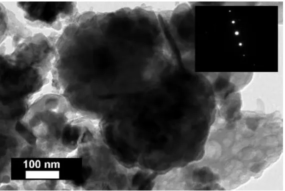
Both bauxite samples have aluminium oxide as a major component, followed by iron oxide
236
while minor amounts of silicon and titanium oxide are also present (Table 2). Calcium oxide is
237
present in the Greek karstic bauxite sample and not in the Ghanaian lateritic bauxite. As a contrast to
238
bauxite samples, bauxite residue’s main component is iron oxide, but there is also present an
239
appreciable amount of unrecovered aluminium oxide. The high amount of calcium oxide is mainly
240
attributed to lime addition in the Bayer process. Sodium oxide content appears from the losses of
241
sodium hydroxide that binds with secondary minerals created during bauxite processing, due to
242
transformation of clay minerals to desilication products and reaction of quartz resulting in the same
243
[61].
244
Greek karst bauxite sample is composed mainly of diaspore, boehmite, hematite, goethite,
245
anatase, calcite and kaolinite. Ghanaian lateritic bauxite sample is composed mainly of gibbsite,
246
hematite, goethite, kaolinite and rutile. Primary mineral phases, those that are already present in
247
bauxite, detected in bauxite residue are hematite, diaspore, boehmite and goethite. Secondary
248
mineral phases, that were formed in the Bayer process, are hydrogarnet, cancrinite, perovskite and
249
gibbsite. Obviously, the content of REEs in the studied samples (Table 3) is too low to be able to
250
detect any REE mineral phases by XRD.
251
Higher REEs concentration between the two bauxite samples is found to be in the Greek karst
252
bauxite (Table 3). The most abundant REE in all samples is cerium (Ce). The resulting bauxite
253
residue has about two times higher REE concentration compared to bauxite feed.
254
Table 2. Major oxide composition of the samples.
255
Karst bauxite Greece
Lateritic bauxite Ghana
Bauxite residue Greece, AoG
wt % wt % wt %
LOI 11.3 29.7 9.2
Al2O3 58.3 55.9 20.2
Fe2O3 21.0 11 44.6
SiO2 2.5 1.2 5.3
TiO2 2.7 1.6 5.7
CaO 1.7 < 0.01 9.1
Na2O 0.4 0.09 2.3
MnO 0.02 0.02 0.04
Table 3. Trace element composition of the samples. Error is given as one standard deviation of a
256
duplicate measurement.
257
Karst bauxite Greece
Lateritic bauxite Ghana
Bauxite residue Greece, AoG
ICP-MS INAA ICP-MS
mg/kg mg/kg mg/kg
La 57 ± 7 19.1 ± 1.3 130 ± 1
Ce 206 ± 8 34 ± 1 480 ± 26
Pr 15 ± 1 n/a 29 ± 2
Nd 53 ± 6 13 ± 1 107 ± 0
Sm 9.8 ± 1.0 2.0 ± 0.2 19.4 ± 0.2
Eu 2.4 ± 0.9 0.8 ± 0.2 4.6 ± 1.1
Tb 2.3 ± 0.5 < 0.5 3.3 ± 0.0
Dy 9.8 ± 0.3 n/a 20.1 ± 0.1
Ho 2.1 ± 0.1 n/a 4.1 ± 0.1
Er 7.2 ± 0.8 n/a 13.3 ± 0.3
Tm < 2 n/a < 2
Yb 7.0 ± 0.4 2.5 ± 0.3 13.8 ± 0.3
Lu < 2 0.4 ± 0.0 2.2 ± 0.0
Y 48 ± 2 n/a 108 ± 2
Nb 55 ± 9 n/a 100 ± 1
Th 51 ± 2 22.7 ± 2.3 105 ± 2
ΣLn1 382.3 854.4
ΣREE2 430.6 962.5
6.2 Precursor REE Phases in AoG’s Bauxite Feed
258
In Table 4 are shown the REE phases that are likely to be introduced to AoG’s production line
259
via the composition of bauxite feed. In Parnassos-Ghiona bauxite profiles (Prossorema and Frussia),
260
detrital rhabdophane and florencite have been identified as LREE phases, whereas detrital churchite
261
and xenotime represent HREE phases [27]. Hydroxylbastnäsite-(La) and -(Nd) were identified in the
262
lowermost bauxite profile samples of Parnassos-Ghiona deposit (Mandri Tsakni) as the only
263
contribution that deployed WDS quantification [23]. Authigenic bastnäsite and parisite group
264
phases were further reported as representatives of authigenic fluorocarbonate LREE minerals in
265
Parnassos-Ghiona bauxite (Pera Lakkos) [62]. As it is not clear on what basis was the distinction of
266
parisite group from the rest of calcium-bearing REE phases made, we categorise it together with
267
other calcium containing LREE fluorocarbonates, synchysite and röntgenite [63]. A recent report
268
questions the earlier identifications of cerium-predominant REE phases, that they could be rather
269
cerium oxides or carbonates, because of the absence of phosphorus and fluorine. Due to the absence
270
of other lanthanides, even the identification as hydroxylbastnäsite-Ce is being questioned [64].
271
Table 4. Precursor REE phases in the bauxite feed of AoG refinery.
272
Parnassos-Ghiona Ghana
Karst Lateriti
c
[23] [27]
[62,65 ] [64]
Presen
t Present LREE
rhabdophane-(Ce) (Ce)(PO4)·H2O +
florencite-(Ce) CeAl3(PO4)2(OH)6 + + +
bastnäsite group Ce(CO3)F +
parisite / synchysite / röntgenite
Ca1–2REE1–3(CO3)2–5F1–
3 + +
hydroxylbastnäsite-(N
d) and -(La) +
Cerianite CeO2 ? +
HREE
1 Sum of lanthanides.
churchite YPO4·2H2O +
xenotime YPO4 + +
In the studied Parnassos-Ghiona bauxite samples, some areas are particularly rich in REE
273
phases (Figure 1). Authigenic cerium-predominant LREE phases in sizes over 10 μm are
274
concentrated into iron and aluminium oxide matrix rather than to alumina-rich pisolithic textures.
275
This is in line with the observations made by Mongelli [25], where he noted that cerium is more
276
fractioned to the bauxite matrix as opposed to ooids (pisoliths). However, the ooids described by
277
Mongelli [25] are controlled by hematite matrix which is different from present observation. This
278
textural fractionation has not been reported in the case of Parnassos-Ghiona bauxite deposit. The
279
LREE phases were identified by μ-Raman spectroscopy being cerianite (CeO2), following the main
280
band at 457 cm-1, when comparing with the reference spectrum R050379 from RRUFF database and
281
those spectra given in the literature sources (Figure 1, b) [60,66]. Based on the analysis of synthetic
282
and natural cerianite specimens, this band can be attributed to the symmetric breathing mode of
283
Ce-O-Ce bond [66,67]. The band at 396 cm-1 present on the reference spectrum R050379, but missing
284
in current experimental spectrum, is not rarely noted in different cerianite Raman spectra [66,67].
285
The band at 396 cm-1 could also be hidden due to the broadening of 457 cm-1 band. Broadening as
286
well as shifting of cerianite main band is noted to occur along with decreasing particle size [68].
287
Notably, the acquired spectrum lacks the presence of Raman bands that could be associated with the
288
occurrence of carbonate (around 1100 cm-1) or hydroxide ions (3400 – 3600 cm-1) [66,69]. The matrix
289
where cerianite is situated, is mainly controlled by hematite, following the Raman bands at 225, 293
290
and 409 cm-1 [60,70]. Zaitsev et al. have demonstrated that fluorine can also be present in cerianite
291
[66]. Fluorine is also present in cerianite of Parnassos-Ghiona bauxite (Figure 1, a). Some of the
292
previously reported REE phases might have been erroneously identified as bastnäsite group phases
293
on qualitative basis when judged only by the presence of fluorine. Thus, current results support the
294
doubts of Mouchos et al. regarding the cerium-predominant phases [64] and evidence is provided
295
for the identification of cerianite in Parnassos-Ghiona bauxite.
296
Figure 1. Cerianite rich area in in Parnassos-Ghiona bauxite shown on (a) backscattered electron
298
(BSE) image with respective EDS elemental maps, and (b) Raman spectra of cerianite and its
299
surrounding matrix compared to reference Raman spectrum [60]. Raman spectrum collected with a
300
532-nm wavelength laser.
301
Authigenic cerium-predominant REE phases were noted to be associated with fissures filled
302
with aluminosilicate matrix that is likely kaolinite (Figure 2). Kaolinite-associated authigenic REE
303
phases have not been reported before, but their presence has been assumed in Parnassos-Ghiona
304
bauxite due to the easily leachable proportion of REEs [71]. It was noted previously, however, that
305
some detrital florencite crystals were encased within clay fragments [27].
306
307
Figure 2. REE phase in Parnassos-Ghiona bauxite associated with aluminosilicate (Al-Si).
308
Regardless of the low REEs concentration in Ghanaian bauxite (Table 3), distinct detrital REE
309
phases are also contained within this lateritic bauxite (Figure 3). The REE minerals have a prevailing
310
content of aluminium, followed by cerium, phosphorus and then other LREEs. Thus, these REE
311
phases can be identified as belonging to the florencite group. In addition, detrital xenotime was
312
identified in the Ghanaian bauxite. Florencite grains are significantly larger (20 – 50 μm) than
313
xenotime grains (1 – 3 μm). The presence of florencite and xenotime group phases in Ghanaian
314
lateritic bauxite implies granitic origin of the bauxite parent material.
315
316
Figure 3. Florencite group LREE phase (Fl) and a zircon grain (Zr) in Ghanaian lateritic bauxite matrix.
317
As a conclusion of this paragraph, it can be said that the main input of LREE phases introduced
318
to the Bayer process in AoG along with the bauxite feed, are LREE fluorocarbonates of the bastnäsite
319
process as phosphate-based groups. Elucidating the REE mineralogy in the Parnassos-Ghiona
321
bauxite deposit deserves a thorough investigation in terms of clearly defining the mineral phases
322
and their spatial distribution along the bauxite profile. The currently existing information is
323
scattered and not uniform.
324
6.3 REE Phases in Bauxite Residue
325
In bauxite residue, REE mineral particles appear in the backscattered electron imaging mode as
326
bright particles. They are contrasting in their brightness from other bauxite residue phases like
327
hematite, diaspore / boehmite, hydrogarnet, titanium dioxides, cancrinite or perovskite (Figure 4).
328
The formerly mentioned phases (hematite, diaspore / boehmite, hydrogarnet, titanium dioxides,
329
cancrinite, perovskite) are distinguishable from each other in EPMA analysis. At a similar brightness
330
level as the REE particles, other heavy mineral particles such as for instance, zircon, chromite, pyrite
331
and chalcopyrite were revealed. Even metallic iron chips with the presence of chromium are present.
332
The latter are thought to originate from the grinding equipment of the alumina refining plant.
333
334
Figure 4. Backscattered electron image of bauxite residue. The bright REE particle in the middle
335
corresponds to the particle depicted on Figure 8 (b) and Figure 11.
336
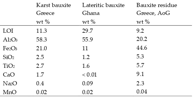
6.3.1 REE Carbonate and Phosphate Phases
337
In the investigated bauxite residue sample, a neodymium and lanthanum predominant particle
338
with the presence of carbon was revealed (Figure 5). Other LREEs like praseodymium and
339
gadolinium were also present. The particle was notably large, more than 40 μm in its longest
340
342
Figure 5. An ancylite group LREE carbonate phase depicted on (a) backscattered electron image with
343
its (b) EDS spectrum and (c) Raman spectrum compared to a reference spectrum of kozoite-(La) [60].
344
Raman spectrum collected with a 532-nm wavelength laser.
345
Raman investigation of this grain resulted in a spectrogram showing a major peak at 1088 cm-1,
346
that can be attributed to symmetric C-O stretching of CO32- (Figure 5 c) [72,73]. Comparison with
347
reference data from RRUFF database resulted in a notably similar match with the kozoite-(La)
348
(La(CO3)(OH)) Raman spectrum [60]. The observed peak at 1088 cm-1 is the most characteristic one
349
for the ancylite group phases [60,72]. Kozoite, belonging to the ancylite mineral group, is
350
dimorphous with hydroxylbastnäsite. In other words, it has identical chemical composition, but
351
different mineral structure. The former occurs in orthorhombic crystal system and the latter in
352
hexagonal [74]. The kozoite-(La) reference spectrum given in the RRUFF database has not yet been
353
confirmed by other identification methods. Therefore, the present identification can’t be regarded as
354
conclusive. However, the absence of other matching spectra and the relative similarity with other
355
ancylite group minerals Raman spectra [60] allows at least suggesting that the investigated particle
356
belongs to ancylite mineral group. Other LREE particles in bauxite residue analysed with Raman
357
spectroscopy did not result in unambiguously interpretable Raman spectra, did not provide any
358
Raman scattering bands or were overwhelmed with fluorescence.
359
The formerly described evidence shows that a part of LREEs can occur as carbonate phases in
360
bauxite residue. Ancylite group minerals have not been identified in any bauxite sample. It could be
361
that they have been reported as hydroxylbastnäsite species because of their identical chemical
362
composition. Generally, REE carbonate phases are expected to be dissolved during sodium
363
hydroxide digestion [32]. Based on the above-mentioned evidence, it is difficult to define whether
364
the LREE carbonate phase is a primary mineral inherited from bauxite that withstood Bayer
365
digestion conditions or is a secondary precipitate form created in the Bayer process. In any case, it is
366
a very rare occurrence type in bauxite residue.
367
In a few cases, LREEs are found as calcium containing phosphate phases in bauxite residue,
368
more specifically as cerium phosphates. It can be seen from the EDS spectrum of an analysed
369
particle, exhibiting a pronounced phosphorus X-ray peak (Figure 6). This and other similar observed
370
grains are cerium predominant. The low amount of LREE phosphate species in bauxite residue is in
371
line with the relative scarcity of phosphate phases in the AoG’s bauxite feed. The composition of
372
these grains resemble rhabdophane-Ce that has been detected in Parnassos-Ghiona bauxite [27]. It is
373
an indication that REE phosphates endure, at least partly, the Bayer process. This is an expected
374
behaviour as REE phosphates don’t dissolve easily in sodium hydroxide, although there are
375
monazite and xenotime [32]. In such processes, sodium hydroxide is more concentrated (40 – 50 %
377
NaOH) [32] than in the Bayer process (12 – 22 % NaOH) [38].
378
379
Figure 6. Cerium phosphate in bauxite residue matrix, shown on (a) backscattered electron image
380
with its (b) EDS spectrum.
381
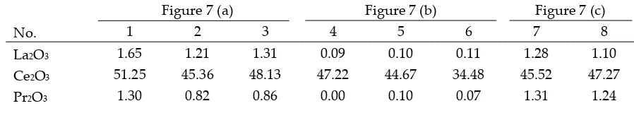
6.3.2 LREE ferrotitanate species, (REE,Ca,Na)(Ti,Fe)O3
382
In bauxite residue, LREE mineral particles that contain calcium, titanium, iron and sodium
383
(Figure 7, Figure 8 Table 5, Table 6) are also found. They further divide into cerium predominant
384
(Table 5) and neodymium-lanthanum predominant particles (Table 6). The number of ions in the
385
mineral formula shown in Table 5 and 6 have been calculated based on a perovskite stoichiometry
386
with three oxygen atoms (ABO3). Alternatively, the number of ions could be calculated by adopting
387
the double perovskite structure with the composition A2B2O6 [75]. Division of the ions between A
388
and B sites is based on previous literature, considering their charges and relative ionic radii [75,76].
389
The chemical composition of these particles is variable, for instance Ce2O3 content ranges from about
390
34 to 51 wt % while TiO2 content ranges from 9 to 24 wt % (Table 5). It can be noted that
391
measurements 1 – 6 in Table 6 are depleted in Fe2O3 content, although the title of this section refers to
392
ferrotitanate species. This effect is explained further in the text below. Such chemical composition
393
which is uncommon for REE phases in bauxite, especially the appreciable presence of sodium,
394
clearly indicates that the LREE ferrotitanates are formed during the Bayer process.
395
396
Figure 7. Backscattered electron images of cerium ferrotitanate grains in bauxite residue matrix, (a),
397
(b) and (c). The indicated quantification spots are reported in Table 5.
398
Table 5. EPMA-WDS quantification (wt %) of cerium predominant ferrotitanate grains (Figure 7).
399
Lower section of the table shows the number of ions in the mineral formula, following the ABO3
400
perovskite structure.
401
Figure 7 (a) Figure 7 (b) Figure 7 (c)
No. 1 2 3 4 5 6 7 8
La2O3 1.65 1.21 1.31 0.09 0.10 0.11 1.28 1.10
Ce2O3 51.25 45.36 48.13 47.22 44.67 34.48 45.52 47.27
Nd2O3 2.48 1.93 2.00 1.41 1.54 1.00 3.30 2.99
TiO2 9.00 9.16 9.44 14.84 21.78 18.56 21.37 23.86
Fe2O3 17.85 22.33 21.98 14.84 8.77 22.87 7.80 7.32
CaO 4.34 4.01 4.13 6.56 8.78 9.91 7.93 9.59
MgO 0.02 0.05 0.03 0.01 0.02 0.22 0.03 0.01
SiO2 0.98 0.97 0.98 1.00 1.21 2.09 1.53 1.59
Na2O 0.90 1.32 0.92 0.92 1.49 2.16 2.01 3.21
Al2O3 1.39 1.43 1.30 1.13 1.12 2.01 1.50 1.39
ThO2 2.68 1.99 2.21 1.36 1.36 1.21 0.02 0.02
Total 93.83 90.58 93.28 89.36 90.94 94.70 93.60 99.58 No. of ions per ABO3 formula
La 0.03 0.02 0.02 0.00 0.00 0.00 0.02 0.01
Ce 0.81 0.73 0.75 0.71 0.61 0.44 0.61 0.58
Pr 0.02 0.01 0.01 0.00 0.00 0.00 0.02 0.02
Nd 0.04 0.03 0.03 0.02 0.02 0.01 0.04 0.04
Ti 0.29 0.30 0.30 0.46 0.61 0.49 0.58 0.60
Fe 0.58 0.73 0.71 0.46 0.25 0.60 0.21 0.18
Ca 0.20 0.19 0.19 0.29 0.35 0.37 0.31 0.35
Mg 0.00 0.00 0.00 0.00 0.00 0.01 0.00 0.00
Si 0.04 0.04 0.04 0.04 0.05 0.07 0.06 0.05
Na 0.08 0.11 0.08 0.07 0.11 0.15 0.14 0.21
Al 0.07 0.07 0.07 0.06 0.05 0.08 0.06 0.06
Th 0.03 0.02 0.02 0.01 0.01 0.01 0.00 0.00
Structural formulas following the ABO3 structure A (REE, Ca,
Na) 1.20 1.11 1.11 1.11 1.10 0.99 1.14 1.20
B (Ti, Fe,
Al, Si) 0.99 1.15 1.12 1.02 0.95 1.24 0.92 0.90
402
Figure 8. Neodymium-lanthanum predominant LREE particles, of which (a) is partly reacted, and
403
(b). exhibits a zonation (I – III) relating to reaction stages with Bayer liquor. Within zone II of (b),
404
deposition of a sodium aluminosilicate phase (Na-Al-Si) is indicated.
405
Table 6. EPMA-WDS quantification (wt %) of a neodymium-lanthanum predominant partly reacted
406
LREE grain (1 – 4) (Figure 8 a) and a LREE ferrotitanate grain (5 – 10) (Figure 8 b). Lower section of
407
Figure 8 (a) Figure 8 (b)
I II III
No. 1 2 3 4 5 6 7 8 9 10
La2O3 23.21 24.93 25.99 25.49 23.89 23.89 10.25 13.87 1.06 7.41 Ce2O3 7.87 8.87 10.57 10.47 5.99 5.46 2.71 3.49 0.58 1.29 Pr2O3 21.91 20.56 20.79 21.90 22.02 20.78 9.41 8.45 1.03 5.60 Nd2O3 35.10 33.73 33.59 34.02 34.92 34.19 14.37 17.22 1.71 8.53 TiO2 3.87 5.85 0.83 3.08 1.44 2.03 27.48 24.13 32.60 40.35 Fe2O3 2.61 2.35 2.20 3.27 6.67 4.07 10.71 12.21 42.44 12.26 CaO 2.11 3.07 1.93 2.56 1.46 1.12 4.42 5.64 17.92 14.16 MgO 0.00 0.00 0.01 0.00 0.11 0.08 0.99 0.02 0.03 0.03 SiO2 0.16 0.29 0.17 0.20 0.38 0.54 2.93 1.25 1.57 2.30 Na2O 0.16 0.62 0.00 0.14 0.00 0.00 5.65 4.46 2.87 6.93 Al2O3 0.28 0.34 0.33 0.25 0.66 0.88 3.21 1.36 1.07 2.13 ThO2 0.00 0.00 0.00 0.00 0.09 0.07 0.05 0.02 0.01 0.04 Total 97.28 100.62 96.40 101.37 97.62 93.11 92.17 92.12 102.89 101.02 No. of atoms per ABO3 formula
La 0.44 0.43 0.52 0.46 0.45 0.47 0.12 0.18 0.01 0.07 Ce 0.15 0.15 0.21 0.19 0.11 0.11 0.03 0.05 0.01 0.01 Pr 0.41 0.35 0.41 0.39 0.41 0.41 0.11 0.11 0.01 0.05 Nd 0.64 0.57 0.65 0.60 0.64 0.65 0.17 0.22 0.02 0.08 Ti 0.15 0.21 0.03 0.11 0.06 0.08 0.67 0.65 0.67 0.81 Fe 0.10 0.08 0.09 0.13 0.26 0.16 0.26 0.33 0.87 0.25 Ca 0.12 0.16 0.11 0.13 0.08 0.06 0.15 0.22 0.52 0.40 Mg 0.00 0.00 0.00 0.00 0.01 0.01 0.05 0.00 0.00 0.00 Si 0.01 0.01 0.01 0.01 0.02 0.03 0.09 0.04 0.04 0.06 Na 0.02 0.06 0.00 0.01 0.00 0.00 0.35 0.31 0.15 0.36 Al 0.02 0.02 0.02 0.01 0.04 0.06 0.12 0.06 0.03 0.07 Th 0.00 0.00 0.00 0.00 0.00 0.00 0.00 0.00 0.00 0.00 Structural formulas following the ABO3 structure
A (REE, Ca,
Na) 1.76 1.72 1.90 1.79 1.72 1.71 0.99 1.08 0.72 0.98 B (Ti, Fe, Al,
Si) 0.27 0.32 0.15 0.27 0.37 0.33 1.15 1.08 1.61 1.18
Some LREE particles were observed that have a relatively small percentage of iron, titanium
409
and sodium oxide content (Figure 8 a, Table 6). Others showed distinct zonation expressed in wide
410
variation in chemical composition as well as in morphological features (Figure 8 b, Table 6).
411
The texture of LREE ferrotitanate grains is anhedral. Secondary electron imaging of bauxite
412
residue “as is” revealed that LREE ferrotitanate grains are partly covered with submicron-sized
413
bauxite residue matrix particulate (Figure 9). At most, aggregates of anhedral globular crystallites
414
can be observed when examining larger particles that exhibit different reaction stages. This can be
415
417
Figure 9. Cerium ferrotitanate particle shown on (a) secondary electron image with its (b) EDS
418
spectrum; gold (Au) peak is from the coating layer on the sample.
419
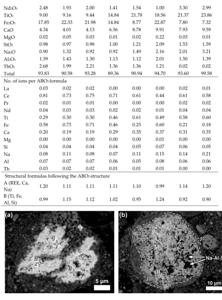
In addition, LREE-containing globular calcium ferrotitanate particles were discerned in
420
nanoscale investigation with HRTEM (Figure 10, Appendix A Table A1). The maximum
421
concentrations of REEs measured in EDS were about 3 wt % of cerium and about 2.5 wt % of
422
lanthanum (Table A1). The quantities of REEs below 1 wt % were regarded as distrustful due to the
423
poor ability of EDS to measure trace constituents. Selected area electron diffraction (SAED) of a
424
LREE bearing particle resulted in a reflection pattern indicating to a well crystallised character. The
425
d-values (d = 0.2564 nm) measured form the patterns resemble the ones of conventional perovskite
426
reference (d = 0.2710 nm) from [121] direction. Most of the calcium ferrotitanate particles were,
427
however, REE-depleted. Other REE-containing phases were not confidently discerned among the
428
fine particulates of bauxite residue during nanoscale HRTEM investigation.
429
430
431
Figure 10. LREE-bearing titanium ferrotitanate observed in the bright field imaging mode of
432
HRTEM. SAED pattern of the particle is inserted to the upper right corner. The pattern is collected
433
from [121] direction.
434
From the previously noted observations, an important conclusion should be made. Calcium
435
ferrotitanate species, likely corresponding mineralogically to perovskite, with low LREE
436
concentration, should not be confused with the REE-barren perovskite sensu stricto (CaTiO3) that is
437
different reaction route. Namely, titanium dioxide phases, especially anatase, react partially with
439
sodium hydroxide and then with lime added to Bayer process and as a result form perovskite sensu
440
stricto [77].
441
When the LREE ferrotitanate particles are relatively large (20 – 30 μm), the variations in their
442
chemical composition can be drawn out by elemental mapping (Figure 11). It is seen in Figure 11,
443
that the highest REEs concentrations are found in the core of the particle. REE concentration
444
decreases towards the edges of the particle. In the bauxite residue matrix surrounding the particle,
445
there are no presence of REEs. The trend is the opposite for titanium, calcium and sodium. Their
446
concentration increases towards the edges of the particle. While the change in titanium
447
concentration is quite gradual from core to edge, there is a more sudden increase of calcium
448
concentration on the rim of the particle. The gradual change of the chemical composition refers to
449
the existence of a solid solution between the LREE predominant and calcium predominant
450
end-members. Another characteristic feature of the reacted LREE ferrotitanate particles is that the
451
outer layer tends to form a distinct calcium ferrotitanate shell around the particle, where LREEs
452
concentrations are low (Ti, Ca and Fe maps on Figure 11). The zone that contains about an equal
453
amount of titanium and REEs (corresponding to zone II on Figure 8 (b), or silica rich area in Figure
454
11, indicated with Na-Al-Si), is also intergrown with a sodium aluminium silicate phase. This likely
455
corresponds to a secondary Bayer process phase, sodalite or cancrinite.
456
457
Figure 11. EPMA-WDS quantitative elemental mapping of a reacted LREE particle. The intensely
458
reacted area is also intergrown with sodium aluminosilicate phase (indicated with Na-Al-Si).
459
Mapped area corresponds to Figure 8 (b).
460
A few considerations can be given about the formation mechanism of such LREE ferrotitanate
461
phases. As mentioned before, the LREE ferrotitanate phases are clearly formed during the Bayer
462
process. The primary LREE phases interact with the Bayer process liquor. The precursor phases
463
possibly belong to the bastnäsite mineral group of REE fluorocarbonates or cerianite as they are the
464
most frequently encountered REE phases in bauxite feed. Bayer liquor consists mainly of sodium
465
aluminate and free sodium in the form of sodium hydroxide [7]. There is commonly, at least in the
466
(1 – 10 mg/l TiO2) present [38,78,79]. The reaction appears to take place in situ on the outer part of the
468
REE particles (III on Figure 8 b). There is no indication if the newly formed phases would be
469
precipitated from the processing liquor. The ions taking part in the reaction (Na, Fe, Ti, Ca) diffuse
470
into deeper parts of the particles as the reaction progresses. Titanium ions diffusion seems to be the
471
most intense. The inner part of the particle (I on Figure 8 b) seems to be only slightly affected by the
472
reaction, as there is only a minor part of titanium and iron present while no sodium is detected.
473
Similarly, the particle on Figure 8 (a) has very low content of titanium and iron as well as sodium.
474
The presence of calcium might be already inherited from the precursor mineral. Thus, the complete
475
particle on Figure 8 (a) as well as inner part (I) of on Figure 8 (b) are neither entirely a primary REE
476
phase nor a newly created LREE ferrotitanate, but an intermediate phase with a deficiency of
477
titanium and iron. The intensely reacted zones II and III (Figure 8 b, Figure 11) are also intergrown
478
with sodium aluminium silicate phases. This might indicate to a gel-like state in the reaction front
479
that allows also other mineral species to be nucleated and formed.
480
The morphology of the inner part resembles the fibrous or acicular radiating morphology
481
described in the case of authigenic LREE phases in bauxites [25]. The middle and outer parts (II and
482
III) exhibit a different morphology with globular crystallites, which are characteristic to the newly
483
formed LREE ferrotitanates. They indicate also to a newly formed mineralogical character. The outer
484
part (III) of the particle represents the last stage of the transformation, where a high amount of
485
calcium and titanium have been deposited with 4 – 23 wt % of REEs also present (Table 6). This latest
486
deposition forms a distinct shell around many of the observed LREE particles.
487
Considering that the grains on Figure 8 are relatively large, it can be assumed that smaller REE
488
particles react entirely and their final products should be something similar to what is observed in
489
the zone III on Figure 8 (b), with calcium prevailing ferrotitanates alongside some presence of REEs.
490
We have shown evidence in support of this claim from the nanoscale HRTEM investigation, where
491
the maximum REE concentrations per particle did not exceed 5 wt %. The particles seen in nanoscale
492
have a similar composition as well as morphology to the area seen in highly reacted zone III on
493
Figure 8 (b).
494
In support of the in situ transformation of LREE minerals in the Bayer process stands the fact
495
that LREEs do not possess soluble species in highly alkaline conditions [80,81]. In a broad sense, an
496
analogous in situ transformation of kaolinite to sodium aluminium hydrosilicate has been described
497
to take place during Bayer process [82].
498
We observed that the LREE species containing sodium, calcium, titanium and iron in variable
499
proportions form a solid solution. The characteristics of the solid solution are expressed on Figure
500
12, where the ionic proportions of the cations are plotted. Region denoted with I on (a) refers to the
501
measurements reflecting the real solid solution. Region denoted with II on (a) are the measurements
502
performed on the transitional phases that are not the final LREE ferrotitanate products. The solid
503
solution characteristic is the most recognizable on A site, where Ca and Na substitute REEs. The
504
correlation coefficient between the substituting cations is 0.947 (b on Figure 12). The endmembers of
505
the series have ideal compositions of (Ca,Na)(Ti,Fe)O3 and (REE,Ca,Na)(Ti,Fe)O3. Because the
506
measured compositions are highly variable, it is not reasonable to report any average chemical
507
composition or formula of the LREE ferrotitanates. Some examples of the formulas can be shown
508
that approach the ideal stochiometric end-members of the series. For the neodymium-lanthanum
509
predominant phases they can be (Ca0.40Na0.36REE0.22)Σ0.98 (Ti0.81Fe0.25)Σ1.05O3 (spot 10 in Table 6) and
510
512
Figure 12. Solid solution character of LREE ferrotitanate series, depicted as ionic proportions of the
513
(a) substitutions of Ca and Na with REE on A site and (b) complete transformation, where (REE + Na
514
+ Th) + (Fe + Al) = Ca + Ti. Region annotated with I on (a) refers to the area of real solid solution,
515
region annotated with II on (a) indicates to the measurements on transitional phases. Adapted after
516
Campbell et al. and Nickel & McAdam [76,83]. The equation is changed for a best description of
517
present situation (i.e. Nb is left out of the equation whilst Th and Al are added to A and B sites,
518
respectively). Figures are based on data from Table 5 and Table 6.
519
The only mineral group containing species corresponding to the currently presented chemical
520
composition (Table 5, Table 6) is the perovskite sensu lato. Perovskite group, also termed as the
521
perovskite supergroup refers to the basic structure of ABX3, where A is a relatively large cation, B is
522
relatively small cation and X is oxygen or another anion [75]. It’s aristotypic mineral structure is
523
cubic. However, due to the extremely wide compositional variations, many structures are possible.
524
Perovskites exhibit extensive solid solutions, where diverse cations can occupy the A and B sites.
525
REE-containing perovskites are well known from the natural systems and there have been a large
526
number of REE perovskites synthesized for many applications [75,84,85]. The compositions
527
measured in present work resemble the end-members of the perovskite sensu stricto (CaTiO3) and
528
loparite ((REE,Na,Ca)(Ti,Nb)O3) [86] solid solution series [75]. Loparite typically occurs in
529
peralkaline igneous rocks, especially in nepheline syenite [86]. As in the peralkaline rocks, there is an
530
excess of sodium in the present investigated system of Bayer process. It can be noted from Table 5
531
and Table 6, that the currently calculated mineral formulas don’t have an ideal stoichiometry as
532
there are deficiencies and excesses of ions on A and B sites. This is a regularly encountered
533
observation for perovskites, as they are considered “defect” structures [75,76]. Excess and deficiency
534
are compensated with the different ion proportions on A and B sites considering the charge balance
535
as well as excess or deficiency of oxygen molecules [75,76,84]. As many of currently analysed spots
536
show approximately 1:1 ratio of A:B sites, oxygen deficiency can be hypothesised to exist. One of the
537
deviations compared to perovskite-loparite natural system, however, is that in current observations,
538
there is no presence of niobium detected. Typically, niobium is a ubiquitous constituent in the
539
natural occurrences of loparite [75]. This is explained by the fact that present LREE ferrotitanates are
540
formed in situ and thus inherit partly the chemical composition of their precursor phases. In the
541
precursor minerals, there is no niobium present (Table 4). Besides, niobium concentration in the bulk
542
sample is low, 100 mg/kg (Table 3). Thus, niobium is not expected to appear in the reaction product
543
either. A second slight deviation is, that we are also observing the presence of iron in the analysed
544
particles. However, iron can generally exist together with titanium on the B site of perovskite /
545
loparite [76], but the nomenclature of iron containing perovskites has not yet been established [75].
546
Regardless of the incomplete nomenclature, perovskites with composition LaFeO3 have been
547
synthesized and characterised [87]. Since we are investigating a technogenic system and not a
548
natural one, it is not uncommon to find some rarely encountered mineral types. Perovskites
549
matching with the currently defined chemical composition have not yet been synthesised, but
550
R² = 0.884
0.00 0.20 0.40 0.60 0.80 1.00
0.00 0.50 1.00 1.50 2.00
C
a,
N
a
REE
(
a
)
I
II
R² = 0.947
0.00 0.40 0.80 1.20
0.80 1.20 1.60 2.00
C
a, T
i
similar ones are for example NaLaTi2O6 or NaCeTi2O6 [75]. Moreover, endmembers of
551
perovskite-loparite series with formulas Na0.5Ce0.5TiO3 and Na0.5La0.5TiO3 have been identified as
552
thermodynamically stable [88]. Among a variety of perovskite synthesizing methods, there exist the
553
wet chemical processes, such as hydroxide-based sol-gel process [85]. Cerium titanates (CeTi2O6)
554
with brannerite structure have been also synthesized [89]. Those titanates, however, do not match
555
with the stoichiometry of current REE ferrotitanates because they have two moles of titanium per
556
one mole of cerium while in the present case the A and B sites have approximately one-to-one ratio.
557
Present findings can also be related to some of the previously existing knowledge about REE
558
phases in bauxite residue. During a nanoscale investigation, a perovskite phase with a general
559
composition of Ca0.8Na0.2TiO3 was described to contain trace amounts of thorium as well possibly
560
cerium and some other trace elements [51]. The authors proposed a minor contribution from a
561
loparite phase related to perovskite to explain the observations. That information compares well
562
with the present analysis. The mentioned perovskite was also crystallographically characterised and
563
matched with perovskite structure reference with some deviations from the conventional data,
564
possibly due to the incorporation of sodium on the A site of perovskite [51]. Cerium and titanium
565
correlating presence was found in a Canadian bauxite residue sample [52]. Bayer process derived
566
REE titanate compounds were mentioned in a patent describing the recovery of REEs from bauxite
567
residue [50]. A recent contribution that investigated strictly the forms of cerium in bauxite residue,
568
did not refer to any relations between cerium and titanium [54]. The former comparison allows to
569
suggest, that the observed LREE ferrotitanate phases presented in this study are not isolated cases
570
for only AoG-s bauxite residue, but instead seem to be a more generic characteristic that occurs in
571
bauxite residues originating from different alumina refineries. The exception presented by Bolanz et
572
al., who hypothesize that the main carrier of cerium is hematite [54], indicates to the need of
573
examining rare earth phases in relation to Bayer process conditions as well as in relation to bauxite
574
feed and REE occurrence forms within bauxite.
575
Current data supports the existence of thorium in perovskite type phases contained in bauxite
576
residue [51]. In fact, compared to the 700 mg/kg thorium concentration estimated in the report by
577
Gamaletsos et al. [51] based on EDS analysis, our WDS quantification results show that thorium
578
concentration in cerium predominant LREE ferrotitanates can reach as high as 2.7 wt % ThO2 (Table
579
5). Also, thorium is mainly associated with cerium predominant phases instead of neodymium and
580
lanthanum predominant ones. This can be explained by the possible existence of cerium in
581
tetravalent oxidation state, which is the same as for thorium. At the same time, practically all the
582
other REEs occur only in the trivalent oxidation state. That inhibits the incorporation of thorium to
583
the mineral structure of neodymium predominant phases. The amount of thorium incorporated to
584
LREE ferrotitanate phases is probably highly dependent on the precursor REE phases in bauxite and
585
the content of thorium within them.
586
Our observations deviate with the work of Gamaletsos et al. [51] in the fact that no niobium or
587
zirconium were detected in the LREE ferrotitanate particles analysed in this work. Considering the
588
reaction mechanism that forms the LREE ferrotitanates, it is a reasonable observation that the
589
aforementioned elements are not present in the reacted REE particles since neither of them is a
590
component of the precursor REE phases. Current work also disagrees with the statement of
591
Gamaletsos et al. [51], that bauxite residue is a very homogeneous material, for which microscale
592
investigations, especially with regards of trace elements, are not feasible to be conducted. As shown
593
in present work, microscale investigations, combined with nanoscale analysis, provide very detailed
594
information about the fate of REE minerals in the Bayer process.
595
6.3.3 Manganese-associated Cerium Oxide or Oxyhydroxide
596
A part of LREE phases were found to be associated with manganese (Mn) in bauxite residue
597
(Figure 13). On Figure 13 and similar observations, LREEs occur as surface adsorbed phases on
598
manganese mineral particles. It may be assumed that they are manganese oxyhydroxides. In the case
599
with cobalt (Co) and nickel (Ni) as well as some magnesium (Mg). Following its chemical
601
composition, the manganese phase could be asbolane (Mn(O,OH)2(Ni,Co)x(O,OH)2 · nH2O) [90].
602
603
Figure 13. Cerium oxide / oxyhydroxide phase associated with manganese particle shown on (a) EDS
604
elemental map with (b) respective spectra.
605
It is common that the named LREE formations have a circular morphology that follows the
606
circular cavities of manganese particles or they surround the manganese grains. The mentioned
607
LREE phases contain cerium as the prevailing element, but other lanthanides as well as calcium were
608
discerned to be present also (Figure 13). Based on semi-quantitative EDS estimation, cerium
609
concentration in this LREE occurrence form is about 35 – 40 wt %.
610
Individual manganese associated LREE particles are small, about 1 μm in size, but
611
agglomerated particles are over 10 μm in dimensions. On occasions, manganese associated LREE
612
phases exhibit remarkable sizes exceeding 50 μm (Figure 14). On the example of the particle
613
depicted on Figure 14, it may be assumed that the growth of this LREE particle was nucleated on a
614
manganese-nickel-cobalt particle and continued to form a cerium and calcium predominant particle.
615
The fine acicular morphology of the particle on Figure 14 indicates that the growth rate of this


![Figure 5. An ancylite group LREE carbonate phase depicted on (a) backscattered electron image with its (b) EDS spectrum and (c) Raman spectrum compared to a reference spectrum of kozoite-(La) [60]](https://thumb-us.123doks.com/thumbv2/123dok_us/7924936.1315862/11.595.152.444.78.287/ancylite-carbonate-backscattered-spectrum-spectrum-compared-reference-spectrum.webp)