A Thesis submitted to The University o f London for the degree of Doctor of Philosophy
Expression Studies o f Recombinant cAMP-specific
Phosphodiesterase Type 4
N a d ia K o rn io tis School o f Pharmacy
Department of Pharmaceutical and Biological Chemistry 29/ 39 Brunswick Square
ProQ uest Number: 10104247
All rights reserved
INFORMATION TO ALL U SE R S
The quality of this reproduction is d ep en d en t upon the quality of the copy subm itted.
In the unlikely even t that the author did not sen d a com plete manuscript
and there are m issing p a g e s, th e se will be noted. Also, if material had to be rem oved, a note will indicate the deletion.
uest.
ProQ uest 10104247
Published by ProQ uest LLC(2016). Copyright of the Dissertation is held by the Author.
All rights reserved.
This work is protected against unauthorized copying under Title 17, United S ta tes C ode. Microform Edition © ProQ uest LLC.
ProQ uest LLC
789 East E isenhow er Parkway P.O. Box 1346
University o f London
Abstract
Expression Studies of Recombinant cAMP-specific
Phosphodiesterase Type 4
By N ad ia K orniotis
Cyclic nucleotides (cAMP and cGMP) are second messenger molecules that play a key role in many physiological processes, cAMP in particular regulates cell growth, cell differentiation, inflammation and glycogen metabolism. cAMP is degraded by phosphodiesterase 3 (PDE3) and phosphodiesterase 4 (PDE4) which belong to a large family o f PDEs consisting o f 12 members. PDE4 isoenzymes which are highly specific for cAMP are most abundant in inflammatory cells making them good clinical targets for the therapy o f asthma and other inflammatory disorders. The design o f potential therapeutics will be helped by the structural analysis o f PDE4.
PDE gene Met^^RDl was cloned and expressed in two systems, bacterial and mammalian. The bacterial system expressed Met^^RDl as a Glutathione S-transferase fusion protein in Escherichia coli. A substantial portion o f phosphodiesterase expressed in bacteria was found to be inactive, insoluble and very highly degraded by proteolytic enzymes. Consequently, PDE4 expression in other systems was investigated. However, the insoluble protein produced was solubilised in urea and used to raise antibodies in rabbits as there were no commercial antibodies available against this protein.
Acknowledgment
The w ork described here would not be made possible without the help and support o f many people. However, my principle debt goes to my husband who showed me support, help and encouragement throughout the project and without whom this work would not be possible or worth doing.
I would like to thank my supervisor Dr Andy W ilderspin for giving me the chance to accomplish such an achievement and for providing me enough supervision and guidance to keep me on the right track.
Many thanks go to the many people that I had the opportunity o f meeting over the last four years at the School o f Pharmacy. In particular I wish to thank Pedro Baptisa, Bela Chopra, Liz Khatri and Seema Sharma for making the School o f Pharmacy a happy and enjoyable environment to work in. My special thanks go to Lynda Hawkins for her technical help and support, to Jurgen for his assistance with DNA sequencing, and to Colin James for his computing expertise. I must also thank Kieran Brickley and Paul Chazot for their help and technical support with antibody production. To my colleagues, Adi Moloudi and Ravinda Sidhu go my best thanks for their help and support in the lab. and to whom I wish all the best for the future.
A word to my financial sponsors the BBSRC and to the Registrar Peggy Stone for her support and backing during the loss o f my mother-in-law a year ago ... THANK YOU.
I would like to thank my current employers at GlaxoSmithKline and my future employer Novartis Institute o f Medical Sciences for their help, support and patience through the past six months and through the coming months.
List of Abbreviations
Approximately
a Alpha
Akt Protein Kinase B
APC Antigen presenting cells
APS Ammonium persulphate
ATP Adenosine triphosphate
P
BetaP-gal p-galactosidase
BHK Baby hamster kidney
bp Base pair
BSA Bovine serum albumin
Ca'+ Calcium ion
CaM Calmodulin
cAMP Cyclic adenosine 3', 5'-monophospate
cDNA Complementary DNA
cGMP Cyclic guanosine 3', 5'-monophosphate
CHO Chinese hamster ovary
Chr Chromosome
cpm Counts per minute
C-terminal Free carboxyl group o f a protein
CREB cAMP response element-binding protein
CRE cAMP-response element
°C Degrees centigrade
Da Dalton
DAB 3', 3-Diaminobenizine
DAG Diacylglycerol
DNA Deoxyribose nucleic acid
dATP Deoxyadenosine triphosphate
dCTP Deoxycytosine triphosphate
Diethylaminoethyl-DEPC dGTP dhfr DMF DMSO DMP dNTP DTT E. coli EDTA EtBr ERK Fab Fc fmole Y g g GAF Diethyl pyrocarbonate Deoxyguanosine triphosphate dihydrofolate reductase Dimethylformamide Dimethylsulphoxide Dimethyl pimelimidate Deoxynucleotide triphosphate Dithiothreitol Escherichia coli Ethylenediaminotetraacetic acid Ethidium bromide Extracellular-signal-regulated kinase Fragment antigen binding
Antibody fragment that is most readily crystallised Femtomole, multiple o f
Gamma Gram
Acceleration due to gravity
cGMP-specific phophodiesterases, cyanobacterial Anabaena adenylyl cyclases, and Escherichia coli transcriptional regulator fh l A
GDP Guanosine 5'-diphosphate
Gi Inhibiting G-proteins
GMEM Glasgow M inimal Essential M edia
Gs Activating G-proteins
GST Glutathione S-Transferase
GTP Guanosine 5'-triphosphate
H Helix
Tritium
h Hour
HESS Hanks’ balanced salts
pH]-cAMP Tritiated cyclic adenosine monophosphate
HRP Horse Radish Peroxidase
HSL Hormone-sensitive lipase
IBMX 3-isobutyl-1 -methylxanthine
IgG Immunoglobulin G
EL-2 Interleukin 2
IP3 Inositol triphosphate
EPTG Isopropyl-p-D-thiogalactopyranoside
IRS-1 Insulin receptor substrate-1
kb or Kb Kilobase pairs
kDa or KDa KiloDalton
K . Michaelis-Menten constant
< Less than
L Litre
Lac Z Reporter gene that encodes for p-gala
LB Luria-Bertani
LBA Luria-Bertani Agar
M Molar, moles per litre
mA Milliamps
Divalent metal ion
MCS M ultiple cloning site
Magnesium ion
|LAg Microgram
ml Millilitre
ml Microlitre
|jM M icromolar
^imole Micromole, multiple o f 10"^
MOPS 3-(N-Marpholino) propane sulphonic
mRNA Messenger RNA
ng Nanogramme
nm Nanomitre
nmole Nanomole, multiple o f 10'^
NO Nitric oxide
N-terminal OD
O N o rO /N Pi PAGE PBS PDE PEST pH PKA pKa PMSF PCR pmole RDI RNA RNase A RNasin rNTP rpm SDS SFV SH3 Sj26 SV X TAE TEMED TLCK TPCK I r is
Triton X-100
Free a-am ino group o f a protein Optical density
Overnight
Inorganic phosphate
Polyacrylamide gel electrophoresis Phosphate buffered saline
Phosphodiesterase
Proline, Glutamic acid, Serine, and Threonine amino acid sequence
-logioPT
Protein Kinase A
Dissociation constant, -logioKa Phenylmethylsulphonylfluoride Polymerase chain reaction Picomole, multiple o f 10’*^ Rat dunce 1
Ribonucleic acid Ribonuclease A Ribonuclease inhibitor
Ribosomal nucleotide triphosphate Revolutions per minute
Sodium dodecyl sulphate Semliki forest virus Src homology 3
Schistosoma japonicum 26 Sindbis virus
Times
Tris, Acetic acid, EDTA buffer
N ,N ,N ’ ,N’ -tetramethyethylethylenediamine Tosyllysine chloromethyl ketone
Tosylphenylalanine chloromethyl ketone Tris-(hydroxymethyl)amino-methane
TS
where ‘poly’ averages 10 ethaxyethenol moieties Tris saline buffer
V Volt
Vh Variable heavy chain
Vl Variable light chain
V„ax M aximum velocity
VS Versus
UCR Upstream Conserved Region
UV Ultraviolet light
(v/v) Volume per volume
(w/v) Weight per volume
X-gal 5-bromo-4-chloro-3-indolyl-P-D-galactopyranoside
Amino Acids and Their Symbols
Ala Alanine
Cys Cysteine
Asp Aspartic acid
Glu Glutamic acid
Phe Phenylalanine
Gly Glycine
His Histidine
He Isoleucine
Lys Lysine
Leu Leucine
M et Methionine
Asn Asparagine
Pro Proline
Gin Glutamine
Arg Arginine
Ser Serine
Thr Threonine
Val Valine
Trp Tryptophan
(Dedicatedto my motHer dn-Caw Cfidssy l(pmiotis wfio sadly is not
Table of Contents
Title Page... i
Abstract...il Acknowledgment...iv
List o f Abbreviations... v
Amino Acids and Their Symbols... x
Dedications...xi
Table o f Contents... xii
List o f Tables and Figures... xx
Chapter 1: General Introduction...1
1.1 Intracellular Messenger Signalling Systems...1
1.1.1 Cyclic Nucleotides... 1
1.1.1.1 cAMP... 1
1.1.1.2 cGMP... 3
1.2 Cyclic Nucleotide Phosphodiesterases...5
1.2.1 Nomenclature...8
1.2.2 Domain Organisation o f Cyclic Nucleotide Phosphodiesterase Enzymes.. 10
1.3 Calcium/ Calmodulin-stimulated PDE 1...12
1.4.1 cGMP-stimulatedPDE2... 14
1.4.2 cGMP-specific/cGM P-bindingPDE5... 15
1.4.3 Photoreceptor PDE6...16
1.4.4 PDEIO...17
1.4.5 P D E ll...17
1.5 cGMP-inhibitedPDE3...18
1.6 cAMP-specific phosphodiesterase PDE4... 20
1.6.1 Genomic Organisation...21
1.6.2 Domain Organisation and the Generation o f Splice Variants... 22
1.6.3 Regulation... 24
1.6.4 Catalytic Unit...25
1.6.5 Rolipram Binding... 28
1.7 R atP D E 4A l(R D l)... 29
1.7.1 Discovery...29
1.7.2 Intracellular Targeting...30
1.7.3 Alignment Studies... 31
1.7.4 Aim o f Study... 34
Chapter 2: General Materials and Methods...36
2.1 Materials... 36
2.1.1 Reagents...36
2.1.1.1 Chemical Reagents...36
2.1.1.2 DNA Reagents...36
2.1.1.3 mRNA Reagents... 37
2.1.1.4 Protein Reagents... 38
2.1.1.5 Phosphodiesterase Assay Reagents... 38
2.1.1.6 Bacterial Growth M edia... 38
2.2 M ethods... 39
2.2.1 Bacterial Manipulation... 39
2.2.1.1 Growth M edia and Stock Solutions... 39
2.2.1.2 E. coli Strains used and Their Genotypes...40
2.2.1.3 Preparation o f Bacterial Competent Cells using Calcium Chloride...41
2.2.1.4 Transformation o f Competent Bacterial Cells... 41
2.2.2 DNA M anipulation...42
2.2.2.1 Plasmid Preparation... 42
2.2.2.1.1 Small Scale Preparation (M iniprep)... 42
2.2.2.1.2 Large Scale Preparation (M idiprep and M axiprep)...42
2.2.2.2 Digestion ofD N A using Restriction Endonuclease Enzymes 44 2.2.2.3 DNA Purification using W izard™DNA Clean-up K it...44
2.2.2.4 DNA Sequencing...45
2.2.2.5 Agarose Gel Electrophoresis for DNA Analysis...46
2.2.3 Protein M anipulation... 47
2.2.3.1 Concentration o f Protein Samples using Chloroform/ Methanol M ethod...47
2.2.3.2 Protein Analysis by SDS PAGE...47
2.2.3.3 Staining o f SDS Polyacrylamide Gels... 49
2.2.3.3.1 Coomassie Brilliant Blue Stain... 50
2.23.3.2 Silver Stain... 50
2.2.3.4 Protein Analysis by Immunoblotting... 51
2.2.3.5 Determination o f Protein Concentration...53
2.2.3.5.1 Bradford Assay...53
2.2.3.5.2 Ultraviolet Adsorption... 54
2.2.3.6 Activity Assay o f Phosphodiesterase using the One-Step Method.. ...55
2.2.3 .6.1 Preparation o f 1 Ox Assay Buffer...55
2.2.3.6.3 Assay...56
2.2.4 Tissue Culture... 58
2.2.4.1 Resuscitation o f Cells From Gaseous Phase Liquid Nitrogen... 58
2.2.4.2 Culturing Cells...59
2..2.4.3 Storage... 59
2.2.4.4 Trypan Blue Test and Cell Counting...59
Chapter 3: Bacterial Expression of GST-Met^RDl Fusion Protein... 61
3.1 Introduction... 61
3.1.1 Glutathione S-transferase (GST) Gene Fusion System... 61
3.1.2 Recombinant Protein Extraction from Bacteria... 64
3.1.3 A im ... 65
3.2 M ethods... 65
3.2.1 Construction o f GST-Met^^RDl (PDE4A1)...65
3.2.1.1 Polymerase Chain Reaction o f M ef^R D l...66
3.2.1.2 Frame Shift Mutation... 67
3.2.1.3 Ligation Reaction o f pGEX-3X-Met^^RDl... 67
3.2.2 Initial Expression Studies o f GST-Met^^RDl Fusion Protein...68
3.2.2.1 Time Course o f GST-Met^^RDl Fusion Protein Expression 68 3.2.2.2 Induction at Different Growth Temperatures... 69
3.2.2.3 Induction at Different Points o f the Bacterial Growth Phase 69 3.2.3 Expression o f GST-Met^^RD 1 Fusion Protein...70
3.2.4 Purification o f GST-Met^^RDl Fusion Protein using Glutathione Agarose
...70
3.3 Results... 71
3.3.1 Cloning o f Me RDI into pGEX-3X Plasmid... 71
3.3.3 Analysis and Purification o f GST-Met^^RDl Protein... 76
3.4 Discussion... 83
Chapter 4:
Generation Of Polyclonal Antibody Against GST-Met^^RDl
Fusion Protein...87
4.1 Introduction... 87
4.1.1 Immune Response System...88
4.1.1.1 Antibody Production...88
4.1.2 Choice o f Antigen Preparation... 92
4.1.3 Choice o f Animal... 93
4.1.4 Immunisation o f Animals... 94
4.1.5 Antibody Purification...95
4.1.6 Aim... 96
4.2 Methods And Results...96
4.2.1 Preparation o f GST-Met^^RD 1 Antigen using Electro-elution... 96
4.2.2 Production ofMet^^RD 1 Polyclonal Antibody in Rabbits... 100
4.2.2.1 Injection o f GST -Met^^RD 1 Antigen into Rabbits...100
4.2.2.2 Boost o f Immunological Response to GST-Met^^RDl Antigen...101
4.2.2.3 Collection o f Test Bleeds... 101
4.2.3 Serum Preparation...102
4.2.3.1 Test Preliminary Bleeds... 102
4.2.3.2 Testing Final Bleeds...102
4.2.4 Purification o f The Polyclonal Antibody... 104
4.2.5 Preparation o f Antibody affinity column... 104
Chapter 5: Optimisation Of Semliki Forest Virus Mammalian Expression
System...110
5.1 Introduction... 110
5.1.1 Semliki Forest Virus Vectors... 113
5.1.2 Production O f Semliki Forest Virus... 113
5.1.3 Semliki Forest Virus Infection... 118
5.1.4 Aim... 119
5.2 Material And Methods... 119
5.2.1 Transient Transfection... 119
5.2.1.1 Transient Transfection using Chemical M ethods... 120
5.2.1.1.1 SuperFect Transfection... 120
5.2.1.1.2 Liposome T ransfection... 120
5.2.1.2 Transient Transfection using Electroporation... 121
5.2.1.3 In Situ Staining o f Cells for p-galactosidase Activity... 122
5.2.2 In vzYro Transcription o f mRNA... 123
5.2.3 Quantification o f the Run-off T ranscripts... 124
5.2.4 Generation O f Semliki Forest Virus (SFV) Containing pSFV-3LacZ 124 5.2.5 Preparation ofBHK-21 Cells For Viral Infection...125
5.2.6 Preparation o f Virus For Infection...125
5.2.7 Infection o f Cells... 125
5.2.8 Viral Titre Determination... 126
5.3 Results...126
5.3.1 In vitro mRNA Synthesis...126
5.3.2 Transfection ofBHK-21 Cells Using SuperFect... 127
5.3.3 Transfection OfBHK-21 Cells Using Liposomes...127
5.3.4 Transfection O f BHK-21 Cells Using Electroporation... 129
5.3.5 Generation o f Semliki Forest Virus Infectious Particles... 135
5.4 Discussion... 139
5.4.2 Generation o f p-galactosidase recombinant Semliki Forest Virus...140
Chapter 6:
The Expression Of
Met^^RDl
Gene In Mammalian Cells Using
Semliki Forest Virus Expression System...142
6.1 Introduction... 142
6.2 Methods... 145
6.2.1 Construction and/« F/Yro Transcription o f pSFV-lM et^^RDl... 145
6.2.2 T ransfection A nd E xpression o f p S F V -lM et^^R D l C o n stru ct in BH K-21 C e lls ... 145
6.2.3 Generation O f pSFV-lMet^^RDl recombinant SFV... 146
6.2.4 Viral Infection... 146
6.2.5 Ion-Exchange Chromatography... 147
6.2.5.1 Q-columns... 147
6.2.5.2 Q-Sepharose... 148
6.2.8 Biochemical Analysis o f mammalian Expressed Met^^RD 1 Protein 149 6.2.8.1 Estimation o f molecular weight by Gel Filtration Chromatography ...149
6.2.8.2 PDE Activity Analysis...149
6.2.82.1 K^^V^^andlCso...149
6.2.9 Immunoaffinity Chromatography... 150
6.2.9.1 Purification Using GST-Met^^RDl antibody column... 150
6.3 Results... 151
6.3.1 Cloning and Expression o f pSFV- IMet^^RD 1 Construct... 151
6.3.2 Generation o f Met^^RD 1 Virus stock...155
6.3.3 Expression o f pSFV-1 Met^^RD 1 Construct using Semliki Forest Virus System... 155
6.3.4 Ion-Exchange Purification o f expressed pSFV- IMet^^RD 1 Construct... 163
6.3.6 Purification o f M e f ^RD1 Using Immunoaffinaty Chromatography... 166
6.4 Discussion... 171
Chapter 7: General Discussion...176
7.1 Introduction... 176
7.2 Bacterial Expression O f Met^^RD 1... 178
7.3 Mammalian Expression O f Met^^RD 1...179
7.4 Future W ork...181
Appendix 1... 183
Appendix 2...184
Chapter 1
List of Tables and Figures
Figure 1.1
Figure 1.2
Table 1.1
Table 1.2
Figure 1.3
Figure 1.4
Figure 1.5
Table 1.3
Figure 1.6
Figure 1.7
Figure 1.8
Figure 1.9
Chapter 2
Table 2.1
Figure 2.1
Figure 2.2
Figure 2.3
Figure 2.4
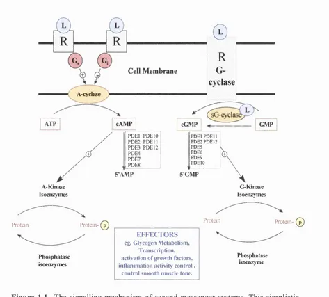
The signalling mechanism o f second messenger systems...4
Structures o f non-selective PDE inhibitors... 7
Nomenclature recom m ended by Beavo, 1988 and Beavo and Rifsnyder, 1990... 9
Nomenclature recommended by Beavo ( 1995)...10
Domain organisation o f cyclic nucleotide phosphodiesterase enzymes 11 The structure o f Vinpocetine PDE 1 selective inhibitor...13
Structure o f Amrinone PDE3 selective inhibitor...20
A summery o f all known PDE4 isoforms...23
A schematic representation o f the three forms o f PDE4 isoenzymes 23 Ribbon diagram o f PDE4B2B catalytic domain (residues 152 to 489)... 27
Structure o f rolipram PDE4 selective inhibitor... 29
Alignment o f catalytic domains o f PDE4B2 (CN4B-Human) and Met^^RDl (CN4Z-Rat)...33
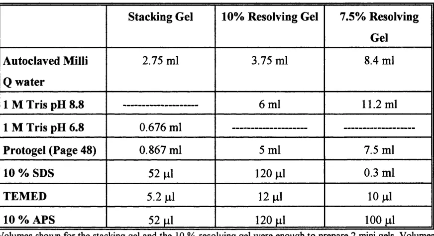
Components o f SDS Polyacrylamide gels...49
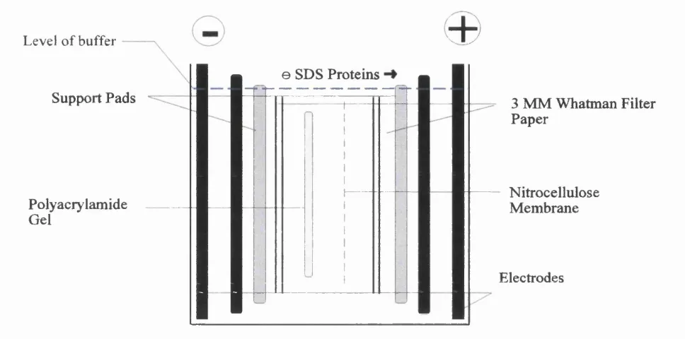
The arrangement o f the blotting sandwich in the transfer tank... 52
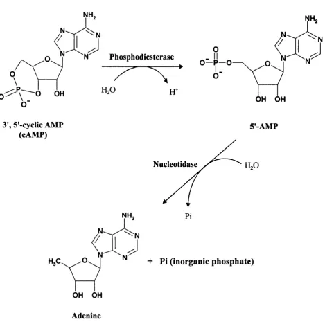
Structural representation o f one-step PDE assay o f PDEs...57
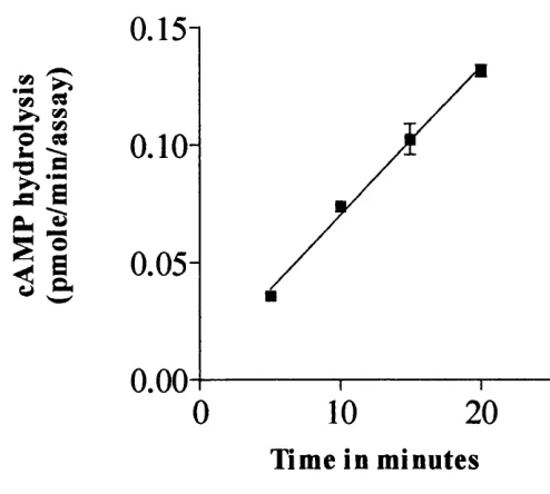
The rate at which PDE4 (Met^^RD 1) hydrolyses cAMP...58
Chapter 3
F ig u re 3.1 A schematic map ofpGEX-3X vector and the multiple cloning site 63 F ig u re 3.2 Primer sequences used in PCR reactions...66
F ig u re 3.3 A schematic diagram to show the cloning o f Mef^RDI gene into pGEX-3X vector...73 F ig u re 3.4 Agarose gel analysis o f mini-preps that have been digested with Bam HI restriction enzyme... 74 F ig u re 3.5 The induction profile o f GST -Met^^RD 1 fusion protein expression 76 F ig u re 3.6 SDS PAGE and Western blot o f GST-Met^^RD 1 time course expression .77 F ig u re 3.7 Effect o f changing the growth temperature o f the bacterial culture on
expression o f GST-Met^^RDl fusion protein... 78 F ig u re 3.8 Effect o f IPTG induction at different points in the growth phase o f the bacterial cells on the expression o f GST-Met^^RDl fusion protein...79 T able 3.1 Expression and purification o f GST-Met^^RD 1 fusion protein...80 F ig u re 3.9 SDS PAGE o f GST-Met^^RDl expression studies... 81 F ig u re 3.10 SDS PAGE and W estern Blot analysis o f Glutathione Affinity Column Purification...82 F ig u re 3.11 Schematic representation o f possible proteolytic sites o n G S T
-Met^^RDl... 85
Chapter 4
F ig u re 4.1 A schematic representation o f immune response represented by the humoral system and the cell mediated system... 89 F ig u re 4.2 Basic structure o f Immunoglobulin proteins...91 F ig u re 4.3 SDS PAGE and Western Blot analysis o f electro-eluted GST-Met^^RDl
fusion protein...99 F ig u re 4.4 Q uantification o f the electro-eluted GST-Met^^RDl protein using
F ig u re 4.6 Western analysis using crude and purified serum...105 F ig u re 4.7 SDS PAGE analysis o f Protein-A agarose Purification GST-Met^^RDl
IgG... 106 F ig u re 4.8 SDS PAGE o f cross-linking o f GST-Met^^RDl antibody to Protein-A Sepharose... 107
Chapter 5
T able 5.1 Proteins expressed fi"om SFV vectors...112 F ig u re 5.1 Maps o f pSFV 1 -3 and pSFV helper 2 vectors and pSFV-3 polylinker 116 F ig u re 5.2 A schematic presentation adapted from Liljestrom and Garoff, 1991 o f the in vivo packaging o f pSFV-1-mouse dihydrofolate reductase (dhfr) mRNA into viral particles... 117 F ig u re 5.3 Ethidium bromide stained agarose gel analysis o f pSFV-3LacZ and Helper 2 mRNA transcribed in vitro...128 T able 5.2 T ransfection efficiencies resulting from transfection o f BHK-21 cells with
either SuperFect or Liposomes... 129 F ig u re 5.4 Effects o f type o f cell line on the transfection efficiency using electroporation...131 F ig u re 5.5 Effects o f changing the amounts o f pSV-Pgal plasmid and pSFV-3LacZ
mRNA on the transfection efficiency o f BHK-21 cells transfected by electroporation...132 F ig u re 5.6 Effects trypsinisation on the transfection efficiency and B H K -21 cells
survival... 133 F ig u re 5.7 Effects o f voltage on the transfection efficiency and survival o f BHK-21
cells transfected by electroporation...134 T ab le 5.3 Viral Stock Storage Conditions...136 F ig u re 5.8 Effects o f the ratio o f mRNA species transfected into BHK-21 cells on the
Chapter 6
T able 6.1 Properties o f PDE4 recombinant proteins produced from mammalian and insect cells... 144 F ig u re 6.1 Polylinker region o f pSFV-1 vector...146 F ig u re 6.2 Agarose gel analysis o f pSFV 1 -Met^^RD 1 RNA transcribed in vitro 153 F ig u re 6.3 W estern blot analysis and PDE activity assay o f pSFV-lM et^^RDl
expressed in BHK-21 cells... 154 F ig u re 6.4 Activation o f two recombinant Met^^RDl viral stocks by a-chymotrypsin at different concentrations... 156 T ab le 6.2 Large scale expression o f Met^^RD 1 in BHK-21 cells using SFV system 158 F ig u re 6.5 Time course o f pSFV-lMet^^RDl construct expression in BHK-21 cells
infected with recombinant Semliki Forest virus... 158 F ig u re 6.6 Intracellular distribution o f the Met^^RD 1 gene product expressed in BHK-21 cells...159 F ig u re 6.6/C Intracellular distribution o f the Met^^RDl gene product expressed in
BHK-21 cells...160 F ig u re 6.7 K inetic characterisation o f Met^^RDl protein expressed in BHK-21
cells... 161 F ig u re 6.8 The action o f R olipram a PDE 4 specific inhibitor on expressed
Met^^RD...162 T able 6.3 Pharmacological properties o f recombinant Met^^RDl and endogenous
PDE (pSFV-1 transfected BHK-21 cells) expressed using SFV system.... 163 T ab le 6.4 Q-Sepharose purification o f Mef^RD 1...164 F ig u re 6.9 Q-Sepharose elution profile... 165 F ig u re 6.10 M olecular weight estimation o f Met^^RDl protein expressed in BHK-21
Chapter 1
General Introduction
1.1 Intracellular Messenger Signalling Systems
Elaborate signalling systems have evolved in higher eukaryotes to enable cells to communicate with one another, so as to co-ordinate and control their behavior for the benefit o f the whole organism. The importance o f such systems has been apparent when these mechanisms fail, resulting in many serious diseases such as cancer.
Intracellular messages could be split into the following categories: 1 ) the generation o f the signal; 2) the removal o f the signal; 3) the direct mediators o f the signal; and 4) the ultimate effects o f the signal (Murray, 1992).
This introduction will give a general overview to the cAMP and cGMP second messenger system giving extra emphases to cAMP signalling pathways and Phosphodiesterase enzymes (PDE), in particular the cAMP-specific (PDE 4) members o f the family.
1.1.1 Cyclic Nucleotides
One o f the oldest and widest studied signals or so called second messengers are the 3 ’,5’ cyclic nucleotides, that include, adenosine 3’,5’ cyclic monophosphate (cAMP) and guanosine 3 ’ ,5 ’ cyclic monophosphate (cGMP). These molecules act as mediators relaying signals initiated by external first messenger molecules, for example hormones, to internal mediators leading to a certain cellular response.
1.1.1.1 cAMP
adrenalin and glucagon stimulate the breakdown o f glycogen and the storage o f glucose in the liver. It was found that these hormones elicit their effects by binding to receptors on the surface o f the cell triggering the formation o f cAMP which stimulates down stream effectors. As these hormones themselves, do not enter the cells, they have been given the name first messengers and consequently cAMP was called a second messenger. Similar experiments revealed that cAMP was present as a second messenger for many other hormones beside adrenalin and glucagon. For example, calcitonin, thyroid-stimulating hormone and noradrenalin.
cAMP has been reported to affect a wide range o f cellular processes such as, the degradation o f storage fuel, the secretion o f acid by the gastric mucosa, dispersion o f melanin pigment granules, aggregation o f blood platelet (Sheth and Colman, 1995), and the opening o f chloride channels (Light et a l , 1990; Lattore et a l , 1991).
The cAMP system is activated by seven helix membrane receptors such as p-adrenergic receptor following the binding o f the hormone adrenalin also known as epinephrine. The hormone-receptor complex stimulate guanine nucleotide-binding proteins (G-proteins) leading to the activation o f membrane bound enzymes called adenylate cyclases. The control o f this enzyme is considered to be important as these enzymes have the sole responsibility o f generating cAMP from ATP (Sutherland et a l , 1962). Control is performed by distinct heterotrimeric guanine regulatory proteins (G-proteins) that relay stimulatory (G J or inhibitory (Gj) signals through a sub-receptor complex (figure 1.1, Gilman, 1984; Krupinski, 1991).
The activation o f the second messenger systems are abolished by the action o f en2ymes called phosphodiesterases (Butcher and Sutherland, 1962). These enzymes hydrolyse cAMP molecules converting them to their corresponding 5’ monophosphates.
For cAMP molecules to function as intracellular mediators, their internal concentration, normally < 10'^ M, must be able to change freely in accordance to stimulus activation (Schramm et al., 1984). cAMP as mentioned before, is produced by adenylate cyclase from ATP, and is rapidly and continuously destroyed by phosphodiesterase by hydrolysing cAMP to 5’-monophosphate. The control o f cAMP levels in cells is achieved by the regulation o f these two enzymes and mostly by adenylate cyclase. The regulation o f these two enzymes are achieved by the presence o f particular isoforms with a certain level o f activity, specific to certain types o f cells.
Other signalling systems have an effect on controlling cAMP levels in cells. This is performed by directly regulating G-protein coupled receptors or adenylate cyclase or both. For example, IP3 signalling molecule which increases calcium ions and diacylglycerol (DAG) levels in cells, causes either, the reduction o f cAMP production by the inhibitory action o f protein kinase C (PKC) on certain G-protein coupled receptors and adenylate cyclase or, the increase in cAMP production by the stimulatory action o f PKC on adenylate cyclase (Houslay, 1998).
1.1.1.2 cGMP
The cGMP system is activated directly by guanylate cyclases which apart from being the sole generators, o f cGMP from GTP, are quite different from the adenylate cyclases. Adenylate cyclases are found exclusively bound to membranes whereas guanylate cyclases can be found both bound to membranes or soluble in the cytoplasm (Drewett and Garbers, 1994; Garbers and Lowe, 1994). Furthermore, it appears that guanylate cyclases are not regulated by G proteins (Drewett and Garbers, 1994).
Cunnick, 1990; Stryer, 1991) and ion channel regulation (Light e t a l , 1990; Lattore et a l , 1991).
A-cydase )
A-Kinase Isoenzymes
Cell Membrane
©
cAMP
PD El PDEIO PDE2 PDE 11 PDE3 PDE 12 PDE4 PDE7 PDE8 5’AMP
R
G-cyclase
cGMP GMPPDEl PDE 11 PDE2 PDE12 PDE5 PDE6 PDE9 PDEIO 5’GMP G-Kinase Isoenzymes
P ro te in P rotein- ( © P ro te in
Phosphatase isoenzymes
EFFECTORS eg. Glycogen Metabolism,
Transcription, activation of growth factoi*s, inflammation activity control,
contr ol smooth muscle tone.
P rotein- (© )
Phosphatase isoenzyme
1.2 Cyclic Nucleotide Phosphodiesterases
Phosphodiesterases (PDE) are a group o f enzymes which were first discovered by Butcher and Sutherland in 1962 not long after the discovery o f cyclic nucleotides. These enzymes played a vital role in characterising adenosine 3 ’ ,5 ’-phosphate which lead to the discovery o f the second messenger system.
PDEs have the important role o f hydrolysing cyclic nucleotide second messengers to the corresponding, inactive 5'-monophosphate counterparts. The degradation o f cyclic nucleotide via these enzymes represents the major route for termination o f cyclic nucleotide action. Other routes for the removal o f cyclic nucleotides are present, for example transport mechanisms that export cyclic nucleotides to the exterior o f cells (Barber and Butcher, 1981). However, these mechanisms only account for a minor fraction o f the total cyclic nucleotides present in cells (Barber and Butcher, 1980; Barber and Butcher, 1981).
PDEs have become more and more important after the discovery o f PDE inhibitors which have been found to reduce or even relieve certain disease symptoms. W ithin only a few years, this field had expanded tremendously as more and more new isoforms have been found. To date twelve PDE families are known and each PDE family consisted o f PDEs that originated from a subfamily o f genes and alternative splice variants (Beavo, 1995).
and levels o f cAMP rise to 10-20 ) liM or higher, PDEs 3 and 4 were thought to be bought
into action as their values were in the micro-molar range. Extremely high cAMP levels were thought to be dealt with by P D E l and PDE2 as their K^, lies in the tens o f micro m olar range. The latter sets o f enzymes were though to act to buffer cAMP concentration.
Different PDEs were also found to be expressed in particular cell types. For example PDE7A (a cAMP-specific PDE) was found to be expressed mainly in cells involved in the immune system and was found to be crucial in T cell activation (Li and Beavo, 1999) and, PDE5 (a cGMP-specific PDE) that was found to be expressed mainly in smooth muscles and was found to be important in the control o f penile erection (Boolell et a l , 1996).
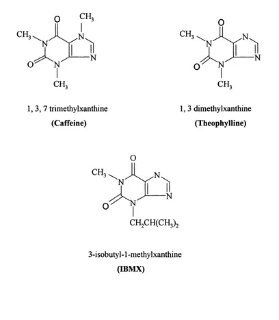
Before the use o f selective inhibitors such as the previously mentioned rolipram and cilostamide, non selective inhibitors such as methylxanthines firstly described by Sutherland and co workers (Sutherland and Rail, 1958; Butcher and Sutherland, 1962) were used to characterise PDEs. These inhibitors which included caffeine and theophylline and later IBMX (figure 1.2), show no preference to PDE isoenzymes and exhibited low inhibitory effects on PDEs. However, these inhibitors represent the first generation o f PDE inhibitors without which none o f the known selective inhibitors today would have been developed.
CH.
CH. CH.
1 ,3 ,7 trimethylxanthine (Caffeine)
1, 3 dimethylxanthine (Theophylline)
O
CH.
3-isobutyl-1 -methylxanthine (IBMX)
1.2.1 Nomenclature
It has been increasingly difficult to name PDEs. This has been due to the multiple isoenzymes o f PDEs being discovered through the years. Previously, workers have tended to number PDE isoenzymes according to their eluting order from an anion-exchange column. This was considered unsatisfactory, as the elution o f the PDEs from the column depended on both the tissue type and the chromatographic conditions used, also each peak may contain more than one PDE isoenzyme. Many other procedures have been introduced to the fractionation mechanisms mentioned above to try and improve the identification o f PDEs. These include the use o f specific assay conditions which will identify fractions containing certain PDE isoenzymes, the use o f selective inhibitors for each o f the PDE isoenzymes, and the use o f immunological identification methods to identify PDE isoenzymes (Beavo, 1988). These methods have not been widely used in the classification approaches because o f their many potential pitfalls.
Table 1.1 Nomenclature recommended by Beavo, 1988 and Beavo and Rifsnyder, 1990.
PDE Family PDE Name Splice Variant
Ca^7 Calmodulin-dependent P D E l 7
cGMP-stimulated PDE II 3
cGMP-inhibited PDE III 4
cAMP-specific, low PDE IV 4
cGMP-specific, cGMP binding PDE V 3
More recently, the ‘diagnostic’ system mentioned above was abandoned and naming of PDEs based upon gene families was adopted (Beavo et al. 1994). This method o f naming provided a more rigorous assessment of family identity based upon primary sequence similarities. The names o f entries assigned to the GenBank were similar to that previously described except for two main changes. The first was the addition of two letters at the beginning of the name to represent the species name and the second was the change of the Arabic numeral from being arbitrary to designate the PDE’s gene family followed by a letter representing the individual gene product within the family. To demonstrate this description an example is shown below:
//.STDE8A2
Homo sapiens (species)
Gene Family (PDE + A rabic num eral) Gene (Capital letter)
Splice v arian t
T able 1.2 Nomenclature recommended by Beavo (1995).
PDE Family Short Name Genes Splice variants
Ca^^ / calmodulin-stimulated PDE 1 3 9+
cGMP-stimulated PDE 2 1 2
cGMP-inhibited PDE 3 2 2+
c AMP-specific, low PDE 4 4 15+
cGMP-specific, cGMP binding PDE 5 2 2
Photoreceptor PDEs PDE 6 3 3
High affinity cAMP-specific PDE 7 3 1
Last updated by the Nomenclature committee on 30*** July 1999.
Since 1995, new PDEs have been identified forming new PDE gene families PD E8, PDE9, PDEIO, PD E l 1 and recently PDE 12. However since the characteristics o f their regulatory properties have not yet been fully elucidated, descriptive names representing their function have not yet been confirmed. The nomenclature shown in table 1.2 was adopted throughout this thesis.
1.2.2 Domain Organisation Of Cyclic Nucleotide Phosphodiesterase Enzymes
Sequence analyses and alignment studies revealed that nearly all mammalian PDEs known today share a conserved region o f approximately 270 amino acids (Bentley and Beavo, 1992). This region which is near the carboxyl terminus o f the enzyme was thought to contain the catalytic domain. The recent structural analysis (Xu etal., 2000) confirmed the involvement o f this region in the catalytic activity o f the enzyme. Conserved histidine residues play an important role in chelating the tightly bound cations (Zn^^ and Mg^^) that are found to be essential for the enzyme’s catalytic activity (Francis et a l , 1994; Turko et a l , 1998; Omburo et a l , 1995; Omburo et a l , 1998; Zhang and Colman, 2000).
containing cGMP-specific phophodiesterases, cyanobacterial Anahaena adenylyl cyclases,
and Escherichia coli transcriptional regulator fhl A (GAF) domains (PDE2, PDE5, PDE6,
PDEIO and PDEl 1 ) and PDE3. Very highly selective inhibitors have been developed for these PDEs creating the need to distinguish these PDEs from PDE4. Thus, information on localisation, domain organisation, regulation and inhibition of each o f the above mentioned PDEs will be discussed.
C a^/ CaM BD
PDEl N
PDE2
PDE3
PDE4
Membrane BD
Catalytic Domain
GAF
UCRl UCR2
PDE5
PDE6 1
GAF
GAF
' a
y subunitFigure 1.3 Domain organisation of cyclic nucleotide phosphodiesterase enzymes.This diagram is not to scale and is not an accurate reflection of size comparisons between members o f the PDE family. Its propose is to show the different structural domains present in PDEs. Abbreviations are; Ca^7 CaM for Calcium ion/ Calmodulin, BD for Binding Domain, GAF for cGMP-specific phophodiesterases cyanobacterial adenylate cyclases, and Escherichia coli transcriptional regulator fhl A, UCR for Upstream Conserved Region. The black region in the catalytic domain o f PDE3 represents a 44 amino acid insertion, y subunit represents the inhibitory PDE6 subunit (see section 1.4.3, page 16)
1.3 Calcium/Calmodulin-stimulated PDE 1
This family o f PDEs are characterised and classified by their sensitively to stimulation by calcium and calmodulin allowing for ‘cross-talk’ between signalling pathways mediated between cyclic nucleotides (cAMP and cGMP) and signalling pathways mediated by calcium ions (Wang et a l, 1990). Generally, PD El isoenzymes hydrolyse both cAMP and cGMP. The rate at which these two cyclic nucleotides are hydrolysed are regulated by two Ca^V calmodulin (Ca^7 CaM)-binding domains present at the N-terminus (regulatory domain) o f PD E l isoenzymes (Novack et ût/., 1991; Sonnenburg et a l , 1995; Kincaid et a l , 1985; Charbonneau et a l , 1991).
To date, three genes encoding PD El have been identified. Using the nomenclature described in section 1.1.2, these genes were called PD E l A corresponding to bovine 61 kDa CaM-PDE, PDE IB corresponding to bovine 63 kDa CaM-PDE (Sharma et a l , 1980; 1984) and PDE 1C possibly corresponding to an isoform identified in the rat testis (Rossi et a l , 1988). The diversity o f PDE Is were further increased by 5' and 3' alternative splicing o f each gene product resulting in the formation o f splice variants (isoforms) that differ in their N and/ or C termini (Michibata et a l , 2001; Yan et a l , 1996; Sonnenburg et a l , 1995). The alternative 5' splicing generates isoforms that have different affinities to Ca^Y CaM due alteration o f the Ca^V CaM-binding domains. For example, PDE1C2 isoform has two o f the Ca^V CaM-binding domains intact whereas the rest o f PDE 1C isoforms (P D E IC I, 3, 4, and 5) have one o f the two Ca^Y CaM domains spliced out making these isoforms less sensitive to Ca^^ when compared to PDE1C2 isoforms (Yan e t a l , 1996).
Members o f the PD E l family are also regulated by phosphorylation. Generally phosphorylation occurs at the N-terminal regions o f the PDE proteins by subtype specific kinases. For instance, PD E l A and PDE 1C isoenzymes are phosphorylated by c AMP- dependent kinases whereas, PDE IB isoenzymes are phosphorylated by calmodulin- dependent kinase II (Sharma and Wang, 1986; Hashimoto et a l , 1989). The phosphorylation o f PD El isoenzymes have been found to reduce the affinity o f these enzymes to Ca^V CaM (Beavo, 1995; Yan et a l , 1996). M ore recently, PD E l isoenzymes have been reported to be regulated by degradation by m-calpain which are Ca^^-dependent cysteine proteases through a PEST m otif present in some PD E l isoforms such as PD E l A2 (Kakkar et ah, 1999).
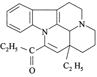
There are large and diverse compounds that are capable o f inhibiting PD E l. These compounds have been divided into two categories according to their inhibiting effect on the enzyme. The first category included those inhibitors that acted on the enzyme’s activity directly and the second included inhibitors that acted on the CaM stimulation o f the enzyme. In this study Vinpocetine (figure 1.4), which is a potent vasodilating agent, was used (Hagiwara et a l , 1984). This inhibitor was found to act at the catalytic site o f PDE Is. This inhibitor was found to be highly selective to P D E l isoenzymes unlike other inhibitors o f the same category for example, Zaprinast and 8-MOMX which were found to act on PDE5 with a similar potency to PD E l (Ahn et a l , 1992).
1.4 PDE Families Containing GAF Domains
PDE2, PDE5, PDE6, PDEIO and the recently identified PD E l 1 have been identified to include a non-catalytic region at their N-terminus having high specificity to cGMP (Soderling and Beavo, 2000; Soderling et a l, 1999; Fawcett et a l, 2000; Mou et a l, 1999; Fujishige et a l , 1999; Fujishige et a l , 2000; Yuasa et a l , 2000). This domain (GAF), binds to cGMP and leads to an allosteric stimulation o f the enzyme’s activity (Granovsky e t a l , 1998; Corbin and Francis, 1999; Tanaka era/., 1991; Sonnenburg a/., 1991), The acronym GAF is derived from the names o f three proteins that contained this conserved sequence; cGMP-specific phophodiesterases, cyanobacterial adenylyl cyclases, and Escherichia coli transcriptional regulator fh l A (Aravind and Ponting, 1997; Shabb and Corbin, 1992). Despite the conservation o f this domain in the three proteins, the function o f this domain it appears to be different in each o f the three proteins.
1.4.1 cGMP-stimuIated PDE2
To date there has been only one gene identified for this family o f PDEs making them one o f the smallest PDE families known. Two 5' splice variants have been identified which differ in their N-terminal regions (Rosman et a l , 1997). One o f these isoforms has been found to be cytosolic and the other was found to be anchored to the plasma membrane by virtue o f its N-terminal region as identified in hepatocytes (Marchmont and Houslay, 1980a). This intracellular targeting o f these isoforms and their functional relevance is not yet fully understood. However, it supports the previous notion that intracellular distribution o f PDEs in cells has fundamental importance.
stimulation and hydrolysis o f cAMP. The decrease in cAMP levels may lead to deactivation o f the cAMP signalling pathway. PDE2 isoenzymes present in particulate fractions have been shown to be phosphorylated by protein kinase C which is activated by Ca^^. Furthermore, PDE2 isoenzymes have been shown to be involved in the ‘cross-talk’ between certain lipid signalling pathways (Geoffroy et a l , 1999).
1.4.2 cGMP-specific/ cGMP-binding PDE5
Until the cloning o f cDNA for this isoenzyme (McAllister et a l , 1993), members o f this family were thought to belong to the photoreceptor PDE6. This was due to similarity in their kinetics, cGMP-binding and size characteristics to members o f PDE6 isoenzymes (Beavo, 1995). Sequence alignment studies revealed less than 60% identity between these two sets o f isoenzymes (Beavo, 1995). Consequently members o f PDE5 were placed in a separate family.
Only one gene product has been reported for this family and two splice variants have been identified with distinct N-terminal regions (Kotera et a l , 1998; Kotera et a l , 1999a). PDE5 isoenzymes are highly specific to cGMP hydrolysis with values o f approximately 5.6 pM and show no significant hydrolysis activity with cAMP (Thomas et a l , 1990a). cGMP has been found to bind to both the catalytic domain and to the two cGMP-binding (GAF) allosteric domains found at the N-terminal end o f the proteins (Thomas et a l ,
1990b; McAllister e t a l , 1995).
protein (Thomas et al^ 1990b). However, the function o f this dimérisation is not well understood.
Recently there have been much interest in PDE5 due to the inhibition o f these PDEs by Viagra which is used in the treatment o f erectile dysfunction. Other PDE5 inhibitors are also available such as Zaprinast, M Y 5445 and SK7F 96231 (Beavo and Reifsnyder, 1990; Nicholson et al., 1991; Murray and England, 1992).
1.4.3 Photoreceptor PDE6
At present, there are at least three gene products described for this family o f PDE6
(Hurwitz et a l , 1985), products o f which are expressed exclusively in the rod and cone photoreceptors o f the retina. These photoreceptors consist o f PDE6 arranged as a multisubunit protein o f two types o f catalytic subunits a and P which are products o f PDE6A and B genes respectively, bound to two inhibitory y subunits. PDE6 isoenzymes are highly specific to cGMP hydrolysis and both the kinetic activity and subunit assembly are regulated by the presence o f GAF domains at their N-terminus (Mou et a l , 1999). It has been described that the binding o f cGMP to the GAF domain induces the dramatic increase in the affinity (300 fold) o f PDE6 (a p ) to its inhibitory y subunits (D ’Amours and Cote, 1999).
1.4.4 PDEIO
The PDEIO family represent one o f the newly discovered groups so description names for these enzymes have not yet been assigned as little is known regarding their regulation and cellular targeting. At present products o f one gene have been isolated from human fetal lungPD ElO A l and PDE 10 A2 (Fujishige era/., 1999; Kotera era/., 1999b). These isoforms have been found to hydrolyse both cAMP and cGMP with K^ values o f 0.26 pM and 7.2 pM respectively, and values for cGMP twice that o f cAMP. Thus, these PDEs have exhibited properties similar to cAMP PDEs and cGMP inhibited PDEs (Fujishige et al., 1999). Sequence alignment studies revealed similarity o f PD E l OA with both PDE2 and PDE5A. These similarities being at the catalytic domains and the GAF cGMP binding- domains (Fujishige et al., 1999). However, the cAMP- and cGMP-dependent protein kinase phosphorylation sites present in PDE5, which are found to be important in the regulation o f these PDEs, were absent in PDE 10 A. These have been found to be replaced by sites phosphorylated by protein kinase C suggesting regulation by this enzyme (Fujishige etal., 1999).
1.4.5 PD E ll
1.5 cGMP-inhibited PDE3
Two gene products have been identified to belong to the PDE 3 family (Meacci et a l , 1992; Taira era/., 1993). Using the nomenclature described in section 1.1.2, these isoforms were called PDE3A and PDE3B. The first isoform PDE3A has been found to be abundantly expressed in smooth muscle, platelets, and cardiac tissues whereas, PDE3B isoforms have been shown to be expressed in adipocytes and liver (Beavo, 1995). M embers o f PDE3 are capable o f hydrolysing both cAMP and cGMP with values in the range o f 0.1-0.8 pM and for cAMP 4-10 times higher than that for cGMP therefore conveying specificity to cAMP (Degerman et a l , 1997; Beavo, 1995). Furthermore, cGMP has been found to bind at the catalytic domain and not at the N-terminal regulatory domains as in PDE2. Therefore, cGMP acts as a potent inhibitor for cAMP hydrolysed by PDE3 enzymes. The PDE3 ‘cross-talk’ between cGMP mediated by nitrogen oxide (NO) stimulation o f guanylate cyclase and cAMP signalling pathways is expected to increase cAMP levels due to the inhibitory effect o f cGMP on the enzyme (Lugnier et a l , 1999).
The catalytic domain in PDE3 isoenzymes differ from the rest o f the PDE families by an insertion o f 44 amino acids (Degerman et a l , 1997). This insertion, which has distinguished the PDE3 catalytic domain from the rest o f the PDE families, interrupts the first o f the two putative Zn^^ binding domains that have been identified to be present in PDE catalytic domains (figure 1.3) (Degerman et a l , 1997; Francis et a l , 1994; Meacci e t a l , 1992; Taira era/., 1993). The presence o f the 44 amino acid insertion in the catalytic domain o f PDE3 has lead to assumption to the presence o f subfamilies within PDE3 however the exact implication to substrate interactions as well as inhibitor interaction has yet to be elucidated (Degerman et a l , 1997).
Using PDES selective inhibitors, PDES isoenzymes have been implicated in the regulation o f key biological processes, such as lipolysis, glycogenolysis and cardiac contractility (Manganiello et a l , 1995; Beebe et a l , 1985). For example, PDESB has been shown to mediate the inhibitory effect o f insulin on lipolysis in adipocytes. This is perform ed by insulin activation o f 1RS-1, Phosphatidylinositol-S- kinase (PIS-kinase) andPDK signalling pathways and the final phosphorylation o f PDESB isoenzymes through Akt (Ahmed et a l , 2000). The activation o f PDESB by phosphorylation causes the increase in cAMP hydrolysis leading to the fall in cAMP levels in the cell. This in turn reduces the activity o f PKA which leads to the de-phophorylation and deactivation o f hormone-sensitive lipase (HSL) and the consequence decrease in hydrolysis o f stored triglyceride. Specific PDES inhibitors have found to induce mycocardial contractility, vascular and airway smooth muscle relaxation (Beavo and Reifsnyder, 1990; W eishaar era/., 1987;K om asera/., 1996; M anganiello et a l , 1995). PDES isoenzymes in these tissues have been shown to be phosphorylated via PKA in vitro however, the mechanism o f regulation o f these isoforms in intact cells is still poorly understood (Degerman et a l , 1997).
F ig u re 1.5 Structure o f Amrinone PDE3 selective inhibitor.
1.6 cAMP-speciflc phosphodiesterase PDE4
The focus o f this thesis is the study o f cAMP-specific Phosphodiesterases. PDE4 isoenzymes are highly specific to cAMP, insensitive to cGMP and are also very highly sensitive to the anti-depressant drug rolipram (Yamamoto et a l , 1984). The PDE 4 family which, contribute to the Tow activity in many cells, have been found to be present in the brain where they are thought to contribute to the regulation o f processes that control mood, emesis, and olfactory sensory transduction (De Mazancourt and Giudicelli, 1988; Davis, 1984). Members o f PDE4 family have been found in the lung (Torphy and Cieslinski, 1990), tracheal smooth muscle (Shahid et a/., 1991) and macrophages (Barnes, 1995) it is likely that PDE4 may be involved in the regulation o f inflammation. Indeed, PDE4 inhibitors have been found to suppress various functions o f inflammatory cells (Torphy and Undem, 1991; Torphy et a l , 1994; Palfreyman and Souness, 1996). Therefore, there is the potential o f utilising these PDE inhibitors in the therapy o f asthma, allergy and other inflammatory diseases (Torphy and Undem, 1991).
Previous work on the Drosophila dunce gene mutant allowed the discovery o f four mammalian subtypes belonging to cAMP-specific PDEs (Beavo and Reifsnyder, 1990; Beavo et a l , 1994). cDNA o f four subtypes encoded by four genes (A, B, C, and D) have been cloned and expressed in various laboratories (Livi et a l , 1990; McLaughlin et a l ,
found to produce at least six different splice variants arising from alternative mRNA splicing. PDE isoforms as well as their splice variants are regulated at several levels, including transcription, splicing, and in subcellular localisation (Bolger, 1994). It has been hypothesized that mRNA levels are transcriptionally regulated by cAMP via cAMP- response element (CRE) found in the promoter region o f the gene. Moreover, differential splicing or degradation o f mRNA is also regulated by cAMP. Recently Houslay and colleagues have found a splice variant encoded by rat PDE4A locus (R D I), to contain a membrane binding region (Shakur et a l, 1993). This finding demonstrates that differential splicing o f PDE enzymes contribute to the regulation o f the subcellular localisation o f the enzyme (Bolger, 1994).
In this section members o f PDE4 will be extensively reviewed giving extra emphases on their domain organisation, regulation as well as rolipram binding.
1.6.1 Genomic organisation
As mentioned previously, four PDE4 genes have been reported (PDE4A, B, C, and D). In humans, these genes are distributed on three different chromosomes; c h rl9 p l3 .1 (PDE4A gene) (Sullivan e t a l , 1998; Horton e t a l , 1995), chrl (PDE4B gene) (Szpirer e t a l , 1995; Milatovich et a l , 1994), c h rl9 p l3 .2 (PDE4C) (Sullivan et a l , 1999), and chr5 (PDE4D) (Milatovich et a l , 1994). These genes have been reported to be large approximately 50 kb in length and complex consisting o f 18 or more exons at one instance (Sullivan et a l ,
1998; Sullivan e ra /., 1999).
1.6.2 Domain Organisation and the Generation of Splice Variants
Analysis performed on the amino acid sequences o f the different PDE4 subtypes have revealed three distinct highly conserved regions. These being two ‘upstream conserved regions’ o f 60 amino acids and 80 amino acids in length better known as U C Rl and UCR2 respectively located at the N-terminus o f the isoenzymes, and the catalytic domain located near the carboxyl terminal which is conserved throughout all known PDE families. Highly variable regions called LR l and LR2 were also identified. LR l is localised between U C Rl and UCR2 whereas LR2 is localised between UCR2 and the catalytic domain. The function o f these regions are thought to provide a subtype specific means o f regulating the enzyme’s activity by altering U C R l and UCR2 interaction with the catalytic domain (Houslay,
1998).
Up to 18 different splice variants have been reported to be encoded by the four PDE4 genes. These splice variants have been characterised according to the presence or absence o f U C R l to form the long or the short isoforms respectively (Bolger, 1994; Bolger et a l , 1994) as well as the truncation o f UCR2 to form supershort isoforms (Sullivan et a l , 1998) (figure 1.6). Recently, it has been suggested that N-terminal regions which include U CRl and in a lesser extent UCR2, may be responsible for the localisation o f PDE4 and for the regulation o f the enzymatic activity (Muller et a l , 1996; Jacobitz et a l , 1996). In addition, it has been established that rolipram and cAMP do not bind to the same region in the PDE4 isoenzymes. Rolipram has been identified to interact within the region that lies amongst U C R l and UCR2 whereas, cAMP has been found to interact exclusively with the catalytic domain (Jacobitz et a l , 1996). All PDE4 isoforms known to date are summarised in table
Table 1.3 A summery of all known PDE4 isoforms.
Human Rat Isoform Type
4A1 (hRD l) 4A1(RD1) Supershort
4A4B (pde46) 4A5 (rpde6 ) Long
4A7 (2el) 4A7 catalytically inactive
4A8 4A8(rpde39) Long
(TM3) (TM3) Long (putative)
4A10 4A10 Long
4B1 4B1 (DPD) Long
4B2 4B2 Short
4B3 4B3 Long
4B4 4B4 Long
4C1 4C1 Long
4C2 4C2 Long
4C3 4C3 Long
4D1 4D1 Short
4D2 4D2 Supershort
4D3 4D3 Long
4D4 4D4 Long
4D5 4D5 Long
Adapted from Houslay, 2001.
UCRl UCR2 Catalytic Domain
Long N
LRl LR2
Short N
Supershort N
Figure 1.6 A schematic representation o f the three forms of PDE4 isoenzymes.
1.6.3 Regulation
Regulation o f PDE4 isoenzymes is found to be extremely complex. There does not appear to be endogenous allosterism or small regulator molecules that regulate PDE4 isoenzymes. However, PDE4 isoenzymes are found to be regulated at the transcription level as well as phosphorylation by a number o f different types o f protein kinases. In Sertoli cells, Conti and co-workers (Swinnen et a l, 1989b) have found a 100 fold increase in PDE4D2 mRNA following prolonged exposure to cAMP. More recently, the same group (Sette et a l ,
1994b) have demonstrated that PDE4D3 are regulated by phosphorylation by protein kinase A. It has been suggested that transcriptional regulation provides a longer term response whereas, regulation by phosphorylation provides short term regulation (Beavo, 1995). It is not clear if both types o f regulation are present in the same cell or even if it is the same PDE4 isoenzyme that is being regulated (Beavo, 1995). In Sertoli cells for example, it has been demonstrated that both types o f PDE4 regulation are present (Beavo, 1995).
Recently, Houslay and co-workers have demonstrated using two hybrid analysis and pull down assays that U C R l binds to UCR2 (Beard et a l, 2000). This supported the notion that U C R l and UCR2 form some sort o f regulatory module that can influence the structure and the function o f the catalytic domain. The mapping for possible interaction sites was performed using several deletion studies. These identified two arginine residues (Arg 98 and Arg 101 in PDE4D3) present at the C-terminus o f the U C R l bound to negatively charged residues (Asp 149, Glu 147 and Glu 146 in PDE4D3) present at the N-terminus o f UCR2 by ionic pair formation (Houslay et a l, 1998; Beard et a l, 2000). However, more interacting residues may be involved in the formation o f the UCR1-UCR2 regulating module as it has been demonstrated that U C Rl can bind to a truncated UCR2-PDE4 catalytic unit complex (Beard et a l , 2000). Phosphorylation by protein kinase A, may change the conformation o f this regulating module. Indeed phosphorylation o f serine 54 (PDE4D3) in the m otif Arg-Arg-Glu-Ser-Trp present in the U C R l region or mutation o f Ser 54 to Asp has been demonstrated to hinder UCR1-UCR2 interaction (Beard et a l ,
A better understanding o f the regulatory action o f the UCR1-UCR2 regulator modules came about with the study o f extracellular-signal-regulated kinase (ERK) phosphorylation o f the PDE4 catalytic domain (MacKenzie et al., 2000; Baillie et a l, 2000). These kinases which have been shown to play a key role in many physiological processes such as, those associated with cell activation, growth, survival, and differentiation appear to elicit different phosphorylation effects on different forms o f PDE4 isoenzymes. It has been shown that ERK phosphorylation elicits activation o f the short forms o f PDE4 (e.g. PDE4D1 andPDE4B2) which lack the U CRl region but elicit inhibitory effects o f the long forms which contain both U C R l and UCR2 regions (figure 1.6) (Baillie et a l, 2000). Thus it has been suggested that both U C R l and UCR2 are responsible in orchestrating the functional consequence o f the phosphorylation o f the catalytic domain by ERK (MacKenzie e t a l , 2000', Baillie e t a l , 2000). Furthermore, the switching in the functional output performed by U C R l and UCR2 were found to be operated by the N-terminal portion o f UCR2 as the removal o f this region in the supershort form PDE4D2 isoform, ablated the stimulatory effect o f ERK phosphorylation, as seen in the short form (MacKenzie et a l , 2000; Baillie et a l , 2000).
1.6.4 Catalytic Unit
This region was initially identified as mentioned earlier according to sequence homologies between different PDE families. The importance o f this region in activity was later confirmed as a single mutation caused the loss o f activity (Jin et a l , 1992; Jacobitz et a l ,
1996; Jacobitz et a l , 1997). Mutation studies also identified putative Me^^ binding sites which are considered to be important for catalysis (Omburo et a l , 1998; Jin et a l , 1992; Jacobitz et a l , 1997). The outer limits o f the catalytic domain were identified using truncation analysis (Houslay et a/., 1998; Horton et a l , 1995; Jacobitz et a l , 1996; Owens et a l , 1997). For example, the catalytic domain in PDE4A4B was found to span from residues 332 or 365 to residue 680 (Houslay et a l , 1998).
which is equivalent to residues 351 -727 on PDE4 A4B defined by truncation analyses. This structural analysis showed that the catalytic domain consisted o f 17 a helices which subdivided to three sub-domains (figure 1.7). This sub-division o f the catalytic domain has provided the possibility o f adopting distinct conformational states and thus, explaining how different PDE4 isoforms exhibited different susceptibilities to inhibition by the PDE4- selective inhibitor rolipram when bound to other proteins (McPhee et al., 1999; Yarwood et a l , 1999), or phosphorylated by PKA or ERK (Hoffmann et a l , 1998; Alvarez et a l ,
1995), or bound to membranes (Huston et a l , 1996; Souness et a l , 1992). This supports the notion that different PDE4 conformers are present. The presence o f the three sub- domains means that changing the orientation o f one or more o f the sub-domains to accommodate a certain conformation which is favorable for a particular function can be made easier. This could be achieved for example by changing the orientation o f one o f the domains in relation to another.
Using the data provided by this crystal structure, a model was proposed for the binding of cAMP to the catalytic domain. This suggested that cAMP bound in an antiparallel conformation with the adenine base inserted into the lipophilic pocket formed from Leu 393 and He 396 present in between helices 13 and 14, lie 410 and Phe 414 present in helix 14 and Phe 446 present in helix 15a (figure 1.7 and figure 1.9). This allowed the phosphate group to interact with the two Me^ ' and the 1 -N and 6-NH2 groups to interact with Gin 443 in helix 15a through hydrogen bonding (Xu et a i, 2000).
Figure 1.7 Ribbon diagram o f PDE4B2B catalytic domain (residues 152 to 489). This diagram is based on information obtained from the crystal structure of PDE4B2 catalytic domain analysed by (Xu et al., 2000) and drawn using the computer program MOLSCRIPT (Kraulis, 1991). M El is shown as a grey sphere reported to be occupied by a zinc ion (Xu
et a i, 2000) and ME2 is shown as a blue sphere. The N-terminus sub-domain o f the
molecule (residues 152 to 274) is coloured pink, the mid sub-domain (residues 274 to 348) is coloured green and the C-terminus sub-domain (residues 348 to 489) is coloured yellow. H is an abbreviation for helix.
1.6.5 Rolipram Binding
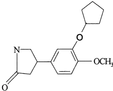
Rolipram (figure 1.8) was originally developed as an anti-depressant. The clinical use o f this prototypical PDE4 inhibitor and other analogs were limited due to its many side effects such as, nausea, emesis, gastric acid secretion, and central nervous system activation.
rolipram from S. cervisiae using heat shock assays (Pilla et al., 1993). These studies revealed that helices 14 and 15 play a key role in conferring sensitivity to rolipram.
o
0Œ.
F ig u re 1.8 Structure o f rolipram PDE4 selective inhibitor.
1.7 RatPDE4Al(RDl)
1.7.1 Discovery
(Henkel-Tigges and Davis, 1989) lead to the prediction that RD I could be involved in mood regulation similar to that reported earlier for Drosophila dunce.
Alignment studies revealed that R D I, which corresponded to PDE4A1 using recent nomenclature, shares great similarity (~ 76%), mostly present in the catalytic domain (Henkel-Tigges and Davis, 1989). These studies also revealed great differences in sequences at the N- and C-terminus, suggesting the presence o f domains that define the regulation and cellular localisation o f these isoenzymes. Indeed that was the case.
1.7.2 Intracellular Targeting
1.7.3 Alignment Studies
PDE4B2B 69 SVSEMASNKFKRMLNRELTHLSEMSRSGNQVSEYISNTFLDKQNDVEIPSPTQKDREKK- 127 Met^®RDl 2 6 MLNRELTHLSEMSRSGNQVSEYISNTFLDKQNEVEIPSPTPRQRAFQQ 73
, H e lix -0 H elix -1
PDE4B2B KKQQLMTQISGVKKLMHSSSLNN--T^ISRFGVNTENEDHLAKELEDLNKWGLN 17 9 Met^®RDl PPPSVLRQSQPMSQITGLKKLVHTGSLN--- Ttlv P R F GVKTDQEDLLAQELENLSKWGLN 130
H elix -2 H e lix -3 H e lix -4 H e lix -5 H e lix -6 PDE4B2B IFNVAGYSHNRPLTCIMYAIFQERDLLKTFRISSDTFITYMMTLEDHY-HSDVAYPiNSLH 238 Met^®RDl IFCVSEYAGGRSLSCIMYTIFQERDLLKKFHIPVDTMMMYMLTLEDHY-HADVAYBNSLH 18 9
H e lix -6 H e lix -7 H e lix -8 H e lix -9 PDE4B2B AADVAQSTHVLLSTPALDAVFTDLEILAAIFAAAIHDVDHPGVSNQFLINTNSELALMYN 2 98 Met^®RDl AADVLQSTHVLLATPALDAVFTDLEILAALFAAAIHDVDHPGVSNQFLINTNSELALMYN 24 9
H e lix -1 0 H elix -1 1 H e lix -1 2 PDE4B2B DESVLENHHLAVGFKLLQEEHCDIFMNLTKKQRQTLRKMVIDMVLATDMSKHMSLLADLK 358 Met^^RDl DESVLENHHIAVGFKLLQEENCDIFONLSKROROSLRKMVIDMVLATDMSKHMTLLADLK 30 9
H e lix -1 2 P -h a ir p in H e lix -1 3 H e lix -1 4
PDE4B2B TMVETKKVTSSGVLLLDNYTDRIQVLRNMVHCADLSNPTKSLEL RQWTDRIMEE FQQG 418 Met^RDl TMVETKKV TSSGVLLLDNYSDRIQVLRNMVHCA')LSNPTKPLEL ROWTDR1M A E FFOOG 3 69
H e lix -1 4 H e lix -1 5 a H e lix -1 5 b H e lix -1 6
PDE4B2B DKERERGMEISPMCDKHTASVEKSQVGFIDYIVHPLWETWADLVQPDAQDILDTLEDNRN 47 8 Met^^RDl DRERERGMEISPMCDKHTASVEKS VGFIDYIVHPLWETWADLVHPDAQDILDTLEDNRD 42 9
H e lix -1 6 I H e lix -1 7
PDE4B2B WYQSMIPQSPdpPLDEQ NRDCQGLMEKFQFELTLDEEDSEGPEKEG 524 ( 40) 564
Met^fiRDl W Y H S A I R Q S P a P P L E E E P - - G G L G H P S L P D K F O F E L T L E E E E E E D S L E V P 477 (133) 610