Acta Cryst. (1982). B38, 523-531
The Structure of a Hydrated 1 : 2 Complex of Adenylyl(3'-5')adenosine-Proflavine
Hemisulphate
BY HUEY-SHENG SHIEH* AND HELEN M. BERMAN't"
The Institute for Cancer Research, Fox Chase Cancer Center, Philadelphia, Pennsylvania 19111, USA
AND STEPHEN NEIDLE,t GARRY TAYLOR~ AND MARK SANDERSON§
Department of Biophysics, Kings College, University of London, 26-29 Drury Lane, London WC2B 5 RL, England
(Received 19 February 1981; accepted 27 July 1981)
523
Abstract
The 1:2 complex (M r = 1410.28) between adenylyl(3'- 5')adenosine (ApA), C20H25NIoOIoP, and proflavine hemisulphate, 2C 13H ~2N~ - . SO42-, crystallizes with 16.5 water molecules in the space group P21212 and unit-cell parameters a = 32.157 (5), b = 21.450 (4) and c = 10.175 (1) A; V = 7018.4 A 3, Z = 4, d c = 1.33, d m = 1.32 Mgm -3,/z(Cu Ka) = 1.32 mm -~, F(000) = 2980. Crystal data were measured up to 20 = 120 ° with Cu Ka radiation. The structure was determined by a multisolution phase method and refined by a blocked- mode full-matrix least-squares procedure. The final R factor is 0.118 for 3507 observed data. The dinucleo- side phosphate, ApA, in this structure has a very unusual conformation which is not found in other oligonucleotide structures. The backbone of ApA is extended and each adenine ring is hydrogen bonded to another symmetry-related one forming an adenine- adenine base pair. Each base pair is sandwiched by proflavine cations which also stack with each other. Solvent molecules lie in the continuous channels between columns of stacked heterocyclic rings.
Introduction
The study of small-molecule-nucleic-acid interactions has been concerned mostly with the intercalative mode of binding of planar chromophores between base pairs in double-helical DNA and RNA (Lerman, 1961; Neidle, 1979). However, these molecules, some of which are drugs, are also known to bind to single-
* Present address: Physical Science Center, Monsanto Company, 800 N. Lindberg Boulevard, St. Louis, MO 63166, USA.
t Authors to whom correspondence should be addressed.
~: Present address: Molecular Biology Laboratory, Birkbeck College, Malet Street, London WC1E 7HU, England.
§ Present address: Department of Organic Chemistry, State University, 2300 Leiden, The Netherlands.
stranded nucleic acids such as synthetic polyribo- nucleotides (Dourlent & H61+ne, 1971) and naturally occurring tRNA (Urbanke, R6mer & Maass, 1973). The exact nature of that binding is not well understood at present because of the conformational flexibility of RNA. In this study, we present a high-resolution X-ray crystallographic analysis of the structure of proflavine complexed with the non-self-complementary dinucleo- side phosphate, ApA (Neidle, Taylor, Sanderson, Shieh & Berman, 1978). There have been many solution studies on this dinucleoside phosphate (Ts'o, Kondo, Schweizer & Hollis, 1969; Lee, Ezra, Kondo, Sarma & Danyluk, 1976; Evans & Sarma, 1976) in an attempt to determine the extent of its conformational flexibility and whether or not it obeys the 'rigid nucleotide principle' (Yathindra & Sundaralingam, 1973). It has been suggested (Berthod & Pullman, 1973) that this principle pertains only in certain crystalline environ- ments. We show in this study that in the presence of proflavine, the conformation of the dinucleoside phosphate has many unusual features.
Experimental
Deep-red rectangular prismatic crystals were grown from an aqueous solution of ApA and proffavine hemisulphate in a 1:2 ratio. The UV spectrum of a washed crystal clearly indicated complex formation. A crystal of size 0.45 × 0.3 × 0.2 mm was cut from a bigger crystal and sealed inside a capillary tube for all subsequent X-ray measurements.
524 HYDRATED A D E N Y L Y L ( 3 ' - 5 ' ) A D E N O S I N E - P R O F L A V I N E HEMISULPHATE on counter statistics and instrumental instability (Stout
& Jensen, 1968). 3507 reflections with I > 2.33aj were considered to be significant and used for structure refinement. The occurrence of so many unobserved reflections (2188) is probably due to the disordered water network discussed in a subsequent section.
Initial attempts to solve the structure with MULTAN (Main, Woolfson & Germain, 1971) were unsuccessful. The structure was solved by an alternative direct- methods approach. The phases of the hkO reflections were obtained by a multisolution method (Sheldrick, 1976) using 12 unknown phases in the starting set. The phases for the best set, as judged by a negative quartet test, were then used as starting points in a tangent- formula phase expansion using the full. three- dimensional set of E values above 1.4. The resulting E map showed two full proflavine rings, a phosphate group and most of the atoms comprising the adenine bases. Two consecutive difference Fourier syntheses revealed the rest of the structure. In an independent manner, use of superposition methods on this problem also determined the structure. The P atom was located from the high-resolution Patterson map (using only those reflections with 0 greater than 30°). A fourfold superposition of the Patterson functions on the four equivalent positions of the P atom revealed most of the structure.
This is a highly solvated structure. Lack of strong interactions among some of the solvent molecules makes them highly disordered. Therefore, the locations of these solvent molecules were difficult to determine and were sometimes ambiguous. A combination of least-squares refinement and difference Fourier syn- theses was carried out step by step to determine unequivocally all 24 positions occupied by 16.5 water molecules and so complete the structure.
At one point, careful examination of the calculated and observed structure factors showed that the major discrepancies lay in the weak reflections and the low-resolution reflections. Therefore, 33 reflections with F o < 16.5 and 44 reflections with sin 0/2 < 0.10/k -~ were removed from the refinement. The weighting scheme used for the least-squares minimization was: w -- [o2(F) + 0.005Fo2] -1. Because of the limitation of the computer-memory storage, the structure was divided into several blocks and all refinements were carried out in a blocked-mode full-matrix least-squares procedure. The atomic scattering factors and dispersion cor- rections were taken from International Tables for X-ray Crystallography (1974).
The positions and anisotropic temperature factors of the ApA and proflavine molecules were varied throughout all refinement cycles. The solvent molecules were divided into four categories:
(1) Sulphate ion: In order to account for the electron density, two idealized tetrahedra of O atoms with occupancy factor of 0.5 on each were placed around
the S atom. Their positions were fixed and their isotropic temperature factors were varied in the first few refinements and then fixed. The S atom was refined anisotropically.
(2) Primary water shell" WP(01). WP(02)... WP(11) (where W and P stand for water and primary shell respectively).
There were l l water molecules which made direct hydrogen bonds to either ApA or the proflavines. Their positions and anisotropic temperature factors were refined. The occupancy factors (G) were all assigned 1.0 except WP(01), which was at a special position with G = 0.5. Judging from their rather high temperature factors, the water molecules WP(07),
WP(08), WP(09), and WP(IO) may be disordered. (3) Secondary (S) water shell" WS(O1), WS(02), WS(03). Three water molecules hydrogen bond to water in the primary shell. They were given isotropic temperature factors with G -- 0.5. Both their positions and temperature factors were varied.
(4) Disordered (D) water molecules: WD(01), WD(02) . . . . WD(10). Ten electron density peaks which did not make any unreasonable contacts with all other molecules and some short distances and unusual geometry among themselves were described as disor- dered water molecules. They are located in the weak polar region of the molecular channel. They were assigned isotropic temperature factors with G = 0-5 except WD(09) and WD(IO), which are at special positions, having G -- 0.25. Their positions and temperature factors were refined.
No attempt was made to locate H atoms. However, the positions of 30 non-hydrogen-bonded H atoms were calculated and included in the structure factor calculation.
The refinement converged at R = 0-106 and R w = 0.139 for 3431 selected observed data, and R = 0.118 and R w = 0.171 for all 3507 observed data.* The difference Fourier map at this point showed no electron density greater than 0.42 e/~-3.
* List of structure factors and anisotropic temperature parameters have been deposited with the British Library Lending Division as Supplementary Publication No. SUP 36329 (27 pp.). Copies may be obtained through The Executive Secretary, International Union of Crystallography, 5 Abbey Square, Chester CH 1 2HU, England.
H.-S. S H I E H , H. M. B E R M A N , S. N E I D L E , G. T A Y L O R A N D M. S A N D E R S O N 525
N6
N6
5 ~ C
fi'32
1 2 2 ~ 1 2 ,N7
f-')~6
~1.36
(~
N ~ 6 C4 ~N3
-
1~28
c l'A (a) c I'B (b)
.57
1.44(a9
lll/.•l
3
L 05'A -P-O3'B = 105
L 0tA-P-O2A= 121 ~r.,~.~ ~
(e)
o..._ C 7 ~
CI
(e)
~ . 2 0
~
.50 L 0 f-s-o3'* 121/_ 02'-S-04 '= 129 L.) /- 01- S-05 = 120 L 0 2 - S - 0 4 = 104
(f)
(g)
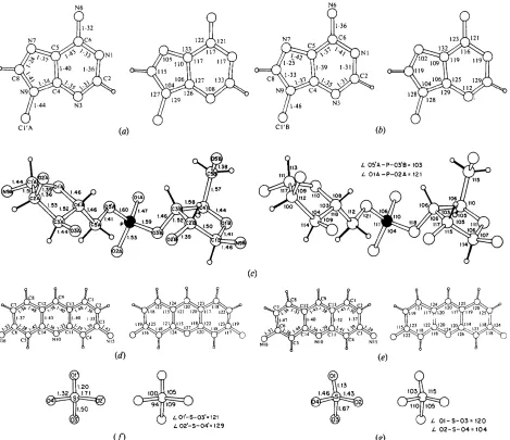
Fig. 2. Numbering system and molecular geometry, showing all covalent bond distances and angles involving non-hydrogen atoms. The 30 H atoms whose positions were calculated and which are included in the structure factor calculations are in the drawing. Those H atoms which participate in the protonation of N(10) positions on proflavine 1 and proflavine 2 and N(I) positions on adenine A and adenine B are not shown since they were never calculated. The average e.s.d, in bond lengths is 0.01 A and in angles is 1 o. (a) Adenine A. (b) Adenine B. (c) Ribose phosphate backbone. (d) Proflavine 1. (e) Proflavine 2. (f) Disordered sulphate 1. (g) Disordered sulphate 2.
The molecular structure
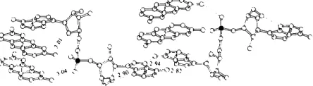
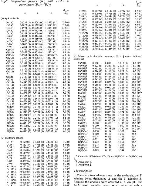
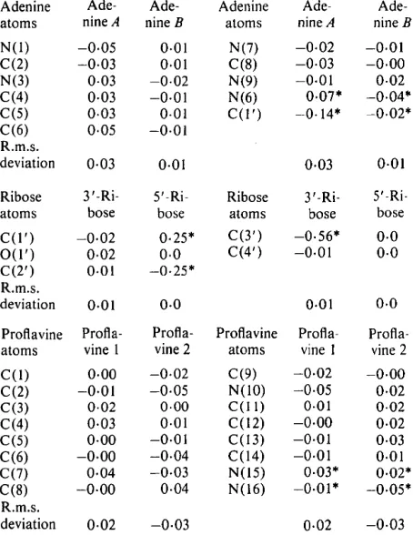
E a c h a s y m m e t r i c unit in the c r y s t a l consists of the dinucleoside p h o s p h a t e A p A in an extended confor- m a t i o n , two proflavine molecules s t a c k e d below an adenine, a sulphate a n i o n a n d several w a t e r molecules f o r m i n g a highly h y d r a t e d 1 : 2 complex. Fig. 1 shows two complexes which interact with each other t h r o u g h base pairing. T h e a t o m i c p a r a m e t e r s are given in Table 1, distances and angles in Fig. 2, m o l e c u l a r planes in T a b l e 2 a n d torsion angles in Fig. 3 and T a b l e 3.
3'-end
r B
I1
,
Fig. 3. Molecular configuration of ApA, illustrating the
526 HYDRATED ADENYLYL(3'-5')ADENOSINE-PROFLAVINE HEMISULPHATE
T a b l e 1.
Fractional coordinates and equivalent iso-
tropic temperature factors
(A 2)with e.s.d.'s in
parentheses (Beq
= ( B u > )T a b l e 1
(cont.)
X I' Z
Beq or
Biso
x y z
Beq o r Blso
(a) ApA molecule
N(1A) 0.1237 (3) 0.5063 (6) 1.2993 (11) 7.7 (6)
C(2A) 0.1623 (4) 0.5309 (6) 1.3143 (15) 7.7 (8)
N(3A) 0.1994 (3) 0.5056 (5) 1.3244 (10) 6.3 (5)
C(4A) 0.1952(3) 0.4434(6) 1.3139(11) 5.6(6)
C(5A) 0.1584 (3) 0.4096 (6) 1.2994 (11) 5.8 (5)
C(6A) 0.1204(3) 0.4444(6) 1.2989(11) 5.2(6)
N(6A) 0.0839 (3) 0.4169 (6) 1-2878 (11) 7.3 (6)
N(7A) 0.1670(3) 0.3474(5) 1.2871 (9) 5.9(5)
C(SA) 0.2055 (3) 0.3439 (6) 1.2942 (13) 6.6 (6)
N(9A) 0.2261 (3) 0.4015 (5) 1.3103 (9) 5.4 (5)
C(I'A) 0.2702 (3) 0.4124 (6) 1.3087 (11) 5.5 (5)
O(I'A) 0.2859 (2) 0.4054 (4) 1.1822 (7) 5.4 (4)
C(2'A) 0.2961 (4) 0.3741 (6) 1.4030 (12) 6.2 (5)
O(2'A) 0.3264 (2) 0.4083 (5) 1.4631 (8) 8.2 (5)
C(YA) 0.3140 (4) 0.3253 (6) 1.3087 (13) 6.2 (5)
O(YA) 0.3531 (3) 0.2990(5) 1.3510(9) 8.5 (5)
C(4'A) 0.3205(3) 0.3613(5) 1.1818(11) 4.8(5)
C(5'A) 0.3193 (4) 0.3268 (7) 1.0584 (12) 7.3 (7)
O(5'A) 0.3191 (2) 0.3671 (4) 0.9493 (8) 5.7 (4)
P 0.3200 (1) 0.3400 (2) 0.8032 (3) 5.6 (1)
O(1A) 0.3157 (2) 0.3941 (4) 0.7158 (7) 6.5 (4)
O(2A) 0.2909 (2) 0.2837 (5) 0.7903 (9) 8-3 (5)
C(I'B) 0.4547 (3) 0.2833 (4) 0.7282 (11) 4.2 (4)
O(I'B) 0.4483 (2) 0.3225 (3) 0.6193 (8) 5.3 (4)
C(2'B) 0.4375(3) 0.3178(5) 0.8439(10) 4.0(5)
O(2'B) 0.4299 (3) 0.2815 (4) 0.9539 (8) 7.0 (5)
C(YB) 0.4008 (3) 0.3539 (5) 0.7890 (12) 5.4 (5)
O(YB) 0.3655 (2) 0.3113 (3) 0.7916 (8) 5.4 (4)
C(4'B) 0.4126 (3) 0.3619 (6) 0.6394 (13) 5.7 (6)
C(5'B) 0.4256 (4) 0.4321 (7) 0.6229 (21) 9.7 (9)
O(5'B) 0.4557 (3) 0.4523 (4) 0.7103 (15) 10.6 (7)
N(1B) 0.6030(3) 0.3735(5) 0.7518(10) 6.5(5)
C(2B) 0.5665(3) 0.4010(5) 0.7512(13) 6.2(6)
N(3B) 0.5293 (3) 0.3759 (4) 0.7429 (10) 5.6 (5)
C(4B) 0.5310(3) 0.3129(5) 0.7384(9) 4.4(5)
C(5B) 0.5675 (3) 0.2780 (6) 0.7409 (10) 5.5 (5)
C(6B) 0.6049(3) 0.3081 (5) 0.7351(11) 5.2(5)
N(6B) 0.6421 (3) 0.2779 (5) 0.7425 (10) 6.3 (5)
N(7B) 0.5570 (2) 0.2139 (4) 0.7326 (9) 4-8 (4)
C(8B) 0.5181 (3) 0.2155 (5) 0.7298(10) 4.5 (5)
N(9B) 0.4992 (2) 0.2707 (4) 0.7357 (9) 4.1 (4)
(b) Proflavine cations
0.1240 (4) 0.4491 0.1631 (4) 0.4735 0.1966 (3) 0.4375 0.1948 (4) 0.3728 0.1149 (4) 0.1843 0.0764 (4) 0.1566 0.0391 (4) 0.1875 0.0399 (4) 0.2511 0.0809 (3) 0.3510 0.1525 (3) 0.2838 0.1560 (4) 0.3453 0.0792 (4) 0.2869 0.1194 (4) 0.3806 0.1165 (3) 0.2503 0.2367 (3) 0.4655 0.0762 (4) 0.0947 0.1335 (5) 0.4926
(7) 0.6281 (13) 7.2 (6)
(6) 0.6366 (13) 6.0 (6)
(6) 0.6405 (10) 5.4 (5)
(6) 0.6341 (11) 5.8(5)
(6) 0.5963 (14) 6.6 (6)
(5) 0.5862 (12) 6.0 (7)
(6) 0.5874 (12) 6.4 (6)
(9) 0.5909 (14) 7.6 (8)
(7) 0.6077 (11) 6.3 (6)
(5) 0.6083 (9) 5.6 (5)
(6) 0.6219 (10) 4.9 (5)
(8) 0.6017 (11) 6.5 (6)
(6) 0.6182(11) 5.1 (5)
(5) 0.6023(11) 5.1(5)
(5) 0.6518 (10) 6.7 (5)
(6) 0.5783 (14) 9-4 (8)
(7) 0.9687 (15) 8.0 (8)
C(1P1) C(2PI) C(3P1) C(4PI) C(5Pl) C(6P1) C(7P1) C(8P1) Cq9P1) N110P1) CqI1PI) C112P1) C113P1) C114PI) Nt 15P1) Ni 16PI) Cq 1P2)
C(2P2) 0.1719(5) 0.5110(6) 0.9732(13) 6.9(7)
C(3P2) 0.2059 (5) 0.4726 (5) 0.9795 (12) 6.4 (7)
C(4P2) 0.1995(3) 0.4086(8) 0.9724(11) 6.7(6)
C(5P2) 0.1093(3) 0.2304(5) 0.9358(11) 5.2(5)
C(6P2) 0.0706 (5) 0.2057 (7) 0.9239 (12) 7.8 (7)
C(7P2) 0.0329 (4) 0.2441 (7) 0.9244 (12) 6.9 (7)
C(8P2) 0.0363 (4) 0.3060 (9) 0.9385 (13) 8.2 (8)
C(9P2) 0.0845 (4) 0.4028 (6) 0.9528 (15) 7.6 (6)
N(10P2) 0.1521 (3) 0.3223 (4) 0.9557 (8) 5.1 (4)
C(IIP2) 0.1598(3) 0.3812(6) 0.9635(11) 5.0(5)
C(12P2) 0.0786(3) 0.3367(8) 0.9462(12) 7.1 (6)
C(13P2) 0.1241 (4) 0.4273 (6) 0.9649 (12) 5.4 (5)
C(14P2) 0.1132(3) 0.2973(6) 0-9464(11) 5.2(5)
N(15P2) 0.2465 (4) 0.4942 (4) 0.9898(10) 8.0 (5)
N(16P2) 0.0639(4) 0.1427(6) 0.9154(12) 9.4(7)
(c) Solvent molecules (the occupany, G, is ½ unless indicated otherwise)
WP(01) 0.000 0.000 0.615 (3) 14.7 (13)
WP(02)t 0.223 (1) 0.247 (1) 0.953 (1) 11.7 (4)
WP(03)+ 0.433 (1) 0.312 (1) 1-226 (2) 18-3 (10)
WP(04)t 0.396 (1) 0.166 (1) 0.923 (2) 19.7 (6)
WP(05)t 0.230 (1) 0.222 (1) 0.582 (2) 16.4 (12)
WP(06)t 0.314 (1) 0.164 (1) 0.911 (2) 17.6 (7)
We(07)t 0-343 (1) 0.530 (1) 1.389 (5) 38.9 (25)
WP(08)t 0.162 (1) 0.023 (1) 1.206 (5) 52.6 (19)
WP(09)t 0.266 (1) 0.591 (1) 1.348 (6) 46.5 (36)
WP(10)t 0.133 (2) 0.040 (2) 0.926 (9) 74.3 (44)
WP(I1)#
0.337 (1) 0.204 (1) 1.506 (3) 26.9 (14)WS(01) 0.449 (2) 0.486 (2) 1-035 (5) 22.8 (17)*
WS(02) 0.475 (1) 0.405 (1) 1.197 (3) 15.1 (9)
WS(03) 0.187 (2) 0.116 (3) 0.875 (7) 26-4 (21)
WD(01) 0.367 (2) 0.159 (3) 1.204 (7) 29.4 (24)
WD(02) 0.419 (1) 0.131 (2) 1.251 (4) 16.1 (10)
WD(03) 0.459 (1) 0.073 (1) 1.239 (3) 14.4 (8)
WD(04) 0.482 (1) 0.037 (2) 1.462 (4) 15.4 (10)
WD(05) 0.489 (1) 0.023 (1) 1.224 (3) 14.1 (9)
WD(06) 0.442 (3) 0.093 (5) 1.416 (9) 34.5 (35)
WD(07) 0.063(1) 0-591(1) 1.211 (2) I0.1(5)
WD(08) 0.023 (1) 0.535 (1) 1.183 (1) 4.9 (3)
WD(09)~ 0.000 0.500 1.196 (6) 17.5 (19)
WD(10)$ 0.000 0.500 1.060 (8) 21.6 (23)
S(04)$ 0.249(1) 0.148(1) 1.230(1) 14.3 (14)
O(ISO4') 0.250 0.104 1-302 14.4
O(2SO4') 0.300 0.169 1.210 26.5
O(3SO4') 0.224 0.205 1.263 12.7
O(4SO4') 0.222 0.128 1. 142 34.3
O(ISO4) 0.242 0.196 1.269 16.7
O(2SO4) 0.277 0.112 1.308 20.2
O(3SO4) 0.266 0.139 i.076 25.7
O(4SO4) 0.209 0.116 1-244 16-3
* Values for WS(01) to WD(10) and O ( l S O 4 ' ) t o O(4SO4) are
Bis o.
+ Occupancy 1. $ Occupancy ~.
The base pairs
H.-S. SHIEH, H. M. BERMAN, S. NEIDLE, G. TAYLOR A N D M. S A N D E R S O N 527 Table
2.
Deviations
(A)
of atoms from the least-squares
planes through the indicated atoms (average e.s.d.
is 0.01 /~,)
Adenine Ade- Ade- Adenine Ade- Ade- atoms nine A nine B atoms nine A nine B
N(I) -0.05 0-01 N(7) -0.02 -0.01
C(2) -0.03 0.01 C(8) -0.03 -0.00
N(3) 0.03 -0.02 N(9) -0.01 0.02
C(4) 0.03 -0.01 N(6) 0.07* -0.04*
C(5) 0.03 0.01 C(I') -0.14" -0.02*
C(6) 0.05 -0.01
R.m.s.
deviation 0.03 0.01 0.03 0.01
Ribose 3'-Ri- 5'-Ri- Ribose 3'-Ri- 5'-Ri-
atoms bose bose atoms bose bose
C(I') -0.02 0.25* C(Y) -0.56* 0.0
O(1') 0.02 0.0 C(4') -0.01 0.0
C(2') 0.01 -0.25*
R.m.s.
deviation 0.01 0.0 0.01 0.0
Proflavine Profla- Profla- Proflavine P r o f l a - Profla-
atoms vine 1 vine 2 atoms vine 1 vine 2
C(I) 0.00 -0.02 C(9) -0.02 -0.00
C(2) -0.01 -0.05 N(10) -0.05 0.02
C(3) 0.02 0.00 C(I 1) 0-01 0-02
C(4) 0.03 0.01 C(12) -0.00 0.02
C(5) 0.00 -0.01 C(13) -0.01 0.03
C(6) -0.00 -0.04 C(14) -0.01 0.01
C(7) 0.04 -0.03 N(15) 0.03* 0.02*
C(8) -0.00 0.04 N(16) -0.01" -0.05*
R.m.s.
deviation 0.02 -0.03 0.02 -0.03
* Atoms not involved in least-squares-plane calculations.
Table
3.
Conformation angles (o) (e.s.d.'s
1-2 °)
Backbone and glycosidic bonds
z(5')
~,(5')12
Y 6
~,(3')
z(3')
C (8 B)-N (9 B)-C ( 1' B)-O( 1' B) C (5' B)-C (4' B)-C (Y B)-O(Y B) C ( 4 ' B ) - C ( 3 ' B ) - O ( Y B ) - P C (3' B)-O (Y B ) - P - O (5'A)
0(3' B)-P-O(5'A)-C(5'A)
P-O(5'A)-C(5'A)-C(4'A)
O(5'A)-C(5'A)-C(4'A)-C(Y A) C(5'A)-C(4'A)-C(YA)-O(YA) O( I'A)-C( I'A)-N(9A)-C(8A)
240 ° 143 262
291 291 177 171
85
71
Ribose, endocyclic bonds
C(4' B)-O( I' B)-C( I' B)-C(2' B) O( I' B)-C( I' B)-C(2' B)-C(3' B) C( I' B)-C(2' B)-C(Y B)-C(4' B) C(2' B)-C(Y B)-C(4' B)-O( I' B) C(3' B)-C(4' B)-O( I' B)-C( I' B) C(4'A)-O( I'A)-C( I'A)-C(2'A)
O( 1 'A)-C ( 1 'A)-C (2'A)-C (3'A)
C( I'A)-C(2'A)-C(Y A)-C(4'A)
C(2'A)-C(YA)-C (4'A)-O(I'A) C ( 3 ' A ) - C ( 4 ' A ) - O ( I ' A ) - C ( I ' A )
-28 34 - 2 6 10
11
3 -24 35 -34 20
negative phosphate group and a protonated adenine much as was found in the structures of ApA+pA + (Suck, Manor & Saenger, 1976) and UpA + (Sussman, Seeman, Kim & Berman, 1972). Since the distances and angles (Fig. 2) have average estimated standard deviations of 0.01 A and 1 ° respectively, it is not possible to utilize them to assign definitely the protonation site. Consideration of the hydrogen- bonding environment (see discussion later) suggests that it is the N(1) of adenine B which bears the positive charge. The distances and angles in both adenine rings are unremarkable with the exception of the C ( 8 ) - N ( 7 ) distances in both rings which are shorter and the N ( 9 ) - C ( 8 ) - N ( 7 ) angle in ring B which is greater than the corresponding values found in other adenine structures.
The adenine rings are each planar (Table 2) within _+0.05 A. Adenine A forms hydrogen bonds to adenine B of a symmetry-related molecule via N(6) and N(7). This hydrogen-bonding pattern was first proposed for acid poly(A +) (Rich, Davis, Crick & Watson, 1961) and occurs in a variety of adenine-containing crystal structures including UpA + (Sussman et al., 1972) and ApA+pA + (Suck et al., 1976). The average deviation of the plane of the base pairs is 0.2 A and the tilt between bases 5 o
The
conformation of ApA
The dinucleoside phosphate adopts an extended conformation (Fig. 3) which results from some unusual values in the torsion angles of the nucleoside segments; this is in contrast to the situation observed in other extended dinucleoside phosphates where only the torsion angles of phosphodiester linkage differ from the normal helical conformations (Sussman et al., 1972).
528 H Y D R A T E D A D E N Y L Y L ( 3 ' - 5 ' ) A D E N O S I N E - P R O F L A V I N E HEMISULPHATE
et al.,
1977; Bermanet al.,
1979; Tsai, Jain & Sobell,1977; Jain, Tsai & Sobell, 1977). The
C(2')-endo
(3' --, 5')C(3')-endo
ordering of the mixed sugar pucker occurs in the crystal structure of the complex between ApU and 9-aminoacridine (Seeman, Day & Rich, 1975), the only other non-intercalated complex where structure has been reported. In the self-complementary ribodinucleoside intercalative complexes with mixed puckering the order is C (3')-endo-C (2')-endo.Some significant conformational angles are pre- sented in Table 3. The a angle of the molecule is 262 °. As has been observed in the structures of pTpT (Camerman, Fawcett & Camerman, 1976) and tRNA
( J a c k , L a d n e r & K l u g , 1 9 7 6 ; H o l b r o o k , S u s s m a n ,
Warrant & Kim, 1978) this high value can be correlated with
C(2')-endo
sugar puckering. When sugars areC(3')-endo
the value of a has always been found to be about 210 ° (Arnott & Hukins, 1969). The value of e is 171 o and together with the increased a is responsible for stretching apart the planes of the bases from the usual 3.4 ,/k to 6.3 ,~. Thetrans
value for this angle is not usually found in 5' nucleotides althoughthere is n o a priori t h e o r e t i c a l b a s i s for this a n g l e to be
always
gauche
(Pullman & Saran, 1976). Thetrans
value has been observed in dGMP (Young, Tollin & Wilson, 1974), the modified nucleotide azaUMP (Saenger & Suck, 1973), and in a few residues of tRNA. Curiously, the otherwise variable fl and 7 angles have almost precisely the values found in R N A 11 (Arnott, Hukins & Dover, 1972), as does 6.(c)
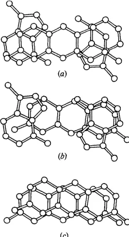
Fig. 4. The stacking patterns of the molecular complex. (a) Proflavine 1/base pair. (b) Base pair/proflavine 2. (c) Proflavine 2/proflavine 1.
Table 4.
Hydrogen bonds and short intermolecular
contacts (~)
Positional parameters of the O atoms of the SO42- ion were not refined. Distances involving these atoms are marked with an asterisk.
(a) lntramolecular h y d r o g e n b o n d s
N ( 3 B ) - • • O ( 5 ' B ) 2 . 9 0 (1)
(b) A p A ~ A p A
N ( 6 A ) . . . N ( 7 B ~) 2 . 9 4 (2) O ( 2 ' A ) . . . O ( I A " ) 2 . 6 1 ( I )
(e) A p A • proflavine, sulphate and water molecules
N ( 7 A ) . . . N ( 6 B ~) 2 - 8 2 (2)
N ( I A ) . • • W D ( 0 7 ) 2 . 8 1 (3) O ( 2 A ) . • • W P ( 0 2 ) 2 . 8 5 (1) N ( 3 A ) . - • W P ( 0 9 ) 2 . 8 3 (3) O ( 2 A ) . . • W P ( 0 5 ) 3 . 1 7 (2) C ( 8 A ) . • • O ( 3 S O 4 ' ) 3 . 0 5 ( 1)* O ( 2 A ) . •. W P ( 0 6 ) 2 . 9 4 (2) O ( I ' A ) . . . N ( 1 5 P 2 ) 3 . 0 1 (1) O ( 2 ' B ) . . . W P ( 0 3 ) 2 . 8 5 (2) O ( 2 ' A ) . • • W P ( 0 7 ) 2 , 7 7 (3) O ( 2 ' B ) - • • W P ( 0 4 ) 2 . 7 2 (3) O ( 3 ' A ) . • • W P ( 0 3 ) 2 . 8 8 (2) N ( I B ) . • • W P ( 0 8 " ) 2 . 9 5 (3) O ( 3 ' A ) . • • W P ( 1 I) 2 . 6 3 (3) N ( 6 B ) . . . O ( 3 S O 4 " ) 2 . 6 6 (I)* O ( I A ) - . . N ( 1 5 P 1 ) 3 . 0 4 (1) N ( 6 B ) . . . O ( 4 S O 4 " ) 3 . 1 3 (I)* O ( I A ) . • • W P ( 0 8 I") 2 . 9 7 (3)
( d ) P r o f l a v i n e --* sulphate and water molecules
N ( 1 0 P I ) . • • W P ( 0 5 ) 2 . 8 4 (2) N ( 1 6 P I ) - • • W P ( 0 7 ' ) 2 - 9 6 (3) N ( 1 5 P 1 ) . . . O ( I S O 4 'ill) 3 . 0 4 (I)* N ( 1 0 P 2 ) . • • W P ( 0 2 ) 2 - 7 9 (1) N ( 1 5 P I ) . . . 0 ( 2 S 0 4 ill ) 3 . 2 0 ( 1 ) * N ( 1 5 P 2 ) . . . O ( 3 S O 4 ILi) 3 . 2 0 ( 1)* N ( 1 6 P I ) - - - W P ( 0 1 ) 3 . 2 0 (1) N ( 1 6 P 2 ) • • • 14'P(10) 3 . 1 3 (6)
(e) S u l p h a t e ~ water molecules
O ( 2 S O 4 ' ) . - • W P ( 0 6 ) 3 . 0 8 (2)* O ( 3 S O 4 ) . • • W P ( 0 2 ) 2 . 9 7 (1)* O ( 2 S O 4 ' ) . - • W D ( 0 1 ) 2 . 1 7 (6)* O ( 3 S O 4 ) . • • W P ( 0 6 ) 2 . 3 4 (2)* O ( 4 S O 4 ' ) . - • W P ( 0 2 ) 3 . 2 0 (1)* O ( 4 S O 4 ) . • • W P ( 0 8 ) 2 . 5 3 (3)* O ( 4 S O 4 ' ) . • • w P ( 0 8 ) 3 . 0 4 (3)*
O ( 4 S O 4 ' ) . • - W S ( 0 3 ) 2 . 9 5 (7)*
( f ) N o r m a l water -, water molecules
W P ( 0 1 ) . • - W S ( 0 2 ~) 2 . 9 1 (3) W P ( 0 7 ) . • • W P ( 0 9 ) 2 . 8 3 (4) W P ( 0 2 ) . • • W S ( 0 3 ) 3 . 1 4 (7) W P ( 0 8 ) . • • W P ( 1 0 ) 3 . 0 2 ( 1 0 ) W P ( 0 3 ) . - • W S ( 0 2 ) 2 . 4 3 (3) W P ( 0 9 ) . - • W S ( 0 3 TM) 2 . 7 8 (9) W P ( 0 4 ) . . . W S ( 0 6 ) 2 . 6 4 (3) W P ( 1 0 ) . - . W S ( 0 1 ' ) 2 . 9 1 (9) W P ( 0 4 ) . • • W D ( 0 1 ) 3 . 0 1 (7) W P ( 1 0 ) . - • W S ( 0 3 ) 2 . 4 4 (9) W P ( 0 4 ) . - • W D ( 0 7 v) 2 . 4 9 (3) W S ( 0 1 ) . - • W S ( 0 2 ) 2 . 5 4 (6) W P ( 0 5 ) . - • W P ( 0 9 ' ) 2 - 9 0 (3)
(g) Disordered water --, disordered water molecules
W D ( 0 3 ) . . . W D ( 0 4 ) 2 . 5 1 (5) W D ( 0 5 ) . . . W D ( 0 6 ) 2 - 8 9 ( 1 0 ) W D ( 0 3 ) . • • W D ( 0 5 " ) 2 . 6 6 (4) W D ( 0 5 ) . • • W D ( 1 0 ~') 2 . 9 5 (9) W D ( 0 4 ) . • • W D ( 0 5 ) 2 . 4 5 (5) W D ( 0 7 ) . • - W D ( 0 9 ) 2 - 8 2 (3) W D ( 0 4 ) . • • W D ( 0 5 '~) 2 . 9 9 (5)
(h) Short c o n t a c t s (<_2.2 A ) in group 1 disordered water molecules I W D ( 0 1 ) to
W D ( 0 6 ) I
W D ( 0 1 ) - • • W D ( 0 2 ) 1 . 8 4 (7) W D ( 0 3 ) - - - W D ( 0 6 ) 1 . 9 3 ( I 0 ) W D ( 0 2 ) . • • W D ( 0 3 ) 1 . 7 9 (5) W D ( 0 4 ) . • - W D ( 0 4 " ) 1 . 9 6 (6) W D ( 0 2 ) . • - W D ( 0 6 ) 2 . 0 1 ( 1 0 ) W D ( 0 4 ) . • • W D ( 0 6 ) 1 . 8 2 ( ! I) W D ( 0 3 ) . • - W D ( 0 5 ) 1 . 4 5 (4) W D ( 0 5 ) . • - W D ( 0 5 'i) 1.21 (4)
(i) Short c o n t a c t s ( ~ 2 - 2 A ) in group 2 disordered water molecules I W D ( 0 7 ) to W D ( 10)J
W D ( 0 7 ) . . . W D ( 0 8 ) 1 . 7 8 (3) W D ( 0 8 ) . • • W D ( I O ) 1 - 6 4 (6) W D ( 0 8 ) . . - W D ( 0 8 '~) 2 . 1 1 (3) W D ( 0 9 ) . - - W D ( 1 0 ) 1 . 3 8 ( 1 0 ) W D ( 0 8 ) . • • W D ( 0 9 ) 1 . 0 6 (2)
( j ) Short stacking c o n t a c t s (_<3.40/~.)
N ( I A ) . . . C ( I P 2 ) 3 . 3 9 (2) C ( 2 B ) . . . N ( 1 6 P V "~) 3 - 3 7 (2) C ( 4 A ) . • • C ( 3 P I *~) 3 . 3 3 (2) C ( 4 B ) . • • C ( 7 P I "~") 3 - 3 3 (2) C ( 6 A ) . . . C ( I P I ~*) 3 . 3 5 (2) C ( 6 B ) . . . C ( 5 P 2 ~' ) 3 . 3 5 (2)
S y m m e t r y c o d e
N o n e x , y , z
(i) - 0 . 5 + x , 0 . 5 - y , 2 - z iv) 0 . 5 - x , - 0 . 5 + ) ' , 2 - z
(ii) xd'. 1 + z (vi) 1 - x. - y , z
(iii) 0 , 5 - x , 0 . 5 + y , 2 - z (vii) - x , 1 - y , z
H.-S. SHIEH, H. M. BERMAN, S. NEIDLE, G. TAYLOR A N D M. S A N D E R S O N 529
The proflavine cations
Both proflavine cations are essentially planar and are protonated at N(10), as found in the crystal structure of proflavine hemisulphate (Jones & Neidle, 1975). They are stacked almost directly on top of one another, 3.4 A apart. Unlike the proflavine molecules in the
complex with CpG (Neidle et al., 1977; Berman et al.,
1979) which are at an angle of 97 ° to each other the angle between the stacked proflavines in this crystal is 0 o (Fig. 4c). The proflavines are symmetrically stacked above and below an adenine-adenine base pair (Fig.
4a,b). The central rings of the proflavines are not involved in this stacking. In this sense the stacking is similar to that found in the intercalated portion of
CpG-proflavine (Berman et al., 1979).
The crystal structure
Proflavine-ApA associations
As shown in Fig. 1 each adenine A base-pairs to adenine B in a symmetrically related molecule to form an extended chain of dinucleoside molecules. The proflavine cations stack above and below each base pair. The sequence of stacked rings is thus proflavine, proflavine, base pair, proflavine, proflavine, . . . in the e direction (Fig. 4). N(15) of proflavine 1 hydrogen bonds to O(1A) of a phosphate group and N(15) of proflavine 2 hydrogen bonds to the ribosyl O(1') (Table 4). Amino-phosphate oxygen hydrogen bonds
were also observed in CpG-proflavine (Berman et al.,
1979). The crystal structure consists of infinite columns of heterocyclic stacked planes extending along e linked
by hydrophilic ribose-phosphate groups, forming
zigzag chains along a (Fig. 5). Along e, trans- lationally-related dinucleosides are connected by a hydrogen bond, O(2'A) of the 5' ribose to the O(1) phosphate O atom (Table 4).
t . a
Fig. 5. A view of the structure perpendicular to the ab plane. Dashed lines indicate hydrogen bonds. The outlines of molecular stacking columns, chain connections, and water channels are shown. Two 2~ axes which are perpendicular to the paper are denoted by 0. The origin of the unit cell is on the 2 z axis coinciding with water molecule PI. Water molecules are named as in Table 1 except that the prefix W is omitted.
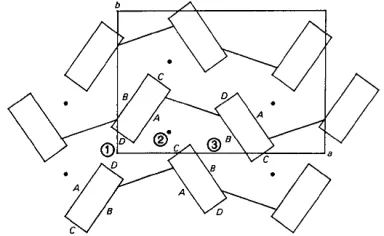
Fig. 6. A schematic representation of the crystal structure. This is an expansion of Fig. 5. The unit-cell outline and the nomenclature of stacking-block faces are shown. The three distinct channel regions are marked (1), (2) and (3). The lines connecting the blocks represent the sugar phosphate chain as shown in Fig. 5. The filled circles represent the sulphate ions.
The solvent structure
In addition to the 1:2 ApA-proflavine complex the asymmetric unit contains a disordered sulphate ion and 24 water molecules. A view of the structure projected
onto the ab plane is shown in Fig. 5. Fig. 6 presents a
simple extended schematic representation of this projection. All the solvent molecules reside in channels between infinite chains of stacked molecules. The major role of solvent in this structure is to hold molecules together between the chains, and to provide additional interactions which aid in maintaining the layers of stacked molecules.
The molecular stacking block, shown in Fig. 6, approximates a rectangular column. Since the pro- flavines are stacked parallel to each other and the sugar puckering is different in base A and base B, the
distribution of polarity on the faces of the rectangular columns is not homogeneous. The four boundaries of the stacking column are designated as faces A, B, C, and D (Fig. 6). A and B are wide faces. Face A is much more polar than face B as all the polar atoms N(10), N(15), and N(16) of the proflavines lie on face A. The exposure of N(3) of adenine A on face C makes it more polar than face D, where N(3) of adenine B is essentially buried in the sugar domain.
530 H Y D R A T E D A D E N Y L Y L ( 3 ' - 5 ' ) A D E N O S I N E - P R O F L A V I N E H E M I S U L P H A T E
P Q ) 1)(4)
N i I 5 P I ) k _ ( ~ " " ( ~ ,> ~ t ' / A O ( ) ' 4 ) ~ ~ , / ~' ) a O(14) . - ~ ~ I ~ , I i
' ,) ~ J ' i
/
i N(10P2)I / " x i ~ ~ L i
v~ p ) ~ - 2-, ~ ? - - - p,~¢ 7T
' N ( I f i P 2 ) ' ~ " ' . / P(6) ' ' " ~ "
21 ~ ~ O ( 2 A ) N(1.4) "" D(71 ) /
6) p(?) __ ~ - - - ~,.~N1341
N ~ I ) N ( I O P I ~ - - - ~ - - ~ . . . P(I)
~O(2
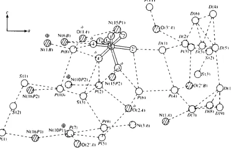
'.4) P(5)Fig. 7. The solvent network in the crystal structure. This view is obtained by rotating 90° along the horizontal axis of Fig. 5. The hydrogen donors or acceptors of the ApA or proflavine molecules are shown as shaded atoms. The symbol + denotes positive charge, with 6-- for partial negative charge.
shows the particular pattern of the charge balance in this structure. Positively charged N(1B)interacts with negatively charged 0 ( 4 ) and 0 ( 4 ' ) of the sulphate ion and O(1 A) of the phosphate group via WP(08). N(10) ÷
of proflavine 1 interacts with O(2A)- via WP(05) while
N(10) ÷ of proflavine 2 interacts with 0 ( 4 ' ) and 0 ( 3 ) o f
the sulphate ion and O(2A)- via WP(02). Thus, all the
charged species in this structure interact with one
another via bridges of neutral water molecules.
This work was supported by grants from NIH GM-21589, CA-22780, CA-06927, RR-05539, and an appropriation from the Commonwealth of Pennsyl- vania (to HMB), from N A T O (to SN arid HMB) and from the Cancer Research Campaign (SP 1384) to SN.
References
large. Three water molecules, WP(01), WS(01), and WS(02) occupy the unique portion of this region. They loosely interact with each other and with N(16) of proflavine 1. The next region (2) has both strong polar faces A and C forming a highly polar channel side. The majority of solvent molecules are in this region. The sulphate ion is in the middle of the channel with its disordered O atoms fully utilized to bind to ApA, the proflavine and other solvent molecules. WS(03) also occupies a pivotal position. Although it does not interact with ApA or proflavines, it connects the sulphate ion, WP(02), WP(10) and WP(09) together and also weakly binds to WP(07). The third region (3) of the channel is confined by two weakly polar and long faces, B and B', related to one another by the second twofold axis. Due to its weak polar surrounding and ample space, all water molecules in this region are very
disordered. WD(01), WD(02), WD(03), WD(04),
WD(05), and WD(06) form one group, while WD(07), WD(08), WD(09), and WD(10) forms the other. These two groups are connected to each other through the interaction WD(05)... WD(10) (2.95 A), and through
the bridge molecule WP(04) [ W D ( O 1 ) . . . W P ( 0 4 )
3.01 A; W D ( 0 7 ) . . . WP(04), 2.49 A]. The attachments of WD(01) to the sulphate ion and WD(07) to N(1) of base ,4 hold these two groups in place. The continuity of a channel is constructed through the 2~ axes as described above. There are no direct contacts among the solvent molecules in different channels. However, as seen on the top right of Fig. 5, one channel interacts
with another via hydrogen bonding among water
molecules with O(3'A) and O(2'B).
The solvent network is shown in Fig. 7. All the
solvent molecules, except WP(11), which had to go
through O(2'A) to interact with other solvent mol- ecules, are hydrogen-bonded to each other. Fig. 7 also
ALTONA, C. (~ SUNDARALINGAM, M. (1972). J. Am. Chem.
Soc. 94, 8205-8212.
ARNOTT, S. & HUKINS, D. W. L. (1969). Nature (London),
224, 886-888.
ARNOTT, S., HUKINS, D. W. L. & DOVER, S. D. (1972).
Biochem. Biophys. Res. Commun. 48, 1392-1399.
BERMAN, H. M., STALLINGS, W., CARRELI.,, H. L., GLUSKER,
J. P., NEIDLE, S., TAYLOR, G. & ACHARI, A. (1979).
Biopolymers, 18, 2405-2429.
BERTHOD, H. & PULLMAN, B. (1973). FEBS Left. 30,
231-235.
BUGG, C. E. & THEWALT, U. (1969). Biochem. Biophys. Res.
Commun. 37, 623-629.
CAMERMAN, N., FAWCETT, J. K. & CAMERMAN, A. (1976).
J. Mol. Biol. 107, 601-621.
DOURLENT, M. & HI~LI~NE, C. (1971). Eur. J. Biochem. 23,
86-95.
EVANS, F. E. & SARMA, R. H. (1976). Nature (London),
263, 567-572.
HOLBROOK, S. R., SUSSMAN, J. L., WARRANT, R. W. & KIM,
S. -H. (1978). J. Mol. Biol. 123, 631-660.
International Tables for X-ray Crystallography(1974). Vol. IV. Birmingham: Kynoch Press.
JACK, A., LADNER, J. E. & KLUG, A. (1976). J. Mol. Biol.
108, 619-649.
JAIN, S. C., TSAI, C. C. & SOBELL, H. M. (1977). J. Mol.
Biol. 114, 317-331.
JONES, A. & NEIDLE, S. (1975). Acta Crvst. B31, 1324-
1333.
LEE, C. H., EZRA, F. S., KONDO, N. S., SARMA, R. H. &
DANYLUK. S. S. (1976). Biochemistry, 15, 3627-3639.
LERMAN, L. S. (1961). J. Mol. Biol. 3, 18-30.
MAIN, P., WOOLFSON, M. M. & GERMAIN, G. (1971).
MULTAN. A Computer Program for the Automatic Solution of Crystal Structures from X-ray Diffraction Data. Department of Physics, Univ. of York, England.
NEIDLE, S. (1979). Prog. Med. Chem. 16, 151-221.
NEIDLE, S., ACHARI, A., TAYLOR, G. L., BERMAN, H. M.,
CARRELL, H. L., GLUSKER, J. P. & STALLINGS, W. C.
H.-S. SHIEH, H. M. BERMAN, S. NEIDLE, G. TAYLOR A N D M. SANDERSON 531 NEIDLE, S., TAYLOR, G., SANDERSON, M., SHIEH, H.-S. &
BERMAN, H. M. (1978). Nucleic Acids Res. 2, 4417-4422.
PULLMAN, B. & SARAN, A. (1976). Prog. Nucleic Acid Res. Mol. Biol. 18, 215-323.
RICH, A., DAVIES, D. R., CRICK, F. H. C. & WATSON, J. D. (1961). J. Mol. Biol. 3, 71-86.
SAENGER, W. t~ SUCK, O. (1973). Nature (London), 242, 610-612.
SEEMAN, N. C., DAY, R. O. & RICH, A. (1975). Nature (London), 253, 324-326.
SHELDRICK, G. M. (1976). SHELX. A system of crystal- lographic programs. Univ. of Cambridge, England. STOUT, G. H. 8~ JENSEN, L. H. (1968). X-ray Structure
Determination, pp. 456-458. New York: Macmillan. SUBRAMANIAN, E., MADDEN, J. J. 8¢. BUGG, C. E. (1973).
Biochem. Biophys. Res. Commun. 50, 691-696.
SUCK, D., MANOR, P. C. & SAENGER, W. (1976). Acta Cryst. B32, 1727-1737.
SUCK, D., SAENGER, W. • VORBRIJGGEN, H. (1972). Nature (London), 235, 333-334.
SUSSMAN, J. L., SEEMAN, N. C., KIM, S.-H. & BERMAN,
H. M. (1972). J. Mol. Biol. 66, 403-421.
TAVALE, S. A. & SOBELL, H. M. (1970). J. Mol. Biol. 48, 109-123.
TSAI, C. C., JAIN, S. C. & SOBELL, H. M. (1977). J. Mol.
Biol. 114, 301-315.
Ts'o, P. O. P., KONDO, N. S., SCHWEIZER, M. P. & HOLLIS,
D. P. (1969). Biochemistry, 8, 997-1029.
URBANKE, C., ROMER, R. 8¢. MAASS, G. (1973). Eur. J. Biochem. 33, 511-516.
WANG, A. H.-J., QUIGLEY, G. J., KOLPAK, F. J., CRAWFORD, J. L., VAN BOOM, J. H., VAN DER MAREL, G. & RICH, A. (1979). Nature (London), 282, 680°686. YATHINDRA, N. t~ SUNDARALINGHAM, M. ( 1973).
Biopolymers, 12, 297-314.
YOUNG, D. W., TOLLIN, P. & WILSON, H. R. (1974). Acta
Cryst. B 30, 2012-2018.
Acta Cryst. (1982), B38, 531-534
Conformational Analysis of Synthetic Androgens. VI. Structure and Crystal Packing of
17fl- H y dro xy- 7fl- methyl-4,14-androstadien- 3-one M onohy drate
BY WILLIAM L. DUAX AND DOUGLAS C. ROHRER
Medical Foundation o f Buffalo, Inc., 73 High Street, Buffalo, New York 14203, USA AND ALBERT SEGALOFF
A lton Ochsner Medical Foundation, New Orleans, Louisiana 70121, USA
(Received 13 January 1981 ; accepted 23 July 1981)
Abstract
The X-ray crystal structure of 17fl-hydroxy-7fl-methyl- 4,14-androstadien-3-one monohydrate was investigated to determine the influence of the 7fl-methyl substituent on the overall conformation. The steroid hydrate (C20H2802.H20) crystallizes in the monoclinic space group P2~ with Z = 2, a = 10.868 (6), b = 8.472 (4), c = 9.838 (4) A,fl = 98.29 (5)°, 2 = 1.5418 A, T = 291 K, V = 8 9 6 . 3 A 3 , p x = 1.18 M g m -3.R = 4.3% for 1972 reflections. Subtle conformational differences between 17fl-hydroxy-7fl-methyl-4,14-androstadien-3- one, 17fl-hydroxy-4,14-androstadien-3-one and 17fl- hydroxy-7fl-methyl-4,14-estradien-3-one are attribut- able to differences in methyl substitution. While the overall shapes of these molecules are very similar, the molecular packings in the crystals of these steroids are entirely different. In contrast to this, the crystal structure of 17fl-hydroxy-7fl-methyl-4,14-androstadien- 0567-7408/82/020531-04501.00
3-one is isomorphous with those of the monohydrates of the most active endogenous androgens, testosterone and dihydrotestosterone. The order of hydrogen-bond lengths and their orientations are remarkably similar in these three structures. Since the hydrophobic surfaces of the molecule are significantly different, the crystal packing in these isomorphs appears to be a function of hydrate formation and the directionally specific hydro- gen bonding mediated by the water molecules in the crystals.
Introduction
The structure determination was undertaken as part of a study of substituent influence on the conformations of modified androgenic steroids. The title compound has approximately 10% of the androgenicity of testos- terone when administered parenterally in the castrate