International Journal of Nephrology and Renovascular Disease 2018:11 321–327
International Journal of Nephrology and Renovascular Disease
Dove
press
submit your manuscript | www.dovepress.com 321
C a s e R e p o Rt
open access to scientific and medical research
Open Access Full Text Article
successful salvage of allograft dysfunction
triggered by transplant renal vein thrombosis
immediately after kidney transplantation:
a case report
shunta Hori tatsuki Miyamoto Keiichi sakamoto takuto shimizu Kazuki Ichikawa Yosuke Morizawa Daisuke Gotoh Yasushi Nakai Makito Miyake tatsuo Yoneda Nobumichi tanaka Katsunori Yoshida Kiyohide Fujimoto
Department of Urology, Nara Medical University, Kashihara, Nara 634-8522, Japan
Background:Transplant renal vein thrombosis (TRVT) is a severe vascular complication and is caused by various factors, including recipient factors, donor factors, immunosuppression regimens, and surgical techniques. Despite adequate interventions, including thrombolytic therapy or surgical thrombectomy, successful salvage of the allograft is often difficult. We observed a case of TRVT induced by compression of the renal vein immediately after intraop-erative abdominal closure.
Case presentation:A 41-year-old male underwent ABO-compatible living kidney transplanta-tion. The donor was his 45-year-old sister, and her right kidney was donated. The allograft had a single artery and vein. One of the preoperative recipient problems was obesity (body mass index, 33.4 kg/m2). Intraoperative Doppler ultrasonography (US) revealed sufficient blood
flow throughout the allograft, and urine output was also observed. After surgery, hematuria was observed; the urine output decreased and serum creatinine levels increased to 7.0 mg/dL. Doppler US showed a decrease in diastolic flow and an elevated resistive index, which were similar findings to those noted in acute rejection. Although steroid pulse therapy was initiated, allograft dysfunction was worsening. On postoperative day 4, surgical exploration revealed TRVT; consequently, thrombectomy was performed. The urine output increased, and serum creatinine levels decreased to 1.8 mg/dL. The cause of TRVT development may be that the transplant renal vein was relatively short, due to the right kidney being compressed by surrounding tissues after abdominal closure, and that TRVT was gradually developing due to stagnant blood flow. Conclusion:Although TRVT is induced by multiple factors, an accurate diagnosis is often difficult. Understanding these factors, including obesity, and considering TRVT as a cause of allograft dysfunction are important during the pre-, peri-, and postoperative periods. Knowledge of TRVT can lead to early and accurate diagnosis and intervention, resulting in better outcomes for the patients with allograft dysfunction.
Keywords:kidney transplantation, surgical exploration, transplant renal vein thrombosis, thrombectomy
Introduction
Kidney transplantation is the best treatment for end-stage renal disease, leading to improved survival rates and quality of life. Despite advancements in managing medical complications, the incidence of surgical complication still amounts to 15.9%, including a 2.7% incidence rate of vascular complications.1 Transplant renal vein thrombosis
(TRVT) is a catastrophic vascular complication, with an incidence rate of 0.1%–3.4%,
Correspondence: Kiyohide Fujimoto Department of Urology, Nara Medical University, 840 shijo-cho, Kashihara, Nara 634-8522, Japan
tel +81 744 22 3051 Fax +81 744 22 9282
email kiyokun@naramed-u.ac.jp
Journal name: International Journal of Nephrology and Renovascular Disease Article Designation: Case report
Year: 2018 Volume: 11
Running head verso: Hori et al
Running head recto: Successful salvage of TRVT DOI: http://dx.doi.org/10.2147/IJNRD.S185520
International Journal of Nephrology and Renovascular Disease downloaded from https://www.dovepress.com/ by 118.70.13.36 on 23-Aug-2020
For personal use only.
Dovepress
Hori et al
resulting in allograft loss and nephrectomy in most cases.1–7
Reportedly, TRVT may be caused by multiple factors, includ-ing recipient factors (age, pre-transplant dialysis modality, and perioperative hemodynamic status), donor factors (age, right kidney, and prolonged ischemia time), immunosup-pression regimens (cyclosporine and mycophenolate mofetil [MMF]), and surgical techniques.2,5,7–11 Clinical presentation
of TRVT is similar to that of rejection or urological com-plications, which are the two most frequent complications immediately after kidney transplantation. Furthermore, the findings of Doppler ultrasonography (US) are also similar to those noted in graft rejection. Thus, diagnosis of TRVT is not easy, and suspicion of TRVT is the most important tool for early diagnosis and intervention, avoiding potential allograft loss. In our case, delayed allograft function that did not improve with steroid pulse therapy was observed, and surgical exploration was performed due to suspected urologi-cal and vascular complications, including TRVT. An early decision to perform surgical exploration and an appropriate intervention could salvage allograft dysfunction induced by TRVT immediately after kidney transplantation.
Case presentation
A 41-year-old man diagnosed with end-stage renal disease, secondary to Gout kidney disease, was hospitalized for pre-emptive living kidney transplantation. He underwent ABO-compatible living kidney transplantation. The kidney donor was his 45-year-old sister, and her right kidney was selected for allograft due to a split renal function with Tc-99m mer-captoacetyltriglycine-3 renography revealing superiority of contralateral kidney function. The right kidney consisted of a single artery and vein. Despite instruction of weight loss,
his preoperative body mass index (BMI) was 33.4 kg/m2;
thus, we handled the surgery with the greatest care to avoid complications. The surgery progressed smoothly (first warm ischemic time: 5 minutes, cold ischemic time: 29 minutes, and second warm ischemic time: 60 minutes), and urine output was observed immediately after reperfusion of the transplant renal artery and vein. Doppler US immediately before abdominal closure revealed sufficient blood flow throughout the allograft, without reversed or absent dia-stolic flow and an obvious elevated resistive index (RI). An allograft biopsy 1 hour after the allograft blood flow was restored, showing no specific changes. He was introduced to an immunosuppression regimen consisting of tacrolimus, MMF, prednisone, and basiliximab. On postoperative day (POD) 1, the serum creatinine levels decreased, the urine output gradually decreased, hematuria was recognized, and Doppler US revealed mild hydronephrosis in the allograft.
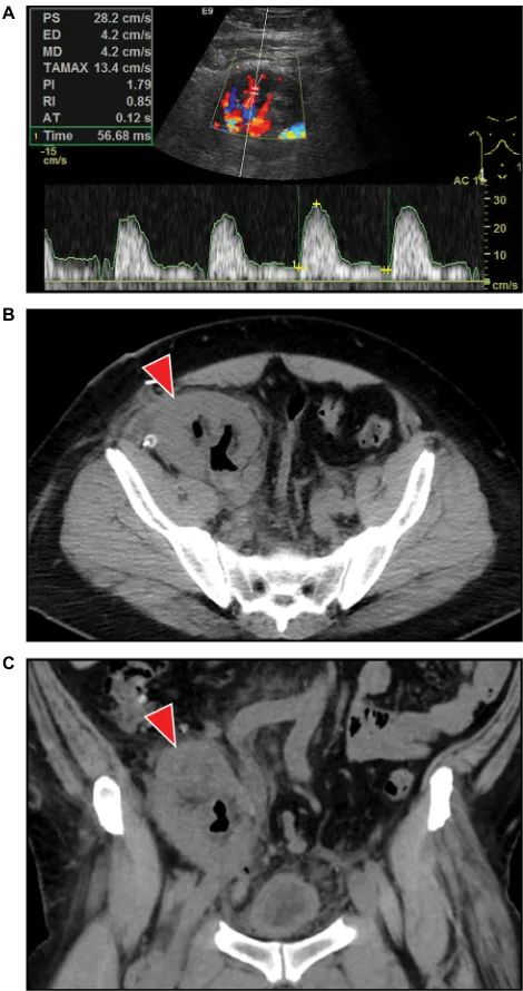
Doppler US on POD 2 showed a decrease in the diastolic flow and an elevated RI without obvious evidence of TRVT (Figure 1A); thus, rejection was considered, and steroid pulse therapy was initiated. Despite this therapy, allograft function did not improve, hydronephrosis worsened, and urine output decreased. Unenhanced computed tomography (CT) on POD 4 revealed a hematoma with air in the urinary tract of the allograft (Figure 1B and C), and serum creatinine levels increased to 7.0 mg/dL. By washing ureteral catheter to check
Figure 1 Representative allograft images of Doppler Us and Ct before surgical exploration.
Notes: Doppler Us revealed a decrease in end-diastolic blood velocity (4.2 cm/s) and an elevated RI (0.85) (A). Ct revealed a hematoma with air in the urinary tract of the allograft (B): coronal image, (C): sagittal image, red arrow: hematoma with air.
Abbreviations: Ct, computed tomography; RI, resistive index; Us, ultrasonography; ps, peak systolic; eD, end-diastolic; MD, mean diastolic; taMaX, time averaged maximum; pI, pulsatility index; RI, resistance index; at, acceleration time.
A
B
C
International Journal of Nephrology and Renovascular Disease downloaded from https://www.dovepress.com/ by 118.70.13.36 on 23-Aug-2020
Dovepress successful salvage of tRVt
for obstraction was the reason why air accumulated in urinary tract. To remove the hematoma with clots in the urinary tract, a ureteral stent was placed, and the allograft was assessed for anastomotic stenosis and compression of the renal artery and vein; then, surgical exploration was performed. Perforation of the pelvis was detected, and evacuation of the hematoma with clots in the urinary tract was performed. Ureteral stent indwelling was performed, and the pelvis was repaired using bioabsorbable sutures. Vascular anastomoses were examined to determine whether there was arterial anastomotic stenosis, kinking of the artery, compression of the vein, and presence of thrombosis. Although there was no evidence of arterial anastomosis and kinks in the renal artery, one of the two branches of the proximal renal vein was compressed by the surrounding tissues, including the allograft, and TRVT was
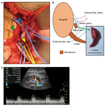
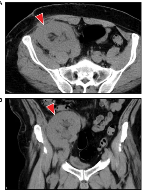
detected. Therefore, subsequent surgical thrombectomy was performed, and Doppler US revealed improved diastolic flow and RI (Figure 2A–C). Postoperative CT also showed that hydronephrosis and the urinary tract hematoma were no longer present (Figure 3A and B). Simultaneously, an allograft biopsy was performed and showed no evidence of rejection, calcineurin inhibitor toxicity, or tubular injury. After surgical thrombectomy, urine output increased, and serum creatinine levels decreased to 1.8 mg/dL (Figure 4).
Ethical approval
Patient provided written informed consent for the publication of his case details and accompanying images. This case report was approved by the institutional review board for Clinical Studies (Medical Ethics Committee ID: NMU-1131).
Figure 2 Representative allograft image and schematic for surgical exploration.
Notes: Representative image of the allograft during surgical exploration. arterial anastomotic stenosis was not observed. the proximal renal vein was branched into two, and one branch was compressed by the surrounding tissues, including the allograft, renal artery, and abdominal wall, resulting in tRVt. In addition, the upper urinary tract of the allograft was occupied by a hematoma with clots and air (A), red arrow: renal artery, blue arrow: renal vein, green arrow: allograft, and yellow arrow: ureter. a schematic of the intraoperative allograft is shown. the trunk of the renal vein had no evidence of thrombosis, and a thrombectomy was performed. In addition, a hematoma with a clot of the upper urinary tract was evacuated (B). Doppler Us showed improved end-diastolic blood velocity (11.7 cm/s) and RI (0.56) (C).
Abbreviations: RI, resistive index; tRVt, transplant renal vein thrombosis; Us, ultrasonography; ps, peak systolic; eD, end-diastolic; MD, mean diastolic; taMaX, time averaged maximum; pI, pulsatility index; RI, resistance index; at, acceleration time.
A B
Allograft
Internal iliac artery
Renal artery
Renal vein
Ureter External iliac vein
Hematoma 10 mm
C
International Journal of Nephrology and Renovascular Disease downloaded from https://www.dovepress.com/ by 118.70.13.36 on 23-Aug-2020
Dovepress
Hori et al
Discussion
TRVT is a rare complication during the early posttransplant period, but it can be a devastating, with a high risk of allograft loss and allograft nephrectomy, even if interventions are performed immediately. Most causes of thrombotic events occurring in the early postoperative period are technical in nature, contrary to thrombotic events occurring 1 month after kidney transplantation, which are associated with rejec-tion.2,4,12,13 Various technical issues are included in the causes
of TRVT, consisting of vein kinking, long veins, multiple veins, and injury of the endothelium.2,12,13 Although surgical
procedures proceeded smoothly, venous anastomosis was carried out gently, and the transplant vein was not too long and did not show kinking during the operation, it was difficult to confirm that there were no complications with the surgical techniques used. Retrospectively, Doppler US immediately after abdominal closure showed a very slight decrease in diastolic blood flow compared to that before the abdominal closure; this might be a sign of TRVT. Generally, the clinical presentation of TRVT is oliguria, hematuria, and a painful
swollen allograft, along with allograft dysfunction as well as the Doppler US typically showing a decrease in diastolic flow and an elevated RI, which are also features of rejection.4,6
Although hematuria with mild hydronephrosis, a decrease in urine output without oliguria, and allograft dysfunction were observed in our case, acute rejection was suspected at first considering the findings of the Doppler US. Despite steroid pulse therapy, allograft function did not improve and, as a result, surgical exploration revealed the presence of TRVT. Repeated Doppler US also did not reach the detection of TRVT in this case. Obesity is one of the reasons. Despite the advancements in Doppler US including image resolu-tion, some vascular complications cannot be diagnosed with
Doppler US even if expert in US examination performed.14
Diagnosing TRVT is very difficult, and suspicion of TRVT is very important for the prevention of allograft loss. In addition, unenhanced magnetic resonance angiography is an option for assessing transplant arteries and veins without damaging the allograft, including the use of contrast material; the quality of the unenhanced magnetic resonance angiography image was equivalent to that of the contrasted image.15 Early diagnosis
leads to successful salvaging of allograft function, and thus various modalities assessing the allograft, including form, surrounding tissues, the urinary tract, and renal vessels, should be used for an accurate diagnosis, followed by early intervention without hesitation.
Reportedly, TRVT can be caused by various factors, including recipient factors, donor factors, immunosup-pression regimens, and surgical techniques.2,5,7–11 Our case
had at least two of these factors (right kidney and usage of MMF), and combination of these factors might result in the development of TRVT. In addition, obesity (BMI >30 kg/m2)
is associated with not only the development of urological complications but also the development of vascular compli-cations including renal artery stenosis and TRVT.16,17 In our
case, the recipient’s preoperative BMI was 33.4 kg/m2, and
the Doppler US immediately after abdominal closure showed a very slight decrease in diastolic blood flow compared to that before abdominal closure. Therefore, we retrospectively sug-gest that the cause of TRVT in our case is that the relatively short renal vein (due to the right kidney) was compressed by surrounding tissues, including the swelling allograft, and the compression was worsened by abdominal closure. Although obesity itself is a risk of the development of cardiovascular events and affects mortality,18 when performing kidney
trans-plantation in obese patients, surgeons must pay attention to the various obesity-related complications. These include not
Figure 3 Representative allograft images of Ct after surgical exploration.
Notes: Ct revealed a loss of the hematoma with air in the upper urinary tract of the allograft. (A): coronal image, (B): sagittal image, red arrow: allograft.
Abbreviation: Ct, computed tomography.
A
B
International Journal of Nephrology and Renovascular Disease downloaded from https://www.dovepress.com/ by 118.70.13.36 on 23-Aug-2020
Dovepress successful salvage of tRVt
only wound complications but also vascular complications, such as artery stenosis and TRVT. Preoperative management of obese patients, including nutritional intervention and exercise programs, could lead to a decrease in perioperative complications, resulting in good prognosis for patients. In addition, to avoid renal vein compression, the use of mesh should be considered. Reportedly, the use of mesh to prevent renal allograft compartment syndrome showed minimal risks and good overall graft and recipient survival rate. Obesity recipients should be taken into consideration to use mesh.19
Although thrombolytic therapy and surgical thrombec-tomy are the most common treatments for TRVT, successful management of salvaging the allograft is often difficult, resulting in allograft loss. Harraz et al20 investigated the
out-come and factors determining the success of allograft salvage with vascular complications, including TRVT, during living kidney transplantations performed on 2,208 consecutive
patients. The study showed that older patients, pre-transplant hypertension, more human leukocyte antigen mismatches, shorter ischemia time, and a longer time to be diagnosed were significantly associated with non-salvage of the allograft after vascular complications, using univariate analysis. Nevertheless, none of these variables were significant using multivariate analysis. Eleven patients out of 23 lived with a functioning graft during a median follow-up of 35 months. With regard to TRVT, 4 patients out of 23 were diagnosed with TRVT. Although three allografts were salvaged (two: thrombolytic therapy, one: surgical thrombectomy), one allograft was lost.20 In our case, surgical thrombectomy was
performed as a result of surgical exploration, resulting in a successful salvage. Considering TRVT as a cause of allograft dysfunction during surgical exploration is important for dealing with the devastating complication with adequate preparation. Repeatedly, preoperative sufficient assessments,
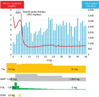
Figure 4 Clinical course of serum creatinine levels and urine output and the immunosuppression regimen.
Notes: on poD 4, the serum creatinine level was 7.0 mg/dL, and urine output decreased; thus, surgical exploration was performed. after evacuation of the transplant renal vein thrombosis and hematoma of the upper urinary tract of the allograft, serum creatinine levels decreased to 1.8 mg/dL, and urine output increased. the immunosuppression regimen was as follows: tacrolimus, MMF, psL, and BXM. at discharge, the doses of each immunosuppressive agent were as follows: 10 mg/day tacrolimus, 1,000 mg/day MMF, and 5 mg/day psL.
Abbreviations: BXM, basiliximab; MMF, mycophenolate mofetil; poD, postoperative day; psL, prednisone; tac, tacrolimus.
0 500 1,000 1,500 2,000 2,500 3,000 3,500 4,000
0
0 5 10 15 20 25 POD
Tac 14 mg 10 mg
MMF 1,000 mg 1,000 mg
PSL 125 mg 5 mg
BXM 20 mg
30 35 40 45 1
2 3 4 5 6 7 8
Steroid pulse therapy (500 mg/day)
Serum creatinine
(mg/dL) Urine output(mL/day)
9
International Journal of Nephrology and Renovascular Disease downloaded from https://www.dovepress.com/ by 118.70.13.36 on 23-Aug-2020
Dovepress
Hori et al
including unenhanced magnetic resonance angiography, can lead to early and accurate diagnosis and intervention, result-ing in successful management of the allograft. In addition, peri- and postoperative management such as low-dose aspi-rin and low-molecular-weight hepaaspi-rin may have benefits in preventing TRVT in high-risk patients, especially in patients with hypercoagulable profiles or those who received kidneys with more than one renal artery.21 In simultaneous pancreas
and kidney transplantation, graft thrombosis is the most common cause of graft loss. The administration of heparin immediately after surgery leads to a significant reduction in graft thrombosis and graft loss.22
In our experience, considering TRVT as a cause of allograft dysfunction can lead to early and accurate diagnosis and intervention, avoiding allograft loss. Multiple imag-ing modalities, includimag-ing unenhanced magnetic resonance angiography, should be used without hesitation. Surgical exploration should also be considered for accurate diagnosis and treatment when the cause of allograft dysfunction is not clarified by various examinations.
Conclusion
We reported a successful salvage of TRVT immediately after kidney transplantation. Diagnosing TRVT is not easy, and suspecting TRVT using various assessment tools that are less harmful to the allograft without hesitation is important. Considering TRVT could lead to early and accurate diagnosis and intervention, avoiding allograft loss.
Acknowledgments
The authors would like to thank the patient for his important contribution to this study. We also thank Saeka Hori (Depart-ment of Radiology, Saiseikai Chuwa Hospital, Nara, Japan) for invaluable help with the interpretation of ultrasonic findings.
Author contributions
All authors made substantial contributions to the acquisition and interpretation of data, critical revision of the manuscript for important intellectual content, approved the final version for publication, and agree to be accountable for all aspects of the work. SH, MM, YN, NT, and KF made substantial contri-butions to the conception and design of the study. SH, TM, KS, TS, KI, YM, DG, TY, and KY performed the treatment.
Disclosure
The authors report no conflicts of interest in this work.
References
1. Eufrásio P, Parada B, Moreira P, et al. Surgical complications in 2000 renal transplants. Transplant Proc. 2011;43(1):142–144.
2. Dimitroulis D, Bokos J, Zavos G, et al. Vascular complications in renal transplantation: a single-center experience in 1367 renal transplantations and review of the literature. Transplant Proc. 2009;41(5):1609–1614. 3. Fathi T, Samhan M, Gawish A, Donia F, Al-Mousawi M. Renal allograft
venous thrombosis is salvageable. Transplant Proc. 2007;39(4): 1120–1121.
4. Bakir N, Sluiter WJ, Ploeg RJ, van Son WJ, Tegzess AM. Primary renal graft thrombosis. Nephrol Dial Transplant. 1996;11(1):140–147. 5. Penny MJ, Nankivell BJ, Disney AP, Byth K, Chapman JR. Renal
graft thrombosis. A survey of 134 consecutive cases. Transplantation. 1994;58(5):565–569.
6. Sanni A, Wilson CH, Wyrley-Birch H, et al. Donor risk factors for renal graft thrombosis. Transplant Proc. 2007;39(1):138–139.
7. Amézquita Y, Méndez C, Fernández A, et al. Risk factors for early renal graft thrombosis: a case-controlled study in grafts from the same donor.
Transplant Proc. 2008;40(9):2891–2893.
8. Palomar R, Morales P, Rodrigo E, et al. Venous graft thrombosis in patients on peritoneal dialysis before transplantation. Transplant Proc. 2007;39(7):2128–2130.
9. Englesbe MJ, Punch JD, Armstrong DR, Arenas JD, Sung RS, Magee JC. Single-center study of technical graft loss in 714 consecutive renal transplants. Transplantation. 2004;78(4):623–626.
10. Verpooten GA, Cools FJ, Van der Planken MG, et al. Elevated plas-minogen activator inhibitor levels in cyclosporin-treated renal allograft recipients. Nephrol Dial Transplant. 1996;11(2):347–351.
11. Cherney DZ, Zaltzman JS. Mycophenolate mofetil causing deep venous thrombosis in a renal transplant patient with factor V Leiden. Nephrol
Dial Transplant. 2001;16(8):1702–1704.
12. Aktas S, Boyvat F, Sevmis S, Moray G, Karakayali H, Haberal M. Analysis of vascular complications after renal transplantation.
Trans-plant Proc. 2011;43(2):557–561.
13. Ponticelli C, Moia M, Montagnino G. Renal allograft thrombosis.
Nephrol Dial Transplant. 2009;24(5):1388–1393.
14. Hori S, Tomizawa M, Maesaka F, et al. Unexpected presentation of allograft dysfunction triggered by page kidney phenomenon imme-diately after kidney transplantation: a case report. BMC Nephrol. 2018;19(1):59.
15. Liu X, Berg N, Sheehan J, et al. Renal transplant: nonenhanced renal MR angiography with magnetization-prepared steady-state free precession.
Radiology. 2009;251(2):535–542.
16. Kamali K, Abbasi MA, Abbasi A, Mortazavi A, Seifee MH. Impact of obesity on urologic complications among unrelated living donor kidney transplants. Indian J Surg. 2010;72(3):211–214.
17. Behzadi AH, Kamali K, Zargar M, Abbasi MA, Piran P, Bastani B. Obesity and urologic complications after renal transplantation. Saudi
J Kidney Dis Transpl. 2014;25(2):303–308.
18. Bozzetto L, Costabile G, della Pepa G, et al. Dietary fibre as a unifying remedy for the whole spectrum of obesity-associated cardiovascular risk. Nutrients. 2018;10(7):E943.
19. Wood LN, Yang W, Annamalai A. Mesh hood fascial closure is a safe alternative to prevent renal allograft compartment syndrome during kidney transplantation. Transplant Proc. 2015;47(6):1845–1849. 20. Harraz AM, Shokeir AA, Soliman SA, et al. Salvage of grafts with
vascular thrombosis during live donor renal allotransplantation: a critical analysis of successful outcome. Int J Urol. 2014;21(10):999–1004. 21. Alkhunaizi AM, Olyaei AJ, Barry JM, et al. Efficacy and safety of low
molecular weight heparin in renal transplantation. Transplantation. 1998;66(4):533–534.
22. Aboalsamh G, Anderson P, Al-Abbassi A, McAlister V, Luke PP, Sener A. Heparin infusion in simultaneous pancreas and kidney transplantation reduces graft thrombosis and improves graft survival. Clin Transplant. 2016;30(9):1002–1009.
International Journal of Nephrology and Renovascular Disease downloaded from https://www.dovepress.com/ by 118.70.13.36 on 23-Aug-2020
Dovepress
International Journal of Nephrology and Renovascular Disease
Publish your work in this journal
Submit your manuscript here: https://www.dovepress.com/international-journal-of-nephrology-and-renovascular-disease-journal
The International Journal of Nephrology and Renovascular Disease is an international, peer-reviewed open access journal focusing on the pathophysiology of the kidney and vascular supply. Epidemiology, screening, diagnosis, and treatment interventions are covered as well as basic science, biochemical and immunological studies. The manuscript
management system is completely online and includes a very quick and fair peer-review system, which is all easy to use. Visit http://www. dovepress.com/testimonials.php to read real quotes from published authors.
Dove
press
successful salvage of tRVt
International Journal of Nephrology and Renovascular Disease downloaded from https://www.dovepress.com/ by 118.70.13.36 on 23-Aug-2020