Review
1
Importance of the androgen receptor signalling in
2
gene transactivation and transrepression for pubertal
3
maturation of the testis
4
Nadia Y. Edelsztein 1 and Rodolfo A. Rey 1,2,*
5
1 Centro de Investigaciones Endocrinológicas “Dr. César Bergadá” (CEDIE) - CONICET – FEI - División de
6
Endocrinología, Hospital de Niños Ricardo Gutiérrez. Buenos Aires, Argentina; nedelsztein@cedie.org.ar;
7
rodolforey@cedie.org.ar
8
2 Departamento de Biología Celular, Histología, Embriología y Genética, Facultad de Medicina, Universidad de
9
Buenos Aires. Buenos Aires, Argentina;
10
* Correspondence: rodolforey@cedie.org.ar; Tel.: +54-11-49635931
11
12
Abstract: Androgens are key for pubertal development of the mammalian testis, a phenomenon
13
tightly linked to Sertoli cell maturation. In this review, we discuss how androgen signalling affects
14
Sertoli cell function and morphology by concomitantly inhibiting some processes and promoting
15
others that contribute jointly to the completion of spermatogenesis. We focus on the molecular
16
mechanisms that underlie AMH inhibition by androgens at puberty, as well as on the role
17
androgens have on Sertoli cell tight junction formation and maintenance and, consequently, on its
18
effect on proper germ cell differentiation and meiotic onset during spermatogenesis.
19
Keywords: Sertoli; meiosis; AMH; blood-testis barrier; spermatogenesis
20
21
1. Introduction
22
At birth, the mammalian testis consists of a series of cords formed by immature Sertoli cells and
23
undifferentiated spermatogonia. At this stage, both cell types proliferate by mitosis. The seminiferous
24
cords are surrounded by peritubular myoid cells and the interstitial compartment, formed by Leydig
25
cells together with developing vasculature and lymph vessels. Alike other organs, the testis
26
undergoes a series of changes throughout its development. These morphological and physiological
27
changes are more notorious during the period spanning from birth until puberty, the prepubertal
28
stage. The length of this critical period varies greatly between species. While some groups, like
29
humans and other primates, have a prepubertal period that lasts years, other mammals such as
30
rodents have a much shorter one that lasts around 45 days, e.g. in the mouse. Despite the variation
31
in their duration, the key changes that occur during this period are consistent across studied species.
32
Testicular maturation is intertwined with the maturation of the hypothalamic-pituitary-gonadal
33
(HPG) axis, characterised by the existence of positive and negative feedback loops that ensure proper
34
gonadal development and function. Several hormones are involved in testicular maturation, such as
35
follicle-stimulating hormone (FSH), luteinizing hormone (LH), oestrogens and androgens.
36
Androgens participate in processes as dissimilar as the regulation of Sertoli cell maturation,
37
Sertoli-Sertoli and Sertoli-germ cell junction involved in blood-testis-barrier (BTB) formation and
38
maintenance, germ cell proliferation and differentiation [1, 2, 3] and spermiation [4]. Androgen action
39
occurs through the androgen receptor (AR), which can act through the classical/genomic or the
non-40
classical/non-genomic pathway [5]. Intuitively, maturation processes occurring in the testes are
41
believed to be the consequence of androgen-induced upregulation of target genes. However, work
42
using high-throughput techniques, like transcriptomic studies based on microarray analyses, clearly
43
indicate that the proportions of androgen up-regulated and down-regulated genes in the testes are
44
similar [6].
45
In this review we will focus on the androgen-dependent changes that take place in the
46
mammalian testis around pubertal onset, using both human and mouse as models, with special
47
interest in Sertoli cell maturation and germ cell meiotic entry.
48
49
2. Sertoli cell maturation during postnatal development
50
Immature Sertoli cells are the main component of the prepubertal testis. They proliferate actively
51
during the early postnatal period in response to FSH [7, 8, 9, 10, 11, 12] and other growth factors [13,
52
14, 15]. The total number of Sertoli cells that is generated during this stage will have a direct effect on
53
sperm production in adult life, since each Sertoli cell is capable of supporting a certain, fixed number
54
of developing germ cells [7, 16, 17, 18, 19].
55
Immediately after birth, Sertoli cells are small and oval. Their size increases during the
56
prepubertal period due to expansion of their cytoplasmatic volume [20, 21]. Concomitantly, Sertoli
57
cells begin to form hemidesmosome-like unions between their basal region and the basal lamina of
58
the tubule [22]. These intercellular unions will ensure the scaffolding of the seminiferous epithelium,
59
which will then support germ cell development throughout spermatogenesis. Therefore, the
60
morphological changes that Sertoli cells undergo as part of their maturation process reflect the
61
changes that germ cells in direct contact with them undergo as well. A key player in this mutual
62
maturation process is the Sertoli cell cytoskeleton, mainly formed by microtubules, actin filaments
63
and vimentin intermediate filaments [23, 24, 25, 26, 27].
64
As for their physiology, prepubertal Sertoli cells produce high levels of anti-Müllerian hormone
65
(AMH), even in the absence of FSH, and begin to express the AR. In humans, the AR is expressed in
66
Sertoli cells at around 12 months after birth [28, 29], whereas in the mouse, Sertoli cells begin to
67
express the AR between 4-5 days after birth [8, 30]. Both the number of AR positive Sertoli cells and
68
the expression levels increase progressively until pubertal onset, when all Sertoli cells express the
69
AR. High expression levels of AMH are a trademark of immature Sertoli cells during the prenatal
70
period and prepuberty. By the time puberty begins, AMH levels start to decline as a direct
71
consequence of androgen action on Sertoli cells. We will expand on the evidence available on AMH
72
inhibition in response to androgens in upcoming sections of this review.
73
Many of the nurse-like and scaffolding roles fulfilled by Sertoli cells are a direct result of the
74
maturation process they undergo from birth to puberty in the mammalian testis. Amongst these
75
changes, the appearance and maintenance of the BTB is of critical importance, since it allows for the
76
creation of two distinct compartments within the seminiferous epithelium and also supports germ
77
cell migration from the basal lamina towards the lumen of the seminiferous tubules [31]. The
78
formation of the BTB is regulated by several hormones, such as FSH and androgens, cytokines and
79
by the presence of the germ cells themselves [32].
80
2.1. Hormonal regulation of Sertoli cell maturation in the postnatal testis
81
After birth, Leydig cells in the interstitial compartment of the testis continue to produce
82
androgens in response to LH, while FSH induces an increase in Sertoli cell proliferation and AMH
83
production [8, 10, 33]. The high AMH production and the lack of Sertoli cell morphological changes
84
typical of maturation occurring at this stage when testosterone production is high reflect a transient,
85
physiological insensitivity to androgens of the Sertoli cell (Figure 1) [8, 28]. Shortly after, e.g. by the
86
6th month in the human male, the HPG axis enters a quiescent period, which results in a decay in FSH
87
and LH levels. This ‘turning-off’ of the HPG axis leads to the disappearance of functional Leydig cells
88
and, therefore, causes a dramatic drop in androgen production. Concomitantly, FSH decay results in
89
cessation of Sertoli cell proliferation. Nevertheless, immature Sertoli cells continue to produce high
90
levels of AMH, which resembles the gonadotrophin-free context production of this hormone
occurring in the foetal gonad [34]. AMH production is a characteristic of immature Sertoli cells and
92
serum AMH is actually used in patients as a biomarker of prepubertal Sertoli cell function [35, 36, 37,
93
38, 39, 40, 41, 42, 43, 44].
94
95
96
Figure 1. Androgen levels, androgen receptor (AR) expression and AMH in the human testis from
97
foetal life to puberty. A-C: During infancy, testosterone levels are high, but they do not induce Sertoli
98
cell maturation because the latter do not express the AR: AMH is high, and germ cells do not enter
99
meiosis. D–F: During the “quiescent” period of the hypothalamic-pituitary-gonadal axis occurring in
100
childhood, most Sertoli cells express the AR (immunohistochemistry), but there are no mature Leydig
101
cells in the interstitial compartment and testosterone is low; therefore, Sertoli cells remain immature.
102
G–I: In puberty and adulthood, the increase in testosterone provokes Sertoli maturation, reflected in
103
the decline of AMH expression, and also in the onset of adult spermatogenesis. HE:
haematoxylin-104
eosin stain; % AR+: percentage of Sertoli cells with positive immunostaining for the AR. AMH
105
(pmol/l) and T (ng/dl) reflect schematic AMH and testosterone serum levels from birth to 20 years of
106
age in the human male. Reproduced with permission from Rey et al. 2009 [29]. Copyright 2009,
Wiley-107
Liss, Inc.
108
109
As previously mentioned, prepubertal immature Sertoli cells begin to express the AR at 12
110
months-old in humans [28] and around 4-5 days after birth in the mouse [8, 30]. The increase in AR
111
expression happens in a testosterone-free environment, thus not inducing Sertoli cell maturation. If
112
due to abnormal conditions, testosterone production is maintained during the expectedly “quiescent”
period, AMH expression is inhibited reflecting precocious Sertoli cell maturation [45, 46]. The signals
114
that induce AR expression in Sertoli cells remain yet to be determined.
115
Reactivation of the HPG axis at puberty results in the reappearance of Leydig cells [47, 48, 49],
116
which are now active and start producing androgens in increasing amounts. This strong
117
intratesticular androgen production is maintained throughout puberty and adulthood. At the onset
118
of puberty, Sertoli cells already show a strong expression of the AR and are, therefore, sensitive to
119
androgen action (Figure 1), which brings about a decline in AMH production as a result of a direct
120
action on Sertoli cells [30, 40, 50, 51, 52, 53, 54]. As a consequence of the Sertoli cell maturation process
121
BTB formation commences [23, 24, 25, 26, 27]. Concomitantly, germ cells enter meiosis and sperm
122
production ensues.
123
124
3. AR signalling in Sertoli cells
125
The AR is a member of the ligand-activated nuclear receptor superfamily, which includes
126
receptors for oestrogens, progestins, glucocorticoids, mineralocorticoids, vitamin D, thyroid
127
hormones and retinoic acid. The AR is encoded by a single copy gene in the X chromosome that is
128
composed by 8 exons [55, 56]. The exon-intron boundaries for this gene are conserved in other steroid
129
receptors, suggesting a common ancestor. The classical nuclear/cytoplasmic AR is a modular protein
130
that consists of three functional domains: an N-terminal domain (NTD), a DNA-binding domain
131
(DBD) and a ligand-binding domain (LBD) [57, 58]. Androgens act through two different
132
mechanisms: the classical/genomic pathway and the non-classical/non-genomic pathway (Figure 2).
133
3.1. Classical pathway of androgen action
134
The classical (or genomic) pathway of androgen action involves the nuclear/cytoplasmic AR
135
(Figure 2). Monomers of this receptor are bound to cytoplasmic heat-shock proteins. Binding between
136
androgens and the AR induce conformational changes that result in the release of these monomers
137
from the heat-shock proteins, an increase in receptor phosphorylation, and homodimer formation
138
and interaction with DNA [57, 58, 59]. The ligand-bound AR dimer can then interact with specific
139
DNA sequences present within the regulatory regions of its target genes, known as androgen
140
response elements (AREs). AREs are usually formed by two palindromic regions 5’-AGAACA-3’
141
joined by a 3-non-defined-base spacer, with the human consensus ARE being
5’-142
AGAACAnnnTGTTCT-3’ [60, 61]. There are both consensus and non-consensus ARE sequences that
143
have been described for known androgen-regulated genes such as Rhox5 [62], Cyp17 [63], Eppin [64]
144
and Tubb3 [65]. This is a relatively slow mechanism, requiring 30 to 45 minutes for transcriptional
145
regulation after androgen stimulation, and additional time is required for the response to be reflected
146
at the protein level [66].
147
Although recent microarray studies have identified similar numbers of up-regulated and
down-148
regulated genes in Sertoli cells during the process of postnatal maturation [67], and especially in
149
response to androgens in Sertoli cells [6, 68, 69], most of the androgen-regulated genes thoroughly
150
studied so far are positively regulated by androgens. Amongst those, Rhox5 (reproductive
homeobox-151
5), formerly known as Pem, is perhaps one of the best characterized androgen-responsive genes [70].
152
Rhox5 is expressed in prepubertal and pubertal Sertoli cells and its regulation has been studied in
153
detail. This gene has two regulatory regions; a distal region that is independent of androgen action
154
and a region within intron 2 that is androgen-dependent and responsible for its expression in both
155
testis and epididymis [71, 72]. Within the intronic regulatory region there are two AREs that act
156
synergistically and respond in an androgen-specific manner [62].
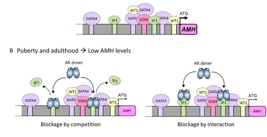
157
The ligand-bound AR can also act indirectly by interacting with other trans-activating factors
158
that are bound to the regulatory regions of their target genes, as is the case for the LH subunits [73]
159
and β [74] genes. This means that AR action is not determined by the presence of ARE sequences.
160
Regardless of the type of interaction between the AR and its target genes, the outcome can be either
positive or negative, meaning that androgens can both stimulate or inhibit the expression of their
162
target genes.
163
164
3.2. Non-classical pathways of androgen action
165
The non-genomic (or non-classical) pathway translates signals into changes in cellular function
166
very rapidly, within second to minutes (Figure 2) [5, 75, 76, 77]. In the Sertoli cell, testosterone
167
stimulation provokes the classic AR to localize near the plasma membrane, where it activates Src
168
tyrosine kinase resulting in phosphorylation of the epidermal growth factor receptor (EGFR).
169
Consequently, the MAP kinase cascade is triggered, including the kinases Raf, MEK and ERK
170
followed by the activation of the p90Rsk kinase, resulting in the phosphorylation of target protein,
171
e.g. the transcription factor cyclic-AMP response element binding-protein (CREB).
172
An alternative pathway, involving a membrane AR, has been described in different cell types
173
[78, 79]. Recently, a member of the ZIP zinc transporter family, ZIP9 has been reported as a membrane
174
AR, unrelated to the classic intracellular AR [80]. There is only one report to date in which the role
175
for ZIP9 is shown in Sertoli cells [81].
176
177
178
Figure 2: Pathways of androgen action in the Sertoli cell. The classical and non-classical pathways
179
of androgen action co-exist in the Sertoli cell. In the cytoplasm the androgen receptor (AR) is bound
180
to heat-shock proteins (HSP). When androgens bind to the AR it causes a conformational change that
181
releases the AR as monomers. In the Sertoli cell, ligand-bound AR monomers can either migrate to
182
the inner side of the cell membrane and interact with Src, thus activating the
non-classical/non-183
genomic pathway of androgen action; or they can translocate to the nucleus and form homodimers
184
that can interact with androgen response elements (ARE) or with other transcription factors (TF), thus
185
activating the classical/genomic pathway of androgen action. Src: Steroid receptor coactivator, EGFR:
186
Epidermal growth factor receptor, MEK: Mitogen-activated protein kinase kinase, ERK: Extracellular
187
signal-regulated kinase, CREB: cAMP response element binding protein. Based on refs. [5, 82, 83, 84,
188
85].
190
4. Androgens and the Sertoli cell
191
As already mentioned, androgen action on Sertoli cells is critical for proper testicular maturation
192
and normal spermatogenesis progression. When the AR is specifically absent from Sertoli cells or it
193
malfunctions, Sertoli cells remain immature, and spermatogenesis is blunted since meiosis does not
194
occur, resulting in infertility. Evidence for these phenotypic characteristics stems from both human
195
[59, 86, 87, 88, 89] and experimental mouse models [8, 90, 91, 92, 93, 94].
196
Sertoli cell maturation in response to androgens involves both upregulation and inhibition of
197
different genes. We will discuss some examples known up to date that show the stimulatory effect of
198
androgens on several BTB tight junction components in Sertoli cells and on meiotic onset in the
199
pubertal testis. We will also expand on the inhibitory effect of androgens on the expression of a key
200
immaturity Sertoli cell marker, AMH.
201
202
4.1. Stimulatory effects of androgens on BTB-related gene expression in Sertoli cells and its role on
203
meiotic onset in the testis
204
The BTB appears at a time when serum gonadotrophins, FSH and LH, are elevated as a result of
205
pubertal reactivation of the HPG axis [95, 96]. While FSH acts directly on the Sertoli cells through its
206
own receptor, LH induces androgen production by the Leydig cells. Androgens act then on Sertoli
207
cells to promote their maturational changes.
208
The BTB divides the seminiferous tubules into two compartments, basal and adluminal, thus
209
creating two distinct microenvironments. The BTB is both a tight [97] and dynamic structure that
210
keeps separate compartments within the seminiferous epithelium while allowing for germ cell transit
211
from basal to adluminal space during spermatogenesis [26, 31, 98]. The mature, fully-formed BTB
212
consists of tight junctions, a testis-specific type of adherent junction known as basal ectoplasmic
213
specializations [22, 99], gap junctions and desmosomes [96, 98, 100, 101, 102] (Figure 3).
214
215
216
Figure 3: The blood-testis barrier. The BTB is formed by intercellular unions between adjacent Sertoli
217
cells. In the presence of androgens, AR-expressing Sertoli cells can mature and express several genes
218
needed for BTB formation, such as Cldn3, Cldn11, Ocln and Tjp1. CLDN3, CLDN11, OCLN and TJP1,
219
together with other proteins and components of the cytoskeleton, such as actin bundles, constitute
tight junctions at the BTB. BTB: Blood-testis barrier, TJP1: Tight junction protein 1. Based on refs. [32,
221
98].
222
223
Tight junctions are formed by claudins, namely claudin-3 (CLDN3) and claudin-11 (CLDN11) in
224
the mouse [103, 104, 105]. Tight junctions interact with the cytoskeleton of Sertoli cells through
225
scaffolding proteins, like Tight junction protein 1 (TJP1, also known as zonula occludens 1 or ZO1)
226
[106] (Figure 3). Cldn3, Cldn11 and Tjp1 are all expressed throughout postnatal development in the
227
mouse testis [107, 108, 109] and their proteins localize to the BTB region from pubertal onset onwards
228
[108, 110, 111]. In mice, the expression of Cldn11 and Tjp1 increases progressively from birth, with a
229
marked increase around day 10 —in coincidence with the upsurge of first meiotic division— and
230
remains elevated throughout adulthood [112].
231
In the gonadotrophin-deficient hypogonadal (hpg) mouse, spermatogenesis is arrested at the
232
prepubertal stage when meiosis has not begun yet, in association with a disorganization of the tight
233
junctions resulting in the lack of a properly formed BTB. This phenotype stems from the lack of
234
maturation of the Sertoli cells in the absence of androgen production due to a disrupted HPG axis
235
[90, 113, 114]. In the tubules of hpg mice there is no CLDN3 expression and CLDN11 is localized to
236
adluminal areas of Sertoli cells. When treated with FSH alone, hpg mice recovered normal CLDN11
237
distribution, but the tight junctions were still unable to function as a proper barrier. On the contrary,
238
treatment with DHT induced normal distribution of CLDN11 and an increase in the expression of
239
both Cldn3 and Cldn11 genes [115].
240
Evidence of androgen-dependency of the BTB for its appearance and maintenance also derives
241
from studies in mice lacking proper AR expression or function. While general defects in BTB
242
formation were initially described in Tfm mice [116], mouse models that either lack AR expression
243
completely (ARKO mice, [92]) or in Sertoli cells only (SCARKO mice, [93, 109]) have provided
244
evidence for many genes potentially involved in BTB formation around pubertal onset and
245
maintenance through puberty and adulthood. Histological and electron microscopy studies showed
246
a clear disruption of the BTB in SCARKO mice [109], and the use of microarrays allowed for the
247
identification of androgen-regulated genes involved in BTB formation [117, 118].
248
The expression of Ocln (Occludin) and Cldn11 is inhibited in the absence of androgen action as
249
seen in SCARKO mice [109, 119, 120, 121], and the same occurs with Tjp1 [122] and Cldn3 [109]. While
250
FSH plays a role in the regulation of Cldn11 expression to a lesser extent than androgens [112], this is
251
not the case for Tjp1, which is strongly inhibited in SCARKO mice but not in FSHRKO mice [122].
252
Changes in gene expression have been shown with a classic RT-qPCR approach [109, 112, 121] and
253
also with RNA-Seq [118].
254
Another example is that of Claudin-13 (Cldn13) and a non-canonical Tight junction protein 2
255
isoform (Tjp2iso3), which have been shown to be downregulated in the SCARKOtm2.1 model [123]. Both
256
Cldn13 and Tjp2iso3 have several putative ARE sequences, mainly with the TGTTCT motif, to which
257
the AR can bind, as seen by ChIP-qPCR. While CLDN13 is part of the Sertoli cell tight junction,
258
TJP2iso3 participates in tricellular junctions. Furthermore, new candidate genes associated with
cell-259
adhesion and cytoskeleton dynamics show altered expression levels in the SCARKO mouse testis,
260
such as Actn3 (actinin-a3), Ank3 (ankyrin 3), Anxa9 (annexin A9) and Scin (scinderin) [109]. However,
261
their involvement in BTB integrity remains yet to be unveiled and much remains to be investigated.
262
Recently it has been shown that dehydroepiandrosterone sulphate (DHEAS) stimulates the
263
expression of Cldn3 and Cldn5 in the mouse Sertoli cell line TM4 through a membrane-bound
G-264
protein-coupled receptor that interacts with Gnα11 and induces phosphorylation of ERK1/2, CREB
265
and ATF1 [124]. This mechanism would mimic the non-classical/non-genomic pathway of androgen
266
action.
267
Coincidentally with the disorganization and delay in BTB formation, there is an incomplete
268
meiosis in the testis of both Tfm and SCARKO mice. The lack of complete meiosis progression in the
absence of the AR specifically in Sertoli cells demonstrates the central role that androgen-signalling
270
through Sertoli cells plays on spermatocyte entry into meiosis [93, 94]. The dynamic nature of the BTB
271
is fundamental for migration of meiotic germ cells from the basal to the adluminal compartment.
272
On the other hand, in a transgenic mouse model with Sertoli cell-specific premature postnatal
273
AR expression [125], Rhox5 levels were elevated. Furthermore, there was a precocious upregulation
274
of tight junction markers Cldn11 and Tjp1 resulting in early BTB and seminiferous tubular lumen
275
formation, associated with premature meiotic onset, shown by increased levels of meiotic markers
276
Dmc1 (DNA meiotic recombinase 1) and Spo11 (SPO11 meiotic protein covalently bound to double
277
strand breaks).
278
The connection between androgen-induced Sertoli cell maturation and germ cell entry into
279
meiosis remains yet to be fully elucidated. A plausible connection could be that the
androgen-280
induced cytoskeletal changes within Sertoli cells might cause changes in the germ cell cytoskeleton
281
itself, thus promoting transition into meiosis and germ cell movement through the BTB. A crucial
282
role for Sertoli cells in the establishment of an immunoprivileged microenvironment at the time of
283
tight junction formation has also been suggested [108, 126]. Whether any of these are the case or not,
284
it is clear that androgen action through the AR on Sertoli cells is pivotal to initiation of meiosis in the
285
pubertal testis, since when the AR is absent there is no complete meiotic progression.
286
287
4.2. Inhibitory effect of androgens on AMH gene expression in Sertoli cells
288
Inhibitory effects of androgens on gene expression have not been as extensively studied as the
289
stimulatory ones, with few examples available to date. Genes coding for WNT5A and podoplanin are
290
down-regulated through unknown molecular mechanisms [54]. Amongst androgen-inhibited genes
291
through AR binding to ARE are the tumour suppressor genes Maspin [127, 128] and Ccnd1 [129].
292
Representing inhibited genes without functional ARE on their promoter regions that rely on AR
293
interaction with trans-activating factors are Ngfr (Nerve growth factor receptor, formerly
294
Neurotrophin receptor p75) [130] and the genes encoding the - [73, 131] and β- [74, 132] subunits of
295
LH.
296
As previously mentioned, the decrease in AMH expression at pubertal onset is indicative of
297
Sertoli cell maturation. Despite the fact that AMH downregulation by androgens has been established
298
a long time ago in all animals studied, including human [50, 53, 86, 133], mouse [8, 45, 46, 134], ram
299
[135, 136], pig [137, 138], stallion [139], bovine [140, 141] and tammar wallaby [142], it has not been
300
until recently that the underlying mechanism of androgen action was described [30].
301
Sertoli cells begin to express AMH early during gonadal development, at 7 weeks in the human
302
embryonic gonad [34] and at 12.5 days post-coitum in the mouse male gonad [143]. The expression of
303
the AMH gene relies on the presence and action of several transcription factors that bind to their
304
promoter, namely SOX9, SF1, WT1, GATA4, AP2 and NFB [134]. AMH transcription is dependent
305
mainly upon SOX9 binding to the promoter, but it also relies on SF1 action. SF1 can bind directly to
306
the AMH promoter and also interact with other transactivating factors, such as SOX8, to induce AMH
307
expression (Figure 4). When SF1 is absent, AMH expression drops dramatically [144]. When
308
interaction between SF1 and SOX8, SF1 and WT1 and/or SF1, SOX9 and GATA4 is disrupted by
309
interaction of DAX1 with SF1, AMH expression is inhibited in Sertoli cells [145, 146]. This inhibitory
310
capacity of DAX1 on AMH, however, has no relation to androgen action, since it has been described
311
at a time when Sertoli cells do not express the AR and are, therefore, insensitive to this type of
312
hormones. At pubertal onset, the androgens testosterone and dihydrotestosterone have a direct
313
negative effect on AMH promoter activity in Sertoli cells. This inhibitory effect involves the proximal
314
region of the AMH promoter and requires the presence of the AR together with at least one intact
315
binding site for SF1 in the promoter of the AMH gene [30]. These findings were shown using a mouse
316
prepubertal Sertoli cell line [147] and suggest that the inhibitory effect of androgens on AMH
317
expression could be due to direct interaction between the AR and SF1 or by the AR blocking SF1
binding sites, thus preventing SF1 from exerting its stimulating action on the AMH promoter (Figure
319
4) [30]. A similar mechanism of action posing an interaction between the AR and SF1 has been
320
described for the androgen-dependent inhibition of the LH β subunit gene [132].
321
Inhibitory action of androgens on gene expression at puberty in the male is not as common as
322
stimulatory effects. AMH is an immaturity Sertoli cell marker that is regulated by androgens in a
323
negative manner, thus presenting itself as a clear example for androgen inhibition.
324
325
326
Figure 4: Underlying mechanism for AMH inhibition by androgens in the pubertal Sertoli cell. (A)
327
Sertoli cells produce high levels of AMH during the prenatal and prepubertal period. This high
328
expression is a direct consequence of SF1, GATA4 and WT1 interaction with their own binding sites
329
on the AMH promoter and also of protein-protein interactions with each other. (B) At pubertal onset,
330
Sertoli cells express the androgen receptor and can respond to androgen action. Androgens inhibit
331
AMH promoter activity through the androgen receptor. Inhibition could be due either to a direct
332
interaction between the ligand-bound AR and the SF1 sites present on the AMH promoter (blockage
333
by competition), or due to a protein-protein interaction between the ligand-bound AR and
promoter-334
bound SF1 (blockage by interaction). In either scenario, the ligand-bound AR prevents SF1 from
335
exerting its stimulatory effect on AMH promoter activity, thus resulting in a decrease in AMH
336
expression. Based on ref. [30].
337
338
5. Concluding remarks
339
Sertoli cells constitute the physical and physiological foundation of the seminiferous epithelium.
340
They are the link between the HPG axis and germ cells and, therefore, sperm production. To ensure
341
their many roles in the adult testis, Sertoli cells must mature in a timely manner and they do so by
342
preparing themselves to respond to androgen action at the right time. Androgens are responsible for
343
the occurrence of several pubertal development-related events in the testis, most of which are known
344
to be dependent on the stimulatory role of androgens.
345
Immature Sertoli cells are impervious to androgen action because they lack AR expression. Once
346
the AR becomes present in Sertoli cells and androgen levels increase at pubertal onset, a consortium
347
of genes —like tight junction-associated genes involved in the formation of the BTB— is upregulated,
348
while others —like AMH— become repressed, depicting together the androgen-dependent process
of Sertoli cell pubertal maturation. As a consequence of androgen action, Sertoli cell maturation sets
350
a favourable environment for germ cell entry to meiosis and full progression of spermatogenesis.
351
352
Author Contributions: Both authors wrote the review and approved the final version.
353
Funding: This review received no external funding.
354
Conflicts of Interest: The authors declare no conflict of interest.
References
356
1 Wang, R.S.; Yeh, S.; Tzeng, C.R.; Chang, C. Androgen receptor roles in spermatogenesis and fertility:
357
lessons from testicular cell-specific androgen receptor knockout mice. Endocr. Rev. 2009, 30, 119-132.
358
2 Ruwanpura, S.M.; McLachlan, R.I.; Meachem, S.J. Hormonal regulation of male germ cell development. J.
359
Endocrinol. 2010, 205, 117-131.
360
3 Verhoeven, G.; Willems, A.; Denolet, E.; Swinnen, J.V.; De Gendt, K. Androgens and spermatogenesis:
361
lessons from transgenic mouse models. Philos. Trans. R. Soc. Lond. B Biol. Sci. 2010, 365, 1537-1556.
362
4 O'Donnell, L.; Nicholls, P.K.; O'Bryan, M.K.; McLachlan, R.I.; Stanton, P.G. Spermiation: The process of
363
sperm release. Spermatogenesis 2011, 1, 14-35.
364
5 Smith, L.B.; Walker, W.H. The regulation of spermatogenesis by androgens. Semin. Cell Dev. Biol. 2014, 30,
365
2-13.
366
6 Zhou, Q.; Shima, J.E.; Nie, R.; Friel, P.J.; Griswold, M.D. Androgen-regulated transcripts in the neonatal
367
mouse testis as determined through microarray analysis. Biol. Reprod. 2005, 72, 1010-1019.
368
7 Orth, J.M. The role of follicle-stimulating hormone in controlling Sertoli cell proliferation in testes of fetal
369
rats. Endocrinology 1984, 115, 1248-1255.
370
8 Al-Attar, L.; Noël, K.; Dutertre, M.; Belville, C.; Forest, M.G.; Burgoyne, P.S.; Josso, N.; Rey, R. Hormonal
371
and cellular regulation of Sertoli cell anti-Müllerian hormone production in the postnatal mouse. J. Clin. Invest.
372
1997, 100, 1335-1343.
373
9 Migrenne, S.; Moreau, E.; Pakarinen, P.; Dierich, A.; Merlet, J.; Habert, R.; Racine, C. Mouse testis
374
development and function are differently regulated by follicle-stimulating hormone receptors signaling during
375
fetal and prepubertal life. PLoS One 2012, 7, e53257.
376
10 Lambert, A.S.; Bougnères, P. Growth and descent of the testes in infants with hypogonadotropic
377
hypogonadism receiving subcutaneous gonadotropin infusion. Int. J. Pediatr. Endocrinol. 2016, 2016, 13.
378
11 Kumar, T.R. Fshb Knockout Mouse Model, Two Decades Later and Into the Future. Endocrinology 2018, 159,
379
1941-1949.
380
12 Urrutia, M.; Grinspon, R.P.; Rey, R.A. Comparing the role of anti-Mullerian hormone as a marker of FSH
381
action in male and female fertility. Expert Rev. Endocrinol. Metab. 2019, 14, 203-214.
382
13 Pitetti, J.L.; Calvel, P.; Romero, Y.; Conne, B.; Truong, V.; Papaioannou, M.D.; Schaad, O.; Docquier, M.;
383
Herrera, P.L.; Wilhelm, D.; Nef, S. Insulin and IGF1 Receptors Are Essential for XX and XY Gonadal
384
Differentiation and Adrenal Development in Mice. PLoS genetics 2013, 9, e1003160.
385
14 Meroni, S.B.; Galardo, M.N.; Rindone, G.; Gorga, A.; Riera, M.F.; Cigorraga, S.B. Molecular Mechanisms
386
and Signaling Pathways Involved in Sertoli Cell Proliferation. Front. Endocrinol. (Lausanne) 2019, 10, 224.
387
15 Neirijnck, Y.; Kuhne, F.; Mayere, C.; Pavlova, E.; Sararols, P.; Foti, M.; Atanassova, N.; Nef, S. Tumor
388
Suppressor PTEN Regulates Negatively Sertoli Cell Proliferation, Testis Size, and Sperm Production In Vivo.
389
Endocrinology 2019, 160, 387-398.
390
16 Orth, J.M. Proliferation of Sertoli cells in fetal and postnatal rats: a quantitative autoradiographic study.
391
Anat. Rec. 1982, 203, 485-492.
17 Russell, L.D.; Ren, H.P.; Sinha Hikim, I.; Schulze, W.; Sinha Hikim, A.P. A comparative study in twelve
393
mammalian species of volume densities, volumes, and numerical densities of selected testis components,
394
emphasizing those related to the Sertoli cell. Am. J. Anat. 1990, 188, 21-30.
395
18 Cooke, P.S.; Hess, R.A.; Porcelli, J.; Meisami, E. Increased sperm production in adult rats after transient
396
neonatal hypothyroidism. Endocrinology 1991, 129, 244-248.
397
19 Hess, R.A.; Cooke, P.S.; Bunick, D.; Kirby, J.D. Adult testicular enlargement induced by neonatal
398
hypothyroidism is accompanied by increased Sertoli and germ cell numbers. Endocrinology 1993, 132, 2607-2613.
399
20 Iczkowski, K.A.; Sun, E.L.; Gondos, B. Morphometric study of the prepubertal rabbit testis: germ cell
400
numbers and seminiferous tubule dimensions. Am. J. Anat. 1991, 190, 266-272.
401
21 Rey, R.A.; Campo, S.M.; Bedecarrás, P.; Nagle, C.A.; Chemes, H.E. Is infancy a quiescent period of testicular
402
development? Histological, morphometric, and functional study of the seminiferous tubules of the cebus
403
monkey from birth to the end of puberty. J. Clin. Endocrinol. Metab. 1993, 76, 1325-1331.
404
22 Russell, L. Movement of spermatocytes from the basal to the adluminal compartment of the rat testis. Am.
405
J. Anat. 1977, 148, 313-328.
406
23 Russell, L.D.; Bartke, A.; Goh, J.C. Postnatal development of the Sertoli cell barrier, tubular lumen, and
407
cytoskeleton of Sertoli and myoid cells in the rat, and their relationship to tubular fluid secretion and flow. Am.
408
J. Anat. 1989, 184, 179-189.
409
24 Vogl, A.W.; Vaid, K.S.; Guttman, J.A. The Sertoli cell cytoskeleton. Adv. Exp. Med. Biol. 2008, 636, 186-211.
410
25 Li, N.; Tang, E.I.; Cheng, C.Y. Regulation of blood-testis barrier by actin binding proteins and protein
411
kinases. Reproduction 2016, 151, R29-41.
412
26 Wen, Q.; Tang, E.I.; Li, N.; Mruk, D.D.; Lee, W.M.; Silvestrini, B.; Cheng, C.Y. Regulation of Blood-Testis
413
Barrier (BTB) Dynamics, Role of Actin-, and Microtubule-Based Cytoskeletons. Methods Mol. Biol. 2018, 1748,
414
229-243.
415
27 Dunleavy, J.E.M.; O'Bryan, M.; Stanton, P.G.; O'Donnell, L. The Cytoskeleton in Spermatogenesis.
416
Reproduction 2019, 157, R53-R72.
417
28 Chemes, H.E.; Rey, R.A.; Nistal, M.; Regadera, J.; Musse, M.; Gonzalez-Peramato, P.; Serrano, A.
418
Physiological androgen insensitivity of the fetal, neonatal, and early infantile testis is explained by the ontogeny
419
of the androgen receptor expression in Sertoli cells. J. Clin. Endocrinol. Metab. 2008, 93, 4408-4412.
420
29 Rey, R.A.; Musse, M.; Venara, M.; Chemes, H.E. Ontogeny of the androgen receptor expression in the fetal
421
and postnatal testis: its relevance on Sertoli cell maturation and the onset of adult spermatogenesis. Microsc. Res.
422
Tech. 2009, 72, 787-795.
423
30 Edelsztein, N.Y.; Racine, C.; di Clemente, N.; Schteingart, H.F.; Rey, R.A. Androgens downregulate
anti-424
Mullerian hormone promoter activity in the Sertoli cell through the androgen receptor and intact SF1 sites. Biol.
425
Reprod. 2018, 99, 1303-1312.
426
31 Smith, B.E.; Braun, R.E. Germ cell migration across Sertoli cell tight junctions. Science 2012, 338, 798-802.
427
32 Mruk, D.D.; Cheng, C.Y. The Mammalian Blood-Testis Barrier: Its Biology and Regulation. Endocr. Rev.
428
2015, 36, 564-591.
33 Lukas-Croisier, C.; Lasala, C.; Nicaud, J.; Bedecarrás, P.; Kumar, T.R.; Dutertre, M.; Matzuk, M.M.; Picard,
430
J.Y.; Josso, N.; Rey, R. Follicle-stimulating hormone increases testicular Anti-Müllerian hormone (AMH)
431
production through sertoli cell proliferation and a nonclassical cyclic adenosine 5'-monophosphate-mediated
432
activation of the AMH gene. Mol. Endocrinol. 2003, 17, 550-561.
433
34 Josso, N.; Lamarre, I.; Picard, J.Y.; Berta, P.; Davies, N.; Morichon, N.; Peschanski, M.; Jeny, R.
Anti-434
Müllerian hormone in early human development. Early Hum. Dev. 1993, 33, 91-99.
435
35 Lee, M.M.; Donahoe, P.K.; Silverman, B.L.; Hasegawa, T.; Hasegawa, Y.; Gustafson, M.L.; Chang, Y.C.;
436
MacLaughlin, D.T. Measurements of serum Müllerian inhibiting substance in the evaluation of children with
437
nonpalpable gonads. N. Engl. J. Med. 1997, 336, 1480-1486.
438
36 Misra, M.; MacLaughlin, D.T.; Donahoe, P.K.; Lee, M.M. Measurement of Mullerian inhibiting substance
439
facilitates management of boys with microphallus and cryptorchidism. J. Clin. Endocrinol. Metab. 2002, 87,
3598-440
3602.
441
37 Lee, M.M.; Misra, M.; Donahoe, P.K.; MacLaughlin, D.T. MIS/AMH in the assessment of cryptorchidism
442
and intersex conditions. Mol. Cell. Endocrinol. 2003, 211, 91-98.
443
38 Lindhardt Johansen, M.; Hagen, C.P.; Johannsen, T.H.; Main, K.M.; Picard, J.Y.; Jorgensen, A.; Rajpert-De
444
Meyts, E.; Juul, A. Anti-mullerian hormone and its clinical use in pediatrics with special emphasis on disorders
445
of sex development. Int. J. Endocrinol. 2013, 2013, 198698.
446
39 Grinspon, R.P.; Loreti, N.; Braslavsky, D.; Valeri, C.; Schteingart, H.; Ballerini, M.G.; Bedecarrás, P.; Ambao,
447
V.; Gottlieb, S.; Ropelato, M.G. et al. Spreading the Clinical Window for Diagnosing Fetal-Onset Hypogonadism
448
in Boys. Front. Endocrinol. (Lausanne) 2014, 5, 51; 51-14.
449
40 Edelsztein, N.Y.; Grinspon, R.P.; Schteingart, H.F.; Rey, R.A. Anti-Müllerian hormone as a marker of steroid
450
and gonadotropin action in the testis of children and adolescents with disorders of the gonadal axis. Int. J. Pediatr.
451
Endocrinol. 2016, 2016, 20.
452
41 Condorelli, R.A.; Cannarella, R.; Calogero, A.E.; La Vignera, S. Evaluation of testicular function in
453
prepubertal children. Endocrine 2018, 62, 274-280.
454
42 Freire, A.V.; Grinspon, R.P.; Rey, R.A. Importance of Serum Testicular Protein Hormone Measurement in
455
the Assessment of Disorders of Sex Development. Sex. Dev. 2018, 12, 30-40.
456
43 Grinspon, R.P.; Gottlieb, S.; Bedecarras, P.; Rey, R.A. Anti-Müllerian Hormone and Testicular Function in
457
Prepubertal Boys With Cryptorchidism. Front. Endocrinol. (Lausanne) 2018, 9, 182; 181-114.
458
44 Grinspon, R.P.; Urrutia, M.; Rey, R.A. Male Central Hypogonadism in Paediatrics – the Relevance of
459
Follicle-stimulating Hormone and Sertoli Cell Markers. European Endocrinology 2018, 14, 67-71.
460
45 Grinspon, R.P.; Andreone, L.; Bedecarrás, P.; Ropelato, M.G.; Rey, R.A.; Campo, S.M.; Bergadá, I. Male
461
Central Precocious Puberty: Serum Profile of Anti-Mullerian Hormone and Inhibin B before, during, and after
462
Treatment with GnRH Analogue. Int. J. Endocrinol. 2013, 2013, 823064.
463
46 Milazzo, J.P.; Bironneau, A.; Vannier, J.P.; Liard-Zmuda, A.; Mace, B.; Nathalie, R. Precocious initiation of
464
spermatogenesis in a 19-month-old boy with Hurler syndrome. Basic Clin Androl 2014, 24, 8.
465
47 Svechnikov, K.; Landreh, L.; Weisser, J.; Izzo, G.; Colon, E.; Svechnikova, I.; Söder, O. Origin, development
466
and regulation of human Leydig cells. Horm. Res. Paediatr. 2010, 73, 93-101.
48 Petersen, P.M.; Seieroe, K.; Pakkenberg, B. The total number of Leydig and Sertoli cells in the testes of men
468
across various age groups - a stereological study. J. Anat. 2015, 226, 175-179.
469
49 Wen, Q.; Cheng, C.Y.; Liu, Y.X. Development, function and fate of fetal Leydig cells. Semin. Cell Dev. Biol.
470
2016, 59, 89-98.
471
50 Rey, R.; Lordereau-Richard, I.; Carel, J.C.; Barbet, P.; Cate, R.L.; Roger, M.; Chaussain, J.L.; Josso, N.
Anti-472
müllerian hormone and testosterone serum levels are inversely related during normal and precocious pubertal
473
development. J. Clin. Endocrinol. Metab. 1993, 77, 1220-1226.
474
51 Grinspon, R.P.; Rey, R.A. Anti-mullerian hormone and Sertoli cell function in paediatric male
475
hypogonadism. Horm. Res. Paediatr. 2010, 73, 81-92.
476
52 Grinspon, R.; Chemes, H.; Rey, R.A. Decline in serum antimullerian hormone due to androgen action in
477
early puberty in males. Fertil. Steril. 2012, 98, e23.
478
53 Hero, M.; Tommiska, J.; Vaaralahti, K.; Laitinen, E.M.; Sipila, I.; Puhakka, L.; Dunkel, L.; Raivio, T.
479
Circulating antimullerian hormone levels in boys decline during early puberty and correlate with inhibin B.
480
Fertil. Steril. 2012, 97, 1242-1247.
481
54 Tanaka, T.; Kanatsu-Shinohara, M.; Lei, Z.; Rao, C.V.; Shinohara, T. The Luteinizing Hormone-Testosterone
482
Pathway Regulates Mouse Spermatogonial Stem Cell Self-Renewal by Suppressing WNT5A Expression in
483
Sertoli Cells. Stem Cell Reports 2016, 7, 279-291.
484
55 Chang, C.S.; Kokontis, J.; Liao, S.T. Molecular cloning of human and rat complementary DNA encoding
485
androgen receptors. Science 1988, 240, 324-326.
486
56 Lubahn, D.B.; Joseph, D.R.; Sullivan, P.M.; Willard, H.F.; French, F.S.; Wilson, E.M. Cloning of human
487
androgen receptor complementary DNA and localization to the X chromosome. Science 1988, 240, 327-330.
488
57 Li, J.; Al-Azzawi, F. Mechanism of androgen receptor action. Maturitas 2009, 63, 142-148.
489
58 Tan, M.H.; Li, J.; Xu, H.E.; Melcher, K.; Yong, E.L. Androgen receptor: structure, role in prostate cancer and
490
drug discovery. Acta Pharmacol. Sin. 2015, 36, 3-23.
491
59 Quigley, C.A.; De Bellis, A.; Marschke, K.B.; el-Awady, M.K.; Wilson, E.M.; French, F.S. Androgen receptor
492
defects: historical, clinical, and molecular perspectives [published erratum appears in Endocr Rev 1995
493
Aug;16(4):546]. Endocr. Rev. 1995, 16, 271-321.
494
60 Khorasanizadeh, S.; Rastinejad, F. Nuclear-receptor interactions on DNA-response elements. Trends
495
Biochem. Sci. 2001, 26, 384-390.
496
61 Denayer, S.; Helsen, C.; Thorrez, L.; Haelens, A.; Claessens, F. The rules of DNA recognition by the
497
androgen receptor. Mol. Endocrinol. 2010, 24, 898-913.
498
62 Barbulescu, K.; Geserick, C.; Schuttke, I.; Schleuning, W.D.; Haendler, B. New androgen response elements
499
in the murine pem promoter mediate selective transactivation. Mol. Endocrinol. 2001, 15, 1803-1816.
500
63 Burgos-Trinidad, M.; Youngblood, G.L.; Maroto, M.R.; Scheller, A.; Robins, D.M.; Payne, A.H. Repression
501
of cAMP-induced expression of the mouse P450 17 alpha-hydroxylase/C17-20 lyase gene (Cyp17) by androgens.
502
Mol. Endocrinol. 1997, 11, 87-96.
503
64 Schauwaers, K.; De Gendt, K.; Saunders, P.T.K.; Atanassova, N.; Haelens, A.; Callewaert, L.; Moehren, U.;
504
Swinnen, J.V.; Verhoeven, G.; Verrijdt, G.; Claessens, F. Loss of androgen receptor binding to selective androgen
response elements causes a reproductive phenotype in a knockin mouse model. Proc. Natl. Acad. Sci. U. S. A.
506
2007, 104, 4961-4966.
507
65 De Gendt, K.; Denolet, E.; Willems, A.; Daniels, V.W.; Clinckemalie, L.; Denayer, S.; Wilkinson, M.F.;
508
Claessens, F.; Swinnen, J.V.; Verhoeven, G. Expression of Tubb3, a beta-tubulin isotype, is regulated by
509
androgens in mouse and rat Sertoli cells. Biol. Reprod. 2011, 85, 934-945.
510
66 Shang, Y.; Myers, M.; Brown, M. Formation of the androgen receptor transcription complex. Mol. Cell 2002,
511
9, 601-610.
512
67 Gautam, M.; Bhattacharya, I.; Rai, U.; Majumdar, S.S. Hormone induced differential transcriptome analysis
513
of Sertoli cells during postnatal maturation of rat testes. PLoS One 2018, 13, e0191201.
514
68 Fietz, D.; Markmann, M.; Lang, D.; Konrad, L.; Geyer, J.; Kliesch, S.; Chakraborty, T.; Hossain, H.;
515
Bergmann, M. Transfection of Sertoli cells with androgen receptor alters gene expression without androgen
516
stimulation. BMC Mol. Biol. 2015, 16, 23.
517
69 Sadate-Ngatchou, P.I.; Pouchnik, D.J.; Griswold, M.D. Identification of testosterone-regulated genes in
518
testes of hypogonadal mice using oligonucleotide microarray. Mol. Endocrinol. 2004, 18, 422-433.
519
70 Shanker, S.; Hu, Z.; Wilkinson, M.F. Epigenetic regulation and downstream targets of the Rhox5 homeobox
520
gene. Int. J. Androl. 2008, 31, 462-470.
521
71 Lindsey, J.S.; Wilkinson, M.F. Pem: a testosterone- and LH-regulated homeobox gene expressed in mouse
522
Sertoli cells and epididymis. Dev. Biol. 1996, 179, 471-484.
523
72 Lee, S.E.; Lee, S.Y.; Lee, K.A. Rhox in mammalian reproduction and development. Clin Exp Reprod Med
524
2013, 40, 107-114.
525
73 Heckert, L.L.; Wilson, E.M.; Nilson, J.H. Transcriptional Repression of the {alpha}-Subunit Gene by
526
Androgen Receptor Occurs Independently of DNA Binding but Requires the DNA-Binding and Ligand-Binding
527
Domains of the Receptor. Mol. Endocrinol. 1997, 11, 1497-1506.
528
74 Keri, R.A.; Wolfe, M.W.; Saunders, T.L.; Anderson, I.; Kendall, S.K.; Wagner, T.; Yeung, J.; Gorski, J.; Nett,
529
T.M.; Camper, S.A.; et al. The proximal promoter of the bovine luteinizing hormone beta-subunit gene confers
530
gonadotrope-specific expression and regulation by gonadotropin-releasing hormone, testosterone, and 17
beta-531
estradiol in transgenic mice. Mol. Endocrinol. 1994, 8, 1807-1816.
532
75 Fix, C.; Jordan, C.; Cano, P.; Walker, W.H. Testosterone activates mitogen-activated protein kinase and the
533
cAMP response element binding protein transcription factor in Sertoli cells. Proc. Natl. Acad. Sci. U. S. A. 2004,
534
101, 10919-10924.
535
76 Shupe, J.; Cheng, J.; Puri, P.; Kostereva, N.; Walker, W.H. Regulation of Sertoli-germ cell adhesion and
536
sperm release by FSH and nonclassical testosterone signaling. Mol. Endocrinol. 2011, 25, 238-252.
537
77 Toocheck, C.; Clister, T.; Shupe, J.; Crum, C.; Ravindranathan, P.; Lee, T.K.; Ahn, J.M.; Raj, G.V.; Sukhwani,
538
M.; Orwig, K.E.; Walker, W.H. Mouse Spermatogenesis Requires Classical and Nonclassical Testosterone
539
Signaling. Biol. Reprod. 2016, 94, 11.
540
78 Konoplya, E.F.; Popoff, E.H. Identification of the classical androgen receptor in male rat liver and prostate
541
cell plasma membranes. Int. J. Biochem. 1992, 24, 1979-1983.
79 Benten, W.P.; Lieberherr, M.; Giese, G.; Wrehlke, C.; Stamm, O.; Sekeris, C.E.; Mossmann, H.; Wunderlich,
543
F. Functional testosterone receptors in plasma membranes of T cells. FASEB J. 1999, 13, 123-133.
544
80 Berg, A.H.; Rice, C.D.; Rahman, M.S.; Dong, J.; Thomas, P. Identification and characterization of membrane
545
androgen receptors in the ZIP9 zinc transporter subfamily: I. Discovery in female atlantic croaker and evidence
546
ZIP9 mediates testosterone-induced apoptosis of ovarian follicle cells. Endocrinology 2014, 155, 4237-4249.
547
81 Bulldan, A.; Dietze, R.; Shihan, M.; Scheiner-Bobis, G. Non-classical testosterone signaling mediated
548
through ZIP9 stimulates claudin expression and tight junction formation in Sertoli cells. Cell. Signal. 2016, 28,
549
1075-1085.
550
82 Heinlein, C.A.; Chang, C. The roles of androgen receptors and androgen-binding proteins in nongenomic
551
androgen actions. Mol. Endocrinol. 2002, 16, 2181-2187.
552
83 Foradori, C.D.; Weiser, M.J.; Handa, R.J. Non-genomic actions of androgens. Front. Neuroendocrinol. 2008,
553
29, 169-181.
554
84 Michels, G.; Hoppe, U.C. Rapid actions of androgens. Front. Neuroendocrinol. 2008, 29, 182-198.
555
85 McHenry, J.; Carrier, N.; Hull, E.; Kabbaj, M. Sex differences in anxiety and depression: role of testosterone.
556
Front. Neuroendocrinol. 2014, 35, 42-57.
557
86 Rey, R.; Mebarki, F.; Forest, M.G.; Mowszowicz, I.; Cate, R.L.; Morel, Y.; Chaussain, J.L.; Josso, N.
Anti-558
müllerian hormone in children with androgen insensitivity. J. Clin. Endocrinol. Metab. 1994, 79, 960-964.
559
87 Rey, R.A.; Belville, C.; Nihoul-Fékété, C.; Michel-Calemard, L.; Forest, M.G.; Lahlou, N.; Jaubert, F.;
560
Mowszowicz, I.; David, M.; Saka, N. et al. Evaluation of gonadal function in 107 intersex patients by means of
561
serum antimüllerian hormone measurement. J. Clin. Endocrinol. Metab. 1999, 84, 627-631.
562
88 Hellmann, P.; Christiansen, P.; Johannsen, T.H.; Main, K.M.; Duno, M.; Juul, A. Male patients with partial
563
androgen insensitivity syndrome: a longitudinal follow-up of growth, reproductive hormones and the
564
development of gynaecomastia. Arch. Dis. Child. 2012, 97, 403-409.
565
89 Hughes, I.A.; Davies, J.D.; Bunch, T.I.; Pasterski, V.; Mastroyannopoulou, K.; MacDougall, J. Androgen
566
insensitivity syndrome. Lancet 2012, 380, 1419-1428.
567
90 Singh, J.; O'Neill, C.; Handelsman, D.J. Induction of spermatogenesis by androgens in
gonadotropin-568
deficient (hpg) mice. Endocrinology 1995, 136, 5311-5321.
569
91 Meachem, S.J.; Wreford, N.G.; Robertson, D.M.; McLachlan, R.I. Androgen action on the restoration of
570
spermatogenesis in adult rats: effects of human chorionic gonadotrophin, testosterone and flutamide
571
administration on germ cell number. Int. J. Androl. 1997, 20, 70-79.
572
92 Yeh, S.; Tsai, M.Y.; Xu, Q.; Mu, X.M.; Lardy, H.; Huang, K.E.; Lin, H.; Yeh, S.D.; Altuwaijri, S.; Zhou, X. et
573
al. Generation and characterization of androgen receptor knockout (ARKO) mice: an in vivo model for the study
574
of androgen functions in selective tissues. Proc Natl Acad Sci USA 2002, 99, 13498-13503.
575
93 Chang, C.; Chen, Y.T.; Yeh, S.D.; Xu, Q.; Wang, R.S.; Guillou, F.; Lardy, H.; Yeh, S. Infertility with defective
576
spermatogenesis and hypotestosteronemia in male mice lacking the androgen receptor in Sertoli cells. Proc Natl
577
Acad Sci USA 2004, 101, 6876-6881.
94 De Gendt, K.; Swinnen, J.V.; Saunders, P.T.K.; Schoonjans, L.; Dewerchin, M.; Devos, A.; Tan, K.;
579
Atanassova, N.; Claessens, F.; Lecureuil, C. et al. A Sertoli cell-selective knockout of the androgen receptor causes
580
spermatogenic arrest in meiosis. Proc. Natl. Acad. Sci. U. S. A. 2004, 101, 1327-1332.
581
95 Vitale, R.; Fawcett, D.W.; Dym, M. The normal development of the blood-testis barrier and the effects of
582
clomiphene and estrogen treatment. Anat. Rec. 1973, 176, 331-344.
583
96 Russell, L.D.; Peterson, R.N. Sertoli cell junctions: morphological and functional correlates. Int. Rev. Cytol.
584
1985, 94, 177-211.
585
97 Dym, M.; Fawcett, D.W. The blood-testis barrier in the rat and the physiological compartmentation of the
586
seminiferous epithelium. Biol. Reprod. 1970, 3, 308-326.
587
98 Cheng, C.Y.; Mruk, D.D. The Blood-Testis Barrier and Its Implications for Male Contraception. Pharmacol.
588
Rev. 2012, 64, 16-64.
589
99 Flickinger, C.; Fawcett, D.W. The junctional specializations of Sertoli cells in the seminiferous epithelium.
590
Anat. Rec. 1967, 158, 207-221.
591
100 Gilula, N.B.; Fawcett, D.W.; Aoki, A. The Sertoli cell occluding junctions and gap junctions in mature and
592
developing mammalian testis. Dev. Biol. 1976, 50, 142-168.
593
101 Pelletier, G.; Dupont, E.; Simard, J.; Luu-The, V.; Belanger, A.; Labrie, F. Ontogeny and subcellular
594
localization of 3 beta-hydroxysteroid dehydrogenase (3 beta-HSD) in the human and rat adrenal, ovary and
595
testis. J Steroid Biochem.Mol Biol 1992, 43, 451-467.
596
102 Pelletier, R.M. The blood-testis barrier: the junctional permeability, the proteins and the lipids.
597
Prog.Histochem.Cytochem. 2011, 46, 49-127.
598
103 Gow, A.; Southwood, C.M.; Li, J.S.; Pariali, M.; Riordan, G.P.; Brodie, S.E.; Danias, J.; Bronstein, J.M.;
599
Kachar, B.; Lazzarini, R.A. CNS myelin and sertoli cell tight junction strands are absent in Osp/claudin-11 null
600
mice. Cell 1999, 99, 649-659.
601
104 Chakraborty, P.; Buaas, F.W.; Sharma, M.; Snyder, E.; de Rooij, D.G.; Braun, R.E. LIN28A marks the
602
spermatogonial progenitor population and regulates its cyclic expansion. Stem Cells 2014, 32, 860-873.
603
105 Chihara, M.; Ikebuchi, R.; Otsuka, S.; Ichii, O.; Hashimoto, Y.; Suzuki, A.; Saga, Y.; Kon, Y. Mice
stage-604
specific claudin 3 expression regulates progression of meiosis in early stage spermatocytes. Biol. Reprod. 2013, 89,
605
3.
606
106 Moroi, S.; Saitou, M.; Fujimoto, K.; Sakakibara, A.; Furuse, M.; Yoshida, O.; Tsukita, S. Occludin is
607
concentrated at tight junctions of mouse/rat but not human/guinea pig Sertoli cells in testes. Am. J. Physiol. 1998,
608
274, C1708-1717.
609
107 Hellani, A.; Ji, J.; Mauduit, C.; Deschildre, C.; Tabone, E.; Benahmed, M. Developmental and hormonal
610
regulation of the expression of oligodendrocyte-specific protein/claudin 11 in mouse testis. Endocrinology 2000,
611
141, 3012-3019.
612
108 Meng, J.; Holdcraft, R.W.; Shima, J.E.; Griswold, M.D.; Braun, R.E. Androgens regulate the permeability of
613
the blood-testis barrier. Proc. Natl. Acad. Sci. U. S. A. 2005, 102, 16696-16700.