Behavior of cells in artificially made cell aggregates and
tissue fragments after grafting to developing hind limb
buds in Xenopus laevis
NOBUTAKA KOIBUCHI* and SHIN TOCHINAI
Division of Biological Sciences, Graduate School of Science, Hokkaido University, Sapporo, Japan
ABSTRACT During vertebrate limb development, the limb bud grows along the proximo-distal (P-D) direction, with the cells changing their adhesiveness. To know whether the position-related differences in cell adhesiveness are actually utilized by morphogenesis to constitute limb struc-tures, we grafted cell aggregates made of dissociated cells derived from different positions and stages of developing hind limb buds into developing hind limb buds and observed the behavior of the cells. Cell aggregates made of dissociated mesenchymal cells from two different origins were implanted in different positions and stages of limb buds or grafted on limb stumps made by cutting. The two grafted cell populations in the aggregate always sorted out from each other, but their patterning of sorting-out was quite different according to the transplanted regions. In summary, cells in the aggregate that have closer positional identity to the transplanted site were always situated at the boundary between host and donor cells. The pattern of sorting-out seemed to be determined by the relative adhesiveness of surrounding cells to the constituent cells of the aggregates. We also transplanted fragments dissected out from different regions along the P-D axis into st. 50 limb buds. The descendants of grafted cells moved distally to the region corresponding to their positional identity and participated in the formation of more distal structures from that point. These results suggest that the difference in cell adhesiveness may probably play a role in arranging cells along the P-D axis of a developing limb bud.
KEY WORDS:
Xenopus, limb development, cell adhesion, cell sorting out
0214-6282/99/$10.00
© UBC Press Printed in Spain www.lg.ehu.es/ijdb
*Address for reprints: Division of Biological Sciences, Graduate School of Science, Hokkaido University, Sapporo 060-0810, Japan. FAX: (81)-11-757-5994. e-mail: nkoib@bio.sci.hokudai.ac.jp
Introduction
Townes and Holtfreter (1955) demonstrated that the dissoci-ated cells of amphibian embryonic organs can reaggregate and sort out to reconstitute semblances of the original structure. Each cell type sorted out to segregate spatially, and their final positions often reflected their embryonic positions. Knowledge of the mecha-nism governing cell sorting-out is thought to be important for understanding normal morphogenesis. Steinberg (1963) proposed the differential adhesion hypothesis to explain the mechanism of sorting-out: if the cells from different tissues are intermixed, the cells rearrange themselves into the most thermodynamically stable pattern according to the differences in strength of their adhesion. The vertebrate limb is an excellent model for studying mecha-nisms of pattern formation. The developing Xenopus and chick limb outgrows along the proximo-distal (P-D) axis. The patterning along the P-D axis is thought to depend on signaling from the apical ectodermal cap (AEC) or apical ectodermal ridge (AER) (Tschumi,
Original Article
1957; Summerbell, 1973). Recently, it has been proposed that the developing vertebrate limb outgrows in the distal direction under the influence of FGFs released from AER (Niswander et al., 1993; Fallon et al., 1994; Mahmood et al., 1995; Christen and Slack, 1997).
develop-structures. This discrepancy is thought to be due to the difference between in vivo and in vitro conditions. The question arose whether the position-related differences in cell adhesiveness are actually utilized by morphogenesis to constitute limb structures.
In this paper, we report that in developing Xenopus limb buds, sorting-out between different positions occurred not only in vitro but also in vivo and that the pattern of sorting-out was determined by the relative adhesiveness of surrounding cells to the constituent cells of the aggregates. It is also described how heterotopically transplanted cells move distally to the region corresponding to their original positional identity and participate in the formation of more distal structures from this point. These results suggest that the difference in cell adhesiveness may probably play a role in arrang-ing cells along the P-D axis in developarrang-ing limbs.
Results
Behavior of cells in aggregates in vivo
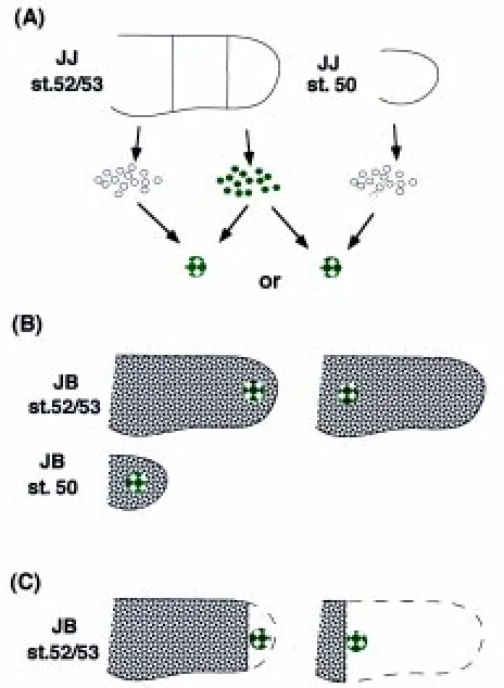
We mixed dissociated mesodermal cells from the distal and the proximal third regions of st. 52/53 limb buds of JJ tadpoles, and pelleted them by centrifugation. To distinguish the origin, the cells derived from the distal regions were labeled with a fluorescent dye (Fig. 1A). The pellet was cultured for 8 h to allow the constituent cells to firmly adhere to each other. The two cell populations were scattered evenly throughout the cell aggregate at this time (Fig. 2). The cell aggregates thus made were cut into small fragments, and each fragment was implanted into the distal or proximal sixth site of a st. 52/53 limb bud in different JB tadpoles (Fig. 1B).
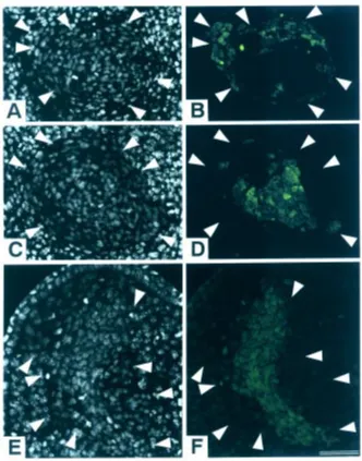
At 36 h after transplantation, the distal cells had already segre-gated from the proximal cells to form small clusters within the cell aggregate. However, there was little difference between aggre-gates grafted at either site (data not shown). At 72 h, the two cell populations had segregated more clearly than before, and the aggregates showed different patterns of segregation depending on the grafted site. While the distal cells existed exteriorly in the cell aggregates implanted into the distal site (n= 5) (Fig. 3A,B), the proximal cells existed exteriorly in the cell aggregates implanted into the proximal site (n= 5) (Fig. 3C,D). Next, we implanted cell aggregates into the middle position along the P-D axis of st. 50 limb buds. The proximal cells existed exteriorly within the cell aggregate (n= 6) at 72 h after implantation. In two out of six cases, cell aggregates extended along the P-D axis, and growth of host limb buds was also observed at this time (Fig. 3E,F). At 96 h, all of the three cell aggregates observed had extended along the P-D axis, but the fluorescent dye had faded, making observation of cell sorting-out in the aggregate impossible (data not shown).
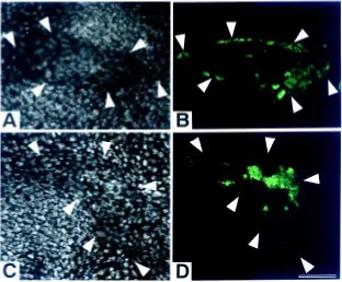
We also implanted cell aggregates derived from st. 50 whole limb buds and the distal third of st. 52/53 limb buds into the distal and proximal sixth sites of st. 52/53 limb buds. Cell sorting-out did not occur between the two cell populations before implantation. At 72 h after implantation, the cells from distal limb buds of st. 52/53 larvae were situated exteriorly in the cell aggregate implanted into the distal site (n= 6) (Fig. 4A,B). On the other hand, in the cell aggregate implanted into the proximal site, the cells from st. 50 limb buds were situated exteriorly (n= 6) (Fig. 4C,D).
Behavior of cells in aggregates grafted on limb bud stumps During limb development, cells change their adhesiveness to other cells, possibly under the influence of AEC. It would be interesting to know the behavior of cells with different positional ing and regenerating limb, with adhesiveness increasing distally.
Generally, in vitro experiments show that cells produced under the influence of AEC or AER during limb morphogenesis have stronger cell adhesiveness than do proximal cells.
Wada et al. (1993) grafted fragments made of dissociated cells from the progress zone (PZ) at different stages and covered with ectodermal jackets on chick limb bud stumps and allowed them to develop into limb-like structures. Early PZ cells were distributed in whole cartilage elements along the P-D axis of the limb-like structures, while late PZ cells participated only in the formation of distal cartilage elements. In this in vivo experiment, late PZ cells, which should probably have stronger adhesiveness, were situated not in the interior region as shown by our in vitro experiment (Koibuchi and Tochinai, 1998) but in the distal region of limb-like
Fig. 1. Procedures for transplantation of cell aggregates. (A) Mesen-chymal cell suspensions, derived from labeled distal thirds and unlabeled proximal thirds of st. 52/53 hind limb buds, or labeled distal thirds of st. 52/ 53 hind limb buds and unlabeled whole st. 50 hind limb buds of JJ tadpoles, were mixed at a 1:1 ratio and lightly spun down. The cell pellets were cultured for 8 h to allow the cells to adhere to each other. The aggregate thus obtained was then trimmed into a lump of about 100 µm in diameter.
only in the boundary region between the host and donor cells within the aggregate.
Distributions of cells originating from the transplanted frag-ments
Although invasion of donor cells into the surrounding host tissue and invasion of host cells into the donor cell aggregate were seldom observed, aggregates transplanted into st. 50 limb buds grew along the P-D axis, with the limb growing in the distal direction. The question arises whether transplanted cells from different regions are involved in making different parts of the developing limb bud growing in P-D direction. To clarify this, we transplanted fragments obtained from three different regions (dis-tal, middle, proximal) of st. 52/53 limb buds into st. 50 limb buds and observed the distribution of donor cells after they reached st. 55/56. To distinguish host and donor cells, we used the JJ and JB nuclear quinacrine marker.
None of the transplanted fragments severely disturbed the cartilage pattern along the P-D axis of the experimental limbs, except for producing supernumerary structures in the distal
ante-Fig. 2. Cell aggregate before implantation. Sections of a JJ cell aggre-gate, made of dissociated mesenchymal cells from the labeled distal and unlabeled proximal third regions of a st. 52/53 hind limb, showing the homogeneous mixing of both cells. Eight hours after mixing. (A) Phase contrast photomicrograph. (B) Fluorescence photomicrograph. Bar, 100
µm.
Fig. 3. Cell aggregates implanted in limb buds at 72h after the operation. Longitudinal sections of grafted cell aggregates made of cells from the labeled distal and unlabeled proximal third regions of st. 52/53 JJ limb buds in JB host limb buds at 72h after transplantation. Aggregates were grafted into the distal (A,B) or proximal sixth region (C,D) of st. 52/53 JB limb buds and the middle position of st. 50 JB limb buds (E,F). (A,C and E) Fluorescence photomicrographs of sections stained with quinacrine. Cells showing the characteristic bright spot in nuclei are host JB cells, and cells showing the uniform darker nuclear staining are grafted JJ cells. (B,D and F) Fluorescence photomicrograph showing the stained distal and unstained proximal cells. Distal region-derived cells are located at the periphery (B) or interior (D,F) of the grafted aggregates. Arrowheads indicate the boundary between the host and grafted tissues. The distal region of the host limb bud is at the top. Bar, 50 µm.
rior region in a few cases (Table 1, Fig. 6D). The transplanted cells were preferentially localized in the anterior side of the limb, probably since the fragments were transplanted from the anterior. Moreover, it was found that distal fragments induced supernumer-ary structures at a higher frequency than did proximal fragments. Since the supernumerary structures that were formed did not alter the order of cartilage pattern along the P-D axis, these cases were also incorporated into the data below.
The transplanted fragment occupied a very limited region (fe-mur or tibia/fibula) in some cases. Except for these cases, frag-ments from the three regions tended to form a continuous stream of cells in the P-D direction and participated in forming both
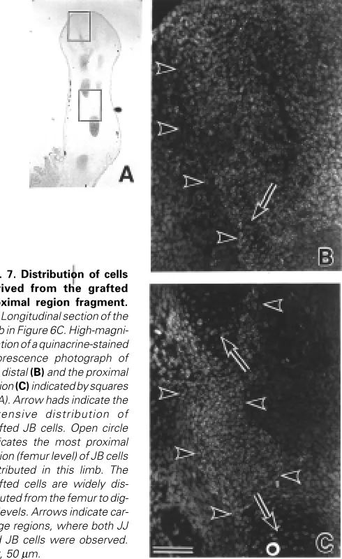
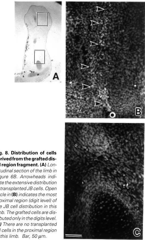
cartilage and connective tissues. In many cases, cells from the proximal region participated in tissue formation over a longer range along the P-D axis than the more distal cells. It appeared that the occupied distributions of grafted cells roughly corresponded to their origin. Fifty-three % (JB into JJ) and 68% (JJ into JB) of the proximal cells participated in tissue formation from the femur or fibula/tibia to digits levels (Fig. 7). Thirty-four % (JB into JJ) and 56% (JJ into JB) of the middle cells participated in tissue formation from the fibula/tibia or fibulare/tibiale to digits levels. Sixty-two % (JB into JJ) and 92% (JJ into JB) of distal cells participated in tissue formation only at fibulare/tibiale or digits levels (Fig. 8). These results are summarized in Table 1.
Fig. 4. Implanted cell aggregates made of cells derived from the distal region of st. 52/53 limb buds and the whole region of st. 50 limb buds. Longitudinal sections of cell aggregates made of labeled distal third region cells from st. 52/53 JJ limb buds and unlabeled cell from the whole region of st. 50 JJ limb buds grafted into the distal (A,B) and proximal sixth region (C,D) of st. 52/53 JB limb buds at 72h after implantation. (A and C) Sections stained with quina-crine. (B and D) Fluorescence photomicrographs showing distal region cells of stage 52/53 limb buds located at the periphery (B) or interior (D) of the grafted aggregates. Arrowheads indicate the boundary between the host and grafted tissues. The distal region of the host limb bud is at the top. Bar, 50 µm.
D, digits.
f/t, tibiale and/or fibulare. F/T, fibula and/or tibia. Fe, femur.
TABLE 1
DISTRIBUTION OF CELLS ORIGINATING FROM TRANSPLANTED JJ OR JB TISSUE FRAGMENTS IN DEVELOPED JJ OR JB HOST LIMB
JJ to JB JB to JJ
Distribution Distal Middle Proximal Distal Middle Proximal
of cells
D 7 (54%) 1 (11%) 8*** (62%) 4* (27%)
D+f/t 5** (38%) 3 (34%) 1 (8%) 4* (27%)
f/t 1 (8%)
f/t+ F/T 1(8%) 1 /7%)
D+f/t + F/T 2* (22%) 2 (15%) 1 (7%) 3 (20%)
D+f/t + F/T+Fe 1 (11%) 7 (53%) 5* (33%)
F/T 4 (30%) 1 (7%)
F/T+Fe 1 (8%) 2 (13%) 4 (27%)
Fe 2 (22%) 1 (8%) 1 (8%) 2 (13%) 3 (20%)
Total 13 9 13 13 15 15
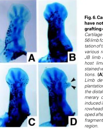
Fig. 6. Cartilage patterns that have not been disturbed by grafting of tissue fragments.
Cartilage patterns of a st. 55/ 56 limb formed after transplan-tation of tissue fragments from various regions of a st 52/53 JB limb bud into a st. 50 JJ host limb bud. Alcian blue-stained whole-mount prepara-tions. (A) Control. (B and D)
Limb developed after trans-plantation of a fragment from the distal region. (D) Supernu-merary digit-like structure is induced in the anterior site (ar-rowheads). (C) Limb devel-oped after transplantation of a fragment from the proximal region.
region at old limb buds did not sort out from the cells from distal region at young limb bud, showing that they have the similar positional identity (Wada and Ide, 1994). The present results showed that not only did sorting-out between cells of different origins occur in vivo but, more importantly, that the pattern of sorting-out was modified according to the transplanted region in the limb bud. Observation of cell aggregates implanted into st. 50 limb buds and into the proximal region of st. 52/53 limb buds showed that cells derived from a st. 50 limb and the proximal region of a st. 52/53 limb were always situated exteriorly; aggregate made derived from a st. 50 limb and the proximal regions of st. 52/53 limb showed very weak sorting-out (data not shown), showing that they have similar positional identity. On the other hand, in the same cell aggregates implanted into the distal region of st. 52/53 limb buds, the cells from the distal region of st. 52/53 limb buds were situated exteriorly. It was reported that in a cell aggregate made of cultured single Xenopus mesenchymal cells, cells derived from a more distal position tended to be situated interiorly (Koibuchi and Tochinai, 1998). The results of the present study showed that the inside-outside patterning of sorted cells was not determined simply by the relationship between cell populations within the cell aggregate but also by the interaction with surrounding host cells. As a conclusion, cells in the aggregate that have closer positional identity to the implanted site are always situated exteriorly to adhere in order to the surrounding host cells.
Wada et al. (1993) grafted fragments made of dissociated cells from the PZ at different stages and covered with ectodermal jackets on chick limb bud stumps and allowed them to develop into limb-like structures. The positions of cells in the limb-like structures reflected their prospective positions along the P-D axis of limb buds at removal, and the arrangement of cells along the P-D axis seemed to be reconstructed. One possible explanation for the arrangement of cells along the P-D axis shown by Wada et al. (1993) is that early cells sort out in order to adhere to host proximal stump cells, and late cells are pushed out distally by early cells. We grafted an aggregate of dissociated cells that did not have an ectodermal covering onto a limb bud stump. The graft was quickly
Fig. 5. Cell aggregates grafted on limb stumps. Longitudinal sections of cell aggregates made of the labeled distal and unlabeled proximal third regions of st. 52/53 JJ limb buds grafted on JB host limb bud stumps at 72h after grafting. Grafting was made by cutting the distal (A,B) or the proximal site (C,D)of st. 52/53 JB limb buds. (A and C) Sections stained with quinacrine. (B and D) Fluorescence photomicrographs showing the distri-bution of proximal and distal cells in the grafted aggregates. Arrowheads indicate the boundary between the host and grafted tissues. The ectoder-mal cells covering the grafts are of host origin. The distal region of the host limb bud is at the top. Bar, 50 µm.
Discussion
covered with a host ectoderm, restoring the AEC (Dent, 1962). Many of the distal cells were situated in the proximal region of the aggregate grafted on the distal stump, and the proximal cells were situated in the proximal region of the aggregate grafted on the proximal stump. These results suggest that the arrangement of grafted cells along the P-D axis is also determined by the adhesive-ness of host and graft cells, as was observed in the above experiment. On the other hand, in the cell aggregates grafted on the distal stump, the grafted distal cells existed not only in the proximal region but also in the distal region beneath tip epidermis (Fig. 5A,B). It was shown that FGF-2 influenced the movement of developing chick limb bud cells (Li et al., 1996). Since the Xenopus limb bud was shown to regenerate an AEC expressing fgf-8 on the stump-grafted cell aggregate (Yokoyama et al., 1998), some of the distal cells might move distally under the influence of FGFs from the regenerated AEC on only the distal stump. Although it is not clear at present why no similar cell movement to AEC occurred on the proximal stump, it may be related to the fact that AEC regenerated on the distal stump earlier than the proximal. It should be further investigated to find out an exact reason.
We transplanted mesenchymal fragments obtained from differ-ent levels along the P-D axis of st. 52/53 limb buds into st. 50 limb
buds and observed the distribution of donor cells in the developed limbs about 14 days later, using the JJ and JB cell marker. In some cases, all of the cells derived from the transplanted fragment solely occupied a small restricted area in the proximal region. It was reported that carbon particles, inserted in the Xenopus limb bud at st. 50 to construct a fate map, were found only in the proximal region (Tschumi, 1957). Therefore, it seems that the cell population found only in the proximal position may have failed to associate with host tissues. There, the cell population was treated as inert material and consequently remained in the transplanted site.
Except for these cases, cells that originated from the trans-planted fragments occupied different distributions along the P-D axis, according to their origin (Table 1). While fragments from the proximal region tended to participate in the construction of not only proximal but also distal regions, fragments from the distal regions tended to participate only in distal region formation. The results showing that the transplanted cells occupy different distributions along the P-D axis according to their origin are in reasonable agreement with the results of previous studies. Crawford and Stocum (1988) demonstrated that when forelimb blastemas from the wrist, elbow, or upper arm were grafted into the blastema-stump junction of a hindlimb regenerating from the midthigh, the grafts migrated distally to the corresponding level of the host hindlimb and then regenerated a new structure, in regenerating axolotl limb. It was also shown that cells that respecified proximal positional identity by retinoic acid stayed in more proximal regions than control cells in regenerating newt limbs and in developing chick limb buds (Pecorino et al., 1996; Tamura et al., 1997). The present results indicate that donor cells moved distally to their original position of the limb buds if the grafting was made in a st. 50 limb bud. On the other hand, when fragments from the distal region were transplanted into the prospective femur level of st. 52/53 limbs, the donor fragment-derived cells did not migrate distally. These cells were found only in femur levels of st. 55/56 limbs (data not shown).
Vargesson et al. (1997) constructed a fate map of the chick limb bud using lipophilic dye. The cells labeled beneath the AER formed a stream running proximodistally between the AER and proximal region of the bud. Some of these cells gradually left the region beneath the AER and entered a differentiation pathway in the proximal region, while other cells remained and proliferated in the region beneath the AER, and their descendants formed a stream running proximodistally. Their results are similar to the results of the present study that show the formation of a continuous stream of grafted cells in the P-D direction. The present results indicate that transplanted cells not only move distally to become situated in the original position from where they were derived but also produce more distal cells. The transplanted cells may remain in the region beneath the AEC until cells that have close positional identity appear. The fact that the transplanted proximal cells produced distal cells reflects that these cells are capable of regeneration.
Although further investigation is needed to clarify what deter-mines cells under the AEC or AER to remain in that region or to leave, it can be hypothesized that the behavior of the cells is determined by the cell adhesiveness. While the cells that have more proximal positional identity tend to adhere to proximal cells and consequently leave the region underneath the AEC or AER, cells that have more distal positional identity are pushed out or move distally. If this is in fact true, even if cells produced under the
influence of the AEC or AER are in a disordered arrangement, the above mechanism may ensure the construction of the correct order of cells along the P-D axis for pattern formation along the P-D axis of the limb bud, which is important for normal development.
Materials and methods
Animals
The animals used in the present study were from the J strain of the South African clawed frog Xenopus laevis, designated as JJ, and an interspecific hybrid (JB) between X. laevis females (JJ) and X. borealis (BB) males. The cell nuclei of JJ and JB can be easily distinguished by differential quinacrine stainability described by Thiébaud (1983). Larvae were anaesthetized in 0.05% MS222 prior to all operations and were allowed to develop at 23±1°C. Developmental stages of larvae were determined according to the normal table of Nieuwkoop and Faber (1956).
Grafting of cell pellets
The design of the experiment is depicted in Figure 1. To prepare cell pellets for implantation, the distal and proximal thirds of hindlimb buds at st. 52/53 and whole hindlimb buds at st. 50 (JJ) were dissected out. The mesodermal cells were isolated by collagenase treatment and labeled with 5 (and 6)-carboxyfluorescein diacetate succinimyl ester (CFSE) (Molecular
probes, USA), if required, according to the method described elsewhere (Koibuchi and Tochinai, 1998). The cell suspensions derived from two different regions or stages, one of which was labeled with CFSE, were mixed at a ratio of 1:1. The mixed cell suspensions were then pelleted down in an Eppendorf tube by brief centrifugation at 400g. They were further incubated for 8 h at 28°C in 70% Leibovitz’s L-15 medium (GIBCO BRL, NY, USA) supplemented with 10% FCS to allow the cell pellet to make a more tightly compacted aggregate. Each pellet was then trimmed into a fragment of about 100 µm in diameter with 27-gauge injection needles (Terumo, Tokyo, Japan) used as knives (Fig. 1A). Grafting sites were made in the hind limb buds of JB tadpoles at different regions or stages. A slit for inserting an aggregate was made with an injection needle at the distal or proximal sixth site of st. 52/53 limb buds and middle position along the P-D axis of st. 50 limb buds (Fig. 1B). A st. 52/53 limb bud was cut off to make a limb stump available for grafting an aggregate at the distal and proximal sixth sites along the P-D axis (Fig. 1C). At various periods after the operation, the grafted whole limb buds were fixed in Carnoy’s fixative, embedded in paraffin, and serially sectioned at 5 µm in thickness. After observation of the distribution of labeled and unlabeled cells in the un-stained sections under a fluorescence microscope, the sections were stained with quinacrine to distinguish donor cells (JJ) and host cells (JB).
Transplantation of tissue fragments
Mesodermal tissues from different regions along the P-D axis (distal, middle, proximal) of st. 52/53 hindlimb buds were dissected out and cut into small pieces of about 100 µm in diameter with injection needles for transplantation. To prepare a grafting site in the host limb bud, a cut was made with an injection needle from the anterior direction at the middle position along the P-D axis of a st. 50 limb bud. A fragment was then inserted into the slit. The reciprocal host-donor combinations were made between either JJ and JB, or JB and JJ. The tadpoles that were operated on were allowed to develop to st. 55/56, and the experimental limb buds were fixed in Carnoy’s fixative. They were then transferred to 0.1% Alcian blue in acid alcohol for 2 days. After excessive Alcian blue removal, the limb buds were dehydrated in 100% alcohol and then cleared in methyl salicy-late for analyzing the formed cartilage patterns. After taking a photograph, each limb bud was embedded in paraffin, serially sectioned at 5 µm, and stained with quinacrine to distinguish donor and host cells.
Acknowledgments
We are very grateful to Dr. Naoyuki Wada for his valuable criticism of the manuscript.
References
CHRISTEN, B. and SLACK, J.M.W. (1997). FGF-8 is associated with anteroposterior patterning and limb regeneration in Xenopus. Dev. Biol. 192: 455-466.
CRAWFORD, K. and STOCUM, D.L. (1988). Retinoic acid coordinately proximalizes regenerate pattern and blastema differential affinity in axolotl limbs. Development 102: 687-698.
DENT, J.N. (1962). Limb regeneration in larvae and metamorphosing individuals of south african clawed toad. J. Morphol. 110: 61-77.
FALLON, J.F., LOPEZ, A., ROS, M.A., SAVAGE, M.P., OLWIN, B.B. and SIMANDL, B.K. (1994). FGF-2: Apical ectodermal ridge growth signal for chick limb develop-ment of the tetrapod limb. Science 264: 104-107.
IDE, H., WADA, N. and UCHIYAMA, K. (1994). Sorting out of cells from different parts and stages of the chick limb bud. Dev. Biol. 162: 71-76.
KOIBUCHI, N. and TOCHINAI, S. (1998). Existence of gradient in cell adhesiveness along the developing Xenopus hind limb bud, shown by a cellular sorting-out experiment in vitro. Dev. Growth. Differ. 40: 355-362.
LI, S., ANDERSON, R., REGINELLI, A.D. and MUNEOKA, K. (1996). FGF-2 influ-ences cell movements and gene expression during limb development. J. Exp. Zool. 274: 234-247.
MAHMOOD, R., BRESNICK, J., HORNBRUCH, A., MAHONY, C., MORTON, N., COLQUHOUN, K., MARTIN. P., LUMSDEN, A., DICKSON, C. and MASON, I.
Fig. 8. Distribution of cells derived from the grafted dis-tal region fragment. (A) Lon-gitudinal section of the limb in Figure 6B. Arrowheads indi-cate the extensive distribution of transplanted JB cells. Open circle in (B) indicates the most proximal region (digit level) of the JB cell distribution in this limb. The grafted cells are dis-tributed only in the digits level.
(1995). A role for FGF-8 in the initiation and maintenance of vertebrate limb bud outgrowth. Curr. Biol. 5: 797-806.
NARDI, J.B. and STOCUM, D.L. (1983). Surface properties of regenerating limb cells: evidence for gradation along the proximodistal axis. Differentiation 27: 13-28.
NIEUWKOOP, P.D. and FABER, J. (1956). Normal table of Xenopus laevis. (Ed. Daudin), North-Holland, Amsterdam.
NISWANDER, L., TICKLE, C., VOGEL, A., BOOTH, L. and MARTIN, G.R. (1993). FGF-4 replaces the apical ridge and directs outgrowth and patterning of the limb. Cell 75: 579-587.
PECORINO L.T., ENTWISTLE A. and BROCKES J.P. (1996). Activation of a single retinoic acid receptor isoform mediates proximodistal respecification. Curr. Biol. 6: 563-569.
STEINBERG, M.S. (1963). Reconstruction of tissues by dissociated cells. Science 141: 401-408.
SUMMERBELL, D., LEWIS, J.H. and WOLPERT, L. (1973). Positional information in chick limb morphogenesis. Nature 244: 492-496.
TAMURA, K., YOKOUCHI, Y., KUROIWA, A. and IDE, H. (1997). Retinoic acid changes the proximodistal developmental competence and affinity of distal cells in the developing chick limb bud. Dev. Biol. 188 : 224-234.
THIÉBAUD, C.H. (1983). A reliable new cell marker in Xenopus. Dev. Biol. 98: 245-249.
TOWNES, P.L. and HOLTFRETER, J. (1955). Directed movements and selective adhesion of embryonic amphibian cells. J. Exp. Zool. 128: 53-120.
TSCHUMI, P.A. (1957). The growth of hindlimb bud of Xenopus laevis and its dependence upon the epidermis. J. Anat. 91: 149-173.
VARGESSON, N. CLARKE, J.D., VINCENT, K., COLES, C., WOLPERT, L. and TICKLE, C. (1997). Cell fate in the chick limb bud and relationship to gene expression. Development 124: 1909-1918.
WADA, N. and IDE, H. (1993). Sorting out of limb bud cells in monolayer culture. Int. J. Dev. Biol. 38: 351-356.
WADA, N., UCHIYAMA, K. and IDE, H. (1994). Cell sorting out and chondrogenetic aggregate formation in limb bud recombinant and in culture. Dev. Growth. Differ. 35: 421-430.
YOKOYAMA, H., ENDO, T., TAMURA K. YAJIMA, H. and IDE, H. (1998). Multiple digit formation in Xenopus limb bud recombinants. Dev. Biol. 196 : 1-10.