CSEIT1831490 | Received : 16 Feb 2018 | Accepted : 27 Feb 2018 | January-February-2018 [(3) 2 : 202-209]
International Journal of Scientific Research in Computer Science, Engineering and Information Technology © 2018 IJSRCSEIT | Volume 3 | Issue 2 | ISSN : 2456-3307
202
Retinal Image Processing Using Neural Networks
For Disease Prediction
R. Venkatesan*1, E. Saranya2
1PG Scholar & Computer Science and Engineering, Anna University/Sir ISSAC Newton college of Engineering
and Technology, Nagapattinam, Tamilnadu, India
2Assistant Professor of Computer Science and Engineering, Anna University/Sir ISSAC Newton College of
Engineering and Technology, Nagapattinam, Tamilnadu, India
ABSTRACT
In medical field, diagnoses of diseases are competently carried out by using the image processing. Human eye is an important organ that reacts to light and has several purposes. The eye has a number of components but it is not limited to the cornea, iris, pupil, lens, retina, and macula, optic nerve, choroid and vitreous. Retinal images play vital role in several applications such as disease diagnose and human recognition. Retinal image analysis is particularly a complicated task because of the variability of the images in terms of the color, the morphology of the retinal anatomical pathological structure and the existence of particular features in different patients, which may lead to an erroneous interpretation. Image processing techniques were used for dark object detection to analyze the condition of the input image, to enhance the input image in order to make it suitable for processing of the retinal image, to improve visibility of Microaneyrysm in color fundus images. K-Nearest algorithm is used to detect the blood vessels effectively for segmentation process and Deep Neural algorithm is used to classify the diagnose the diseases such as stroke, heart attack and cardio vascular disease by segmenting optic disc and to predict retinal disease using Ellipse Fitting method. Experimental results show that good accuracy in disease prediction.
Keywords : Image Processing, Eye Components, Disease Diagnosis, Diabetic Prediction
I.
INTRODUCTION
Nowadays diseases related to eye are increasing and many people fell in to blindness. Image processing is the area which leads with image analysis and which involves the study of feature extraction, segmentation and classification. The process of recognizing the vessel patterns that are used to analyse the vessels in retinal image. Diabetic retinopathy one of the complicated disease, which affects to the retina and outcome is the total blindness. Segmentation is the method of identifying regions of pixels in an image to find out the correlation with objects. The retina is internal part of the eye. In the centre of retina, there is the optic disk,
considered as one of the most promising solution for disease diagnosis in the future.
The blood vessels network is an important anatomical structure in human retina, which is use to recognize different types of disease. However, manual detection of blood vessels is not simple because the vessels in retina image are complex and have low contrast. For retinal anatomy ophthalmologist uses an ophthalmoscope. The programmed mining of blood vessels in retinal images is one of the important step in computer aided diagnosis and treatment of diabetic retinopathy, glaucoma, arteriosclerosis, obesity, retinal artery occlusion and hypertension. Retinal images are influenced by all the factors that affect the body vasculature in general. The human eye is a unique region of the human body where the vascular condition can be directly observed. In addition to fovea and optic disc, the blood vessels contribute one of the main features of a retinal fundus image and worldwide major diseases such as diabetes, hypertension, and arteriosclerosis noticeably affect several of its properties. Further, certain eye diseases such as choroid neovascularization and retinal artery occlusion also make changes in the retinal vasculature. As per previous statement, the segmentation of blood vessels in retinal images can be a valuable aid for the detection of diabetic retinopathy and glaucoma diagnosis Segmentation can be done by supervised, unsupervised or semi supervised. Here using semi supervised segmentation method because of easy to use labelled data and unlabelled data together. Semi supervised segmentation have much applications in medical image data sets.
An automated segmentation and inspection of retinal blood vessel features such as diameter, colour and tortuosity as well as the optic disc morphology allows ophthalmologist and eye care specialists to perform mass vision screening exams for early detection of retinal diseases and treatment evaluation. This could prevent and reduce vision impairments; age related
diseases and many cardiovascular diseases as well as reducing the cost of the screening. The basic retinal image processing is shown in figure 1.
Figure1. Retinal Image Processing
II.
RELATED WORK
available datasets have been used to test the performance of the suggested method.
Manvir Kaur,et.al, [2] analysed the process on images of retina with the help of Digital Image Processing (DIP) tool in which images are detected and then processed. At last we describe the problem of detecting edges in images as a diabetic retinopathy (DR), macular degeneration and glaucoma. The edge detection problem can be separated into three stages: filtering; detection; and tracing and images separated with the application of different algorithm based on local pixel characteristics which can control the degree of Gaussian smoothing. Filtered images are then applied to a simple edge detection algorithm which evaluates the edge fuzzy association value for each pixel; based on local image characteristics. Diabetic retinopathy is a complication of diabetes and is a major cause of blindness in developed countries. The patients might not notice a loss of vision until it became too severe, hence early diagnosis and timely treatment is vital to delay or prevent visual impair and even blindness. Retinal vessel segmentation can simplify screening for retinopathy by reducing the number of false positive results in micro aneurysm detection and may serve as a means of image registration from the same patient taken at different times by delineating the location of the optic disc and fovea. However, manual detection of blood vessels is not simple because the vessels in a retinal image are complex and have low contrast. Detecting abnormalities such as venous looping or beadings is critical for early treatment as they are in most cases indication of potentially sight-threatening retinopathy. In order to utilize these useful characteristics of retinal blood vessels this is very important to obtain their locations and shapes accurately. Blood vessels appeared as networks of either deep red or orange-red filaments that originated within the optic disc and were of progressively diminishing width.
Shingling Wang,et.al,. [3] presented the hybrid method based upon convolutional neural network
(CNN) and ensemble random forests (RFs) for automatic retinal blood vessel segmentation. We first employed a set of pre-processing steps to correct the non-uniform illumination of retinal images and to improve vessel contrast. We then used CNN to extract a set of hierarchical features which are not only invariant to image translation, scaling, skewing and other distortions, but also contain image based multi-scale information of the geometric structure of retina. We finally trained ensemble RFs to obtain a vessel classifier. The whole pipeline of the proposed method is trainable and automatic. Although these supervised methods have achieved satisfactory segmentation results in some scenarios, there are still some issues. In hand-designed feature extraction approaches, the features must be very carefully predefined before classification, making feature detection very time-consuming and tedious. Most importantly, hand-designed features have to be redesigned for datasets with different characteristics.
In contrast, the approaches based upon feature learning can extract features automatically from the raw images. Moreover, CNN is supervised feature learner able to learn complex invariances such as scale and rotational invariance. However, the classification mechanism in ANN or its variants is fairly simple usually nonlinear activation functions are employed in the last output layer to predict patterns, which results in the low performance of ANN.
automatically locating the optic nerve and blood vessel in retinal images, using graph cut method. To segment and determine the blood vessels and optic disc from retinal images, the input retinal images are read which then under goes pre-processing, then to find the optical disc segmentation and blood vessel segmentation. The colour based segmentation and gradient method used for blood vessel segmentation for achieving exceptional performance in segmenting the blood vessel and Hough circular transform is used for optical disc segmentation. For rule based validation is applied to validate proper optical disc segmented or not similarly validation of blood vessel segmentation.
Roberto Annunziata,et.al,. [5] proposed in painting technique has a different focus. Indeed, neither texture synthesis nor a visually plausible image is needed. Our goal is to fill structures such as exudates in retinal images so that, when vessel enhancement is applied, the number of nearby false positives is greatly reduced. This goal is only achieved if exudates are filled in a smooth way that reduces or eliminates possible edges. A multiple-scale Hessian-based enhancement is applied to detect retinal vessels. This technique is fast and has proven to be effective when detecting vessels of normal eyes. The key idea of the proposed method is to apply Hessian-based enhancement after exudate in painting. Moreover, it yields the best performance on pathological images, the target of most automated retinal image analysis tools. Indeed, a vessel segmentation algorithm is usually the first step for the automated detection of eye diseases. In order to be used in clinical practice, these methods should be robust enough to analyse pathological and non-pathological images without requiring user interaction. We propose a fully automated algorithm.
III.
EXISTING METHODOLOGIES
Considering both data has been validated to boom the retinal imaging accuracy extensively. There are two essential classes such as vessel or non-vessel
utilizing functions: to extract some sort of features (e.g., texture, colour, and shape features), and to at once use pixels in a small neighbourhood for joint type assuming that these pixels normally percentage the same magnificence membership. Existing algorithms are derived as follows
A. Mrf Model
The MRF model, which combines retinal parts with vessels, is widely used in classification. It can provide an exact feature representation of pixels and their neighbourhoods. The basic principle of MRF is to integrate spatial correlation information into the posterior probability of the spectral structures. Based on the maximum posterior probability principle, the classic MRF model can be expressed as follows ρ(x_i )=-1/2 ln|Σk|-1/2(x_i-m_k )^T ∑_k^(-1)[[(x]_i-[m]_k)]-β∑_∂i[1-δ(ω_ki,ω_∂i )] --- Eqn(1)
where m_k and Σk are the mean vector and covariance matrix, respectively, of class k and the neighbourhood and class of pixel i are represented by ∂_and ωk , respectively. The constant parameter β, called the weight coefficient is used to control the influence of the spatial term. According to Equation (1), the MRF model can be separated into two components: the vessel term and non-vessel term.
B. Deformable Models
The recent one of the methods of contour detection is deformable models or snake. A snake is an active contour model that is manually initiated near to the contour of interest. This contour model deforms according to some criteria and image features to finally stay to the actual contour(s) in the image. An energy function is formulated to obtain an estimate of the quality of the mode in terms of its internal shape, and external forces e.g. underlying image forces and user-constraint forces. The energy function integrates a weighted-linear combination of the internal and external forces of the contour.
First order derivative term v_s (s) make the snake behave like a membrane and represent the elastic energy of the contour. The second order derivative term v_ss (s) makes the snake act like a thin plate and represents the contour bending energy. Decreasing αelasticity allows the contour to develop gaps, while increasing α_elasticity increases the tension of the model by reducing its length. Decreasing αbending allows the active contour model to develop corners, and increasing α_bending increases the bending rigidity, making the contour smoother and less flexible. Setting either of the weighting coefficients to zero permits first and second order discontinuities respectively. The external energy term ϵ_image represents the energy due to image forces like lines, edges and terminations of line segments and corners.
∈_image^* (v(s))=α_line (s) ϵ_line (v(s))+α_edge (s) ϵ_edge (v(s))+α_term (s) ϵ_term (v(s)) ---Eqn(2) Existing approaches only consider vessel segmentation and cannot analyse vessel classification.
IV.
PROPOSED FRAMEWORK
The macula, the optic disc, and the blood vessels are the main anatomical features of the retina. A macula in the eye is a small spot where vision is keenest in the retina. The macula of the retina is the light-sensitive layer of tissue at the back of the eye. Blood vessels are the elastic tube or passage in the body through which blood circulates. Examination of blood vessels in the eye allows detection of eye diseases such as glaucoma and diabetic retinopathy. Traditionally, the vascular network is mapped by hand in a time-consuming process that requires both training and skill. Automating the process allows consistency, and most importantly, frees up the time that a skilled technician or doctor would normally use for manual screening. Implement automatic process to examine the blood vessels to identify the cardio vascular diseases in retinal images. It utilizes the concept of active contours to remove noise, enhance the image, track the edges of the vessels, calculate the perimeter of vessels and identify the cardio diseases. Implement neural network model to
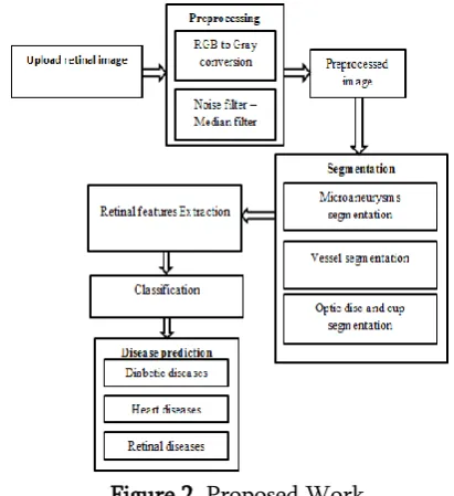
linear discriminant analysis, contrast limited adaptive histogram equalization, green channel extraction ad Ellipse fitting method. Ellipse fitting method involves in feature extraction. Contrast limited adaptive histogram equalization is used to enhance the image. After the extraction of vessel from retinal fundus image, they are removed by this technique. Finally ellipse operations are applied to the resulted image to detect the optic disc boundary. The cup segmentation is performed in the green channel which is extracted from the input image. Watershed transformation is used for cup segmentation. Cup to Disc Ratio is calculated after determining the optic disc and cup boundary. The Normal cup to disc ratio range is from 0.1 to 0.4. If the cup to disc ratio exceeds 0.4 then it indicates the abnormal condition that is the presence of glaucoma. The robust process provides automated results without any user intervention. The proposed work is shown in.
Figure 2. Proposed Work
A.CNN Based Classification
CNNs represent feed-ahead neural networks, which encompass numerous combinations of the convolutional layers, max pooling layers, and completely related layers and Take benefit of spatially community correlation with the aid of manner of manner of implementing a nearby connectivity pattern among neurons of adjacent layers. Convolutional layers alternate with max
pooling layers mimicking the individual of complex and easy cells in mammalian visible cortex. A CNN consists of one or more pairs of convolution and max pooling layers and in the long run ends with completely associated neural networks. The hierarchical structure of CNNs is progressively proved to be the maximum green and a success way to research seen representations. The fundamental project in such visual responsibilities is to model the intra-elegance appearance and form variation of objects. The hyper-spectral records with loads of spectral channels can be illustrated as 2D curves. We can see that the curve of every magnificence has its very own visible form which is different from other lessons, although it is extraordinarily hard to differentiate some classes with human eye. We recognize that CNNs can accomplish aggressive or even higher performance than man or women in some visible troubles, and its capability inspires us to have a look at the opportunity of applying CNNs for blood vessel segmentation. The CNN varies in how the convolution and max pooling layers are realized and how the nets are trained.
trainable parameters among this accretion and layer F3. Consequently, the architecture of our proposed CNN classifier definitely has 20 × (𝑘1 + 1) + (20 × 𝑛3 + 1) × 𝑛4 + (𝑛4 + 1) × 𝑛5 trainable parameters. Classifying a detailed blood pixel wishes the corresponding CNN with the aforementioned parameters, wherein 𝑛1 and 𝑛5 are the spectral channel length and the wide variety of output classes of the statistics set, respectively. In our experiments,
𝑘1 is higher to be ⌈𝑛1/nine⌉, and 𝑛2 = 𝑛1−𝑘1+1. 𝑛3 may be any wide variety among 30 and forty, and 𝑘2 = ⌈𝑛2/𝑛3⌉. 𝑛4 is about to be 100.These alternatives won't be the satisfactory however are in effect for widespread retinal information. In our architecture, layer C1 and M2 may be analysed as a trainable attribute extractor to the input retinal records, and layer F3 is a trainable classifier to the characteristic extractor. The output of subsampling is the real characteristic of the authentic statistics. In our proposed CNN structure, 20 capabilities may be extracted from every original retinal pixels, and each function has 𝑛3 dimensions. This network varies affording to the channel size and the number of output classes of input retinal data. Therefore, our proposed work overcomes irregular boundaries separation in retinal image classification with vessels and optic disc features.
V.
CONCLUSION
We developed a new novel framework for disease classification to extract blood vessel information. Vessel Features are extracted as multi attributes profiles and we reduced the dimensionality by using supervised features extraction method such as median filter. And implementing CNN segmentation for improves the accuracy in results. The Proposed framework is considerably examined on extensively used blood vessel statistics to provide better accuracies. In addition, the new approach achieves better classification accuracies than other extensively used classification strategies, with acceptable CPU processing time. We emphasize that the proposed system is fully computerized, that's a exceedingly
acceptable characteristic. In future, we can extend the framework to improve the accuracy in various kinds of datasets and try to analyse parallel processing approach and include other performance metrics.
VI.
REFERENCES
[1]. Abdallah,Mariem Ben,et al. "Automatic extraction of blood vessels in the retinal vascular tree using multiscale medialness." Journal of Biomedical Imaging 2015 (2015): 1. [2]. Kaur,Manvir,and Rajneesh Talwar. "Automatic
Extraction of Blood Vessel and Eye Retinopathy Detection." European Journal of Advances in Engineering and Technology 2.4 (2015): 57-61.
[3]. Wang,Shuangling,et al. "Hierarchical retinal blood vessel segmentation based on feature and ensemble learning." Neurocomputing 149 (2015): 708-717.
[4]. Vidyashree,M. R.,M. V. Usha,and vtu ewit. "Locating the optic nerve and blood vessel in a retinal images using graph partition method." (2015).
[5]. Annunziata,Roberto,et al. "Leveraging multiscale hessian-based enhancement with a novel exudate inpainting technique for retinal vessel segmentation." IEEE journal of biomedical and health informatics 20.4 (2016): 1129-1138.
[6]. N. Srivastava,G. Hinton,A. Krizhevsky,I. Sutskever,and R. Salakhutdinov,"Dropout: A simple way to prevent neural networks from overfitting," The Journal of Machine Learning Research,vol. 15,no. 1,pp. 1929-1958,2014. [7]. Y. Bengio,P. Lamblin,D. Popovici,H. Larochelle
et al.,"Greedy layer-wise training of deep networks," Advances in neural information processing systems,vol. 19,p. 153,2007.
Comput. Methods Prog. Biomed. vol. 108,no. 1,pp. 407-433,Oct. 2012.
[9]. J. Staal,M. D. Abràmoff,M. Niemeijer,M. A. Viergever,and B. van Ginneken,"Ridge-based vessel segmentation in color images of the retina," Medical Imaging,IEEE Transactions on,vol. 23,no. 4,pp. 501-509,2004.