Western University Western University
Scholarship@Western
Scholarship@Western
Electronic Thesis and Dissertation Repository
7-11-2019 9:30 AM
Quantitative analysis of the kinematics of the foot during gait with
Quantitative analysis of the kinematics of the foot during gait with
respect to barefoot running shoes using a Multi-Segment Foot
respect to barefoot running shoes using a Multi-Segment Foot
Model
Model
Lisa Y. Oikawa
The University of Western Ontario
Supervisor Dickey, James P.
The University of Western Ontario
Graduate Program in Kinesiology
A thesis submitted in partial fulfillment of the requirements for the degree in Master of Science © Lisa Y. Oikawa 2019
Follow this and additional works at: https://ir.lib.uwo.ca/etd
Part of the Biomechanics Commons
Recommended Citation Recommended Citation
Oikawa, Lisa Y., "Quantitative analysis of the kinematics of the foot during gait with respect to barefoot running shoes using a Multi-Segment Foot Model" (2019). Electronic Thesis and Dissertation Repository. 6487.
https://ir.lib.uwo.ca/etd/6487
This Dissertation/Thesis is brought to you for free and open access by Scholarship@Western. It has been accepted for inclusion in Electronic Thesis and Dissertation Repository by an authorized administrator of
Abstract
Kinematics of the foot during static and dynamic tasks are technically challenging to accurately measure, making it difficult to evaluate their contribution to running-related injuries. Motion capture can detect changes in running mechanics, such as with differing footwear. Habitual barefoot runners and/habitual minimalist shoe runners underwent a biomechanical evaluation of their foot movement during running using reflective markers and optical motion capture. A five-segment foot model was used to compare motions between the different parts of the foot while running barefoot and wearing Vibram Five
Fingers™ (VFF) barefoot mimicking shoes. Supination/pronation in the forefoot was larger but not significant between habitual shod and habitual VFF runners. In contrast, the other foot motions (forefoot spreading/rising, hindfoot pronation/supination and hindfoot adduction/abduction) were not significantly different between the groups of runners. Therefore it could be possible that transitioning from a regular running shoe to a barefoot mimicking minimalist shoe would cause the foot to adopt a more supinated gait cycle.
Lay Summary
Movement of the foot due to it's complexity during still and moving tasks are challenging to measure. This makes it difficult to compare movements to running related injuries. This study took a look at Vibram Five Fingers and shod runners using a segmented foot model to see if differences were evident. The study used reflective markers and motion caption cameras to compare motion between the two groups of runners. There were no differences seen between the two groups of runners.
Keywords
Table of Contents
Abstract ... ii
Table of Contents ... iii
Glossary & Nomenclature ... vi
Chapter 1: Background & Introduction ... 1
1.1 Statement of purpose ... 1
1.2 Introduction ... 2
1.3 Foot anatomy ... 3
1.3.1 Bones of the foot ... 3
1.3.2 Functional units of the foot ... 6
1.3.3 Major joints of the foot and ankle ... 7
1.3.4 Ligaments of the ankle and foot ... 12
1.4 Gait cycle ... 14
1.4.1 Biomechanics of gait ... 14
1.4.2 Walking gait cycle ... 15
1.4.3 Running gait cycle ... 17
1.5 Running ... 20
1.5.1 Running patterns ... 20
1.5.2 Barefoot runner’s patterns ... 21
1.5.3 Shod Runner’s patterns ... 22
1.5.4 Impact force & injury ... 22
1.6 Injury ... 22
1.6.1 Training errors ... 23
1.6.2 Intrinsic & extrinsic factors to injury ... 23
1.6.3 Mechanisms behind injury ... 24
1.7 Anatomy of the running shoe... 24
1.7.1 Heel counter & wedge ... 25
1.7.2 Motion control ... 26
1.7.3 Shoe use ... 27
1.7.4 Lacing design of running shoes ... 27
1.8 Barefoot simulation shoes ... 27
1.8.1 Pressure and ground reaction force during barefoot running ... 27
1.8.2 Stride frequency and impulse ... 27
1.8.3 Characterization of a barefoot running shoe ... 28
1.8.4 Hazards of barefoot running ... 29
1.9 Studies and kinematic outcomes ... 29
1.9.1 Previous studies ... 29
1.9.2 Current testing methods ... 30
1.9.3 Vibram Five Fingers & Barefoot running ... 31
1.9.4 Summary of kinematic outcomes ... 31
Chapter 2: Kinematic Models ... 33
2.1 Kinematic models ... 33
2.1.1 Fluoroscopy and X-ray ... 33
2.1.2 Optical motion capture... 33
2.1.3 Video analysis ... 34
2.1.4 Electromagnetic tracking ... 34
2.1.6 Skin motion artifact ... 35
2.2 Multi-segment foot model ... 36
2.3 Windowing shoes ... 42
Chapter 3: Methods ... 44
3.1 Experimental equipment ... 44
3.2 Motion analysis equipment ... 45
3.3 Experimental procedures ... 47
3.3.1 Calibration ... 47
3.3.2 Helen Hayes marker set ... 49
3.3.3 Cluster marker setup ... 50
3.4 Static trial ... 51
3.6 Vibram Five Finger Shoes... 54
3.7 Dynamic trials ... 55
3.8 Kinematic Analysis ... 56
3.8.1 Medial Longitudinal Arch (MLA) ... 56
3.8.2 Forefoot pronation/supination ... 57
3.8.3 Hindfoot pronation/supination ... 58
3.8.4 Kinematic Variables... 59
3.9 Post Processing ... 59
Chapter 4: Results... 61
4.1 Habitual shod and habitual VFF running barefoot ... 62
4.1.1. MLA (barefoot) ... 62
4.1.2 Forefoot in the frontal plane (barefoot) ... 63
4.1.3 Forefoot in the transverse plane (barefoot) ... 64
4.1.4 Hindfoot in the frontal plane (barefoot) ... 65
4.1.5 Hindfoot in the transverse plane (barefoot) ... 66
4.2 Habitual shod and habitual VFF running in VFF ... 67
4.2.1 MLA (VFF) ... 67
4.2.2 Forefoot in the frontal plane (VFF) ... 68
4.2.3 Forefoot in the transverse plane (VFF) ... 69
4.2.4 Hindfoot in the frontal plane (VFF) ... 70
4.2.5 Hindfoot in the transverse plan (VFF) ... 71
Chapter 5: Discussion & Conclusions... 73
5.1 Discussion ... 73
5.2 MLA Kinematics ... 73
5.3 Forefoot kinematics ... 74
5.4 Hindfoot Kinematics ... 76
5.5 Limitations ... 78
5.6 Strengths ... 79
5.7 Conclusion ... 79
References ... 80
Appendix A... 87
Appendix B... 91
Appendix C ... 92
Appendix D ... 93
Summary of kinematic outcomes ... 93
Appendix E ... 97
List of Tables
Table 1 Cluster names and locations of the multi-segment foot model ... 39 Table 2 Bony landmarks digitized for each segment used to define segment fixed axes. Note that the medial and lateral forefoot segments share landmarks (Jenkyn, et al. 2009; Jenkyn & Nichol, 2007). ... 40 Table 3.1 Demographic data of eight subjects of the study describing group, average
running mileage per week, sex, age at time of data collection, weight and height. ... 44 Table 3.2 Positions of reflective markers based off of the Helen Hayes marker set. ... 49 Table 3.3 Multi-segment foot model marker set and their respective locations. The same marker locations were used for each subject and for each condition. ... 50 Table 3.4 Virtual marker definitions of 5 MSFM triad clusters; Hallux, 1st metatarsal
(midfoot), navicular (medial forefoot), 5th metatarsal (lateral forefoot) and calcaneus. Origin markers, long axis markers and plane markers were used consistently through each subject using static trials to define virtual marker locations. ... 52 Table 3.5 Cluster markers used in creating two vectors for each foot segment. Each set of vectors allows for a separate coordinate system to be defined. ... 53 Table 3.6 Stylus landmarks used to calculate bone locations. ... 54 Table 4.1 Habitual shod runner trials compared to habitual VFF runners in barefoot (BF) and VFF conditions. ... 72
Glossary & Nomenclature
abduction (ABD) movement towards the midline adduction (ADD) movement away from the midline
axis of rotation straight line going through fixed points of a rotating rigid body
BF barefoot
distal further away from a point of attachment or origin dorsiflexion (DF) flexion of the ankle towards the body
eversion abduction of the ankle
extension increasing the angle of the joint flexion decreasing the angle of the joint
frontal plane defined as separating the body into front and back forefoot strike (FFS)
gait
characterized by initial ground contact on the forefoot (first 1/3 of the foot) proceeded by transferring weight to the heel manner of walking or running
inversion (inv) when the ankle supinates and the foot rolls towards the midline
kinematics study of motion of the body without regard to the forces that are producing the motion
kinetics study of forces that cause motion lateral away from the midline of the body
Matlab software used to write code to read and analyse data medial situated towards the midline of the body or attachment or
origin medial longitudinal arch
(MLA) arch formed at the bottom of the foot midfoot strike (MFS)
mm
characterized by initial ground contact on the midfoot (middle 1/3 of the foot)
millimetre
proximal closer to the point of reference
plantar flexion movement where the toes are pointed downwards towards the sole
posterior towards the back of the body
pronation combination movement of eversion, dorsiflexion and abduction
rearfoot strike (RFS)
midsagittal plane
characterized as initial ground contact on the rearfoot (rear 1/3 of the foot)
anatomical plane that runs through the midline and divides the body into left and right
superior higher when referring to a point of reference
supination combination movement of inversion, plantar flexion and adduction
tendon tissue connecting muscle to bone
transverse plane separating the body into an upper half and a lower half valgus a deformity in which an anatomical part is turned outward
varus a deformity in which an anatomical part is turned inward toward the midline of the body to an abnormal degree
VFF Vibram Five Fingers running shoes
Chapter 1: Background & Introduction
1.1Statement of purpose
The purpose of this research was to quantify the relative motion between the forefoot and hindfoot in participants with healthy, non injured feet during running. Participants were divided into two groups. One group habitually ran in traditional running shoes (more than
10 kilometers per week), and the second group habitually ran in Vibram Five Fingers™ or
barefoot (more than 10 kilometers per week).
This study used optical motion capture and a multi-segment foot model to individually track the segments of the hindfoot, midfoot, medial forefoot, lateral forefoot and hallux. The optical motion capture system used skin-mounted auto-reflective markers organized into rigid triad clusters. At least one cluster was attached to each foot segment. The multi-segment foot model and optical motion capture system have been previously validated in our laboratory (Jenkyn & Nicol, 2007). The multi-segment foot model measuresthe degrees of freedom motion between the five foot segments so that the function of the foot can be compared between subject types and footwear conditions.
This study had two hypotheses. The first hypothesis was that the kinematics of habitually shod runners are significantly different than habitual Vibram Five Fingers™ runners when running barefoot. The second hypothesis was that the kinematics of habitually shod runners are significantly different than habitual Vibram Five Fingers™ when running in
1.2 Introduction
For several centuries, shoes have been used to provide protection for the soles of the feet, traction between the feet and the ground, for motion control and stability, and for
dispersion of ground reaction forces (Mcpoil, 2000). Runners typically strike the ground 600 times per kilometer (Milner, Ferber, Pollard, Hamill, & Davis, 2006; Pohl, Hamill, & Davis, 2009; van Gent et al., 2007). Each time the foot strikes the biomechanical loading is approximately 1.25 times body weight during walking and 2 to 3 times body weight during running (Lieberman et al., 2010; Waetjen, Parker, & Wilken, 2012). Thus, due to the
repetitive nature of these foot strikes, any imbalance in foot and gait mechanics may result in a running related injury.
The high loads associated with running, and the associated risk of injury, catalyzed research studies looking into barefoot and shod running. The article by Lieberman et al. (2010) was one of the first published studies that compared the differences between habitually
barefoot and shod runners. It sparked a lot of controversy in the running community. In a
2014 Runner’s World interview, Dr Lieberman stated that changing your running
mechanics affects more than just your footstrike; it can also change your joint moments, the cadence and tissue loading (Lovett, 2014). He goes on to say that there is a need for future studies examining different aspects of running.
Incidences of running related injuries and overuse injuries such as plantar fasciitis, stress fractures, tendonitis and shin splints have increased over time as the number of runners in Canada have also increased (van Gent et al., 2007) with the incidence of running related injuries range from 19-79% in any given year (van Gent et al., 2007). Despite advancements in running shoe technology and design (increases in cushioning, alterations to materials, changes flexibility and stability) rates of repetitive strain injuries vary little from year to year (Richards, Magin, & Callister, 2008; Van Mechelen, 1992) and therefore perhaps variations in shoe material are not the sole contributor to overuse injuries.
There are many different types of shoes that will attract the attention of various runners (Klettler, 2005). These include a variety of colours, styles, types, foams, gels, cushioning, air pockets, lacing, etc. Running shoe prices range from $50-$225 with features varying in the addition of motion control (midsole stabilization), cushioning systems (gels and air
pockets), and differing lacing techniques (Klettler, 2005). Although there are a lot of
features to choose from, mileage is another factor (Mcpoil, 2000). Mileage affects how shoes can function over time, during inclimate weather and over varying distances and thus, it is important to regularly change your shoes (Mcpoil, 2000; Taunton et al., 2002).
surface, the forefoot has a decreased amount of inversion. Another study determined that there was an increase in variability in footstrike patterns between hard and soft surfaces (Lieberman et al., 2015). The hard surface was described as a dirt field with compacted soil and a soft surface was the same dirt field that was disrupted 10 cm down to loosen up the terrain (Lieberman et al., 2015). Habitually barefoot runners were more likely to rearfoot strike on soft surfaces, and habitually shod runners were more likely to forefoot strike on soft surfaces (Lieberman et al., 2015). Barefoot runners were significantly more likely to use more than one foot strike type on varying terrain (72%), compared to shod runners (32%) (Lieberman et al., 2015). The difference in footstrike patterns between hard and soft surfaces were not significantly different between habitually shod and barefoot runners, however, there was a significant difference in the variability of foot strike on the soft surface between all the runners.
Running shoes that have a decreased heel-toe offset are thought to encourage more of a midfoot strike or even a forefoot strike (Mcpoil, 2000). Barefoot mimicking or minimalist running shoes were introduced in an effort to achieve a forefoot strike (Altman & Davis, 2012; Curran & Tozer, 2010; Nunns, House, Fallowfield, Allsopp, & Dixon, 2013; Warne & Gruber, 2017; Zhang, Paquette, & Zhang, 2013). Barefoot mimicking running shoes are a technology that entered the market in 2005. This barefoot running trend started with the introduction of barefoot mimicking running shoes such as the Nike Free (Nike, Inc.,
Beaverton, Oregon) and most notably, the Vibram Five Fingers™ (Vibram, Albizzate, Italy). A lawsuit was launched against Vibram in 2012 as they claimed that the shoes prevented common injuries and worked to strengthen the feet (Tucker, 2014). Vibram settled this claim and refunded their customers between $20-$50 per pair as their claims could not be supported by science (Tucker, 2014).
In contrast, true barefoot running would consist of running with nothing on the soles of the feet (Murphy, Curry, & Matzkin, 2013). The goal of a minimalistic shoe is to mimic barefoot running via little-to-no cushioning, no support on the upper mesh portion, no heel-toe offset on the sole, and little-to-no laces that constrain forefoot spreading (Bonacci et al., 2013; Willy & Davis, 2013) .
Runners are likely to run in barefoot if they have grown up barefoot running such as tribesmen in remote locations in the world, those who have transitioned from minimalist shoes into full barefoot running, and those who believe it is better for the feet. Many
runners currently wear or have tried training in minimalist shoes. Therefore, in theory, the use of barefoot mimicking shoes such as the Vibram Five Fingers™ (VFF), should yield similar outcomes in foot strength and movement to an individual running truly barefoot.
1.3 Foot anatomy
1.3.1 Bones of the foot
phalanges (14), metatarsals (5), cuboid, cuneiforms (3), sesamoids (2), navicular, talus and calcaneus (Figure 1.1). The sesamoid bones of the foot are oval shaped bones (two on each foot) that lie under head of the first metatarsal and are embedded within the tendons
(Martin & McFerran, 2014). They act to bear weight from the first metatarsal and also act as a pulley during the toe off phase of the gait cycle, creating leverage during walking and running (Martin & McFerran, 2014). This is important when considering barefoot running, as this mechanism is what propels the body forwards. There are 3 phalanges in each toe except for the hallux, which has two. The phalanges act to stabilize the foot (Nordin & Frankel, 2001). Of the metatarsal bones, the hallux is the shortest and thickest and acts to propel the body forward, thus bearing the most body weight during walking, running and barefoot running (Nordin & Frankel, 2001). The phalanges are connected to the midfoot via the metatarsals. The metatarsals form the forefoot and connect the tarsals to the toes (Nordin & Frankel, 2001). Five of seven tarsals are found in the midfoot via the cuneiforms (3), the navicular and the cuboid.
The hindfoot consists of the remaining two tarsal bones: the talus and the calcaneus. The calcaneus is the largest tarsal bone and forms the heel of the foot (Nordin & Frankel, 2001). The talus is located directly above the calcaneus and forms the pivot point of the ankle joint (Nordin & Frankel, 2001). It enables the movements of inversion and eversion of the ankle. The calcaneal tuberosity is located at the rear of the calcaneus and serves as the attachment of the Achilles tendon (Nordin & Frankel, 2001). This area is covered by a fat pad makes contact with the ground during weight bearing activity (Nordin & Frankel, 2001). Sometimes, the bones of the midfoot and hindfoot (7) are call the tarsus. Their irregular shapes allows them to interlock to allow for a highly stable weight bearing structure
Figure 1.1: The 26 bones of the foot and ankle (shown in the transverse plane from above) (adapted from Nordin & Frankel, 2012).
The talus articulates superiorly with the tibia and fibula (talocrural joint) and inferiorly with the calcaneus and navicular (subtalar joint). The cuboid articulates with the navicular and lateral cuneiform medially, the calcaneus proximally and the base of the 4th and 5th metatarsal bases distally.
The articular surface of the navicular can be divided into three facets. Each facet articulates with the intermediate, medial and lateral cuneiform. The intermediate cuneiform
articulates with the medial cuneiform medially, and the base of the 2nd metatarsal distally and lateral cuneiform laterally. The medial cuneiform articulates with the base of the 1st and 2nd metatarsals distally and the intermediate cuneiform laterally. The lateral cuneiform articulates with the 3rd metatarsal distally, the 2nd metatarsal medially and the 4th
Interphalangeal joints are formed between the proximal and middle articulating surfaces of the phalanges as well as between the middle and the distal articulating surfaces of the phalanges. All articulating surfaces are covered in hyaline cartilage, which provides a smooth, lubricated surface to allow of joint and relatively friction-free movements. This cartilage compresses when loaded, which allows for the articular surfaces to spread out forces acting within the joints (Nordin & Frankel, 2001). Articulations between the cuneiforms, navicular and cuboid are gliding joints where small amounts of rotation can occur (Nordin & Frankel, 2001). Between the cuneiforms, there is a small amount of vertical motion that can alter the shape of the transverse arch. The medial longitudinal arch (MLA) consists of the calcaneus, talus, navicular, cuneiforms and the first three metatarsals
(Nordin & Frankel, 2001). The MLA is the highest arch of the foot (Nordin & Frankel, 2001). The MLA is supported via tibialis anterior and posterior, flexor hallucis longus and flexor digitorum longus. The lateral longitudinal arch is lower than the MLA and consists of the calcaneus, cuboid, 4th and 5th metatarsals (Nordin & Frankel, 2001). The lateral longitudinal arch is supported by peroneus brevis and longus and some parts of flexor digitorum longus. The transverse arch is located perpendicular to the longitudinal arches and consists of the cuneiforms, cuboid and all five metatarsals (Nordin & Frankel, 2001). The transverse arch is supported via peroneus longus. The phalangeal joints are located internally between the proximal and middle phalanges and the middle and distal phalanges. They are uniaxial and thus allow only for flexion and extension. The proximal phalange is concave whereas the distal phalange is convex.
1.3.2 Functional units of the foot
The forefoot acts to propel the body forward (Nordin & Frankel, 2001). This is done via the hallux and the attached sesamoids as this spring like structure creates the forward motion of the body during gait (Nordin & Frankel, 2001). The tibialis anterior acts to dorsiflex the ankle and assists with foot inversion (Nordin & Frankel, 2001). Peroneus longus acts to evert the foot and assist to plantarflex the ankle (Nordin & Frankel, 2001).
The fifth metatarsal provides the attachment for the peroneus brevis (Nordin & Frankel, 2001). There are two sesamoid bones located under the 1st metatarsal on the medial and lateral side (Nordin & Frankel, 2001). These are located between the 1st metatarsal and the tendon of flexor hallucis brevis (Nordin & Frankel, 2001). Peroneus brevis acts to evert the foot and assists in ankle plantar flexion (Nordin & Frankel, 2001). Flexior hallucis brevis acts to plantarflex the hallux during propulsion (Nordin & Frankel, 2001).
The midfoot which consists of the cuneiforms (3), navicular and cuboid serves to invert and evert the foot as well as plantarflex the ankle (Nordin & Frankel, 2001). Inversion occurs via tension on the attachment of tibialis posterior to the navicular, and eversion occurs via the tension in the peronus longus that passes over the cuboid (Nordin & Frankel, 2001).
1.3.3 Major joints of the foot and ankle
The foot connects to the rest of the body at the ankle joint (talocrural joint) and the subtalar joints (talocalcanealnavicular joint). The ankle joint is the articulation between the tibia and fibula with the talus below. The fibula forms the lateral malleolus (bony point on lateral side of ankle) and the tibia forms the medial malleolus. The bony protrusions articulate with the medial and lateral sides of the talus which allows the talus to pivot (Nordin & Frankel, 2001).
The subtalar joint is the articulation between the talus and the calcaneus (Figure 1.1). The hindfoot and midfoot allow for movements of inversion and eversion. The ankle or
talocrural joint has allows the movements of plantar flexion and dorsiflexion (Figure 1.2). Plantar flexion is as the motion of pointing the foot downwards. Dorsiflexion is the motion of lifting the foot upwards (Nordin & Frankel, 2001) (Figure 1.2).
Figure 1.2: The movements of dorsiflexion and plantar flexion. Dorsiflexion involves lifting the foot up towards the anterior shin and plantar flexion involves pointing the toe downwards. This motion is fundamental throughout the gait cycle, used in order
to propel the body forward during each swing phase
Figure 1.3: Movements of the talocrural joint; plantar flexion and dorsiflexion (adapted from Nordin & Frankel, 2012).
The axis of the ankle joint is oriented between the malleoli (Nordin & Frankel, 2001), and cab be located by palpation. Movement about the ankle joint determines the plantar flexion and dorsiflexion of the ankle. Movement occurs in all three planes and occurs about the oblique axis (Nordin & Frankel, 2001).
Figure 1.4: Movement of the subtalar joint; adduction and abduction, inversion and eversion (adapted from Nordin & Frankel, 2012). The subtalar joint is made up of the talus and the calcaneus. The subtalar joint allows for pronation and supination of the
foot.
The transverse tarsal joint consists of movement between two pairs of bones. The first pair being movement between the talus and the navicular and the second pair being movement between the calcaneus to the cuboid (Nordin & Frankel, 2001). This joint is very important to the overall function and movement of the foot as it allows for inversion and eversion of the midfoot relative to the hindfoot (Nordin & Frankel, 2001). The calcaneocuboid joint is located on the lateral side and the talonavicular joint is located on the medial side.
The calcaneocuboid joint is consists of the calcaneus and the cuboid. The anterior calcaneal surface is saddle shaped (convex transversely and concave vertically), and the articular surface of the cuboid is also saddle shaped, however it is convex vertically and concave transversely.
The longitudinal movement at the midtarsal joint occurs along an axis that passes through the postero-lateral calcaneus and the top of the cuboid. Movement here can be classified as pronation-abduction and supination-adduction when the calcaneus is fixed. The transverse axis consists of movement of dorsiflexion-abduction and plantar flexion-adduction when the talus and calcaneus are fixed.
more importantly, the ability to adapt to uneven surfaces when running or walking. In contrast, when the subtalar joint is supinated, the midtarsal joint is locked, which creates rigidity. This rigidity is what allows the foot to propel the body forward during locomotion.
Medial longitudinal arch (MLA)
The medial longitudinal arch consists of the calcaneus, talus, navicular, cuneiforms and the first three metatarsals. It is supported via the calcaneonavicular ligament, plantar fascia and the plantar ligament. The MLA dampens the ground reaction forces through arch deflection. In specific, the plantar fascia and associated ligaments contract and expand with unloading and loading to take the foot from a load bearing platform to a rigid lever for propulsion (Kindred, Trubey, & Simons, 2011).
During the loading phase of stance, the MLA deflects interiorly, allowing muscles
surrounding the arch to store energy and be used during push off (Fukano & Fukubayashi, 2009). The windlass effect involves tightening of the plantar fascia, which is a thick band of connective tissue that spans the calcaneus to the metatarsal heads (Figure 1.5). The plantar fascia originates on the medial tuberosity of the calcaneus and inserts on the
metatarsophalangeal plantar plates (Dugan & Bhat, 2005; Nordin & Frankel, 2001). The windlass mechanism describes tightening the plantar fascia due to the extension of the hallux and pulling calcaneus and metatarsals closer, creating a structural support to propel the body forward. The midfoot is locked via the internal rotation of the lower leg segment driving the head of the talus medially and the cuboid and navicular subtly change their relative positions. It is also thought that the windlass mechanism aids in transferring energy and momentum from the gastrocnemius and soleus to the foot to propel the body forward on toe off (Kindred et al., 2011).
The lateral longitudinal arch
The lateral longitudinal arch is formed by the calcaneus, cuboid, and metatarsals four and five. This arch runs parallel and laterally to the MLA. This arch is very shallow and for people with a high arch (or pes cavus) foot type, this arch may touch the ground. This arch is not as flexible as the MLA and it transmits weight during locomotion.
Transverse arch
Figure 1.5 The Windlass Mechanism. The toe extends, causing tension in the plantar fascia, resulting in a raised arch.
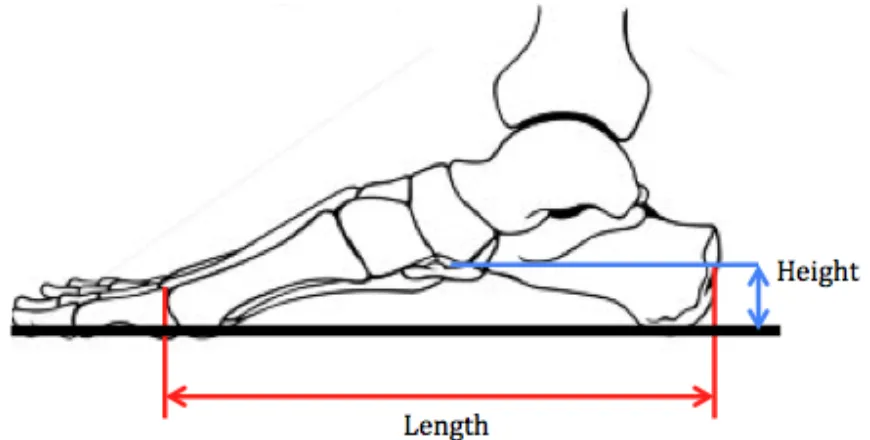
MLA height to length ratio
The most appropriate way to measure the MLA position has been debated. A simple method uses the length and the height of the MLA to characterize its shape via the truncated foot length. The truncated foot length is measured by the length from the heel to the first metatarsal head. This has been shown to be a valid approximation of MLA structure compared to radiographic images of the bones of the arch (Saltzman, Nawoczenski, & Talbot, 1995). The arch height to arch length ratio is commonly reported (Saltzman et al., 1995)(Figure 1.6). This ratio was used in the model validation study by Jenkyn & Nicol (2007).
As the arch goes up, the height to length ratio increases (narrows) and as the arch goes down, the height to length ratio decreases (widens). In both walking and running gait, as the foot reaches toe off, the arch narrows (height to length ratio increases). Researchers and clinicians find that the height to length ratio of the MLA is an important measure and it is often used in the determination and classification of foot pathologies and biomechanical gait analyses (Caravaggi et al., 2018).
and potential damage to soft tissues (Nachbauer & Nigg, 1992). Typically, arch height determines susceptibility to injury (Mei-Dan et al., 2005). Pes planus or flat footed people are more likely to pronate; over time this can lead to foot pain, heel pain and plantar fasciitis (Mei-Dan et al., 2005). Mei-Dan et al., (2005) found that those with low arches are more likely to have a higher rate of ankle sprains than in normal arched patients (55% to 39%). High arched or pes cavus people are more likely to stand supinated which can also lead to plantar fasciitis (Mei-Dan et al., 2005).
Figure 1.6 Medial Longitudinal Arch (MLA) height to length ratio (height/length) (Adapted from (Saltzman et al., 1995)). Here, height was the distance from floor to
the inferior aspect of the talar head. Length was the distance from the posterior surface of the calcaneus to the anterior surface of the first metatarsal head (Saltzman
et al., 1995).
1.3.4 Ligaments of the ankle and foot
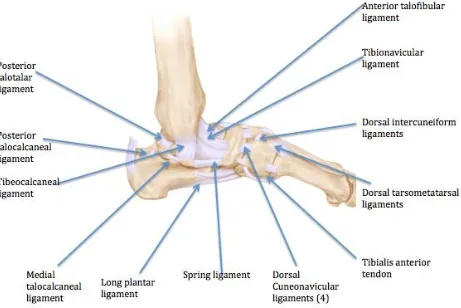
Ligaments are a form of connective tissue which hold bones together. Each of the joints and articulations previously defined are held together via ligaments. There are several
ligaments that hold the ankle together. The anterior and posterior talofibular ligament connect the talus to the fibula in the front and back via the fibular malleolus to the talus at the front of the lateral articular facet. The talofibular ligaments prevent the foot from excessively sliding forward relative to the tibia. In reference to injury, this is the most commonly injured ligament (Oae, Takao, Uchio, & Ochi, 2010).
The deltoid ligament is made up of the tibiocalcaneal ligament, the anterior and posterior tibiotalar ligaments and the tibionavicular ligaments. The tibionavicular ligament connects the tibia to the navicular via the navicular tuberosity to the medial malleolus and the
tibiocalcaneal ligament connects the tibia to the calcaneus via the sustentaculum tali on the calcaneus to the medial malleolus. The deltoid ligament provides stability to the hindfoot.
The spring ligament is attached to the anterior sustentaculum tali of the calcaneus to the plantar surface of the navicular. The function of the spring ligament is to hold up the bones of the MLA.
Figure 1.7 Medial view of the tendons and ligaments of the foot and ankle.
Within the midtarsal joint, there are five supporting ligaments (Figure 1.7). Stability for the calcaneocuboid joint comes from the plantar calcaneocuboid joint, the bifurcate ligament and the dorsal calcaneocuboid ligament. The talonavicular joint receives stability solely from the talonavicular ligament, and the cuboidnavicular joint is stabilized solely by the cuboidnavicular joint.
Metatarsophalangeal joints are supported and stabilized via metatarsophalangeal collateral ligaments. These ligaments limit plantar and dorsiflexion. The 1st metatarsal is also
metatarsals are stabilized on the medial and lateral sides via the metatarsoglenoid ligaments. Interphalangeal joints are stabilized both medially and laterally via interphalangeal collateral ligaments.
1.4 Gait cycle
Walking and running are cyclic activities where the legs and feet repeat specific movement patterns (Nordin & Frankel, 2001). The gait cycle is the repeating pattern of gait, such as from initial ground contact with one leg to the subsequent ground contact with the same leg. There are
different gait cycles that characterize walking and running. However, for both walking and running, one leg moves forward and strikes the ground, the body moves forward over top and then that leg is swung forward while the other leg is on the ground. Walking and running are characterized by the relative proportion of time that each leg spends on the ground (Nordin & Frankel, 2001). During running, both legs will spend less time on the ground, and there are two phases of double limb no support. In contrast, at least one leg is always on the ground during the gait cycle of walking.
1.4.1 Biomechanics of gait
The components of gait and the gait cycle require coordination of many movements of the legs including the hip, knee and ankle and foot (Nordin & Frankel, 2001). At initial contact, it is essential for the lower limb to be stable to allow for proper force absorption (Dugan & Bhat, 2005). This is provided by the hip adductors that remain contracted through the whole gait cycle (Dugan & Bhat, 2005).
In order for heel off to occur in the stance phase, controlled movement of the tibia occurs which allows dorsiflexion (Dugan & Bhat, 2005). It is during this phase that pelvic rotation occurs in order to create some external rotation of the stance limb (Dugan & Bhat, 2005). It is this external rotation that inverts the calcaneus and supinates the foot (Dugan & Bhat, 2005).
Toe off is caused by the contraction of the gastrocnemius and soleus to create supination in heel off that creates the rigid subtalar joint (Dugan & Bhat, 2005). In combination with the tightening of the plantar fascia, the intrinsic muscles of the foot play a large role, such as the abductor hallucis, flexor hallucis brevis, abductor digiti minimi and flexor digiti minimi (Dugan & Bhat, 2005). Muscles that cross the transverse tarsal joint also act to stabilize and solidify this joint (Dugan & Bhat, 2005). If knee flexion at this stage is not enough for the foot to clear the ground, the quadriceps muscle is activated to ensure the knee has enough flexion (Dugan & Bhat, 2005).
1.4.2 Walking gait cycle
For each leg, walking and running are both made up of a stance phase and the swing phase (Dugan & Bhat, 2005). For walking, the stance phase accounts for 60-65% of the walking cycle while the swing phase accounts for the remaining 35-40% of the cycle (Dugan & Bhat, 2005; Nordin & Frankel, 2001) (Figure 1.8). Since each leg and foot spends more time in stance phase than in swing phase, there are two portions of the walking gait cycle where both feet are on the ground and are impacted by the opposing ground forces (Nordin & Frankel, 2001). This is called double limb support (Nordin & Frankel, 2001) and it accounts for approximately 24% of the gait cycle (12% for each instance). The rest of the gait cycle is therefore single leg support with only one foot on the ground.
The stance phase for each leg begins at initial foot contact (heel strike) followed weight acceptance (Perry, Thorofare & Davids, 1992). Of these two events, weight acceptance is more demanding on the foot (Perry et al., 1992) and would be considered the foot strike, causing the most demand on the foot and thus, the shoe. These first two events of stance phase occur during double limb support. Single limb support consists of the next two events of stance phase: mid-stance and terminal stance events.
Midstance phase consists of the single limb support that happens after double limb support (Perry et al., 1992). One foot remains planted firmly on the ground as the other foot
prepares for the swing phase (Perry et al., 1992). The swing phase consists of the pre swing, initial swing, mid swing and finally, terminal swing. Terminal stance is what propels the body forwards (Perry et al., 1992). After terminal stance, there is another occurrence of double limb support (Perry et al., 1992). The limb advances and prepares the body for the next gait cycle.
Figure 1.8: Gait cycle break down into stance and swing. The stance phase is broken down into weight acceptance and single limb support and the swing phase is also
known as limb advancement (adapted from Perry et al., 1992).
commences right after initial contact and continues until the foot is flat (Perry et al., 1992). This is characterized by controlled ankle plantar flexion, knee flexion and hip stabilization (Perry et al., 1992). Shock absorbing in this stage creates stability and allows for the
continuation of forward movement (Perry et al., 1992). There is approximately 10° of ankle plantar flexion and subtalar valgus rolling the heel to flat foot (Perry et al., 1992).
The next stage of the gait cycle is from 10-30% of the gait cycle where the ankle is in dorsiflexion, knee extension and hip stabilization (Perry et al., 1992). This is where the body weight goes over the planted foot and contributes to limb and trunk stability (Perry et al., 1992). Midstance phase is defined as the other foot is lifted and continues until the body weight is aligned over the forefoot (Perry et al., 1992). Both the heel and forefoot remain on the ground however the tibia rotates above the ankle rocker.
After midstance, from 30-50% of gait cycle and begins with a heel rise and a free forward roll of the body forward (Perry et al., 1992). This phase continues until body weight is transferred over to the other foot or until heel strike of the other foot (Perry et al., 1992). The forefoot will be the only body weight support as the heel rises and the ankle is now in 5-10° of dorsiflexion.
Pre-swing occurs at 50-60% of the gait cycle and is defined with knee flexion (Perry et al., 1992). It begins with initial contact on the opposite foot and ends with toe off (Perry et al., 1992). The weight transfer is the cause of the high heel rise seen (Perry et al., 1992).
Initial swing occurs from 60-73% of the gait cycle and is defined by knee and hip flexion (Perry et al., 1992). The purpose of this stage is for foot clearance and subsequent limb advancement (Perry et al., 1992). The stage begins with the foot lifting off the floor and ends with the opposite foot swinging (Perry et al., 1992).
Mid swing occurs from 73-87% and has the purpose of limb advancement and foot
clearance from hip flexion and ankle dorsiflexion (Perry et al., 1992). This phase starts with the swinging limb in line with the planted limb and ends with the tibia vertical on the stance leg (Perry et al., 1992).
Figure 1.9: One complete gait cycle during walking shown in percentage as A) the stance phase and B) the swing phase for one foot (adapted from Nordin & Frankel,
1992).
1.4.3 Running gait cycle
The running gait cycle is different from the walking gait cycle. The stance phase has a decreased duration, accounting for 40% of the running cycle, while the swing phase takes up more of the gait cycle, accounting for 30% (Nordin & Frankel, 2001). In comparison to walking, there is another distinct component. Between the stance and the swing phase are two series of float phases, each encompassing 15% of the gait cycle (Nordin & Frankel, 2001) (Figure 1.9). The float phase is the time during running when both feet are off the ground (Nordin & Frankel, 2001).
Toe off allows the body to go into it’s first phase of floating. This is where both legs are off of
the ground using momentum from the big toe to still move forwards followed by the other leg on the ground, starting the gait cycle again. After mid swing comes terminal swing and lastly, initial contact occurs once again. Terminal swing is the follow through forward of the swing that allows the foot to touch the ground again and start the gait cycle again.
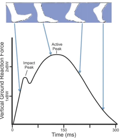
More specifically, during the initial contact to midstance (Figure 1.9), the foot is supinated (Bates, Osternig, Mason, & James, 1978; R. A. Mann, Baxter, & Lutter, 1981). Compared to walking, after heel strike there is no plantar flexion at the ankle as the ankle immediately goes into dorsiflexion which is what allows the foot to move into pronation (R. Mann & Inman, 1964). Proper absorption during this initial contact is attributed to a conjunction of cartilage compression, joint motion and muscle contraction (Dickinson, Cook, & Leinhardt, 1985). At this stage hip and knee flexion play a large role in the absorption of ground reaction forces (R. A. Mann et al., 1981). For those who consistently run in running shoes, the initial peak force or impact peak occurs at around 10% of the stance and lasts less than 30ms (Hreljac, 2004). Also known as a braking force, this is the initial force used to slow the body down upon heel strike(Goss & Gross, 2012). In the ankle, movement of the subtalar joint allows for the ankle to absorb and dissipate many forces during running. As the subtalar joint pronates within the first 20% of the stance phase, the transverse tarsal joint axes become parallel. Accordingly the forefoot and thus the forefoot is now more mobile and can adjust to uneven terrain (Rodgers, 1988).
During the period from midstance to toe off, or the propulsion part of the stance phase, dorsiflexion occurs as the foot is now fixed to the ground (Dugan & Bhat, 2005). A
maximum in dorsiflexion is seen here as soon as the tibia has gone past the ankle (Dugan & Bhat, 2005). It is also important to note that as maximum dorsiflexion and thus pronation is reached, there are many forces acting on the knee (Dugan & Bhat, 2005). As these forces come to their maximum the quadriceps and hamstrings will contract thus stabilizing the knee (Dugan & Bhat, 2005). Heel lift can occur after maximum pronation as the pelvis is forced to rotate causing the tibia to rotate externally, the calcaneus to rotate internally and supination occurs forcing the heel off of the ground (Dugan & Bhat, 2005).
Acceleration of the stance leg is initiated as the foot is now in plantar flexion (Adelaar, 1986). This plantar flexion causes the foot to go into supination, locking the transverse tarsal joint and creating a rigid base for propulsion (Dugan & Bhat, 2005). This propulsion is where the second maximum in ground reaction force occurs (Dugan & Bhat, 2005) (Figure 1.8).
Initial swing occurs as the body is propelled forward and the ground reaction forces pass posterior to the knee, forcing the knee to flex (Dugan & Bhat, 2005). Knee flexion that occurs here cancels out the need for the ankle to become dorsiflexed in order for the foot to clear the ground (Dugan & Bhat, 2005).
flex, thus the need for contraction of the quadriceps to protect the knee joint (Dugan & Bhat, 2005). The second peak force known as the active peak occurs at about 0-75% of the stance phase and lasts considerably longer (up to 200ms) (Hreljac, 2004). This is due to the
amount of force used to propel the body forward into the float phase, where both feet are off of the ground (Goss & Gross, 2012). Once the foot has touched the ground the body is prepared to commence another gait cycle.
Figure 1.10 Running gait cycle as adapted from Onupuu, 1994. During running there are two series of float phases seen where both feet are off of the ground in comparison to walking where there are none. There is also an increase of time spent
in the swing phase and a decrease of time spent in the stance phase.
Figure 1.11 Vertical ground reaction forces seen during one gait cycle (adapted from Nordin, 2012). There are two instances were GRFs peak, once on impact and the
second towards the end of stance phase, as the body is propelling forwards.
1.5 Running
1.5.1 Running patterns
There are three distinct running patterns. The first is a forefoot strike, which can be characterized by initially striking on the forefoot, followed by rocking and transferring weight to the heel (Lieberman et al., 2010). As the foot approaches the ground the ankle is plantarflexed, just like hindfoot or heel strike runners, however forefoot strikers will generally land on the outer edge of the foot landing under the 4th and 5th metatarsals. The hip and knees will remain flexed. After the initial strike, the ankle will naturally begin to dorsiflex as the weight and load are dispersed throughout the foot. This loading will force the foot to flatten and thus the arch begins to stretch. As the foot and ankle prepare to toe off, the foot will start to evert and pronation begins. Pronation is a combination of eversion, dorsiflexion and arch flattening (Dugan & Bhat, 2005). The ankle will then plantarflex as a result of the contraction of the calf muscles and the toes flex propelling the body into float phase.
and knees are still in a flexed position, the ankle is dorsiflexed and runners typically land on the heel or just below the ankle joint. Once the initial contact has taken place the ankle will plantarflex however the arch remains unloaded as the forefoot comes down with the help of the muscles of the anterior shin. The foot will then evert and the foot finally comes to a flattened position, loading the arch. The foot is now in pronation so that the Achilles and calf muscles can now plantarflex the ankle, flex the toes and propel the body into the air into float phase.
The third running pattern is a midfoot strike. It is characterized by the foot landing flat or the midfoot hitting the ground upon initial contact followed by transferring weight to the heel (Lieberman et al., 2010). Midfoot strikers have the same amount of hip and knee flexion as both forefoot and hindfoot strikers. In regards to the initial contact, contact is going to take place when both the heel and forefoot land simultaneously. As the initial strike happens, the foot is already flattened so the arch is loaded immediately and the ankle goes into plantar flexion and propels the body forwards into float phase and prepares the body for another gait cycle.
It is also important to note that there is a decreased ground contact time seen in runners with a forefoot strike and midfoot strike (De Wit, De Clercq, & Aerts, 2000; C Divert et al., 2008; Nunns et al., 2013). Forefoot striking also increases pressure put on the metatarsal heads (Altman & Davis, 2012). This decrease in ground contact time can be attributed to the fact that during forefoot striking and midfoot striking there is less dorsiflexion at the ankle. With less dorsiflexion occurring, the ankle becomes flattened quicker and thus the arch becomes loaded sooner. This means that the foot is able to plantarflex and propel the body forward sooner preparing the legs for the next gait cycle.
Although, characterizations in strike patterns have been studied in relation to energy expenditure and injury rates, some studies have shown that there is no direct benefit from transitioning a strike pattern either from forefoot to rearfoot, or from rearfoot to forefoot (Hamill & Gruber, 2017). Perhaps, changing footstrike pattern can actually lead to injuries sustained from stressing the feet (Hamill & Gruber, 2017). This study identifies that foot strike pattern appears to depend on the type of run, as running longer distances may cause a rearfoot strike and faster, shorter runs may cause a forefoot strike (Hamill & Gruber, 2017). On a harder surface, runners were less likely to rearfoot strike, have a higher step cadence and thus, were able to run faster (Lieberman et al., 2015).
1.5.2 Barefoot runner’s patterns
Experienced long distance barefoot runners most often have similar forefoot running patterns (Lieberman et al., 2010). Forefoot runners will exhibit a flatter foot placement (ie. more plantar flexion, less dorsiflexion) and the flatter the foot, the smaller pressure on the heel (De Wit et al., 2000). The ankle is more plantarflexed at landing (Squadrone &
has also been shown that barefoot runners exhibit a higher cadence or stride rate compared to traditional shod runners (Moody, Hunter, Ridge, & Myrer, 2018).
Runners are likely to run barefoot if they have grown up barefoot running such as
tribesmen in remote locations in the world, those who have transitioned from minimalist shoes into full barefoot running, and those who believe it is better for their feet. Many runners currently wear or have tried training in minimalist shoes. This has motivated the purpose of this study: to see whether barefoot mimicking shoes are significantly different than running in regular cushioned running shoes.
1.5.3 Shod Runner’s patterns
Shod runners are runners that wear running shoes that are typically cushioned. They often initially strike the ground with the hindfoot, followed by the ball of the foot (Lieberman et al., 2010). Research suggests the reason that shod runners rearfoot strike is that this may have a lower metabolic cost associated for longer distances (Hamill & Gruber, 2017).
Rearfoot runners strike the ground in inversion and change to eversion at midstance (Stackhouse, Davis, & Hamill, 2004). At heel contact, the heel and forefoot will be slightly supinated, that is, inversion, dorsiflexion of the ankle and flattening of the arch (Rodgers, 1988). At midstance, the foot will be pronated from 55-85% of support phase and the foot will supinate and return to neutral around 70-90% of support (Rodgers, 1988). The foot will be supinated for push-off (Rodgers, 1988) and the ankle is more dorsiflexed upon landing (Squadrone & Gallozzi, 2009).
1.5.4 Impact force & injury
Impact forces upon initial contact cause sudden, large forces to travel up the body (Milner et al., 2006; Pohl et al., 2009; van Gent et al., 2007) (Figure 1.11). Repetitive high loading on the joints and tissues are thought to explain many injuries such as tibial stress fractures and plantar fasciitis. The ground reaction force experienced during running can be nearly
double that of walking, such as 1.5-2 times body weight during running (Milner et al., 2006). This initial peak force is seen in the first 50ms of the heel strike (Bobbert, Schamhardt, & Nigg, 1991). During the strike phase, the internal leg muscles are lengthened, creating forces and stress (Bobbert et al., 1991).
Although there is a decreased force associated with barefoot running, shorter strides leads to an increased cadence or frequency of foot strike, so any potential decreases in force upon impact associated with barefoot running could be counteracted by the increased frequency of foot strike and thus, potential injury (Collier, 2011). A study by Shih et al., (2013), found that barefoot runners had a significantly higher cadence than shod runners.
1.6 Injury
One study, described 2002 running injuries and determined that the four most common sites of injury ranked from highest to lowest were the knee (42.1%), foot and ankle
(16.9%), lower leg (12.8%), hip and pelvis (10.9%) with the top three injuries in this study were patellar femoral pain syndrome, iliotibial band friction syndrome, and plantar fasciitis (Taunton et al., 2002). As the feet are exposed to approximately 10,000 steps an hour, poor biomechanics, such as excessive pronation or supination during heel strike, can lead to injury over time (Macera et al., 1989).
In a study done by Messier et al., 2018, 300 runners were studied over the course of a 12 month training period. The findings were that at least one injury occurred to 66% of the runners within the initial 12 months and more interestingly, more women than men were injured (Messier et al., 2018). The most common sites for injuries were the knee and foot (Messier et al., 2018).
1.6.1 Training errors
Training errors play a large role in injury occurrence (Shorten, 2000). These are most acknowledged as anything that causes an increase load being placed on the body, such as sudden increases in duration and intensity, constant intense training, a single intense training day or race and/or sudden return (Shorten, 2000). Training errors can be
attributed to many running injuries, specifically repetitive strain injuries (Shorten, 2000). Cross training, small mileage increases (of about 10% per week) and most importantly rest can help to prevent overtraining, training errors and repetitive strain injuries (Shorten, 2000). A study by Altman & Davis (2016), was one of the first scientific study to look at habitually shod and barefoot runners over the course of a year and track rates of injury. The study noted that there was a significantly lower number of injuries in the barefoot group, although when normalized for distance, there was no significant difference between the two groups (Altman & Davis, 2016).
1.6.2 Intrinsic & extrinsic factors to injury
Running injuries can be caused by a variety of intrinsic and extrinsic factors (Taunton et al., 2002). Intrinsic factors are those that are concerned with the runner themselves, such as experience or anatomical limitations, while external factors are training errors, old shoes and unpredictable running terrains (Taunton et al., 2002).
Extrinsic factors are those that are concerned with the environment, such as running terrain, type (i.e. long distance, sprinting, racing, etc.), and age of shoe (Taunton et al., 2002). Running terrain includes running on trails, sand, concrete and asphalt. If the terrain is bumpy or sloped to one side for a large part of a run, over a prolonged period of time the hips could be uneven, causing repetitive stress on one side of the knee, causing pain such as iliotibial band syndrome (Taunton et al., 2002). Running on sand inhibits the ability to effectively arch the foot required to propel the body forward (Taunton et al., 2002). This is because as the body is arching the foot trying to pronate to excel forward, the sand
underneath starts to give, causing the arch to work harder to propel. Concrete is going to be harder to run on for the body, as there is very little give and therefore any force put on the concrete will come right back up into the lower limbs (Taunton et al., 2002). This repetitive stress can cause things such as shin splints, iliotibial band syndrome, etc. Asphalt has a little more give to it as it is not as dense. This means that as the body exerts force, some of it can be dissipated into the ground and less comes back to the lower limbs (Taunton et al., 2002).
1.6.3 Mechanisms behind injury
High instantaneous loading, (during heel strike and toe off), high ground reaction forces and lower medial longitudinal arch heights were shown to be some key factors in the development of plantar fasciitis (Pohl et al., 2009). As ground reaction forces and loading increases, so does the strain placed on the plantar fascia, potentially resulting in plantar fasciitis over time (Pohl et al., 2009).
Excessive varus and valgus movements, eversion, and tibial rotation have been suggested to cause injury in long distance runners (James, Bates, & Osternig, 1978; B. M. Nigg, 2001). Excessive movements can occur in any form; shod, barefoot or in barefoot mimicking shoes can lead to repetitive strain injuries (B. M. Nigg, 2001). Correcting alignment of the lower limb and ankle through proper running shoe design, orthotics or other inserts could correct these risk factors (B. M. Nigg, 2001). Having correct alignment of the body can reduce or eliminate excessive movements (B. M. Nigg, 2001).
As the majority of the population does run shod, studies concentrate on this group of runners. Within the past few years as barefoot and minimalist running becomes more popular, more studies have emerged that looked into barefoot running and minimalist running. One study by Decker, Torry, Wyland, Sterett, & Steadman, 2003, has shown that barefoot running and minimalist shoes cause a decrease in hip and knee flexion which have been linked to increases seen in ACL injuries. This decrease in hip and knee flexion can be attributed to the decrease in stride length, suggesting that the lower leg is more centred beneath the body on foot strike (Soares et al., 2018). Another study by Sinclair, Butters, & Stainton, 2018, found that running in barefoot and minimalist shoes cause an increase in knee adduction on instantaneous loading. This medial knee compartment loading is a strong predictor for knee osteoarthrits (Sinclair et al., 2018).
1.7 Anatomy of the running shoe
motion control. The cushioned midsoles are made of EVA foam with inserts of gels, dual density foam, air or fluids in attempts to increase cushioning (Mcpoil, 2000). These are seen is models such as the Nike Air. The use of cushioning systems in modern day running shoes, help to attenuate impact and ground reaction forces acting on the foot (Mcpoil, 2000). However, cushioning systems do not completely absorb forces causing the body to bear the remaining impact repeatedly over the course of a bout (Mcpoil, 2000).
Heel wedges are made up of polymeric foam and the outsole is made of hard rubber (Frederick, 1984). Reinforcements are common throughout the rest of the shoe, mostly in places such as the toe, heel and midsole (Mcpoil, 2000). This helps lengthen the longevity of the shoe and thus, the time to replace is also lengthened. Running shoe design has the ability to affect both speed and energetics based on factors of age, model, materials and quality of the shoe (Frederick, 1984).
1.12 Anatomy of the standard running shoe. A strong shank of a shoe can increase torsional strength and aide in motion control.
1.7.1 Heel counter & wedge
Most running shoes are designed with a high wedge at the heel, located underneath the midsole (Lieberman et al., 2010) (Figure 1.12). This forces the runner to have a rear foot strike (Lieberman et al., 2010). The heel strike is forced as there is a heel-toe offset whereas the heel is higher than the forefoot, naturally guiding the runner to heel striking. Many
shoes have arch supports and/or stiff soles that decrease the runner’s ability to strengthen
such as plantar fasciitis (Lieberman et al., 2010). Although heel wedges are shown to potentially be the cause of some injuries, shoes have a heel wedge as this is where shock absorbing materials are found, providing cushioning to the heel. This cushioning is vital as repeated impacts such as those found in exercise and more specifically running help protect the heel.
The heel counter supports the back of the ankle and Achilles during running and activity and is located between the heel tab and the midsole (Figure 1.12). The heel counter aids in ensuring a good fit between the shoe and the foot, preventing excessive motion during dorsiflexion of the foot at heel strike also known as motion control (Mcpoil, 2000).
1.7.2 Motion control
Both anti pronation and motion control has been a factor in running shoe design since the
early 1980’s (Shorten, 2000). It was originally incorporated into shoes via heel wedging or flaring the heel (Stacoff, Nigg, Reinschmidt, van denBogert, & Lundberg, 2000). Motion control aids in preventing the foot from excess supination or pronation of the foot during ground contact. The foot needs a level of pronation in order to propel the body forward as the heel strikes, the foot pronates, and then supinates along the outside edge and ends in pronation by pushing off of the big toe. This motion control is accomplished by adding stiffer cushioning, heel counters, insoles and wedges (Shorten, 2000). Repeated and excessive pronation or supination of the foot during running can result in repetitive strain injuries such as plantar fasciitis and iliotibial band syndrome (Shorten, 2000). Individuals with pes planus, or flat feet are more likely to over pronate, requiring a stabilizing shoe, while individuals with pes cavus, or high arches are more likely to have reduced pronation requiring a cushioned shoe (Shorten, 2000).
Another form of motion control comes from traction of the outersole (Mcpoil, 2000). Without traction, activities performed could be dangerous especially over difficult terrains (Mcpoil, 2000). This suggests the idea that shoes should be monitored on a regular basis for wear, as treads will degrade over use, time, and frequency, thus signalling to the user to replace them.
Another motion control factor is the motion control over the rear and midfoot (Mcpoil, 2000). This is because foot pronation is controlled by heel stabilization and midfoot
1.7.3 Shoe use
Mcpoil, (2000) suggests that runners be vigilant about recording shoe usage. Under each step, the midsole of the shoe undergoes stress and compression (Mcpoil, 2000). Over the course of a run, the compression can decrease midsole shock effectiveness by 25-30% (Mcpoil, 2000).
Many people are not aware of the miles they run, weather they train in or surfaces trained on (Mcpoil, 2000). All of these factors have an impact on shoe durability and lifespan (Mcpoil, 2000). Wearing shoes that have had too many miles run on them mean that the cushioning systems may be ineffective at helping to dissipate ground reaction forces, thus more force is acting on the joints (Mcpoil, 2000). This could have implications for injury so runners should be vigilant about recording mileage to help decrease the likelihood of injury.
1.7.4 Lacing design of running shoes
The way that shoes are laced and the placement of the lacing eyelets are an important design of all running shoes (Hong, Wang, Li, & Zhou, 2011). Most importantly, laces allow the foot to be secured into the foot bed or the sole of the shoe and against the heel counter (Hong et al., 2011). This also allows the individual user to create a custom fit to the shoe and increases comfort (Frey, 2000).
It has been suggested the purpose of laces are to create a better fit and more importantly, distribute stress evenly over the dorsum of the foot (Hong et al., 2011). Hong et al. (2000), also found that lacing conditions have a significant influence on shoe-foot coupling during running. That is, the better laced they are for a specific foot type, the better the shoe will fit. The same goes for the more the lacing technique does not match a foot type, the worse the shoe will fit.
1.8 Barefoot simulation shoes
1.8.1 Pressure and ground reaction force during barefoot running
Ground reaction force is the force the body exerts when the foot strikes the ground. Barefoot running can be characterized as having increased loading rates due to minimal external protection and shock absorption as compared to running shoes (De Wit et al., 2000). The flatter the foot is upon impact, the smaller the pressure on the heel as the foot better dissipates forces upon impact (De Wit et al., 2000). This suggests that perhaps barefoot runners aim to flatten their feet as they run to diminish heel impact (De Wit et al., 2000). There is also evidence that barefoot running creates the highest vertical ground reaction forces (De Wit et al., 2000). These high vertical ground reaction forces repeated over time can be a leading cause of injury for both barefoot runners and minimalist runners.
1.8.2 Stride frequency and impulse
Pressure under the toes was significantly higher with VFF compared to barefoot running (Squadrone & Gallozzi, 2009). Perhaps this can be attributed to the fact that the VFF have a thick rubber sole, limiting it’s ability to dissipate pressure compared to running barefoot.
This could have injury implications for the metatarsals, as increases in pressure over time can be a factor in the development of a repeated strain injury.
1.8.3 Characterization of a barefoot running shoe
Barefoot running shoes or minimalist shoes or barefoot mimicking shoes seek to create a thinner, more flexible rubber sole (Lieberman et al., 2010). This sole acts to protect the foot from any abrasions from the ground (Figure 1.13). Bonaccini, et al. (2013), classify
minimalist shoes as having a low profile, increased sole flexibility, reduced offset between the heel and forefoot (<4 mm), little motion control, lightweight and the minimalist part really coming from having very little cushioning and motion control (Bonacci et al., 2013; Cauthon, Langer, & Coniglione, 2013). This small heel-toe offset results in runners adopting a forefoot strike pattern. Other characteristics a light outer sole and little or no heel counter (Mcpoil, 2000). VFF do have an upper, however it acts solely to hold the foot to the foot bed, as does the Velcro strap. The Velcro strap also holds the heel to the back of the shoe so the toes do not slide forward during running.
Figure 1.13 VFF barefoot mimicking minimalist shoes. These shoes typically contain little to no support however offer a protective barrier between the sole of the foot
Nigg, (2009), suggests that there is indirect evidence that barefoot running strengthens various muscles crossing at the ankle joint as well as big muscle groups (i.e. quadriceps and hamstrings) and small muscle groups (i.e. peroneus longus and soleus)(B. Nigg, 2009). Running shoes use more energy as the acceleration and deceleration power required with the weight of a shoe, energy used to deform the shoe during running, energy used to rotate the shoe on ground contact, energy absorbed in the midsole and energy lost in stiffness of the joints of the foot (B. M. Nigg, 2001; Warburton, 2001).
1.8.4 Hazards of barefoot running
The largest external hazard of running barefoot (and in barefoot simulated (i.e. minimalist) running shoes) is the potential of injury caused by debris (Murphy et al., 2013). This
includes nails, glass, wood chips (slivers) and stones (Squadrone & Gallozzi, 2009). A study by Lieberman et al., (2016) found that habitually barefoot runners exhibit significantly more injuries to the plantar surface of the foot.
In general, it is thought that those transitioning from a cushioned midsole to either barefoot running or barefoot simulated running shoes may take a longer time than those simply transitioning from a minimalist shoe to barefoot running (Murphy et al., 2013). In order to properly transition to barefoot running, barefoot or minimalist shoes should slowly be integrated into the workout, just as a new exercise would be (Murphy et al., 2013).
1.8.5 Why transition to barefoot running?
Barefoot enthusiasts and historical barefoot runners claim that running barefoot strengthens the intrinsic muscles of the feet and that shod running makes the intrinsic muscles of the feet potentially weaker (Rossi, 1999; Wikler, 1961). It is thought that even with greater improvements in cushioning and motion control, injury rates are not declining (Lieberman et al., 2010).
Theories behind advantages of barefoot running include; a reduction in impact forces (C Divert, Mornieux, Baur, Mayer, & Belli, 2005; Caroline Divert, Baur, Mornieux, Mayer, & Belli, 2005; Squadrone & Gallozzi, 2009), reduced oxygen consumption (Burkett, Kohrt, & Buchbinder, 1985; Catlin & Dressendorfer, 1979), increased proprioception (Lieberman et al., 2010), and increased intrinsic muscles (Rao & Joseph, 1992).
1.9 Studies and kinematic outcomes
1.9.1 Previous studies