Copyright © 1998 by the Genetics Society of America
Genetics 148: 139–149 ( January, 1998)
A Mutation in
Paramecium tetraurelia
Reveals Functional and Structural
Features of Developmentally Excised DNA Elements
Kimberly M. Mayer,* Kazuyuki Mikami
†and James D. Forney*
*Department of Biochemistry, Purdue University, West Lafayette, Indiana 47907-1153 and †Research Institute for Science Education, Miyagi University of Education, Aoba-ku, Sendai 980, Japan
Manuscript received April 22, 1997 Accepted for publication September 18, 1997
A B S T R A C T
The excision of internal eliminated sequences (IESs) from the germline micronuclear DNA occurs dur-ing the differentiation of a new macronuclear genome in ciliated protozoa. In Paramecium, IESs are gen-erally short (28–882 bp), AT rich DNA elements that show few conserved sequence features with the excep-tion of an inverted-terminal-repeat consensus sequence that has similarity to the ends of mariner/Tc1 transposons (Klobutcher and Herrick 1995). We have isolated and analyzed a mutant cell line that
can-not excise a 370-bp IESs (IES2591) from the coding region of the 51A variable surface protein gene. A sin-gle micronuclear C to T transition within the consensus sequence prevents excision. The inability to excise IES2591 has revealed a 28-bp IES inside the larger IES, suggesting that reiterative integration of these ele-ments can occur. Together, the consensus sequence mutation and the evidence for reiterative integration support the theory that Paramecium IESs evolved from transposable elements. Unlike a previously studied Paramecium IES, the presence of this IES in the macronucleus does not completely inhibit excision of its wild-type micronuclear copy through multiple sexual generations.
AT rich, single-copy sequences that are invariably flanked by 59-TA-39 dinucleotides, only one copy of which remains in the macronuclear DNA after excision (Steele et al. 1994). No significant open reading
frames have been found inside the eliminated regions. In the micronuclear DNA, IESs frequently interrupt coding regions and their precise removal is required to maintain an open reading frame in the transcription-ally active macronuclear genes. Based on the number of IESs found so far, it is estimated that as many as 65,000 per haploid genome are removed during macro-nuclear development (Duharcourtet al. 1995).
The determination of the macronuclear and micro-nuclear DNA sequences for different variable surface protein genes has allowed the comparison of evolution-arily related IESs in Paramecium. Variable surface pro-tein genes are a family of related genes that encode abundant, high molecular weight (250–300 kD) sur-face proteins which are mutually exclusive in their ex-pression (reviewed in Preer 1986; Caron and Meyer
1989). Alignment of the micronuclear A and B surface protein gene sequences revealed that the positions of at least three IESs are conserved between the two genes. Comparisons between the sequences show that only the first 4 or 5 nucleotides at each end are identi-cal, and the remaining sequence shares little recogniz-able similarity (Scottet al. 1994a). Even the size of the
eliminated DNA is not conserved; IES4578 in the A
gene is 882-bp in length but its corresponding IES in the B gene is only 416-bp.
Despite this general lack of conserved sequence
fea-Corresponding author: James D. Forney, Purdue University, Depart-ment of Biochemistry, 1153 Biochemistry Bldg., West Lafayette, IN 47907. E-mail: [email protected]
P
ROGRAMMED DNA rearrangements occur in awide variety of eukaryotic organisms. Examples range from mating type switching in the yeast, to the re-combination of immunoglobulin genes in mammals. Among all the eukaryotic phyla, some of the most dra-matic and extensive examples of DNA rearrangement occur in ciliated protozoa. These organisms undergo massive developmentally controlled DNA rearrange-ments as a normal part of their sexual reproductive cy-cle (reviewed in Prescott 1994; Coyne et al. 1996;
Klobutcher and Herrick 1997).
Like other ciliates, Paramecium tetraurelia contains two morphologically and functionally distinct types of nuclei. The micronuclei are transcriptionally silent and contain the germline genome. In contrast, the ampli-fied DNA in the macronucleus is transcriptionally active and, thus, determines the phenotype of the cell. During sexual reproduction, the old macronucleus is destroyed and a new macronuclear genome is created from the micronuclear DNA. Several types of developmentally regulated DNA rearrangements occur as part of this process, including fragmentation of the germline chro-mosomes, de novo addition of telomeres, and DNA splic-ing events that eliminate thousands of non-codsplic-ing se-quences called IESs (internal eliminated sese-quences).
140 K. M. Mayer, K. Mikami and J. D. Forney
tures, a detailed statistical analysis revealed that Para-mecium IESs share a defined, albeit poorly conserved, terminal-inverted-repeat consensus sequence of 8-bp at each end of the IES (Klobutcher and Herrick 1995).
Interestingly, the consensus sequence is closely related to the terminal-inverted-repeats of mariner/Tc1 trans-posons (Klobutcher and Herrick 1995), a large
fam-ily of transposable elements found in organisms rang-ing from insects to fish (reviewed in Robertson 1995).
This observation provided the first indirect evidence that Paramecium IESs evolved from a specific class of transposons. The transposon evolution theory assumes that the ciliate host appropriated control of the exci-sion activity which it supplies in trans; thus, the se-quence of each element is constrained only to maintain the cis-acting sequences required for excision (K lo-butcher and Herrick 1997). Additional support for
this theory is provided by studies of Euplotes crassus, an-other ciliate with IESs bordered by 59-TA-39 dinucle-otides. In this organism, micronuclear specific trans-posons called Tec1 and Tec2 share identity with the terminal-inverted-repeat consensus of Paramecium IESs as well as the mariner/Tc1 transposon family (K lo-butcher and Herrick 1995). Despite considerable
data from comparative sequence analysis, no study has provided direct evidence that the terminal-inverted-repeat consensus in Paramecium or Euplotes is func-tionally important for IES excision.
In addition to cis-acting signals in the micronuclear DNA, there is increasing evidence that DNA rearrange-ments in the new macronucleus are influenced by the content of the old macronucleus. The first evidence for this effect was obtained from cell lines that have a macronuclear deletion of the A surface protein gene but contain wild-type micronuclei (Epstein and F or-ney 1984; Kobayashi and Koizumi 1990). Despite the
presence of normal micronuclei, the macronuclear de-letion is inherited by the new macronucleus after sex-ual reproduction. The effect is gene specific, and it is controlled though the cytoplasm by a trans-acting signal originating in the old macronucleus. More recently, the effect of the old macronucleus has been observed for
Paramecium IESs. Duharcourt et al. (1995) showed
that the presence of an IES in the old macronucleus in-hibits the excision of the corresponding micronuclear IES during the next round of sexual reproduction. This inhibition is IES specific and copy-number dependent; macronuclei containing more copies of the IES are bet-ter able to inhibit developmental excision. The remark-able specificity of the effects of the old macronucleus have led to models that propose nucleic acid interac-tions between RNA or DNA from the old macronucleus
and the DNA in the developing macronucleus (Meyer
and Duharcourt 1996a,b; Forneyet al. 1996). It is
un-clear whether the inhibitory effect of IESs in the macro-nucleus is a general property shared by most Parame-cium excised elements or only a small subset.
We took a classical genetics approach to identify mu-tations in cis-acting elements which disrupt IES exci-sion. In theory, our approach could also isolate muta-tions in trans-acting elements important in the effect of
the old macronucleus on DNA rearrangements. The A
surface protein gene, which contains 7 IESs within its coding region, is an excellent target for mutagenesis of IES excision. Failure to remove any IES will create ei-ther a frameshift or a stop codon and prevent A surface protein expression. We selected mutants that were un-able to express the A surface antigen, then analyzed the resulting collection of A2 cell lines for mutants defec-tive in the removal of IESs from the A gene coding region. The first mutant we identified fails to remove IES2591 from the A gene. Molecular and genetic analy-sis of this cell line demonstrated that a micronuclear mutation consisting of a single base-pair change within the terminal-inverted-repeat sequence is responsible for the defect in excision. In addition, this mutant cell line has revealed the presence of a 28-bp IES inside IES2591. The small internal IES has ends which match the inverted consensus sequence including the flank-ing 59-TA-39 repeats, and it is removed from the mutant during macronuclear development even though the re-maining portion of IES2591 is not excised. Evidence for internal IESs in Paramecium supports the transpo-son evolution hypothesis because it implies that IESs are mobile elements that can integrate into other IESs. This is analogous to the reiterative integration of some classes of transposons (San Miguelet al. 1996; Bryan et al. 1990). Although the presence of either the wild-type or the mutant version of IES2591 in the macro-nucleus has a partial inhibitory effect on the excision of its wild-type micronuclear copy, the inhibition is not maintained through several sexual generations as ob-served for the previously studied example of IES inhibi-tion (Duharcourtet al. 1995). The combination of
ev-idence for internal IESs and a functionally important base pair in the consensus sequence shared with the
mariner/Tc1 transposon family supports the hypothesis that Paramecium IESs have evolved from transposable elements.
M AT E R I A L S A N D M E T H O D S
Cell lines, media, and growth conditions: P. tetraurelia stock 51 is homozygous for the 51A surface antigen gene. Line 51ND was derived from stock 51 and contains a Mendelian mutation that prevents trichocyst discharge but is wild-type at the A locus. Line d12 was originally derived from stock 51 and contains macronuclear and micronuclear deletions of the 51A gene (Rudmanet al. 1991). All cells were cultured in a
0.25% wheat-grass medium buffered with 0.45 g/L sodium phosphate and supplemented with 0.25 mg/L stigmasterol. The medium was inoculated with a nonpathogenic strain of Klebsiella pneumoniae 1–2 days prior to use. All cell lines were maintained at 278 and cultured as described by Sonneborn
(1970).
Paramecium IES Excision Mutation 141
expressing A surface protein were concentrated by centrifu-gation into a volume of 50 ml. The cells were added to an equal volume of 0.15 mg/ml nitrosoguanidine (1-methyl-3-nitro-1-nitrosoguanidine) dissolved in Dryl’s solution (2 mm
sodium citrate, 1.0 mm Na
2HPO4, 1.0 mm NaH2PO4, 2 mm CaCl2). After mutagenesis, the cells were divided into 24 dif-ferent tubes and starved to induce autogamy. The resulting homozygous cells were cultured for 8–10 fissions at 348 to in-duce A expression and then treated with anti-A serum. The surviving cells were grown an additional 8–10 fissions at 348, and again treated with anti-A serum. Finally, clonal lines were isolated and scored for A expression.
Total DNA isolation: Two-hundred ml of cell culture (1000 cells/ml) were pelleted in pear-shaped flasks, re-suspended in 0.4 ml of culture fluid, then quickly squirted into 0.8 ml of low EDTA lysing solution (10 mm Tris pH 9.5; 50 mm EDTA;
1% SDS; 0.1 mg/ml proteinase K) and incubated at 658 for 10 min. The DNA was then extracted once with phenol:chloro-form (1:1) and once with chlorophenol:chloro-form before being precipi-tated with 0.1 volume 3 m NaOAc and 2.5 volumes 95%
EtOH. The DNA pellets were re-suspended in 100 ml 13 TE (10 mm Tris pH 8.0, 1 mm EDTA).
Southern blot analysis: Southern blot analyses were per-formed according to the method of Sambrooket al. (1989).
In Paramecium, macronuclear DNA is approximately 250 times more abundant than micronuclear DNA; thus, a South-ern blot of total genomic DNA is essentially an analysis of macronuclear DNA. Total genomic DNA was digested with XbaI and EcoRI, then run on a 1.2% agarose gel. The gel was blotted onto a nitrocellulose filter (Schleicher & Schuell, Keene, NH), which was dried, then washed in a solution con-taining 103 Denhardt’s, 0.2 m phosphate buffer, 0.1% SDS,
and 53 SET (13 SET is 0.15 m NaCl, 30 mm Tris, and 2 mm
EDTA) at 658 for 1 hr. The filter was incubated in hybridiza-tion soluhybridiza-tion (13 Denhardt’s, 0.02 m phosphate buffer, 53
SET, 0.25% SDS) for 1 hr at 658 before the labeled probe (pSA8.8R, a plasmid clone containing the EcoRI fragment of the wild-type macronuclear A gene from 21052 to 17026; see Figure 1) was added. After incubating at 658 overnight, the fil-ter was washed three times for 30 min each in a solution con-taining 0.23 SET, 0.1% SDS, 0.1% sodium pyrophosphate, and 25 mm phosphate buffer at increasingly stringent
temper-atures (658, 688, 728, respectively).
PCR amplification: Macronuclear amplification products of the area surrounding IES2591 in the A gene were obtained using primers 2460 (59-GGCATGTAGAAGTGCAA-39) and 2638 (59-GGCATTAAGCTTGTGTC-39). Micronuclear ampli-fication products were obtained using the 2460 primer plus a primer (d28) overlapping part of the 28-bp deletion (59 -GCTTTTAAACTTATGAATCAAG; Figure 1). Approximately 5 cells (5 ml) were added to 5 ml of 1.0% NP40. This was placed at 658 for 10 min and 928 for 3 min, then 10 ml 103 buffer (15 mm MgCl
2, 250 mm KCl, 100 mm Tris pH 8.8), 10 ml 2.0 mm dNTPs, 2 ml each primer, 5 U Taq DNA polymerase,
and 65 ml H2O were added to give a 100 ml reaction. PCR con-sisted of 30 cycles of 928 for 1 min, 508 for 1.5 min, and 728 for 2 min, followed by a final elongation cycle of 728 for 5 min.
Sequencing: The PCR product from AIM-1 was amplified
in two separate reactions and purified on an agarose gel. The DNA was extracted from the agarose using a QIAGEN gel pu-rification kit (QIAGEN, Chatsworth, CA). The purified PCR product from each reaction was cloned into pUC119, then se-quenced using the Sequenase dideoxy kit (version 2.0; United States Biochemical, Cleveland, OH). The PCR products from two F2 mutant lines were amplified, cloned, and sequenced at the Purdue Sequencing Center. The PCR products from 6 other F2 mutant lines, the micronuclear PCR products from AIM-1 and the PCR products from post-autogamous cells
in-jected with wild-type IES2591 were sequenced directly using the ThermoSequenase dideoxy kit (Amersham, Arlington Heights, IL).
Genetic crosses: Mating and the induction and scoring of autogamy were carried out as described by Sonneborn
(1970). A cross between two Paramecium cell lines produces heterozygous F1 exconjugant clones with identical micronu-clear genotypes. Homozygous F2 lines are obtained by induc-ing autogamy in the F1 clones. For each gene locus, half of the resulting F2 lines are homozygous for the allele found in the one parent and the other half are homozygous for the al-lele found in the other parent. Hence, a normal Mendelian mutation would segregate with a 1:1 ratio in the F2 genera-tion. The Mendelian marker ND (non-discharge) was used to distinguish between the two parents and to indicate the proper exchange of nuclei. True conjugation was confirmed by the appearance of the trichocyst discharge trait in F1 cells from both sides of the cross and by its 1:1 segregation in the F2 (verified by chi-square analysis). Expression of A surface protein was used as a marker for identification of the parental cytoplasm of the F1 progeny as previously described (Epstein
and Forney 1984).
Micronuclear transplantation: Wild-type micronuclei were transplanted into amicronucleate AIM-1 cells using the meth-ods of Koizumi (1974) and Mikami and Koizumi (1982) as
summarized in Scott et al. (1994b). The successfully
trans-planted recipient cells were cultured for several days, and then autogamy was induced to create a new macronucleus. Presence of the ND phenotype confirmed that the AIM-1 ami-cronucleate cell was successfully renucleated with a micronu-cleus that contained a wild-type A gene. Transplanted cells were examined by whole-cell PCR to detect excision of the IES. A second autogamy was induced in each cell line to de-tect increased levels of excision of the IES during macronu-clear development. After several days of growth, the cells were again scored for presence of the ND mutation and examined by whole cell PCR.
Macronuclear transformation: Macronuclear
transforma-tion was performed as described by Godiska et al. (1987).
DNA was dissolved in 13 TE (10 mm Tris-HCl pH 8.0, 1 mm
EDTA) at a concentration of about 1 mg/ml. Cells were coin-jected with p947-18 (containing an SphI-BglII fragment of the wild-type micronuclear A gene from 12393 to 12971) and pPVX-neo, containing the aminoglycoside 39 -phosphotrans-ferase-II gene under the control of the Paramecium calmodu-lin gene promoter (Haynes et al. 1995).
Scoring for A serotype and trichocyst discharge:Expression of the A serotype was scored by mixing 100 ml of cells (approxi-mately 100 cells) and 100 ml of anti-A serum diluted 1:100 in Dryl’s solution (2 mm sodium citrate, 1.0 mm Na
2HPO4, 1.0 mm NaH
2PO4, 2 mm CaCl2).
Trichocyst discharge was scored by mixing 20–30 cells with an equal volume of saturated picric acid (z10 ml of each). The discharge of the trichocysts creates a fuzzy halo surround-ing the cell when observed under 4003 magnification (S on-neborn and Schneller 1979). Cells were scored as either D
or ND.
R E S U LT S
se-142 K. M. Mayer, K. Mikami and J. D. Forney
quence of the gene. Our genetic selection for IES mu-tations was based on the observation that failure to ex-cise any IES from the A gene will inhibit expression of the A protein because the IES will introduce either a frameshift or a stop codon.
A population of Paramecium tetraurelia stock 51 (hereafter referred to as wild-type) cells was muta-genized with nitrosoguanidine, and then induced to create new homozygous micronuclei and macronuclei by the self-fertilization process called autogamy. Cells were cultured at 348 to induce A expression, and then treated with anti-A serum, killing the A-expressing cells and selecting for A2 cell lines. We analyzed each of the resulting 30 cell lines by genomic Southern blot (data not shown). The Southern hybridization pattern of one of these cell lines was consistent with the presence of IES2591 in the macronuclear genome (Figures 1 and 2), and it was subsequently named AIM-1 (A gene IES
Mutation).
AIM-1 contains a single base change in the terminal-inverted-repeat and reveals an internal IES: Whole-cell PCR was used to amplify the region containing IES2591. Primers on either side of IES2591 (primers 2460 and 2638) amplified a single band 178-bp in size from wild-type cells, but the same primers produced a fragment of about 550-bp (178-bp 1 370-bp of IES) when used with AIM-1 cells. We sequenced two clones made from separate whole-cell PCR amplifications of
AIM-1. Comparison of the mutant sequence to the known wild-type sequence revealed a single C to T tran-sition mutation within the conserved terminal repeat sequence (Figure 3A). This base change is located in a position that is generally conserved as G in the consen-sus (Figure 3B) and although A and C residues are found in this position in other IESs, only one of the published Paramecium IES sequences contains a T in this position (Vayssie et al. 1997).
Figure 1.—Maps of wild-type and AIM-1 micronuclear and macronuclear copies of the A surface antigen gene. The wild-type
micronuclear A gene contains seven IESs in the coding region, plus two in the 59 non-coding region. The position of each IES is indicated by a number corresponding to the macronuclear sequence numbered from the start of translation of the A gene. The AIM-1 version of IES2591 contains a single C to T base pair mutation that renders it incapable of excision, shown as a C–T in the AIM-1 macronuclear map. The EcoRI and XbaI sites are used in diagnostic Southern blots discussed in the text. The black box in-side IES2591 represents a newly identified 28-bp IES that is excised from both the AIM-1 and wild-type cells during macronuclear development. The arrows represent PCR primers used in analyses.
Figure 2.—Southern blot of
Paramecium IES Excision Mutation 143
In addition to the single base change, the macronu-clear DNA of AIM-1 contains a deletion of 28-bp inside IES2591. This deletion is flanked by 59-TA-39 repeats and the ends have a reasonable match to the terminal-inverted-repeat consensus (Figure 3B). The sequence features of this deletion suggest that it is an IES located inside IES2591. It was revealed only because excision of the larger 370-bp IES was inhibited. Using one primer located outside IES2591 and one primer that over-lapped the 28-bp macronuclear deletion, we amplified the micronuclear DNA from IES2591 in the AIM-1 line. The sequence of this segment confirmed that the 28-bp missing from the macronucleus of AIM-1 is present in the micronuclear DNA (Figure 3A). The PCR product contained the same base change found in macronu-clear DNA, confirming that the DNA was amplified from the AIM-1 strain. Although we cannot conclu-sively demonstrate that the product was amplified from micronuclear DNA as opposed to a non-spliced macro-nuclear copy, the results clearly indicate that the 28-bp deletion is the result of a DNA processing event and not a micronuclear deletion of the sequence. There-fore, the 28-bp sequence inside IES2591 meets the cri-teria for a Paramecium IES.
The AIM-1 mutation shows Mendelian segregation:
Micronuclear mutations are expected to show typical Mendelian inheritance in a genetic cross. Nevertheless, due to effects of the old macronucleus on the DNA re-arrangements in the developing macronucleus the macronuclear and micronuclear genotypes do not al-ways correspond. The only previous example of a
muta-tion effecting IES excision, called mtFE, does not
ex-hibit simple Mendelian inheritance because the presence of a 222-bp IES in the macronucleus inhibits the excision of the corresponding micronuclear IES whether the micronuclei are mutant or wild-type (Meyer and Keller 1996). This results in a
non-Men-delian maternal pattern of inheritance in which all the F1 from the mutant parent are unable to excise the IES. A cross between AIM-1 and wild-type cells was per-formed to (1) demonstrate that the nucleotide muta-tion is correlated with defective IES excision and (2) determine whether the presence of the IES in the macronucleus of AIM-1 inhibits the excision of its cor-responding wild-type micronuclear copy.
We mated homozygous AIM-1 cells to a homozygous cell line that contained a recessive Mendelian marker called trichocyst non-discharge (51ND) and was wild-type at the 51A locus. F1 cell lines from twelve mated pairs were scored for A gene expression and trichocyst non-discharge. Because conjugation between two cells results in F1 progeny with identical micronuclear
ge-nomes, true exconjugants are heterozygous (ND/1)
and therefore trichocyst discharge. Southern hybridiza-tion of total genomic DNA was performed to deter-mine whether each F1 contained the IES in its macro-nucleus. Southern analysis of three pairs is shown in Figure 4. All F1 lines expressed the A surface protein and contained both the wild-type and mutant versions of the A gene in the macronucleus. The presence of both the wild-type and mutant bands was confirmed with whole-cell PCR (data not shown). This is
consis-Figure 3.—(A) Sequence of IES2591 in wild-type and AIM-1 micronuclear DNA and AIM-1 macronuclear DNA. The IES
144 K. M. Mayer, K. Mikami and J. D. Forney
tent with the Mendelian inheritance of a recessive mi-cronuclear nucleotide change.
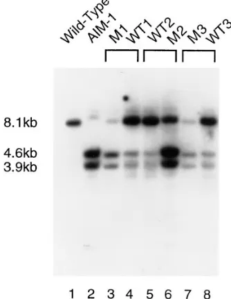
Although the Southern hybridization of F1 progeny indicates that all exconjugant lines contain both the wild-type (IES2) and mutant (IES1) bands, there are differences in the amounts of each product in different cell lines. These differences in the ratio of excised and non-excised IES from the F1 macronuclear genomes may result from an effect of the old macronucleus on
DNA excision. It is possible to differentiate between F1 progeny from the AIM-1 parent and the wild-type (51ND) parent by scoring cells with anti-A serum a few cell divisions after conjugation. Heterozygotes from the AIM-1 parent tend to remain A2 and those from the wild-type parent remain A1. Examination of the South-ern shows that those F1 which originate from the wild-type (51ND) parent have similar amounts of excised and non-excised product, but those from the AIM-1 parent generally contain much less of the wild-type product (for example, Figure 4, compare lanes 3 and 4). Although not conclusive, the results are consistent with the ability of the IES in the macronucleus to par-tially inhibit the excision of the wild-type allele. Such an inhibitory effect has been demonstrated for a 222-bp IES in the G surface protein gene (Duharcourt et al. 1995), but in those experiments the IES completely
inhibited excision of its micronuclear counterpart. Evi-dence presented in the next two sections provides addi-tional support for the conclusion that the AIM-1 ver-sion of IES2591 does not completely inhibit exciver-sion of its wild-type micronuclear copy.
Selected crosses were followed into the F2 genera-tion by the inducgenera-tion of autogamy, which results in a completely homozygous micronuclear genome. A typi-cal micronuclear mutation in Paramecium should show 1:1 segregation in the F2. Each F2 line was scored for trichocyst non-discharge, and then analyzed using PCR to determine the presence or absence of IES2591 in the macronucleus. The results of three representative crosses are shown in Table 1. Both the presence of the IES in the macronucleus and trichocyst non-discharge segregated 1:1. To determine whether the nucleotide mutation segregates with the inability to excise IES2591, we selected eight independent mutant F2 lines, amplified the IES region using PCR, and se-quenced the PCR products. All eight lines contained
Figure 4.—Southern blot of F1 cell lines from three
sepa-rate AIM-1 to wild-type matings. Genomic DNA from each F1 cell line was digested with EcoRI and XbaI. The blot was probed with the wild-type 8.1-kb macronuclear EcoRI A gene fragment contained in the plasmid pSA8.8R described in the text. “M” denotes AIM-1 exconjugant, “WT” denotes wild-type exconjugant.
TABLE 1
Inheritance of phenotypes in the F2 generation from three representative AIM-1 (mutant) to wild-type (51ND) matings
Pair no. and
descendant cytoplasm
No. of descendants with PCR band Marker segregation
Mutant Wild-type Both x2 P D ND x2 P
1
Mutant 6 5 2 0.09 .0.5 4 9 1.92 .0.1
Wild-type 3 3 0 0.00 .0.995 4 2 0.66 .0.1
2
Mutant 2 1 3 0.33 .0.5 3 3 0.00 .0.995
Wild-type 4 8 0 1.33 .0.1 8 4 1.33 .0.1
3
Mutant 8 2 1 3.60 .0.05 6 5 0.09 .0.5
Wild-type 2 6 2 2.00 .0.1 7 3 1.60 .0.1
Paramecium IES Excision Mutation 145
the mutant base change, confirming that the inability to excise IES2591 segregates with the base change.
Some F2 lines contained both wild-type and mutant PCR bands. These were taken through autogamy to the F3 generation, at which point many resolved to a single macronuclear genotype. Cell lines that did not resolve by the F3 generation were not included in the chi-square analysis but are listed in Table 1. The reason for these “mixed” lines is not clear. Some may result from partial inhibition of wild-type IES excision due to macronuclear copies of the IES; alternatively, under some conditions the mutant allele may be excised with a low efficiency. The complex nature of the F2 data was illustrated by the results from one unusual mated pair (data not shown) which showed 1:1 segregation of ex-cised and non-exex-cised products in the F2 generation from progeny derived from the wild-type parent (51ND), yet only non-excised products were obtained from the mutant parent (AIM-1). This non-Mendelian effect occurred even though the F1 progeny contained both the excised and non-excised products (data not shown). Despite some complexity within the F2 data, the results clearly demonstrate that the C to T base change in the IES is a micronuclear mutation which in-hibits IES excision.
Excision of the 28-bp IES inside IES2591 is detected in wild-type cells: The Mendelian inheritance of AIM-1 contrasts with the strong macronuclear effect reported for cell lines containing the 222-bp IES from the G sur-face protein gene (Duharcourt et al. 1995). Two
pos-sibilities to explain this difference were considered. Either wild-type IES2591, when present in the macro-nucleus, is not capable of completely inhibiting the excision of its micronuclear counterpart, or the 28-bp that are missing from the macronuclear copies of IES2591 in AIM-1 prevent the inhibitory effect. To dis-tinguish between these possibilities we attempted to create a stable IES1 cell line from a wild-type cell by in-jecting cloned wild-type copies of IES2591 into the old macronucleus and inducing autogamy to create a new macronucleus. Cells were coinjected with pPXV-NEO, which provides resistance to G418 (Haynes et al. 1995),
and a plasmid containing IES2591 plus 198-bp and 380-bp of DNA from the left and right flanking regions, re-spectively. After autogamy whole-cell PCR was per-formed to examine the macronuclear DNA products. Each cell line contained both the wild-type (IES2) and mutant (IES1) PCR bands indicative of incomplete re-moval of the IES during macronuclear development (Figure 5). After a second autogamy, all of the cell lines contained the wild-type band (some also retained a fraction of IES1 copies), signifying that the epigenetic effect of the IES in the macronucleus is unstable (Fig-ure 5). In other words, IES2591 is able to inhibit its own excision, but is unable to maintain this inhibition over several cycles of sexual reproduction.
Interestingly, when the PCR products from the cells
after one autogamy were run on a high-percentage aga-rose gel, we were able to discern at least two distinct bands running between 500–550-bp in size (Figure 5). The most abundant band matched the size of the wild-type IES2591 plus flanking sequence from 12460 to
12638 (548-bp). This product may be amplified from plasmid DNA contained in fragments of the old macro-nucleus that are still present in exconjugants. The plas-mid DNA in the old macronuclear fragments is easier to detect by PCR because it is present in higher copy number than endogenous macronuclear DNA. A mi-nor band occasionally observed above the most abun-dant product was not investigated. We believed that the band below the most abundant represented IES2591 without the 28-bp internal IES; in other words, the smaller IES was excised from a fraction of the macronu-clear copies of IES2591. Direct sequencing of the smaller PCR band confirmed that it was missing the 28-bp internal IES. Therefore, the internal 28-28-bp IES is not merely an aberrant splicing product in the mutant cell line; it also occurs (at least under some conditions) in wild-type cells.
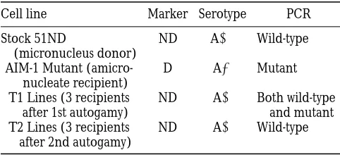
Transplantation of a wild-type micronucleus into AIM-1 does not result in stable inhibition of IES2591 ex-cision: Micronuclear transplantation is an alternative method used to determine the effect of the old macro-nucleus on DNA rearrangements in the developing
Figure 5.—PCR after the first and second round of
146 K. M. Mayer, K. Mikami and J. D. Forney
macronucleus. The idea is to remove the micronuclei from the mutant cell and replace them with a wild-type micronucleus. After induction of autogamy, the new macronucleus should be wild-type unless the old macronucleus inhibits the normal excision events. We used 51ND as the micronuclear donor and an amicro-nucleate AIM-1 cell line as the recipient. The first auto-gamy after transplantation resulted in cell lines (T1) that showed the non-discharge phenotype of the donor and were able to express A, signifying IES removal in the macronucleus (Table 2). However, when these cells were analyzed by PCR, they showed both wild-type and mutant bands in their macronucleus, indicating that IES removal was incomplete (data not shown). Because the micronuclear IES sequence is wild-type in these cells, the retention of some copies of the IES in the new macronucleus must be caused by the presence of the IES in the recipient cell macronucleus (the AIM-1 macronucleus). After the second autogamy (T2 lines), each cell line contained only the wild-type band (data not shown). We conclude that IES2591 in AIM-1 (which lacks the 28-bp internal IES) can partially in-hibit its own excision; however, this effect is neither complete nor stable.
D I S C U S S I O N
Significance of the inverted-terminal-repeat consen-sus sequence: The elimination of internal regions of DNA followed by rejoining of the flanking sequences is a common event during ciliate macronuclear develop-ment. Although DNA splicing is common to all ciliates, there is increasing evidence that different types of elim-inated elements have different sequence requirements for excision. Thus far, all the eliminated elements ex-amined in Paramecium and Euplotes are bounded by 59-TA-39 repeats and are therefore referred to as TA IESs. TA elements share several common features, and they are rather different from the features of the best-studied IESs, those in Tetrahymena (discussed below).
In Euplotes, the TA IESs include both large 5.3-kb elements, called Tec1 and Tec2, that have the
charac-teristics of transposons (Jahn et al. 1993), and short
(31–539-bp) generally unique sequences that have no significant open reading frames. The presence of 59 -TA-39 direct repeats, frequent inverted repeats, and large IESs that appear to be transposable elements led to early suggestions that short IESs in Euplotes and other hypotrich ciliates are related to transposable ele-ments (Herrick et al. 1985; Klobutcher and Jahn
1991). Although only short (28–882-bp) IESs have been identified in Paramecium, the availability of nu-merous sequences from these elements has allowed a statistical analysis that identified the inverted-terminal-repeat consensus (Klobutcher and Herrick 1995).
The similarity of this sequence to the terminal repeats of mariner/Tc1 transposons argued that the two types of elements are related, but the lack of an in vivo or in
vitro excision assay has prevented a direct experimental
investigation of the cis-acting sequences required for TA IES excision. The AIM-1 mutant has now provided a link between the conserved inverted-terminal-repeat se-quence and its function in IES excision.
The AIM-1 mutation, a C to T transition in the fifth position of the consensus sequence, is consistent with our expectations based on the analysis of Paramecium IESs. None of the known Paramecium IESs match the mutant allele of IES2591. Although A and C residues are occasionally found in the fifth position of the con-sensus (predominantly G), none of the original 20 IESs analyzed by Klobutcher and Herrick (1995) had T
in the fifth position. Interestingly, a recent report showed that an IES inside the intron of a Paramecium centrin gene does contain a T in the fifth position (V a-yssie et al. 1997). This suggests that there is no absolute
prohibition of T and that other nucleotides in the con-sensus may compensate for a poor concon-sensus match at position 5.
Effect of the AIM-1 mutation on DNA excision:
The single base mutation on the left end of IES2591 in the AIM-1 cell line results in retention of the IES in the macronuclear DNA even though the right end of the IES has a wild-type sequence. The alternative outcome, a broken end healed by the addition of telomere se-quence, is clearly not the predominant product, de-spite the precedence for such healing events in Para-mecium. For example, the A gene chromosome is
broken and healed in the d48 strain (Forney and
Blackburn 1988). A connection between DNA
elimi-nation and telomere formation in ciliates has been sug-gested previously (Blackburn and Karrer 1986; Amar
1994). Although in Tetrahymena it is clear that a 15-bp chromosome breakage sequence specifies chromo-some fragmentation (Yao et al. 1990), the connection
between IES excision and telomere formation remains a possibility in Paramecium. It is interesting to note that in vitro excision assays of Tc1 transposons show that mutations which prevent double-strand cleavage at one end of an element do not effect cleavage at the wild-TABLE 2
Phenotypes of strains for micronuclear transplantation
Cell line Marker Serotype PCR
Stock 51ND
(micronucleus donor)
ND A1 Wild-type
AIM-1 Mutant (amicro-nucleate recipient)
D A2 Mutant
T1 Lines (3 recipients after 1st autogamy)
ND A1 Both wild-type and mutant T2 Lines (3 recipients
after 2nd autogamy)
Paramecium IES Excision Mutation 147
type end (Vos et al. 1996). If cleavage at one end of an
IES is prevented by a mutation but occurs at the other end of the IES, it should result in double-strand breaks. Although we have no evidence for broken ends in AIM-1, Paramecium may be a useful organism to identify such events in vivo. The developmentally controlled excision of IESs means that large numbers of excision events can be examined easily as opposed to the relatively rare excision events of transposons. Also, the ability to heal broken DNA ends during macronuclear development makes it likely that broken ends would be maintained. If double-strand breaks cannot be found it may suggest models for the mechanism of IES excision in Parame-cium.
Currently, we do not have any quantitative measure of the AIM-1 mutation’s effect on excision of IES2591. Although we cannot detect any spliced product in AIM-1 (K. Mayer, unpublished results), the inhibition by
cop-ies of the IES in the old macronucleus may enhance the effect of the nucleotide mutation. In fact, this inhi-bition may have allowed us to isolate the mutant line. Even if the first macronucleus formed after mutagene-sis contained some correctly excised products, the non-excised copies of the IES could increase the inhibition of IES excision after the next round of autogamy, thus strengthening the A2 phenotype. The analysis of F1 progeny (Figure 4) shows that without the influence of macronuclear inhibition (progeny from wild-type par-ent) excision is still inhibited, but it cannot determine whether a portion of the excised product came from the mutant allele. Measurement of the amount of mu-tant allele that can be excised will require a cross be-tween AIM-1 and the d12 cell line which has a deletion of the A gene in the macronucleus and micronucleus (Rudman et al. 1991).
Reiterative integration of IES elements and evolu-tion: The AIM-1 mutation revealed the presence of a 28-bp IES inside IES2591. We do not consider this a cryptic splice product because two internal splice sites are used rather than a deletion involving one internal site paired with the non-mutant end of IES2591. In ad-dition, we showed that the internal 28-bp IES can be ex-cised independently in wild-type cells. This result has also been obtained by S. Duharcourt and E. Meyer
(personal communication). In a series of quantitative experiments that examine the ability of different IESs to inhibit excision of their micronuclear counterpart they demonstrated independent excision of the 28-bp IES inside IES2591. Their results indicate that, in fact, all detectable macronuclear copies after autogamy are missing the 28-bp internal IES. This demonstrates that excision of the internal IES cannot be inhibited by cop-ies in the old macronucleus, and also suggests that the presence of the PCR product containing the entire IES in our experiments (Figure 5) may be the result of am-plification of injected plasmid DNA present in old macronuclear fragments.
Although previous examples of Tec transposons within other Tec transposons have been described in Euplotes (Baird et al. 1989; Krikau and Jahn 1991),
the presence of small internal IESs presents additional questions regarding the evolution and function of these DNA elements. The most explicit model for IES evolution has proposed that transposons initially spread throughout the micronuclear genome, and then began to shrink in size after the excision function
was assumed by the host (Klobutcher and Herrick
1997). The existence of the internal 28-bp IES cannot be explained easily by this model. There is no obvious selective pressure for an internal IES to maintain its cis-acting excision sequences since it is already inside an eliminated region. Therefore, if the 28-bp IES was orig-inally an ancient insertion of a large transposon when it inserted into IES2591, it should have lost the ability to be excised from the genome long before it reached its current size of 28-bp.
Nevertheless, there are several possible explanations for the presence of the small internal IES. It could rep-resent a random sequence that is utilized due to its sim-ilarity with the inverted-terminal-repeat consensus. Al-though this remains a possibility, many close matches to the consensus can be identified within regions of
macronuclear DNA that are never excised (K
lo-butcher and Herrick 1995). Therefore, if IESs can be
created by chance, it is probably a special property of those located within a functional IES, perhaps due to the recruitment of the excision machinery to the larger IES.
Alternatively, the internal IES could have originated as a full-length transposon that inserted into another transposon, disrupting an important cis-acting element (an internal promoting sequence). This model predicts that over evolutionary time the internal IES has con-served its excision function because it is required for excision of the larger IES. Excision of the internal IES regenerates the required site. This explanation is
com-patible with the Klobutcher and Herrick (1997)
model for IES evolution and predicts that the 28-bp IES is removed prior to excision of the larger IES. Unfortu-nately, at this time we have no detailed information about the excision intermediates for this or any other Paramecium IES.
Finally, it is possible that the 28-bp IES is a recent in-sertion into IES2591. Small (28-bp) mobile IESs could in part explain the rapid evolution of these excised ele-ments (reviewed in Prescott 1994; Klobutcher and
Herrick 1997). Of the four IESs common to both the A and B surface protein genes, only one is the same size
in both genes (Scott et al. 1994a). Insertion or
148 K. M. Mayer, K. Mikami and J. D. Forney
the most recent integrations, the older insertions hav-ing diverged so that they are no longer capable of inde-pendent excision.
Klobutcher and Herrick (1995) have noted that
Paramecium IESs are approaching a minimum size of 28–29-bp. The AIM-1 cell line illustrates how mutations which prevent IES excision could increase the fre-quency of small IESs in the genome. Regardless of how the internal 28-bp IES originated, the mutation causes the incorporation of additional DNA into the macronu-clear genome, and the resulting elimination event is 28-bp rather than 370-bp. Although this is unlikely to be common in coding regions, it could occur with rea-sonable frequency within intergenic regions.
Comparison of Paramecium IES to other types of IES: The best-studied eliminated elements in any cili-ate are those in Tetrahymena (reviewed in Coyne et al.
1996). Unlike Paramecium and Euplotes, Tetrahymena IESs are neither bounded by 59-TA-39 repeats nor do they contain an inverted-terminal-repeat consensus. They are generally flanked by 4–8-bp direct repeats and range in size from a few hundred base pairs to over 13-kb. Elimination of the M2 element is dependent on two types of cis-regulatory elements: a polypurine tract (A5G5) located about 45-bp outside of the deleted
re-gion which specifies the deletion border, and internal promoting sequences which are required for excision (Godiska et al. 1993). Analysis of an element called
mse2.9 has also provided evidence that a short se-quence outside the excised element is required for ex-cision (Li and Pearlman 1996). These elements do not
resemble any known transposons and do not appear to be related to the TA IESs in Paramecium. Although Tetrahymena IESs undergo alternative deletion events, the products result from joining different sequences to one constant end (Austerberry and Yao 1988). There
are no reports of internal IESs.
Macronuclear inhibition of IES excision: The mech-anism responsible for the inhibition of IES excision by macronuclear copies of the element is unknown. Du-harcourt et al. (1995) showed that a 222-bp IES in the 51G surface protein gene inhibited the excision of its
corresponding micronuclear copy, but did not inhibit any other IESs examined. This macronuclear mutation
(IES1) shows stable inheritance through multiple
rounds of sexual reproduction (autogamy) even though the micronuclear genome is wild-type. Mating this cell line with a normal (IES2) cell line results in F1 prog-eny that are IES1 if they originate from the IES1 par-ent and IES2 if they originate from the IES2 cell line.
The presence of IES2591 in the macronucleus does not have such a dramatic influence on excision. Some excision of the IES occurs in F1 progeny from the AIM-1 parent. In addition, substitution of a wild-type micronu-cleus for the AIM-1 micronumicronu-cleus does not result in sta-ble inheritance of the IES1 phenotype in the macro-nuclear genome. Finally, transformation of the old
macronucleus with cloned wild-type IES2591 was able to inhibit IES excision during formation of the next macronucleus, but the trait was not stable into the next sexual generation. S. Duharcourt and E. Meyer have
performed a comprehensive study of the ability of vari-ous IESs to inhibit excision of their micronuclear coun-terparts (personal communication). Their results also indicate that inhibition of IES2591 is not as stable as inhibition of the 222-bp IES in 51G. One possible explanation for the difference in stability is the pres-ence of the internal 28-bp IES. Duharcourt et al.
(1995) showed that an internal deletion of 147-bp from the 222-bp IES eliminated the macronuclear inhibition of excision. Thus, the AIM-1 macronuclear copy of IES2591, which contains a 28-bp deletion, may be less efficient in macronuclear inhibition. If, in addition, the 28-bp IES does not efficiently inhibit itself (perhaps due to its small size), it would be difficult to keep a sta-ble IES25911 cell line.
The only other known micronuclear mutation af-fecting IES excision is called mtFE (Meyer and Keller
1996). This pleiotropic mutation affects mating type determination as well as several other phenotypic char-acteristics and inhibits the excision of the 222-bp IES in the 51G gene. Although the molecular defect in this strain is not known, it has been suggested that mtFE
en-codes a trans-acting factor required for excision of a subset of IESs, at least one of which is involved in mat-ing-type determination.
In lieu of an in vivo or in vitro excision assay, the iso-lation of mutations that prevent IES excision may pro-vide the best opportunity to investigate the structure and function of IESs in Paramecium. The apparent flexibility of the inverted-terminal-repeat consensus suggests that it may be possible to isolate intragenic suppressors of the AIM-1 mutation. These suppressors could be mutations in other positions of the consensus that compensate for the original mutation. We may also identify genetic loci encoding trans-acting factors that are required for excision of one or a few IESs as pro-posed for mtFE. These would appear as IES1 A gene
mutants in which the mutation is unlinked to A.
We thank Michael Ku for performing the microinjection of
plas-mid DNA. This research was supported by a National Science Foun-dation predoctoral fellowship DGE-9253915-03 to K.M.M. and a Na-tional Science Foundation grant MCB-9506009 to J.D.F. This is paper 15454 from the Purdue Agriculture Experiment Station.
L I T E R AT U R E C I T E D
Amar, L., 1994 Chromosome end formation and internal sequence
elimination as alternative genomic rearrangements in the ciliate Paramecium. J. Mol. Biol. 236: 421–426.
Austerberry, C. F., and M.-C. Yao, 1988 Sequence structures of
two developmentally regulated, alternative DNA deletion junc-tions in Tetrahymena thermophila. Mol. Cell. Biol. 8: 3947–3950. Baird, S. E., G. M. Fino, S. L. Tausta and L. A. Klobutcher,
Paramecium IES Excision Mutation 149
transposonlike element is removed during macronuclear devel-opment. Mol. Cell. Biol. 9: 3793–3807.
Blackburn, E. H., and K. M. Karrer, 1986 Genomic
reorganiza-tion in ciliated protozoans. Annu. Rev. Genet. 20: 501–21. Bryan, G., D. Garza and D. Hartl, 1990 Insertion and excision of
the transposable element mariner in Drosophila. Genetics 125: 103–114.
Caron, F., and E. Meyer, 1989 Molecular basis of surface antigen
variation in paramecia. Annu. Rev. Microbiol. 43: 23–42. Coyne, R. S., D. L. Chalker and M.-C. Yao, 1996 Genome
downsiz-ing durdownsiz-ing ciliate development: nuclear division of labor through chromosome restructuring. Annu. Rev. Genet. 30: 557– 578.
Duharcourt, S., A. Butler and E. Meyer, 1995 Epigenetic
self-regulation of developmental excision of an internal eliminated sequence in Paramecium tetraurelia. Genes Dev. 9: 2065–2077. Epstein, L. M., and J. D. Forney, 1984 Mendelian and
non-Mende-lian mutations affecting surface antigen expression in
Parame-cium tetraurelia. Mol. Cell. Biol. 4: 1583–1590.
Forney, J. D., and E. H. Blackburn, 1988 Developmentally
con-trolled telomere addition in wild-type and mutant paramecia. Mol. Cell. Biol. 8: 251–258.
Forney, J. D., F. Yantiri and K. Mikami, 1996 Developmentally
controlled rearrangement of surface protein genes in
Parame-cium tetraurelia. J. Eukaryot. Microbiol. 43: 462–467.
Godiska, R., K. J. Aufderheide, D. Gilley, P. Hendrie, T. Fitzwater
et al., 1987 Transformation of Paramecium by microinjection of a cloned serotype gene. Proc. Natl. Acad. Sci. USA 84: 7590–7594. Godiska, R., C. James and M.-C. Yao, 1993 A distant 10 bp
se-quence specifies the boundaries of a programmed DNA deletion in Tetrahymena. Genes Dev. 7: 2357–2365.
Haynes, J. W., K.-T. Ling, Y. Saimi and C. Kung, 1995 Induction of
antibiotic resistance in Paramecium tetraurelia by the bacterial gene APH-39-II. J. Eukaryot. Microbiol. 42: 83–91.
Herrick, G., S. Cartinhour, D. Dawson, D. Ang, R. Sheets et al.,
1985 Mobile elements bounded by C4A4 telomeric repeats in
Oxytricha fallax. Cell 43: 758–768.
Jahn, C. L., S. Z. Doktor, J. S. Frels, J. W. Jaraczewski and M. F.
Krikau, 1993 Structures of the Euplotes crassus Tec1 and Tec2
elements: identification of putative transposase coding regions. Gene 133: 71–78.
Klobutcher, L. A., and C. L. Jahn, 1991 Developmentally
con-trolled genomic rearrangements in ciliated protozoa. Curr. Opin. Genet. Dev. 1: 397–403.
Klobutcher, L. A., and G. Herrick, 1995 Consensus inverted
minal repeat sequence of Paramecium IESs: resemblance to ter-mini of Tc1-related and Euplotes Tec transposons. Nucleic Acids Res. 23: 2006–2013.
Klobutcher, L. A., and G. Herrick, 1997 Developmental genome
reorganization in ciliated Protozoa: the transposon link. Prog. Nucl. Acid Res. Mol. Biol. 56: 1–62.
Kobayashi, S., and S. Koizumi, 1990 Characterization of
non-Men-delian and Mennon-Men-delian mutant strains by micronuclear transplan-tation in Paramecium tetraurelia. J. Protozool. 37: 489–492. Koizumi, S., 1974 Microinjection and transfer of cytoplasm in
Para-mecium. Exp. Cell Res. 88: 74–78.
Krikau, M. F., and C. L. Jahn, 1991 Tec2, a second transposon-like
element demonstrating developmentally programmed excision in Euplotes crassus. Mol. Cell. Biol. 11: 4751–4759.
Li, J., and R. E. Pearlman, 1996 Programmed DNA rearrangement
from an intron during nuclear development in Tetrahymena
ther-mophila: molecular analysis and identification of potential
cis-act-ing sequences. Nucleic Acids Res. 24: 1943–1949.
Meyer, E., and S. Duharcourt, 1996a Epigenetic regulation of
programmed genomic rearrangements in Paramecium aurelia. J. Eukaryot. Microbiol. 43: 453–461.
Meyer, E., and S. Duharcourt, 1996b Epigenetic programming
of developmental genome rearrangements in ciliates. Cell 87: 9–12.
Meyer, E., and A.-M. Keller, 1996 A Mendelian mutation affecting
mating-type determination also affects developmental genomic rearrangements in Paramecium tetraurelia. Genetics 143: 191–202. Mikami, K., and S. Koizumi, 1982 Nuclear transplant studies of the
determination of mating type in germ nuclei of Paramecium
tet-raurelia. Exp. Cell Res. 137: 397–402.
Preer, J. R., Jr., 1986 Surface antigens of Paramecium, pp. 301–339
in The Molecular Biology of Ciliated Protozoa, edited by J. G. Gall.
Academic Press, NY.
Prescott, D. M., 1994 The DNA of ciliated Protozoa. Microbiol.
Reviews 58: 233–267.
Robertson, H. M., 1995 The Tc1-mariner superfamily of
trans-posons in animals. J. Insect Physiol. 41: 99–105.
Rudman, B., L. B. Preer, B. Polisky and J. R. Preer, Jr., 1991
Mutants affecting processing of DNA in macronuclear develop-ment in Paramecium. Genetics 129: 47–55.
Sambrook, J., E. F. Fritsch and T. Maniatis, 1989 Molecular
Clon-ing: A Laboratory Manual, Ed. 2, Cold Spring Harbor Laboratory,
Cold Spring Harbor, NY.
San Miguel, P., A. Tikhonov, Y. K. Jin, N. Motchoulskaia, D. Z ak-harov et al., 1996 Nested retrotransposons in the intergenic
regions of the maize genome. Science 274: 765–768.
Scott, J., C. Leeck and J. Forney, 1994a Analysis of the
micronu-clear B type surface protein gene in Paramecium tetraurelia. Nu-cleic Acids Res. 22: 5079–5084.
Scott, J. M., K. Mikami, C. L. Leeck and J. D. Forney, 1994b
Non-Mendelian inheritance of macronuclear mutations is gene spe-cific in Paramecium tetraurelia. Mol. Cell. Biol. 14: 2479–2484. Sonneborn, T. M., 1970 Methods in Paramecium research, in
Meth-ods in Cell Physiology, edited by D. M. Prescott. Academic Press,
NY. 4: 242–339.
Sonneborn, T. M., and M. V. Schneller, 1979 A genetic system for
alternative stable characteristics in genomically identical ho-mozygous clones. Dev. Genet. 1: 21–46.
Steele, C. J., G. A. Barkocy-Gallagher, L. B. Preer and J. R. Preer,
Jr., 1994 Developmentally excised sequences in micronuclear
DNA of Paramecium. Proc. Natl. Acad. Sci. USA 91: 2255–2259. Vayssie, L., L. Sperling and L. Madeddu, 1997 Characterization of
multigene families in the micronuclear genome of Paramecium
tetraurelia reveals a germline specific sequence in an intron of a
centrin gene. Nucleic Acids Res. 25: 1036–1041.
Vos, J. C., I. D. Baere and R. H. A. Plasterk, 1996 Transposase is
the only nematode protein required for in vitro transposition of Tc1. Genes Dev. 10: 755–761.
Yao, M.-C., C.-H. Yao and B. Monks, 1990 The controlling
se-quence for site-specific chromosome breakage in Tetrahymena. Cell 63: 763–772.