Copyright 0 1994 by the Genetics Society of America
Mutations in
the
Gene for a tRNA That Functions
as
a Regulator
of
a
Transcriptional Attenuator
in
Bacillus
subtilis
D.
B. Garrity
and S . A. Zahler
Section of Genetics and Development, Cornell University, Ithaca, New York 14853
Manuscript received December 27, 1993
Accepted for publication March 9, 1994
ABSTRACT
It has been proposed that uncharged tRNA molecules may act as positive regulatory factors to control the expression of a number of operons in Bacillus subtilis and related bacteria by interacting with leader sequences to cause antitermination. In this study we report the isolation and characterization of regulatory mutations that modify one of the tRNA molecules predicted to have such a regulatory role. Three different alleles of the B. subtilis leucine tRNAgene leuGwere found that resulted in higher expression of the ilu-leu
biosynthetic operon. Each resulted in a base change in the D-loop of the leucine tRNA molecule with the anticodon 5’-GAG3’ (leucine tRNbAG). Experiments with strains that are diploid for mutant and wild-type alleles suggested that both charged and uncharged tRNA molecules may interact with leader sequences to control expression of the operon.
B
ACTERIA have evolved elaborate mechanisms to regulate gene expression in response to changes in physiological conditions and nutrient supply. The regu- lation of genes involved in amino acid biosynthesis in the gram-negative bacterium Escherichia coli has been stud- ied in considerable detail. In recent years there has been increasing interest in the mechanisms that control the expression of corresponding genes in Bacillus subtilis,a gram-positive soil bacterium that occupies a very dif- ferent ecological niche. In this report we describe mu- tations in a leucine tRNA gene that cause overexpression of the B. subtilis ilv-leu operon, which contains genes for branched-chain amino acid biosynthesis. Our analy- sis suggests that the operon is controlled by an attenu- ation mechanism in which a particular tRNA species helps to determine the frequency of transcriptional ter- mination at a site in an untranslated leader sequence.
The ilv-leu operon of B. subtilis contains seven genes required for the biosynthesis of leucine, isoleucine and valine (VANDEYAR 1987), preceded by a 482-bp untrans- lated leader sequence. Transcription of the operon is about 30-fold higher when cells are grown in limiting leucine than when they are grown in excess leucine
(GRANDONI et al. 1993). Attenuation of transcripts within this leader accounts for most of the negative effect of excess leucine on expression of the operon (GRANDONI
et al. 1992, 1993).
GRUNDY and HENKIN (1993,1994) have proposed that a number of the aminoacyl-tRNA synthetase genes and amino acid biosynthetic operons of B. subtilis and re- lated bacteria are regulated by a common mechanism in which uncharged tRNA molecules act as positive r e p - lators by interacting directly with leader mRNA to pro- mote transcriptional antitermination. Mutational analy- sis of the leader of the B. subtilis tyrosyl-tRNA synthetase
Genetics 137: 627-636 (July, 1994)
gene ( t y r S ) led them to propose this novel model of transcriptional attenuation.
This proposed mechanism differs from the exten- sively studied mechanisms that regulate the amino acid biosynthetic operons of E. coli. Translation of upstream open reading frames is an essential component of the
E. coli attenuation mechanisms (LANDICK and YANOFSKY
1987). In the GRuNDYand HENKIN model, the mechanism by which tRNA molecules influence transcriptional ter- mination is independent of translation. Analysis of the
ilv-leu operon leader sequence suggests that it is regu- lated by this mechanism (GRUNDY and HENKIN 1994;
GRANDONI et al. 1993).
In the leader region of each of the operons that
GRUNDY and HENKIN analyzed they identified a potential RNA secondary structure that could serve as a transcrip tional terminator. Upstream of the terminator is a po- tential stem-loop structure that contains an unpaired bulge with a triplet sequence (the specifier) that they believe is a site of interaction with the anticodons of regulatory tRNA molecules (GRUNDY and HENKIN 1993). In the t y S operon they showed that this site determines the specificity of the regulation. The tyrS leader has the tyrosine codon 5’-UAG3’ (UAC) at the specifier posi- tion. Changing the triplet to UUC, which codes for phenylalanine, altered the regulation so that transcrip tion of the operon became dependent on starvation for phenylalanine instead of tyrosine. They noted in their papers that the leucyl-tRNA synthetase ( l e d ) and ilu-
leu operons both have the specifier triplet CUC that codes for leucine. Their model predicts that tRNA mol- ecules that recognize CUC control expression of both of the operons. In this paper we describe our analysis of mutations within a leucine
tRNhAG
gene (ZeuG) ofTABLE 1
B. subtilis strains
Strain Genotype Reference
CU1065 CU1962' CU4609 CU4645 CU4670 CU4694 CU4785 CU4831 cu4933
CU4936 cu4937
CU4987 CU4993 CU5016 CU5022 CU5023 cu4934",b
CU4949'Zb
trpC2 [metB5] sup-3
ilvN-lacZ-erm leuBl6 trpC2 (SPP)
ilvN-lacZ-erm leuBl6 trpC2 leuGI (SPP) ilvN-lacZ-erm leuBl6 trpC2 leuG2(SPP) ilvN-lacZ-erm leuBl6 trpC2 leuG5 (SPP)
ilvN-lacZ-erm leuB16 metB5 leuGI (SPj3)
metB5 (SPP c2 deL2::Tn917)
ilvN-lacZ-erm leuBl6 trpC2 leuGI (SPP[sup-3 leuF' leuG']) [metB5] (SPP[sup-3 leuF' leuG'])
ilvN-lacZ-erm leuB16 trpC2 (SPp[pCV-2])
ilvN-la&-erm leuBl6 trpC2 leuGI (SPP[pCV-2])
ilvN-lacZ-cat lys-3 [metB5/ (SPP[sup-3 leuF+ leuG']) ilvN-lacZ-cat metB5 (SPP[leuFf leuGI])
ilvN-lacZ-erm leuBI6 leuFI AleuG-neo trpC2 (SPP)
ilvN-lacZ-erm leuBI6 AleuG-neo trpC2 (SPP)
ilvN-lad-erm leuBI6 trpC2 leuGI (SPP[leuF+ AleuG-neo]) ilvN-la&-erm leuBI6 trpC2 leuGI (SPS[leuFI AleuGneo])
This laboratory This laboratory This laboratory This work This work This work This laboratory This laboratory This work
G mand ZAHLER (1993)
This work This work This work This work This work This work This work This work
Additional strains are listed in Table 4.
'
The metB5 mutation in CU1962, CU4934 and CU4949 is suppressed by the sup-3 suppressor, and the strains are prototrophic for methionine.~~
These strains carry SPP prophages that contain the entire trnS operon.
operon. Our analysis of the mutations supports the Grundy and Henkin model.
The mutations in the leuG gene were isolated in a screen of B. subtilis mutants that overexpress the ilv-leu operon. The mutants were identified as suppressors of a phenotype associated with an insertion of a transposon in ilvN, the second gene of the ilv-leu operon. B. sub-
tilis strains with Tn 91
7
inserted into the iEvN gene have a leucine-sensitive phenotype (VANDEYAR 1987). In the presence of leucine, expression of the operon is so low that strains carrying the ilvN:Tn917 insertion are aux- otrophic for isoleucine andvaline. We isolated and char- acterized mutations that allow ilvN:Tn917-containing strains to grow on minimal medium with leucine. The mutations mapped to three loci on the B. subtilis chro- mosome. Leucine-resistant mutants in the ilv-leu termi- nator sequence and in the structural gene for leucyl- tRNA synthetase (Zeus) have previously been described(GRANDONI et al. 1993; VANDER HORN and ZAHLER 1992). In this report we discuss our analysis of leucine-resistant mutants that have point mutations within the leucine tRNA gene leuG. The 1euG gene is located in tmS, an operon containing seven tRNA genes (GARRITY
and ZAHLER 1993). The leuG gene codes for leucine
tRNbAC
Another tRNA gene in this operon, leuF, codes for leucine tRNAu,. We constructed a mutation that changed the anticodon of the EeuFgene from 5'-UAG3' to 5'-GAG3'; it suppresses the phenotype of the leuG mutations. We discuss the significance that the leuF and leuG mutations have on the Grundy and Henkin model.MATERIALS AND METHODS
Media: Bacterial strains were grown on plates of tryptose blood agar base (TBAB; Difco) or Spizizen minimal medium with 0.5% glucose, 20 pg/ml tryptophan, 1 pg/ml biotin, 10
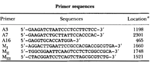
TABLE 2
Primer sequences
Primer Sequences Location a
A3 5'-GAAGATCTAATCCCTCCTTCTCC-3' 1198 A7 5'-GAAGATCTGCTTATTCCACCCAC-3' 2301
A16 5'-GAGGTGCACCATGGA-3' 465
MI 5"AGGACTTGAATTCCGCCACGACCGCGTGA-3' 1660 MI, 5'-TGGCGGAATTCAAGTCCTCTCGGCCGCA-3' 1748
MI,, 5'-CTACGGATCCTCAGTCTAGCGCGTCTG-3' 1521
'
The approximate locations where primers match sequences inthe trnS operon are indicated in Figure 2. The coordinates refer to
positions in the previously published trnS sequence (Figure 3,
GARRITY and ZAHWR 1993). The 3' ends of the primers correspond to
nucleotides in the published sequence at the indicated positions.
Primers A3 and A7 contain BgZII (AGATCT) sites added to their 5'
ends. MI,, matches sequences in l e d including the leuFl mutation,
and has a BamHI (GGATCC) site at its 5' end. MI and M,, are
complementary at their 5' ends.
p~ MnCl,, and 17 g/liter purified agar (HARWOOD and CUTTING
1990). Additives included either 50 pg/ml ampicillin, 5 pg/ml chloramphenicol (CAM), 10 pg/ml neomycin, 40 pg/ml me- thionine, 100 pg/ml leucine, 70 pg/ml isoleucine, or 30
pg/ml valine. Plates for selecting for erm (the MlsR phenotype) contained 1 pg/ml erythromycin and 25 pg/ml lincomycin (MLS)
.
5-Bromo4~hloro-3-indolyl P-Dgalactopyranoside (X-gal; Sigma) was added to some plates to a concentration of80 pg/ml. Liquid cultures were grown in Luna-Bertani broth
(LB) (HARWOOD and CUTTING 1990) or in synthetic medium
(see below). Cultures were aerated at 37".
The ibN-EacZreporter gene: The effects that different mu- tations have on expression of the ilv-leu operon were deter- mined by measuring P-galactosidase activity of strains carrying a transcriptional fusion of the operon with the lacZ gene of
E . coli. Fusions were constructed as described by PERKINS and
YOUNGMAN (1986). Sequences containing the lacZ gene were inserted into a transposon located in ilvN, the second gene of the
ilv-Zeu operon. Constructs with two different antibiotic markers were used. i1vN:Tn 91 7[ZucZ-em] confers resistance to erythromy- cin and lincomycin. ilvN:Tn917[ZucZcat] confers resistance to
leuG Mutations of B. subtilis 629
u A
U G
G C A
A
C
A
G r C C = G G = C G = C G = C U - A
G = C
A
G G u u u c c L I n r G C G G G G C
C G
A G
U C
J
U A G
G
1.ur1
leuF
C W G
u ' A
G
u U G
A
C C
G = C
A
C = C G = C G = C U - G
1 o u G S C = C G = C
A A A u u c u c c
v u
G GG G A G G I I u I1 II
u G A
G
P F P
LGcAl
A
A c G c G u # U u C C = G u - G
U - A 'Ic
G = C eC G = C 0-A
A A - 0 c;Jc
l e u G 1
l r u G 3 A I] G G A
l e u G I l e u G 2
0 G A A u
G A G
leuG
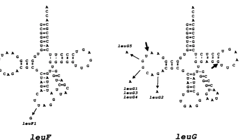
FIGURE 1.-The predicted sequences and secondary structures of the tRNAs coded by leuFand leuG. Post-transcriptional modi- fications other than the addition of CCA to the 3' ends of the tRNAs are not shown. The large arrows indicate the extent of the AleuG deletion; a 1-kb fragment of DNA carrying neo replaced the deleted region to make AleuG-neo.
walactosidase assays: Cultures were grown in Spizizen minimal medium with 0.5% glucose, 20 pg/ml tryptophan, 100 pg/ml leucine, 70 pg/ml isoleucine, 30 pg/ml valine, 1 pg/ml biotin, and 10 pM MnCl, (HARWOOD and CUTTING 1990). One milliliter of overnight culture was used to inoculate 50 ml of medium in 300-ml Klett flasks. The flasks were aerated at 37". Samples were removed at time points during logarithmic growth and frozen immediately on dry ice. Assays were per- formed as described by PLATKO et al. (1990).
Bacterial strains, plasmids and primers: B. subtilis strains are indicated in Table 1. Primers are listed in Table 2. Plasmid pCV-2 (YOUNGMAN et al. 1989) was a gift from P. YOUNGMAN. The neo gene of pUBllO was derived from pMK3 (SULLIVAN et al. 1984) received from the Bacillus Genetics Stock Center. E. coli strains used were JM109 recAl supE44 endAl hsdRl7gyrA96 relAl thi A(1ac-pro) F'[traD36 ProAB' lacP lacZAM15I (YANISCH-PERRON et al. 1985) and JM2r- recAl supE44 endAl hsdRl7 gyrA96 relAl thi A(1ac-pro) F'LtraD36 proAB+ lacP lacZAMIS] mcrAB serB:Tn5 (WEINER 1986).
Bacterial transformation: The method of
HANAHAN
(1983) was used to transform E. coli strains. The method of CWC and VANDER HORN (1990) was used to transform B. subtih strains.Isolation of leucineresistant mutants: Five-milliliter samples of LB were inoculated with isolated colonies of CU4609 ilvN-lac2 leuBl6 trpC2 and aerated at 37" until cul- tures were slightly turbid. Cells were pelleted, washed in saline- citrate (0.1 M NaCl, 0.01 M sodium citrate), and plated on mini-
mal agar medium with 160 pg/ml leucine. Mutants leuG2 through leuG5 were obtained after treatment with the muta- gen N-methyl-K-nitro-N-nitrosoguanidine (MNNG; Al-
dridge). Pellets of the exponentially growing cells were resus- pended in 100 pg/ml MNNG in saline-citrate, aerated for 30 min at 37" and washed in salinecitrate. One tenth milliliter of washed cells was used to inoculate 5-ml tubes of LB. Cultures were aerated at 37" until turbid and plated as before. Leucine- resistant colonies were patched to TBAB containing X-gal, and blue (Lac+) colonies were chosen.
Gene mapping: Leucine-resistant mutations were mapped relative to known genes on the B. subtilis chromosome using
standard methods of transformation and transduction with phage PBSl (CUTTING and VANDER HORN 1990). Five mutations were tightly linked to the sup-3 nonsense suppressor. Sequenc- ing polymerase chain reaction (PCR) products derived from the five mutant strains showed that each had a single base-pair change in a particular leucine tRNA gene located in the same operon as the sup-3 mutation. The operon has been named trnS (GARRITY and ZAHLER 1993). The leucine tRNA gene was named leuG; it codes for leucine
tRNbAC
DNA sequencing: Plasmid DNA segments were sequenced using Sequenase kits (U.S. Biochemical Corp.). PCR products were sequenced using femtomole sequencing kits (Promega) after purification from low melt gels using Magic PCR preps (Promega).
DNA isolation: Small amounts of plasmid DNA (1-3 pg) were isolated using Magic Minipreps (Promega). Larger amounts (50-500 pg) were purified by CsCl gradient centrifu- gation (MANIATIS et al. 1982).
AZeuG The method described by HIGUCHI (1990) was used to amplify a DNA fragment from the trnS region in which 49 bases internal to the ZeuGgene, including the entire anticodon a r m , were deleted. The deletion was named AleuG (Figure 1). The deletion creates an EcoRI restriction site within the re- mainder of the leuG gene. The fragment of about 1.1 kb was cloned into the BamHI site of pBluescript (Stratagene). Plas mid pDG218 was obtained (Figure 2).
Primers used include A3, A7, MI, and
Mil.
Primer sequences are indicated in Table 2. Approximate positions where primers hybridize to sequences in trnSare indicated in Figure 3. Details of the procedure can be found in (GARRITY 1994). The am- plified product included sequences between primers A3 and A7 with sequences between primers MI and MI, deleted.Scal
1 EcoRV Sa /I
EcoR I
Scal
Xbal
Seal
//\
EcoRlEcoRl
Kpnl
Sal1
1
EcoRVEcoR I
Seal
\
In
p D G 2 3 4Sal1
i Xbal
Afragment of DNAfrom an XbaI and ScaI digest of pDG224 was ligated to a DNA fragment from an XbaI and ScaI digest of pDG213 (Figure 2). Plasmid pDG213 was described previ- ously (GARRITY and ZAHLER 1993). It is a derivative of pBM22 with a 4.2-kb insert from the region of the trnS operon. The resulting plasmid, pDG234, has the AleuG-neo deletion in- serted into the trnkontaining fragment of pDG213. CU4645 ilvN-lacZ leuBl6 trpC2 leuGl was transformed with pDG234 linearized by ScaI, and NeoR colonies were isolated. A NeoR CamS transformant was designated CU5016 ilvN-lacZ leuBl6 trpC2 AleuG-neo.
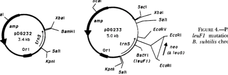
leuFl: Primer M,,, is complementary to sequences in the leuFgene except that it has a base substitution to change the anticodon from UAG to GAG. A PCR product was obtained using primers A16 and MI,, with template DNA from strain CU1065 trpC2. The product was cutwith BamHI and ScaI and ligated to pBluescript cut with EcoRV and BamHI. The result- ing plasmid was designated pDG232 (Figure 4). A fragment of DNA derived from a digest of pDG224 cut with BstYI and XbaI
was ligated to pDG232 cut with BamHI and XbaI. Plasmid pDG233 was obtained. It contains the region of the trnS operon with AleuG-neo and the leuFl mutation. CU4987 ilvN-
lac2 metB5 (SPP[le@ leuGl]) (see below) was transformed with ScaIdigested pDG233. NeoR colonies were obtained. CU4609 ilvN-lac2 leuBl6 trpC2 was transformed with DNA from one of the NeoR transformants, selecting for NeoR. One of these was designated CU4993 ilvN-1ucZ leuBl6 trpC2 leuFl
A leu G-neo.
SPP lysates: All SPP phages in this study have the c2
FIGURE 2.-Plasmids used to construct the AleuG-neo deletion. The construction ofpDG213 was previouslyreported (GARRITY and ZAHLER 1993). The neomycin resistance gene ne0 is derived from pUBllO (BRON 1990). cat is the chloramphenicol resistance gene of pC194 (BRON 1990).
temperature-sensitive repressor gene. Lysates were made by heat induction as previously described (WEINER 1986).
SPP[pCV-O]: This is a recombinant SPP phage that con- tains the vector used to construct SPP[sup-3 leuIiC leuG’] (see below). CU4831 metB5 (SPP[c2 Tn9171) was transformed with pCV-2 linearized with SstI. CamR transformants were selected.
SPP[supJ 1euF leuG+J The isolation of a recombinant SPP phage with a cloned insert including the trnS operon was previously reported ( G m a n d ZAHLER 1993). The insert car- ries the sup3 ochresuppressor mutation in a lysine tRNA gene located in hnS. The W a n d leuGgenes are wild-type in the cloned operon. In our previous work the phage was called SPPS. In this paper we designate the phage SPP[sup3
leuF+ leuGI.
SPP[leuF leuGl
J
Genetic recombination was used to iso- late a derivative of SPP[sup-3 l e u P leuG+] in which the sup-3 marker was replaced by its wild-type allele and the wild-type leuG allele was replaced by the leuGl mutation. The construc- tion was carried out by congression [cotransformation of un- linked loci; see STREIPS (1991)l.Strain CU4949 ilvN-lacZ lys-3 metB5 (SPP[sup-3 le& leuG+]) is phenotypically Met’ because the sup-3 ochre sup- pressor suppresses the metB5 nonsense mutation. This strain was transformed with excess DNA from CU4785 leuGl iZvN-
leuG Mutations of B. subtilis 63 1
In Lys 1euP I e u G
H
W
T I IS c a I B s t Y I EcoRV S a l I
A 1 6
-b
A 3 -b
FIGURE 3.-Diagram of the trnS operon showing the location of primers used in this study. The sequence of the region was previously published (GARRITY and ZAHLER 1993). The promoter is shown as P , the terminator as T.
Scal
Scal
3.4 kb
Or1 Salt
Kpnl
auxotroph was isolated. The new strain was called CU4987 ilvN-1acZ metB5 (SPP[leup leuGI]). The insert in the SPP prophage had lost the sup-3 mutation and gained the leuGl marker.
SP@[leuFl AleuG-neo] and S P @ [ l e u p AleuG-neol: These are derivatives of SPP[leuF+ l e u G l ] in which the leuGl allele was replaced by AleuG-neo. The heterogenote CU4987 ilvN- lacZ metB5 (SPP[leuF+ leuGI]) was transformed with DNA extracted either from CU5016 iluN-1acZ leuBl6 trpC2 AleuG- neo, or from CU4993 ilvN-LacZ leuBl6 trpC2 leuFl AleuG-neo. Colonies were selected on TBAB with neomycin and Xgal.
The CU5016 transformation gave 104 blue colonies and 5 white colonies. The recipient CU4987 has two copies of trnS, one on the recombinant SPP phage, the other at its chromo- somal location. Selecting for NeoR can give transformants in which either the chromosomal or phage copy of leuG was re- placed by AleuG-neo. Disrupting the phage copy of leuG made the cell haploid for leuG'. Disrupting the chromosomal copy of leuG made the cell haploid for leuG1. This accounts for the blue and white colonies isolated in the transformation. There is much more homology between the donor DNA and the recipient's chromosomal trnS region than with the cloned fragment in SPP. We predicted that the white colonies should have AleuG-neo inserted into the phage. Colonies lysogenized with SPP lysates made from the white colonies did indeed ac- quire neomycin resistance. The phage was called S P P [ l e u P AleuG-neo]. A strain carrying this phage is CU5022 ilvN-1acZ leuBl6 trpC2 leuGl (SPP[leuFt AleuG-neo]).
The CU4993 transformation selecting for NeoR gave 1 blue colony and 171 white colonies. This is because CU4993 has the leuF1 mutation tightly linked to A leuGneo, so that most of the transformants that acquire the deletion also received leuF1. The colonies were white whether the deletion disrupted the chromosomal or phage copy of leuG, because leuFl suppresses leuG1. We mixed the white colonies together and made a single SPP lysate. Some of the phage in this lysate transferred the neo gene to recipients. An SPP lysate made from one of
EcoRl
FIGURE 4.-Plasmids used to transfer l e d 1 mutation with AleuG-neo onto B. subtilis chromosome.
the the
these neomycin-resistant strains was shown to transfer neomy- cin resistance to other strains along with other markers on the phage. The phage was designated SPP[leuFl AleuG-neo]. A bacterial strain carrying it is CU5023 ilvN-1acZ l e u B l 6 trPC2 leuGl (SPP[leuFl AleuG-neo]).
Strain verification: Sequencing of PCR products was used
to confirm the genotypes of some of the strains contructed in this work. Details of the strain verification are available
(GARRITY 1994). Itwas confirmed that strains CU4993 ilvN-lac2 leuBl6 V C 2 W l AleuG-neo and CU5023 ilvN-hcZ h B 1 6 trpC2 leuGl (SPp[euFI AleuGneo]) both contain the W l mutation and the AleuG-neo deletion. Strain CU5040 ilvN-lac2 leuB16
leuFt
leuC(SPP[leuFt leuGl]) carries the leuG1 mutation.RESULTS
The leuG1-leuG5 mutations: B . subtilis strains that carry the ilvN-lacZ insertion are auxotrophic for isole- ucine and valine when grown in the presence of excess leucine (VANDEYAR 1987). We selected mutations that permitted growth in the presence of leucine and caused overexpression of the ilv-leu operon, indicated by the formation of blue colonies on media with excess leucine and X-gal. Five mutations (of about 300 that were tested) that affect the regulation of the ilv-leu operon were closely linked to the trnS operon (GARRITY and ZAHLER
1993). Each of the mutants had a base substitution in the leuG gene. The predicted sequence of the leucine
tRNbAG
molecule coded by the leuG gene is shown in Figure 1. Each of the mutations was a transition in the D-loop of the tRNA. Three of the mutations (leuG1, leuG3 and leuG4) were identical.TABLE 3
walactosidase assays
Fold
Strain Genotype Dta Activityb expression
CU4609 l e d leuG+ ( S P P ) 40 ? 4 1
CU4670
49.1
l e d leuG2 ( S P P )
CU4694
49.3
l e d leuG5 ( S P P ) 198
? 22 5
52.3 296 ? 32 7.5
cu4933
95.0 751 2 104 19
cu4937
leuF+ leuGl (SPP[sup-3 l e d leuG']) 54.4 80 ? 5 2
le@+ leuGI (SPP[pCV-2]) cu4939
84.3 595 2 79 15
CU5016
le&+ leuGI (SPP[leuF+ l e u G I ] ) 107.0 923 ? 81 23 l e d AleuGneo (SPP)
CU5022
86.6
le&' leuGl (SPP[leuF+ AleuGneo])
CU5023
66.6 494 ? 61 12
le& leuGl (SPP[leuFI AleuGneo]) 59.4 90 ? 6 2
The effects of leuFand leuG mutations on expression of the ilu-leu operon. All of the strains contain the iluN::Tn917[lacZ-erm] reporter gene.
They also have the trpC2 and l e u B l 6 mutations. Different let#, leuG and SPP alleles are indicated. Strains were grown in minimal medium with
qThe doubling time (Dt) is in minutes.
the activity measured for CU4609.
cu4645 l e d leuGl ( S P P )
105 2 23 2.5
tophan, glutamate, isoleucine, valine and leucine.
&Galactosidase activity is reported in Miller units. Fold expression was determined by dividing the Pgalactosidase activity of each strain by
excess leucine. Strains with the leuG mutations and the
ilvN-lucZ transcriptional fusion produce blue colonies on
TBAB (complex) plates with Xgal. Strains wild-type for
leuG produce white colonies. The effects that the differ-
ent leuG mutations have on expression of the ilvleu
operon were determined by measuring Pgalactosidase expression of strains with the ilvN-hcZ reporter gene dur- ing logarithmic growth in minimal medium with excess leucine (Table 3).
The mutations vary in the degree to which they affect the expression of the operon. The leuGl mutation has the greatest effect. Expression of the ilvleu operon is 19-fold higher in the leuGl mutant strain in the presence of excess leucine than it is in the
leuG
strain. The mutations also affect the growth rate of the cells, which was significantly reduced in the mutants that had the greatest effect on the expression of the ilwleu operon. The doubling time of theleuGl mutant was 1.9 times greater than the doubling time
of the
leuG
strain. The effect on growth rate was seen in minimal and in complex media. The leuGl mutant strains produce small colonies on TBAB plates.The leuGl and leuG5 mutations are located in G
residues that are highly conserved among tRNA genes
(KIM 1979). Based on the structure that has been de- termined for some tRNA genes it has been suggested that the conserved G residues interact with bases in the pseudouridine loop to stabilize the three- dimensional structure of the tRNA. These mutations might disrupt the structure of the tRNA and cause the molecule to be non-functional. The fact that the
leuG5 mutation has a much less severe phenotype
than leuGI suggests, at least for leuG5, that the mu- tant tRNA is partly functional.
The leuG+ d e l e is partly dominant over leuG1: The
trnS operon was cloned onto a recombinant SPP phage
(GARRITY and ZAHLER 1993). Phage SPP[sup-3 leuP
leuG'] (previously called SPPS) contains a fragment of
DNA with the entire trnS operon. It has the nonsense
suppressor mutation sup-3 in a lysine tRNA gene of trnS and the wild-type leuF and leuG alleles.
Lysogens formed by lysogenizing strains with the re- combinant SPP phages are diploid for the trnS operon. We lysogenized CU4645 ilvN-1acZ leuBl6 trpC2 leuGl
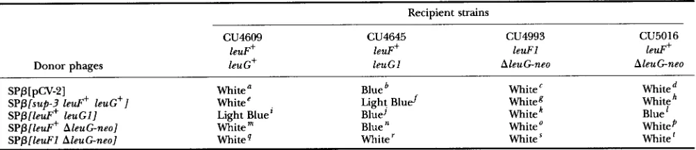
with SPP[sup-3 leuP leuG+]. The resulting heterog- enotic strain, CU4933, produced light blue colonies on TBAB X-gal plates (Table 4). This suggested that the
ZeuG+ allele partly complemented the leuGl mutation.
To be certain that the presence of sup-3 in CU4933 was not responsible for the decrease in ilv-leu expres- sion, we constructed a heterogenote with the genotype
le& leuG+/SPP[le& ZeuGl]. We lysogenized B. sub-
tilis strains carrying the ilvN-lac2 reporter gene with
phage SPP[leuP leuGI]. When we lysogenized a leuGI
strain we got lysogens that formed dark blue colonies on TBAB Xgal plates. When we lysogenized a leuG' strain with the phage the resulting lysogens formed light blue colonies on TBAB X-gal ('CU4938, Table 4).
We saw a similar light blue phenotype when we plated either of the reciprocal leuG+/leuGl diploid strains on TBAB X-gal plates: CU4645 lysogenized with SPP[sup-3
l e u P l e u G + ] , or CU4609 lysogenized with SPP[leuP
l e u G l ] (fCU4933 and 'CU4938, Table 4). This shows
that the wild-type leuG' allele partly complements the
leuGl mutation. We assayed the Pgalactosidase activity
of CU4933 ilvN-lac2 leuBl6 trpC2 leuG1 (SPPlsup-3
l e u P ZeuG']) grown in minimal medium with leucine,
isoleucine and valine to compare ilv-leu operon expres- sion in the diploid strain with the leuG' and leuGl hap- loid strains (Table 3). As stated before, a leuGl strain was shown to express the ilv-leu operon 19-fold higher than
a leuG+ strain. CU4933, the leuG+/leuGl diploid strain,
expressed the operon only 2-fold higher than wild type. As a control we constructed a leuGl strain that was ly- sogenic for a recombinant SPP phage that does not have the extra copy of tmS. This strain, CU4937, expressed the
l e u G Mutations of B . s u b t i l i s 633
TABLE 4
Colony colors of SPP lysogens
Recipient strains
CU4609 CU4645 c u4993 CU5016
leuF' leuF+ leuFl le*+
Donor phages leu G+ leuGI AleuG-neo AleuGneo
SPP[pCv-21 White' Blue' White Whited
S P p [ s u p 3 l e d + leuG'] Whitee Light Bluef Whiteg White
SPp[leuFi leuGI] Light Bluei Blue] White Blue'
SPP[leuF+ AleuGneo] Whitem Blue White
'
WhitePSPP[leuFl AleuGneol White" Whiter White White
The color phenotypes of B . subtilis strains plated on TBAB with X-gal. Recipient cells were lysogenized with donor SPP lysates. Lysogens were selected on TBAB with either chloramphenicol or neomycin. Purified colonies were plated on TBAB X-Gal plates with the same antibiotic. Phage lysates were made from strains CU4936 (SPp[pCV-2]), CU4934 (SPP[sup-3 le&+ l e u G + ] ) , CU4987 (SPP[leuF+ l e u G I ] ) , CU5022 (SPP[leuF+
AleuG-neo]), and CU5023 (SPP[leuFl AleuG-neo]). The phage carrying pCV-2 does not have the trnS operon. The recipient strains all have the
ilvN-lacZ-erm fusion.
The following strain numbers were assigned: 'CU4936, kU4937, %U5031, kU5035, eCU4932, fCU4933, gCU5032, kU5036, *CU4938, 'CU4939, 'CU5040, 'CU5037, mCU5029, nCU5022, 'CU5033, pCU5038, %U5030, %U5023, 'CU5034, kU5039.
The leuG gene is not essential: We constructed a plas- mid containing trnS in which 49 bp of the leuG gene were deleted and replaced with a neo gene ( AleuGneo)
.
We initially expected that the gene would be essential. No other gene that codes for leucine t R N A , - A G has been reported for B. subtilis (GREEN and VOLD 1993). How- ever, we found that we could transfer the deletion di- rectly from the plasmid to a trnS haploid strain by trans- formation. PCR and Southern transfer experiments verified that the resulting strains are haploid for the re- gion around leuG and carry only AleuGneo. We con- clude that the leuG gene is not essential for B. subtilis. However, haploid strains carrying AleuGneo grow more slowly than LeuG+ strains on all media. The doubling time of strain CU5016 ilvN-1acZ leuBl6 trpC2 AleuG- ne0 grown in minimal medium with isoleucine, valine, and leucine was87
min (Table 3). Strain CU4609 ilvN- lacZ leuBl6 trpC2 leuG+ grown in the same medium had a doubling time of 49 min. We conclude that there must be another tRNA gene in B. subtilis that codes for a tRNA that can translate 5'CUC-S' and 5'-CUU-3' codons. The lower growth rate of strains with theA
leuG neo deletion indicates that the gene for the other tRNA is probably not highly expressed. The growth rate of the strain with AleuGneo in the minimal medium is similar to the growth rate of the LeuGIcontaining strain (com- pare CU5016 and CU4645 in Table 3). It is probable that the growth defect of the leuG1-containing strains is caused by a reduced ability to translate 5'-CUG3' and 5'-CUU-3' codons.Haploid strains that contain the AleuG-neo deletion and the ilvN-lac2 reporter gene do not have the over- expression phenotype that we saw with the leuG transi- tion mutations. Strain CU5016 ilvN-1acZ leuBl6 trpC2 AleuGneo forms small white colonies on TBAB X-gal plates. Lysogenizing CU5016 with SPP[le@ leuGI] produced colonies that were blue on TBAB with X-gal
('CU5037, Table 4). PGalactosidase assays indicate that CU5016 expresses the ilv-leu operon about 2.5-fold higher than CU4609 ilvN-1acZ leuB16 trpC2 leuG+ while CU4645 ilvN-1acZ leuB16 trpC2 leuGl expresses the operon about 19-fold higher than the leuG+-carrying strain (Table 3).
The leuFl mutation suppresses the leuGl mutation:
We created a plasmid that contains the region of trnS with a mutation in the leuF gene, which codes for leucine tRNAu,. We mutated the gene in vitro so that the anticodon for the tRNA was changed from UAG to GAG. The new mutation was called leuFl. The plasmid with leuF1 also contains the AleuG-neo deletion. We transferred the leuFl and AleuG-neo mutations to B. subtilis strains by transforming cells with linearized plas- mid DNA and selecting for NeoR. A haploid strain con- taining leuFl and AleuG-neo was lysogenized with SPP[leup leuGI]. The resulting strain produced white colonies on TBAB X-gal plates (kCU5040, Table 4). The isogenic strain without the leuFl mutation makes blue colonies ('CU5037, Table 4). The result shows that the leuFl mutation suppresses the leuGl mutation.
The AleuG-neo deletion was transferred onto the SPP phages that carry tmS. Transformation of CU4987 ilvN- lacZ leuBl6 metB5 (SPP[leup leuGl]) with linearized plasmid DNA containing the AleuGneo deletion pro- duced transformants in which the ne0 gene was inserted into either the chromosomal or phage location of tmS. Phage with the AleuG-neo deletion made lysates that could be used to transduce the deletion to other strains. Using two AleuG-neecontaining plasmids, one with 1euFl and the other with l e u p , we were able to make two
X-gal plates ('CU5023, Table
4).
This again indicates that the leuFl mutation partly suppresses the leuGImutation. As a control we lysogenized CU4645 ilvN-lacZ 2euB16 trpC2 leuG1 with SPP[leuF+ AleuG-neo]. The lysogen ("CU5022) produced blue colonies on TBAB X-gal plates. We measured the expression of the ilv-leu
operon in these two strains using the @-galactosidase as- say (Table 3). The l e u p leuGI/SPP[leuF+ AleuG-nee]
strain, CU5022, expressed the ilv-leu operon 12-fold higher than a leuG+ haploid strain. The l e u P l e u G I / SPPlleuFl AleuG-neo] strain, CU5023, expressed the operon only 2-fold higher than wild type.
DISCUSSION
When
B .
subtilis cells are starved for leucine, they overexpress the ilv-leu operon by reading through the transcriptional terminator in the untranslated leader(GRANDONI et al. 1992, 1993). Analysis of the ilv-leu
leader led us to believe that trans-acting factors are in- volved in its regulation. We isolated and characterized mutations unlinked to the ilv-leu operon that caused overexpression of ilv-leu.
The mutations were mapped to two loci: near Zeus
described by VANDER HORN and ZAHLER (1992) and near
sup-3 (present work). All of the mutations near sup-3
that have been tested have base substitutions in the leuG
tRNA gene. The phenotypes of both classes of mutations can be explained by a model in which tRNA molecules control the ilv-leu attenuator independently of either trans-acting regulatory proteins or the translation of an upstream open reading frame.
The similarity of features found in the leaders of the
ilv-leu and tyrS operons suggests that the ilv-leu operon is regulated by a mechanism analogous to that proposed by GRuNDYand HENKIN (1993) for the tyrs operon. Analy- sis of mutations in the tyrS leader led them to conclude that uncharged tyrosine tRNA that recognizes the codon UAC can interact with structures in the tyrS leader RNA to promote antitermination. Similarly, features in the
ilv-leu leader suggest that an uncharged leucine tRNA that recognizes CUC promotes antitermination (GRUNDY and HENKIN 1993). The model predicts that any mu- tation that increases the level of uncharged leucine
in the cell will lead to overexpression of the
ilv-leu operon. The leucine-resistant mutations located in the leuS and leuG genes could all be of this class.
Leucyl-tRNA synthetase, the gene product of leuS, is the enzyme that charges leucine tRNAs. The point mu- tation leuSl results in increased expression of the ilv-leu
operon (VANDER HORN and ZAHLER 1992). Any kind of change in the enzyme that reduced its charging ability might lead to the accumulation of uncharged leucine
tRNA (including leucine
tRNbAG).
The mutations in leuG that cause increased ilv-leu
expression alter the D-loop of leucine tRNbAG. The phenotypes of the LeuG mutations could be the conse-
quence of either the accumulation of uncharged leucine
tRNbAG
molecules due to a defect in charging, or the loss of function of the tRNA in some other pro- cess in the cell. The G residues changed in the leuGmutations are highly conserved in all tRN&. Crystal structure studies of other tRNA molecules suggest that two of these residues may interact with residues in the pseudouridine loop to stabilize the tRNA structure (KIM
1979). Our leuG mutations might alter the tRNA three- dimensional structure, which might in turn affect its ability to be charged by its synthetase. Alternatively, some other function of the tRNA could be affected, such
as its role in the translation of CUC and CUU codons. The failure of the leuG null mutant to overexpress the
ilv-leu operon appreciably supports a model in which the leucine-resistant phenotype conferred by the leuG
point mutations did not reflect a reduced function of the
tRNA in a process such as translation. Our ability to con- struct null mutant strains suggests that there is another
tRNA that also recognizes CUC and CUU codons, al- though a gene for such a tRNA has not been reported yet (GREEN and VOLD 1993).
The codominance of leuGl and leuG+suggests a pre- viously unrecognized aspect of the WAdirected anti- termination mechanism. In a minimal medium with leucine, isoleucine, and valine, a strain carrying the
leuGI mutation expressed the ilv-leu operon about 19- fold higher than a strain with the wild-type allele of leuG
(CU4645 and CU4609 respectively, Table 3). A diploid strain carrying both the leuGl and leuG+ alleles had only 2-fold higher expression than the haploid strain with
leuGf (CU4933 and CU4609, respectively, Table 3). Having an extra copy of the operon with awild-type copy of the leu G gene reduces ilv-leu overexpression greatly. We conclude that the leuG+ allele is partly dominant over the 1euGl mutation.
The phenotype of the leuG+/leuGI strains raises some interesting questions about the interaction of these tRNA gene products with the ilv-leu leader. If our hy- pothesis is correct and the leuGI allele produces mutant leucine
WhAG
that is not efficiently charged, we pre- dict that it should increase the intracellular level of un- charged leucinetRNkAc
both in trnS haploid and dip- loid strains. There is no obvious mechanism by which the presence of the wild-type copy of the leuG gene could improve the charging of leuGI mutant leucine tRNbAG molecules. It is possible that the ZeuG' allele could lower the level of uncharged leucine tRNkAG in the cell by some effect on the expression or turnover of the mutant leuGI leucinetRNbAG,
but we think that this is unlikely. We will discuss our reasoning for this below.leuG Mutations of B. subtilis 635
leader in the diploid as in the haploid. The GRUNDY and HENKIN model predicts that only uncharged tRNA causes antitermination. The fact that ilv-leu expression in the
leuGi/leuG1 diploid strain is much lower than in the
leuG1 haploid strain suggests that both charged and un-
charged leucine
tRNhAG
molecules can interact withthe ilv-leu leader, and perhaps compete for binding. If
both charged and uncharged leucine
tRNhAG
mol- ecules are able to interact with the ilv-leu leader, higher levels of the charged tRNAs could compete with the un- charged tRNAs and reduce their ability to cause anti- termination at the attenuator.As part of our analysis of the leuGl mutation, we con- structed a mutation in another leucine tRNA gene that suppressed the mutant phenotype. The trnS operon has two adjacent leucine tRNA genes. The leuG gene codes for leucine
tRNhAG.
The leuF gene is one of two known identical leucine tRNAuAG genes. We introduced into aleuGl strain a leuF allele (leuF1) with a UAG to GAG
anticodon change, which allows recognition of the CUC specifier codon in the ilv-leu leader. If charged wild-type leucine tRNA,-.AG molecules compete with uncharged
leuGl mutant tRNAs for interaction with the ilv-leu
leader, we thought that other charged tRNAs that can recognize the specifier codon in the leader might also compete with the uncharged leuGl tRNA.
Strains carrying the leuF1, leuG1 and ilvN-lacZ re- porter genes were plated on indicator plates to deter- mine the effect of the mutations on expression of the
ilv-leu operon. Strains with the leuF1 and leuGl muta-
tions produced white colonies. Colonies of isogenic strains without the leuFl mutation were blue. We con- clude that the leuFl mutation suppresses the leuGl mu- tation. This result suggests that charged tRNA molecules that recognize the CUC specifier triplet can compete with uncharged leucine WAGAG for interaction with the
ilv-leu leader.
We would like to address the possibility that either the
leuG+ allele or the leuF1 mutation might influence the
leuGl phenotype through some effect on the expression
or stability of leuGl mutant leucine
tRNkAG
molecules. If this were true, the conclusions based on the domi- nance and suppression results would not be valid. One might argue that creating strains diploid for trnS had some effect on its expression. For example, the cells might have some mechanism that can down-regulate the operon when its copy number is increased. The lower expression of ilv-leu in the leuGi/leuG1 diploid strains could then be explained not as a competition between the two alleles but as a reduction in leuGl expression caused by the down-regulation. A similar explanation could apply to our observations with diploid strains car- rying the leuFl mutation.TO address this question we compared the phenotypes of two strains, CU5022 and CU5023. Each has the chro- mosomal leuGl mutation and the ilvN-lac2 reporter
gene. Each is lysogenic for an SPP phage with a cloned insert of DNA including the trnS operon. In CU5022 the phage has the l e u P allele and AleuG-neo. In CU5023 the phage has the leuFl mutation and AleuG-neo. In CU5022 ilv-leu expression was
12
times higher than ina leuGf haploid strain. In CU5023 the ilv-leu operon was
expressed only twice as much as in the wild-type (Table 3). (A haploid leuGl strain expressed the ilvN-lac2 fu- sion 19-fold more than the wild type.) The expression of
the ilv-leu operon is much higher in CU5022 than in
CU5023. Both strains are diploid for t m S , so having a second copy of the operon does not appear to cause a major reduction in the level of leuGl mutant tRNA in the cell. We conclude that the leuFl mutation s u p presses the leuGl phenotype by a mechanism that does not involve a reduction in the intracellular level of the
leuGl mutant tRNA.
Our results are in general agreement with the model of GRUNDYand HENKINS. Conditions that are likely to lead to increased quantities of uncharged leucine tRNkAG in the cell lead to overexpression of the ilv-leu operon. We strongly suspect that the uncharged tRNA interacts di- rectly with the leader of ilv-leu to increase readthrough at the terminator. We also present evidence suggesting that charged leucine tRNhAG competes with uncharged leucine
tRNhAG
for binding to the leader. We have no evidence concerning proteins that may interact with theilv-leu leader to affect attenuation.
We thank PHILIP YOUNGMAN for providing plasmid pCV-2. We grate- fully acknowledge useful conversations with our colleagues: T. HENKIN,
J. GRANDONI, J. CALVO and P. VANDER HORN. The work was supported by National Institutes of Health grant GM43970 to S.A.Z. D.B.G was recipient of a National Institute of General Medical Sciences traineeship under T32-GM07617.
LITERATURE CITED
BORN, S., 1990 Plasmids, pp. 79-81 in Molecular biological methods
for Bacillus, edited by C. R. HARWOOD and S. M. CUITING. John Wiley & sons, New York.
CUTTING, S. M., and P. B. VANDER HORN, 1990 Genetic analysis, pp. 27-74 in Molecular biological methods for Bacillus, edited by C. R. HARWOOD and S. M. CUTTING. John Wiley & sons, New York.
GARRITY, D. B., 1994 Mutations in the Bacillus subtilis tRNA gene,
leuG, affect the transcriptional regulation of the ilv-leu bio-
synthetic operon. Ph. D thesis. Cornel1 University, Ithaca, N. Y.
GARRITY, D. B., and S. A. ZAHLER, 1993 The Bacillus subtilis ochre
suppressor sup-3 is located in an operon of seven tRNA genes. J.
Bacteriol. 175: 6512-6517.
GRANDONI, J. A., S. A. ZAHLER, and J. M. CALVO, 1992 Transcriptional regulation of the ilv-leu operon of Bacillus subtilis. J. Bacteriol.
GRANDONI, J. A., S. B. FULMER, V. BRIZZIO, S. A. ZAHLER, and J. M. CALVO,
1993 Regions of the Bacillus subtilis ilv-leu operon involved in regulation by leucine. J. Bacteriol. 175: 7581-7593.
GREEN, C. J., and B. S. VOLD, 1993 tRNA, tRNA processing, and ami- noacyl tRNA synthetases, pp. 683-698 in Bacillus subtilis and Other Gram- positive Bacteria: Biochemistly, Physiology, and Mo-
lecular Genetics, edited by A. L. SONENSHEIN, J. HOCH and R.
LOSICK. American Society for Microbiology, Washington, D.C.
ulator of transcription antitermination in B. subtilis. Cell 74:
GRUNDY, F. J., and T. M. HENKIN, 1994 Conservation of a transcription
antitermination mechanism in aminoacyl-tRNA synthetase and
amino acid biosynthetic genes in the Gram-positive bacteria.
J. Mol. Biol. 235: 798-804.
HAIWHAN, D., 1983 Studies of transformation ofEscherichia coli with
plasmids. J. Mol. Biol. 166: 557-580.
HARWOOD, C. R., and S. M. CUITING, 1990 Appendix, pp. 545-550 in
Molecular Biological Methods for Bacillus, John Wiley & Sons, New York.
HIGUCHI, R., 1990 Recombinant PCR, pp. 177-183 in PCR Protocols,
edited by M. A. INNES, D. H. GELFAND, J. J. SNINSKY and T. J. WHITE. Academic Press, New York.
K I M , S., 1979 Crystal structure ofyeast tRNAPhe and general structural features of other t R N A s , pp. 83-100 in Transfer RNA: Structure, Properties, and Recognition, edited by P. R. SCHIMMEL, D. S ~ L L and
J. N. ABELSON. Cold Spring Harbor Laboratory, Cold Spring
Harbor, N.Y.
LANDICK, R., and C. YANOFSKY, 1987 Transcriptional attenuation, pp.
1276-1301 in Escherichia coli and Salmonella typhimurium:
Cellular and Molecular Biology, edited by F. C. NEIDHARDT, J. L.
INGRAHAM, IL B. Low, B. MAGWIK, M. SCHAECHTER et al. American
Society for Microbiology, Washington, D.C.
MANIATIS, T., E. F. FRITSCH and J. SAMBROOK, 1982 Molecular Cloning:
A Laboratory M a n u a l . Cold Spring Harbor Laboratory, Cold Spring Harbor, N.Y.
PERKINS, J. B., and P. L. YOUNGMAN, 1986 Construction and properties
of Tn917-lac, a transposon derivative that mediates transcrip 475-482.
tional gene fusions in Bacillus subtilis. Proc. Natl. Acad. Sci. USA
PLATKO, J. V., D. A. WILLINS and J. M. CALVO, 1990 The ilvZHoperon
of Escherichia coli is positively regulated. J. Bacteriol. 172: 456 3-4570.
SULLIVAN, M. A,, R. E. YASBIN and F. E. YOUNG, 1984 New shuttle vec-
tors for Bacillus subtilis and Escherichia coli which allow rapid
detection of inserted fragments, Gene 29: 21-26.
STREIPS, U. N., 1991 Transformation, pp. 210-214 in Modern Micro-
bial Genetics, edited by U. N. STREIPS and R. E. YASBIN. Wiley-Liss, New York.
VANDER HORN, P. B., and S. A. ZAHLER, 1992 Cloning and nucleotide
sequence of the leucyl-tRNA synthetase gene of Bacillus subtilis.
J. Bacteriol. 1 7 4 3928-3935.
VANDEYAR, M. A., 1987 Studies on the ilvBNC genes o f Bacillus sub-
tilis. Ph.D. Thesis. Cornell University, Ithaca, N.Y.
YOUNGMAN, P., H. POTH, B. GREEN, K. YORK, G. OLMEDO et al., 1989 The
SPP cloning system, pp. 77-80, in Regulation ojProkaryotic De-
velopment, edited by I. SMITH, R. A. SLEPECKYand P. SETLOW. Ameri- can Society for Microbiology, Washington, D.C.
WEINER, M. P., 1986 Characterization of bacteriophage H2. Ph.D.
Thesis, Cornell University, Ithaca, N.Y.
YANISCH-PERRON, C., J. VIERA and J. MESSING, 1985 Improved
M13 phage cloning vectors and host strains: nucleotide
sequences of the M13 mp18 and pUC19 vectors. Gene 33:
83: 140-144.
103-119.