Copyright © 1998 by the Genetics Society of America
Genetics 148: 815–825 ( February, 1998)
Conserved Regions of the
timeless
(
tim
) Clock Gene in Drosophila Analyzed
Through Phylogenetic and Functional Studies
Andrea Ousley, Khaja Zafarullah, Yifeng Chen, Mark Emerson, Lesley Hickman and Amita Sehgal*
Howard Hughes Medical Institute, Department of Neuroscience, and *Center for Sleep and Respiratory Neurobiology, University of Pennsylvania Medical School, Philadelphia, Pennsylvania 19104
Manuscript received August 1, 1997 Accepted for publication November 3, 1997
A B S T R A C T
Circadian (z24-hr) rhythms in Drosophila melanogaster depend upon cyclic expression of the period (per) and timeless (tim) genes, which encode interacting components of the endogenous clock. The per gene has been isolated from other insects and, more recently, a per ortholog was found in mammals where its expres-sion oscillates in a circadian fashion. We report here the complete sequence of a tim gene from another species, Drosophila virilis. TIM is better conserved than the PER protein is between these two species (76 vs. 54% overall amino acid identity), and putative functional domains, such as the PER interaction domains and the nuclear localization signal, are highly conserved. The acidic domain and the cytoplasmic localiza-tion domain, however, are within the least conserved regions. In addilocaliza-tion, the initiating methionine in the D. virilis gene lies downstream of the proposed translation start for the original D. melanogaster tim cDNA and corresponds to the one used by D. simulans and D. yakuba. Among the most conserved parts of TIM is a region of unknown function near the N terminus. We show here that deletion of a 32 amino acid segment within this region affects rescue of rhythms in arrhythmic tim01 flies. Flies carrying a full-length tim trans-gene displayed rhythms with z24-hr periods, indicating that a fully functional clock can be restored in tim01 flies through expression of a tim transgene. Deletion of the segment mentioned above resulted in very long activity rhythms with periods ranging from 30.5 to 48 hr.
the timeless (tim) and period (per) genes in Drosophila, and the frequency and white collar-2 (a transcriptional ac-tivator) genes in Neurospora (Dunlap et al. 1995;
Sehgalet al. 1996; Crosthwaiteet al. 1997). Recently,
two mammalian genes that function in the circadian system were isolated. The first, Clock, is the gene af-fected in a circadian mutant mouse, and it encodes a novel member of the bHLH-PAS family of transcription factors (Kinget al. 1997; Antochet al. 1997). The
sec-ond is a mammalian ortholog of the per gene (Sunet al.
1997; Teiet al. 1997).
Where characterized, the clock mechanism involves a molecular feedback loop. In Drosophila, RNA and protein levels of the per and tim genes cycle with an
z24-hr period, and the TIM and PER proteins affect expression of their own mRNAs through a feedback mechanism that requires nuclear entry of TIM and PER (Siwickiet al. 1988; Hardinet al. 1990; Zenget al.
1994, 1996; Vosshall et al. 1994; Sehgalet al. 1995;
Hunter-Ensoret al. 1996; Myerset al. 1996). Nuclear
localization of TIM and PER is mediated, at least in part, by association of the two proteins with each other through known interaction domains (Vosshall et al.
1994; Gekakiset al. 1995; Saez and Young 1996). The
Neurospora frequency gene operates in a similar auto-regulatory feedback loop whereby both RNA and pro-tein cycle and the propro-tein inhibits synthesis of its own RNA (Aronson et al. 1994). Finally, while the role of Corresponding author: Amita Sehgal, Department of Neuroscience,
233A Stemmler Hall, University of Pennsylvania, Philadelphia, PA 19104-6074. E-mail: [email protected]
C
IRCADIAN (z24-hr) rhythms are displayed by vir-tually all organisms, ranging from cyanobacteria to mammals (Dunlap et al. 1995; Hall 1995).Circa-dian rhythms, as distinguished from phenomena that are driven solely by cues in the environment, are gener-ated by an internal time-keeping mechanism called the circadian clock. Because the circadian clock is normally synchronized with the environment, models of a circa-dian system include an input pathway that conveys in-formation about the environment to the clock (Eskin
1979). Cues in the environment, such as light, reset the phase of the clock, thereby synchronizing the z24-hr endogenous rhythm with the environmental cycle. The mechanism of resetting has been investigated in at least two systems, and in both, the level of a clock compo-nent changes in response to light, producing a predict-able phase shift (Crosthwaite et al. 1995; Hunter
-Ensor et al. 1996; Lee et al. 1996; Myers et al. 1996;
Zenget al. 1996). A circadian system also includes an
output pathway that relays temporal information from the clock to other sites, effecting the circadian regula-tion of different physiological processes and behavioral activities (Eskin 1979).
816 A. Ousley et al.
the mammalian per gene in the circadian system has not yet been established, it is known that levels of its RNA cycle in the suprachiasmatic nuclei and retina. Both these tissues contain endogenous oscillators (Tosini and Menaker 1996; Turek 1996).
A phylogenetic analysis carried out for per from a number of Drosophila species revealed that much of the coding region (approximately one-third) is not conserved (Colot et al. 1988; Thackeray and K yria-cou 1990). The per gene was also isolated from a
non-Dipteran species, Antheraea pernyi, the giant silkmoth (Reppertet al. 1994). Although the giant silkmoth per
gene is only 39% identical at the amino acid level to the most closely related Drosophila gene, it was able to res-cue circadian rhythms in the arrhythmic per01
Dro-sophila mutant (Levine et al. 1995). Sequences
con-served between the insect per genes, particularly those corresponding to the PAS domain, were used to isolate a mammalian per homolog (Tei et al. 1997). The same
gene was isolated fortuitously by a group studying tran-scripts that mapped to human chromosome 17 (Sun et
al. 1997). Surprisingly, given the low level of homology
between the insect per genes, mammalian per is z44% homologous to Drosophila per (including identical amino acids as well as conserved and neutral substitu-tions; Sun et al. 1997). Conserved regions of PER
in-clude all known functional domains, including the PAS domain and the cytoplasmic localization domain, both of which also mediate the interaction with TIM (Gekakis et al. 1995; Saez and Young 1996; Huang et
al. 1993, 1995).
Since the cloning of the tim gene (Myers et al.
1995), no one has reported the sequence of a tim ho-molog. We undertook a phylogenetic analysis of tim by isolating and characterizing a tim homolog from D.
viri-lis, and also by sequencing selected regions of a tim
ho-molog from D. hydei. In this report, we present the intron/exon organization of the gene and the conser-vation profile of the TIM protein. We found that the amino acid sequence of TIM is more highly conserved overall than that of PER from different Drosophila spe-cies, and that most functional domains of TIM are con-served. In addition to known functional domains, such as the PER interaction domains and the nuclear local-ization signal (NLS), the conserved parts of TIM in-clude an additional region that we show here to be im-portant for function. tim transgenes lacking a 32 amino acid sequence from this region showed aberrant rescue of behavioral rhythms in tim01 flies, while a wild-type tim
transgene restored completely normal behavior.
M AT E R I A L S A N D M E T H O D S
Fly strains: The D. hydei flies were obtained from Carolina Bio-logical Supply Co. (Burlington, NC), and the homozygous yw; Ki
D2-3 Drosophila strain was kindly provided by Paul Hardin
(Texas A&M University, College Station, TX).
Library screening: The genomic library of D. virilis made in l phage EMBL3 was kindly provided by R. K. Blackman
(University of Illinois, Urbana, IL). This genomic library was screened with a 1.5-kb EcoRI fragment, which corresponds to nucleotides 1974–3492 of the D. melanogaster tim cDNA (M y-ers et al. 1995; EMBL/GenBank accession number U37018).
Hybridization was performed under low stringency condi-tions (378, 40% formamide, 63 SSPE, 0.5% SDS, 53 Den-hardt’s solution, and 0.1 mg/ml salmon sperm DNA). Washes were repeated twice at 508 in a 53 SSPE, 0.1% SDS solution. The library screen yielded two recombinant phage clones that were subjected to Southern blot analysis to identify hybridiz-ing restriction fragments. An z4-kb BamHI fragment that showed strong hybridization to the tim probe was subcloned and sequenced in both directions. To extend this sequence in the 59 and 39 directions, we synthesized primers based on the sequence obtained and used these to sequence phage DNA isolated from our l phage clones.
A recombinant phage clone containing the upstream se-quence of the D. melanogaster tim gene was isolated from a ge-nomic library made in EMBL3A (Spradling and Mahowald
1981) as described above, except that more stringent condi-tions were used for the hybridization.
RT-PCR experiments of D. melanogaster tim RNA: For the RT-PCR experiment, total RNA was isolated from fly heads by homogenizing them in 0.5 ml of buffer (150 mM sodium ace-tate, 50 mM Tris, pH 9.0, 5 mM EDTA, pH 8.0, 1% SDS con-taining 1/100th volume diethyl pyrocarbonate) followed by two phenol/chloroform (1:1) extractions and ethanol precip-itation. RNA (in 10 mM dNTP mixture, 20 mM DTT, 13 re-verse transciptase buffer containing 10 mM random hexamers and 2 units/ml RNasin) was heated at 658 for 5 min and then cooled to 428. The reaction mixture was incubated with 1 ml of AMV-reverse transcriptase (10 units/ml, Promega, Madi-son, WI) for 1 hr at 428, and the enzyme was then inactivated at 658. Reverse-transcribed cDNA was amplified by PCR (in 2.5 mM MgCl2, 13 PCR buffer, 2 mM dNTP mixture
contain-ing 5 pmol of each primer and 1 ml of Promega Taq poly-merase) under the following conditions: 958, 3 min; 30 cycles of 95°, 30 sec, 558, 30 sec, 728, 1 min; 728, 10 min. A pair of specific primers, which amplifies a fragment corresponding to nucleotides 839–1501 of the original tim cDNA sequence (Myers et al. 1995; GenBank accession number U37018) and
spans the 32 amino acid sequence (GenBank accession num-ber AF038501; see results), was used to amplify this region
from the reverse-transcribed cDNA and from the original 59 tim cDNA clone.
39 RACE: For the 39 RACE experiment, total RNA was iso-lated and the reverse transcription was done as described above, except that an oligo-dT primer was used (59-[C]13
AAGC[T]17-39) and the reaction was carried out at 378.
Re-verse-transcribed cDNA was amplified by PCR as described above, with some modifications. During the first round of am-plification, a D. virilis-specific primer (59-TTGGCTGCAGT TGGTCAT-39; D. virilis tim sequence, GenBank accession numbers AF038502 and AF040096) and a shorter oligo-dT primer (59-[C]8 AAGC[T]8-39) were used, and the annealing
temperature was 428 for the initial three cycles and 528 for the remaining 47 cycles. An aliquot of this PCR reaction was then reamplified by PCR using a second D. virilis-specific primer in-ternal to the first (59-ATGCGCAGCAAATGCAGCA-39; D. viri-lis tim sequence, GenBank accession numbers AF038502 and AF040096) and the short oligo-dT primer. The conditions were the same as for the first round of amplification, except that the annealing temperature was 528 for all 40 cycles. The amplified fragments were cloned into the pCR2.1 Vector (In-vitrogen, San Diego, CA) and sequenced as below.
primers were used to amplify, by PCR, regions of the tim gene from D. hydei. The primers were designed based on sequences within regions of the tim gene that were highly conserved be-tween D. melanogaster and D. virilis. The primers were 1S (sense primer) (59-AA(G/A) CCI CA(A/G) CA(T/C) CA(G/ A) AA(G/A/) CC-39), 4S (59-GA(T/C) CA(G/A) AT(C/T/A) AA(C/T) AA(T/C) TG(T/C) CT-39), 13S (59-GA(C/T) ATG GA(A/G) CA(C/T/) AT(C/T/A/) GA(T/C/) AC-39), 1A (antisense primer; the exact complement of 1S), and 12A (59 -TC(A/G) TA(G/A) TCI GC(T/C) TCC CA(A/G/T/) AT-39). Advantage KlenTaq polymerase (Clontech Laboratories, Palo Alto, CA) was used to amplify the following D. hydei frag-ments. The primer pairs (13S, 12A) and 4S, 1A) amplified a fragment corresponding to nucleotides 2127–2749 and 74– 1804 of the D. melanogaster tim cDNA (Myers et al. 1995;
Gen-Bank accession number U37018), respectively. The primer pair (1S, 12A) amplified an overlapping fragment corre-sponding to nucleotides 1785–2749 of the D. melanogaster tim cDNA. A pair of specific primers were made based on the D. hydei sequence to amplify a fragment spanning the 1S/1A primer region. PCR amplification using degenerate primers was done (in 1.5 mM MgCl2, 13 PCR buffer, 0.4 mM dNTP
mixture containing 2 pmol of each primer and 1 ml of Taq polymerase) under the following conditions: 958, 3 min; 3 cy-cles of 958, 1 min, 428, 1 min, 728, 2 min; 50 cycles of 958, 1 min, 508, 1 min, 728, 2 min; 728, 10 min. PCR amplification us-ing specific primers was done as described above for 30 cycles, except for omitting cycles 2–4 at a lower annealing tempera-ture. Each fragment was cloned into the pCR 2.1 Vector (In-vitrogen) and sequenced as described below.
Sequencing and analysis: Sequencing was done at the Uni-versity of Pennsylvania Sequencing Facility on an automated sequencer (Applied Biosystems, Foster City, CA) using a dye-terminator and thermal cycling method. The Sequencher program (version 3.0; Gene Codes Corporation, Ann Arbor, MI) was used to create the contigs of independent sequenc-ing reactions. The intron/exon structure of the D. virilis gene was predicted based on its consensus 59 and 39 splice sites and by comparison with the D. melanogaster tim cDNA sequence (Myers et al. 1995). We used the DSPL, FGENED, and FEXD
programs found on the Baylor College of Medicine Gene-finder site (http://dot.imgen.bcm.tmc.edu:9331/gene-Gene-finder/ gf.html) for prediction of intron/exon junctions, and the Gap and BestFit programs in the Genetics Computer Group (GCG) Wisconsin Software package (version 8.0.1-UNIX) was used for pairwise alignments. The PileUp program (GCG) was used to make multiple sequence alignments, and the amino acid sequence similarity plot was generated by the Plot-Similarity program (GCG).
Construction of plasmids: Plasmids for P-element-medi-ated DNA transformation were generP-element-medi-ated by fusing z4.3 kb of sequence upstream of the transcription initiation site of the D. melanogaster tim gene to the D. melanogaster tim cDNA (Myers et al. 1995; see Figure 4 in this paper). Upstream
se-quences were isolated by screening a genomic library made in EMBL3A (Spradling and Mahowald 1981) with the 59
re-gion of genomic clone Ec1 (Myers et al. 1995). An z7 kb
SacI-SacI DNA fragment, which contained a SacI-SwaI frag-ment extending from approximately 24300 to 1180 relative to the transcription initiation site, was isolated from the phage clone.
A full-length tim cDNA was generated by piecing together fragments from partial D. melanogaster cDNA clones (35c and 35g; see Myers et al. 1995). Individual fragments were
succes-sively cloned into pBluescript: first, a BamHI-ApaI fragment from the 39 end, then a SacI-BamHI from the 59 end, followed by the internal BamHI-BamHI fragment. The orientation of the BamHI-BamHI fragment was verified. The resulting cDNA
insert extended from nucleotide positions 1–4955 of the orig-inal cDNA sequence and had a 39 alternatively retained intron inserted after position 3555 (Myers et al. 1995; GenBank
ac-cession number U37018), but it did not include the addi-tional 32 amino acid sequence reported in this paper (see re-sults). This full-length cDNA was then excised using SacI
and KpnI (polylinker sites) and cloned into the pet-17b vector to introduce a SpeI site at the 59 end.
The z4.5-kb SacI-SwaI fragment containing the upstream sequence (see above) was substituted for the 180-bp SacI-SwaI fragment in the tim cDNA pet-17b construct. A SpeI-KpnI frag-ment derived from the resulting construct, which contained the upstream sequence fused to the tim cDNA, was then cloned into the P element vector pCaSper4, which contains a miniwhite gene p(white), thus enabling selection of trans-genic flies by eye color. The resulting pCaSper4 construct was called Tim 1. Another pCaSper4 construct (Tim 4) was made, which was identical to Tim 1, except that it also contained the additional 32 amino acid sequence (see Figure 4). Since the 32 amino acid sequence was contained within a unique SphI fragment in the tim gene, the SphI fragment in the Tim 1 con-struct was replaced with the corresponding fragment from the genomic clone Ec1 (Myers et al. 1995). The resulting
construct was called Tim 4.
Production of transgenic strains: These two constructs were introduced into the Drosophila genome by P-element-mediated DNA. Embryo injections were performed using a yw/yw; Ki D2-3 strain (Robertson et al. 1988), and multiple
Figure 1.—Genomic organization of the tim gene. The
in-tron–exon structure of the D. virilis tim gene and part of the D. hydei tim gene is shown schematically. The D. virilis ge-nomic tim homolog (4682 bp) begins at a consensus cap site (asterisk), extends through a codon corresponding to amino acid 1194 of the D. virilis TIM sequence, and contains 11 ex-ons (black boxes) and 10 intrex-ons (lines between boxes). The translation initiation site (ATG) is at nucleotide position 68. The shaded box represents the sequence obtained from the 39 RACE experiment (see materials and methods), which
Figure 2.—Profile of amino acid similarity between the tim homologs. (A) The amino acid similarity plot was generated from
820 A. Ousley et al.
independent lines carrying each of the two transgenes were derived from the progeny of surviving G0 adults. Each
trans-gene was then introduced into a yw tim01 background. Behavioral assays: Behavioral analysis was done on trans-genic flies carrying only the tim transgene [p(white)tim01/
tim01], on siblings that carried a wild-type copy of the tim gene
(CyO/tim01), on siblings that carried neither the endogenous tim gene nor the transgene (tim01/tim01), and on wild-type flies (yw strain). Locomotor activity rhythms were monitored using the Trikinetics system, and analysis was done exactly as described previously (Sehgal et al. 1994).
R E S U LT S
Cloning of a tim homolog from D. virilis: We screened a D. virilis genomic library under low stringency condi-tions using a D. melanogaster tim cDNA probe that corre-sponds to nucleotides 1974–3492 (Myers et al. 1995).
This probe includes the PER interaction domains of the tim gene, which we assumed would be well-con-served because the TIM interaction domains of the per gene are well conserved between D. melanogaster and
D. virilis (Colot et al. 1988). Two recombinant phage
clones were isolated. An z4-kb BamHI fragment that strongly hybridized with the tim cDNA probe on South-ern blots was subcloned and sequenced. To extend the sequence in the 59 and 39 directions, we synthesized primers based on the sequence obtained and used these to sequence phage DNA prepared from our clones.
Both genomic clones lacked sequences correspond-ing to the 39 end of the D. melanogaster tim gene, which includes the cytoplasmic localization domain (CLD) of TIM (Saez and Young 1996). Since this was a known
functional domain that was reported to be conserved in TIM (Saez and Young 1996), we used a 39 RACE
method to obtain sequences from the 39 end of the
D. virilis tim cDNA. By using a D. virilis-specific primer
that overlapped the coding region in the 39 end of our genomic clone and an oligo-dT primer, we amplified from D. virilis cDNA a fragment that included nucle-otides homologous to the C-terminal 149 amino acids of TIM.
Genomic organization of tim homologs: We charac-terized .4 kb of D. virilis genomic sequence corre-sponding to the tim gene. The schematic in Figure 1 represents z4.7 kb of genomic sequence from the
D. virilis homolog beginning at a consensus cap site
(Cherbas and Cherbas 1993) just upstream of a
me-thionine codon and continuing through a codon corre-sponding to amino acid 1194 of the D. virilis TIM se-quence. The C-terminal, 149 amino acid stretch obtained through the 39 RACE experiment is also indi-cated. The total length of the coding region is 4029 bp, from which we predict a protein of 1343 amino acids.
The genomic organization in Figure 1 shows the in-tron exon structure of tim. The figure shows this orga-nization for the D. virilis tim gene and for part of the
D. hydei tim gene, which was subsequently isolated for
comparison of specific sequences (see below). Our lim-ited data on the organization of the tim gene in D.
mela-nogaster and D. hydei indicate that, where examined, the
position of introns within the gene is conserved. We found that the positions of introns corresponding to in-trons 2–4 of the D. virilis gene are conserved in D. hydei (see Figure 1). Also, the position of an intron corre-sponding to intron 1 in the D. virilis gene is conserved in D. melanogaster. The DNA sequence of introns was not conserved (data not shown).
The predicted translation start site in the D. virilis gene corresponds to a methionine downstream of the proposed start site in the D. melanogaster gene (Myers et
al. 1995). This finding is consistent with a recent report
that this methionine may, in fact, also be the start site in some strains of D. melanogaster as well as in other Dro-sophila species (Rosato et al. 1997). A polymorphism
identified in the tim gene from certain D. melanogaster strains, as well as from D. simulans and D. yakuba, gener-ates a premature stop codon shortly after the originally proposed site (Rosato et al. 1997). Use of the
down-stream start site would shorten the D. melanogaster TIM protein by 23 amino acids.
Conservation profile of the TIM protein: The amino acid similarity plot in Figure 2A shows the level of con-servation across the TIM protein. The predicted amino acid sequences of the tim gene from different Dro-sophila species are aligned in Figure 2B, with domains relevant to our analysis of TIM indicated. We found that TIM is more highly conserved between D. virilis and D. melanogaster than PER is conserved between these two species, with TIM having a 76% overall amino acid identity compared with 54% overall identity for PER. In contrast to the per gene, which has five nonserved domains interspersed within the relatively con-served portion of its coding region (Colot et al. 1988),
the coding region of the tim gene has only two regions of low homology. The stretch of acidic amino acids found in D. melanogaster TIM (Myers et al. 1995) falls
within one such region (Figure 2, A and B).
Most of the known functional domains in TIM are highly conserved. The NLS in D. virilis is identical to that in D. melanogaster, except for the conservative change of a glutamate to an aspartate. Relative to
D. melanogaster, the PER interaction domains in D. virilis
are 80–85% identical at the amino acid level, a level of conservation that is similar to that for the TIM interac-tion domains of per (Colot et al. 1988; Thackeray and
Kyriacou 1990; Saez and Young 1996). The
N-termi-nal region of D. virilis tim also shows a high degree of homology (90% identical) to the D. melanogaster gene although no functional domain of tim has been mapped to this region. Incidentally, a 32 amino acid se-quence within this region was lacking in the original
tim cDNA (see below). The CLD recently mapped to
the C-terminal end of TIM (Saez and Young 1996),
having only 56% amino acid identity between D. virilis and D. melanogaster.
Of note is that the single amino acid mutated in the
timSL allele of D. melanogaster (Rutila et al. 1996), which
occurs just upstream of the first PER interaction do-main, is conserved in D. virilis. This allele contains a missense mutation that converts threonine 494 to an isoleucine. The mutation does not produce a dramatic phenotype by itself (the wild-type rhythm is shortened by z0.5 hr in homozygous timSL lines), but it suppresses
the perl (long period) mutation, shortening the period
by 4 hr and also restoring temperature compensation (Rutila et al. 1996).
Isolation of homologous sequences of the tim gene from D. hydei: Analysis of D. virilis tim provided some interesting revelations about tim structure. Most impor-tantly, perhaps, it revealed that the acidic region in
D. melanogaster, which was thought to be similar to
acti-vation domains of transcription factors (Myers et al.
1995), was not conserved, and that an additional 32 amino acid sequence was present within a highly con-served region. To determine the extent to which these findings were universally applicable, we amplified spe-cific sequences of the tim gene from yet another Dro-sophila species D. hydei. The genomic D. hydei sequence analyzed here begins in the middle of exon 2 in the
D. virilis gene and extends through most of exon 5 (see
Figure 1). Protein-coding sequences within this region are represented in Figure 2B and were included in the profile analysis shown in Figure 2A. The following facts were confirmed by our analysis of the D. hydei se-quence: (1) the acidic region is poorly conserved, (2) the region N-terminal to this is very well-conserved and includes the additional 32 amino acids, (3) the PER in-teraction domains are conserved, and (4) the amino acid mutated in the timSL allele is conserved.
The tim RNA in D. melanogaster contains an additional sequence: The D. virilis TIM sequence contained an extra 32 amino acids within the highly conserved N-ter-minal region of the protein (see Figure 2, A and B). Note that the D. hydei tim gene also contains the addi-tional 32 amino acids; however, the original D.
melano-gaster tim cDNA that was characterized lacked this
se-quence (Myers et al. 1995). To determine whether this
sequence was included in a differentially spliced form of the tim RNA, we carried out RT-PCR experiments. We used a pair of specific primers to amplify a fragment spanning the 32 amino acid region from head RNA of
D. melanogaster (yw strain) and from the original 59 tim cDNA clone (Myers et al. 1995). In Figure 3, lanes 1
and 2 (yw 14 and yw 16) show the fragment amplified by RT-PCR from head RNA collected at Zeitgeber times 14 and 16, when tim RNA is expressed at high levels (Sehgal et al. 1995). The RT-PCR product is of the size
predicted (758 bp) if the fragment includes the addi-tional 32 amino acids. The PCR product amplified from the tim cDNA clone (Figure 3, lane 3) is z100 bp
smaller than the RT-PCR product, as expected, since this cDNA clone lacks the 32 amino acid sequence. Other experiments in which control genomic frag-ments were also amplified confirmed that endogenous tim RNA was z100 bp longer than the tim cDNA (data not shown). Since we failed to amplify a smaller band from head RNA in all experiments, it is unlikely that the 32 amino acid sequence is part of a differentially spliced message. The other possibility is that the shorter version represents a polymorphic form of the
tim RNA that occurs in other strains. However, given
that the 32 amino acid sequence is found in both D.
vir-ilis and D. hydei homologs of tim and that it is important
for tim function (see below), we believe that this is
un-Figure 3.—Presence of an additional sequence in the
D. melanogaster tim RNA. We amplified a region of the tim gene thought to include coding sequences that were lacking in the original D. melanogaster tim cDNA, but which were present in both the D. virilis and D. hydei tim genes (see Figure 2). Ampli-fied products were resolved on a 1% agarose gel and stained with ethidium bromide. Locations of DNA size markers are indicated (far left). The same primer pair was used to amplify a fragment from D. melanogaster wild-type fly head RNA col-lected at Zeitgeber times 14 and 16 (lanes 1 and 2), and from the original D. melanogaster tim cDNA clone (lane 3; Myers et
822 A. Ousley et al.
likely. Thus, we conclude that it is an integral part of the D. melanogaster tim RNA.
A protein of 1389 amino acids was predicted from the original sequence of the Drosophila tim gene (Myers
et al. 1995). The predicted size becomes 1421 after
in-cluding the additional 32 amino acids. Use of the me-thionine in D. melanogaster corresponding to the trans-lation start we report here for D. virilis would shorten TIM by 23 amino acids (see also Rosato et al. 1997).
A tim transgene lacking a sequence in the N-terminal conserved region produces aberrant rhythms in tim01
flies: The functional significance of the conserved re-gion containing the extra 32 amino acid sequence was determined through behavioral rescue experiments. Arrhythmic tim01 flies were transformed with a tim
trans-gene that either contained (Tim 4) or lacked (Tim 1) the 32 amino acid sequence. Each transgene construct consisted of a tim cDNA from D. melanogaster, which in-cluded a 39 alternatively retained intron (Myers et al.
1995) placed under the control of z4.3 kb of sequence upstream of the transcription initiation site in the
D. melanogaster tim gene (see materials and meth-ods). Flies carrying either transgene were examined in
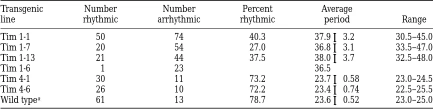
constant darkness after entrainment in 12 hr: 12 hr light/dark cycles for several days. We evaluated two in-dependent lines of flies carrying the Tim 4 transgene (see Table 1). In each of these lines, a high percentage of individual flies were rhythmic (72%), and the rhyth-mic individuals displayed wild-type periods. For the Tim 4-1 line, the average period was 23.7 6 0.58, and for the other line, Tim 4-6, the average period was 23.4 6 0.74. These periods are comparable to those seen in wild-type flies (see Table 1). As expected, tim01
siblings that lacked the transgene were arrhythmic. In Figure 4B, we show the locomotor activity plot and peri-odogram for a representative fly from one of these lines, Tim 4-1.
To determine whether the Tim 1 construct could rescue the arrhythmic phenotype, we evaluated four in-dependent lines of tim01 flies carrying this transgene. In
three of these lines, 27–40% of individual flies showed rescue of behavioral rhythms, but the periods were long, ranging from 30.5 to 48 hr; individual lines had average periods of 36.8–38.0 hr (see Table 1). In Figure 4B, we show the locomotor activity plot and peri-odogram for a representative fly from one of these lines, Tim 1-1. In a fourth line (Tim 1-6), only one of the individuals tested was rhythmic (Table 1). As be-fore, flies carrying a wild-type copy of the tim gene (CyO/tim01 siblings or yw flies) were rhythmic with an
average period of 23.6 6 0.52 (see Table 1). The behav-ioral analysis of the Tim 1 and Tim 4 flies demonstrates that the 32 amino acid sequence in TIM is necessary for restoring wild-type rhythms to tim01 flies.
D I S C U S S I O N
We report here the characterization of tim homo-logues from D. virilis and D. hydei. Our data show that the overall conservation of TIM is higher than that of PER. All known functional domains of TIM are highly conserved (.80% amino acid identity), with the excep-tion of the CLD (Saez and Young 1996), which shows
only 56% amino acid identity between D. virilis and
D. melanogaster. The PER interaction domains in TIM
show the same level of conservation as the TIM interac-tion domains in PER (Colot et al. 1988). We propose
that since interaction between the PER and TIM pro-teins is an integral part of the clock mechanism, there is genetic selection to conserve the interacting do-mains. While the CLD, as currently defined, is less well conserved, there are regions within this domain that are identical between D. virilis and D. melanogaster (see
TABLE 1
Period Estimates and the Percentage of Rhythmic Flies for Different tim Transgenic Lines
Transgenic line
Number rhythmic
Number arrhythmic
Percent rhythmic
Average
period Range
Tim 1-1 50 74 40.3 37.9 6 3.2 30.5–45.0
Tim 1-7 20 54 27.0 36.8 6 3.1 33.5–47.0
Tim 1-13 21 44 37.5 38.0 6 3.7 32.5–48.0
Tim 1-6 1 23 36.5
Tim 4-1 30 11 73.2 23.7 6 0.58 23.0–24.5
Tim 4-6 26 10 72.2 23.4 6 0.74 22.5–25.5
Wild typea 61 13 78.7 23.6 6 0.52 23.0–25.0
Locomotor activity data were obtained in constant darkness for flies from each Tim 1 and Tim 4 line (see text and Figure 4). Period analysis was done as described in Figure 4B. The number of flies that were rhythmic (and arrhythmic) for each line is shown, as well as the percentage of flies that were rhythmic. The average period (6SD) for rhythmic flies and the range in period are given in hours.
Figure 4.—Rescue of activity rhythms in tim01 flies by tim transgenes. (A) Schematic representation of the tim transgenes. The
tim transgene constructs (Tim 4 and Tim 1) were made by ligating an upstream z4.5-kb SacI-SwaI genomic fragment extending from approximately 24300 to 1 180 relative to the tim transcription start site to a tim cDNA (Myers et al. 1995). The tim cDNA
ei-ther included the additional 32 amino acid sequence reported in this paper (in Tim 4) or did not contain the sequence (in Tim 1). The tim cDNA also contained a 39 alternatively retained intron (Myers et al. 1995). The SpeI-KpnI (polylinker sites) fragments
824 A. Ousley et al.
Figure 2B). We also report an additional highly con-served region close to the N terminus. Deletion of a 32 amino acid sequence within this region affects the function of a tim transgene.
This is the first report of rescue of the arrhythmic
tim01 mutant phenotype using a tim transgene. The
wild-type tim transgene, Tim 4, gave wild-type rescue (average period of z24 hr) in a high percentage of in-dividual flies. We also obtained full rescue using a tim construct in which almost all cDNA sequences (up to amino acid 1228) were replaced with corresponding genomic sequences. Full rescue of the tim01 phenotype
contrasts with that often reported for the arrhythmic
per01 phenotype. Rescue of per01 flies by genomic per
constructs has usually resulted in periods that are some-what longer than wild type. Best results are produced with a 13.2-kb per construct that includes z4 kb of up-stream sequences and generates periods of z24.7 hr (Citri et al. 1987). However, fusion of a per cDNA to
the same upstream sequence generated wild-type peri-ods (average 23.8 in one line), suggesting inefficient processing of the genomic constructs (Citri et al.
1987). As mentioned above, we didn’t see any differ-ence in the behavioral rescues effected by a tim cDNA versus a tim genomic construct.
Behavioral analysis of arrhythmic tim01 flies carrying
a tim transgene that either included (Tim 4) or ex-cluded (Tim 1) the additional 32 amino acid sequence reported here demonstrated that this sequence is nec-essary for restoring wild-type rhythms to tim01 flies, and
thus is likely to be part of an important functional do-main. Deletion of this domain does not prevent rescue, as might occur if the protein were rendered nonfunc-tional through instability or incorrect folding, but it lengthens circadian period to levels rarely observed be-fore. A high percentage of arrhythmia accompanies the long-period phenotype, which is also true, although to a lesser extent, of the perl phenotype (unpublished
ob-servations). It may be the case that long periods are as-sociated with arrhythmia, perhaps because of variable expressivity or low penetrance. Interestingly, when
Clock mutant mice are monitored under freerunning
conditions, they display long periods that eventually de-generate into arrhythmicity (Vitaterna et al. 1994).
Since tim was only recently isolated, previous struc-ture-function studies focused on per, and it should be noted that mutagenesis of per does not always produce a phenotype. Deletion of the Gly-Thr repeat in PER has no observable effect on activity rhythms (Yu et al. 1987).
Other mutations in per, such as those in the pers domain
(Baylies et al. 1992), or a 177-bp deletion within
an-other conserved part of per generate short periods (A. Sehgal, unpublished results). Deletion of PER’s NLS,
on the other hand, lengthens circadian period, but not to the extent reported here for the Tim 1 construct (Baylies et al. 1992). As we learn more about the
bio-chemical activities of these proteins, the function of
specific domains and the effects of mutations in these domains will become clear.
Since tim is only the second Drosophila clock gene characterized, the phylogenetic conservation of this gene is encouraging. The per gene was found to be poorly conserved among Drosophila species, and yet the information gleaned from this analysis facilitated the isolation of additional homologs and will likely help the characterization of these homologs. Likewise, we believe that our analysis of tim will be useful for ad-dressing clock mechanisms in other species. The Neu-rospora frequency gene has also been isolated from other species (Merrow and Dunlap 1994), and the
mouse Clock gene appears to be conserved among ver-tebrates (King et al. 1997). Mutations affecting
circa-dian rhythms have now been identified in several other organisms (Ralph and Menaker 1988; Kondo et al.
1994; Millar et al. 1995; Hicks et al. 1996), and the
near future will likely see the isolation of new clock genes or homologs of those already known.
We would like to thank M. W. Young for communicating unpub-lished results and Jeffrey Field for comments on the manuscript. This research was supported by U.S. Public Health Service grants 1F32-NS-09919-01 and 1R01-NS-35703-01A1, by funds from the Amer-ican Cancer Society and National Service Foundation, and in part by a grant from the Pittsburgh Supercomputing Center through the Na-tional Institutes of Health NaNa-tional Center for Research Resources grant 2-P41-RR06009.
L I T E R AT U R E C I T E D
Antoch, M. P., E.-J. Song, A.-M. Chang, M. H. Vitaterna, Y. Zhao et al., 1997 Functional identification of the mouse circadian Clock gene by transgenic BAC rescue. Cell 89: 655–667.
Aronson, B. D., K. A. Johnson, J. J. Loros and J. C. Dunlap, 1994 Negative feedback defining a circadian clock: autoregula-tion of the clock gene frequency. Science 263: 1578–1584. Baylies, M. K., L. B. Vosshall, A. Sehgal and M. W. Young,
1992 New short period mutations of the Drosophila clock gene per. Neuron 9: 575–581.
Cherbas, L., and P. Cherbas, 1993 The arthropod initiator: the cap-site consensus plays an important role in transcription. In-sect Biochem. Mol. Biol. 23: 81–90.
Citri, Y., H. V. Colot, A. C. Jacquier, Q. Yu, J. C. Hall et al., 1987 A family of unusually spliced biologically active tran-scripts encoded by a Drosophila clock gene. Nature 326: 42–47. Colot, H. V., J. C. Hall and M. Rosbash, 1988 Interspecific
com-parison of the period gene of Drosophila reveals large blocks of non-conserved coding DNA. EMBO J. 7: 3929–3937.
Crosthwaite, S. K., J. J. Loros and J. C. Dunlap, 1995 Light-induced resetting of a circadian clock is mediated by a rapid in-crease in the frequency transcript. Cell 81: 1003–1012.
Crosthwaite, S. K., J. C. Dunlap and J. J. Loros, 1997 Neuro-spora wc-1 and wc-2: transcription, photoresponses, and the ori-gins of circadian rhythmicity. Science 276: 753–754.
Dunlap, J. C., J. J. Loros, B. D. Aronson, M. Merrow, S. C rosth-waite et al., 1995 The genetic basis of the circadian clock:
identification of frq and FRQ as clock components in Neuro-spora. Ciba Found. Symp. 183: 3–17.
Eskin, A., 1979 Identification and physiology of circadian pacemak-ers. Fed. Proc. 38: 2570–2572.
Gekakis, N., L. Saez, B. A. Delahaye, M. P. Myers, A. Sehgal et al., 1995 Isolation of timeless by PER protein interaction: defective interaction between timeless protein and long-period mutant PERL. Science 270: 811–815.
mecha-nisms of biological clocks. Trends Neurosci. 18: 230–240. Hardin, P. E., J. C. Hall and M. Rosbash, 1990 Feedback of the
Drosophila period gene on circadian cycling of its messenger RNA levels. Nature 343: 536–540.
Hicks, K. A., A. J. Millar, I. A. Carre, D. E. Somers, M. Straume et al., 1996 Conditional circadian dysfunction of the Arabidopsis early-flowering 3 mutant. Science 274: 790–792.
Huang, Z. J., I. Edery andM. Rosbash, 1993 PAS is a dimerization domain common to Drosophila period and several transcription factors. Nature 364: 259–262.
Huang, Z. J., K. D. Curtin and M. Rosbash, 1995 PER protein in-teractions and temperature compensation of a circadian clock in Drosophila. Science 267: 1169–1172.
Hunter-Ensor, M., A. Ousley and A. Sehgal, 1996 Regulation of the Drosophila protein timeless suggests a mechanism for reset-ting the circadian clock by light. Cell 84: 677–686.
King, D. P., Y. Zhao, A. M. Sangoram, L. D. Wilsbacher, M. Tanaka et al., 1997 Positional cloning of the mouse circadian Clock gene. Cell 89: 641–653.
Kondo, T., N. F. Tsinoremas, S. S. Golden, C. H. Johnson, S. K ut-suna et al., 1994 Circadian clock mutants of cyanobacteria.
Sci-ence 266: 1233–1236.
Lee, C., V. Parikh, T. Itsukaichi, K. Bae and I. Edery, 1996 Resetting the Drosophila clock by photic regulation of PER and a PER-TIM complex. Science 271: 1740–1744.
Levine, J. D., I. Sauman, M. Imbalzano, S. M. Reppert and F. R. Jackson, 1995 Period protein from the giant silkmoth Anther-aea pernyi functions as a circadian clock element in Drosophila me-lanogaster. Neuron 15: 147–157.
Merrow, M. W., and J. C. Dunlap, 1994 Intergenic complementa-tion of a circadian rhythmicity defect: phylogenetic conservacomplementa-tion of structure and function of the clock gene frequency. EMBO J.
13: 2257–2266.
Millar, A. J., I. A. Carre, C. A. Strayer, N. Chua and S. A. Kay, 1995 Circadian clock mutants in Arabidopsis identified by lu-ciferase imaging. Science 267: 1161–1163.
Myers, M. P., S. K. Wager, C. S. Wesley, M. W. Young and A. S eh-gal, 1995 Positional cloning and sequence analysis of the
Drosophila clock gene, timeless. Science 270: 805–858.
Myers, M. P., K. Wager-Smith, A. Rothenflugh and M. W. Young, 1996 Light-induced degradation of TIMELESS and entrain-ment of the Drosophila circadian clock. Science 271: 1736–1740. Ralph, M. R., and M. Menaker, 1988 A mutation of the circadian
system in golden hamsters. Science 241: 1225–1227.
Reppert, S. M., T. Tai, A. L. Roca and I. Sauman, 1994 Cloning of a structural and functional homologue of the circadian clock gene period from the giant silkmoth Antheraea pernyi. Neuron 13: 1167– 1176.
Robertson, H. M., C. R. Preston, R. W. Phillis, D. Johnson-Schlitz, W. K. Benz et al., 1988 A stable genomic source of P element transposase in Drosophila melanogaster. Genetics 118: 461–470. Rosato, E., A. Trevisan, F. Sandrelli, M. Zordan, C. P. Kyriacou et
al., 1997 Conceptual translation of timeless reveals alternative initiation methionines in Drosophila. Nucleic Acids Res. 25: 455–457.
Rutila, J. E., H. Zeng, M. Le, K. D. Curtin, J. C. Hall et al.,
1996 The timSL mutant of the Drosophila rhythm gene timeless
manifests allele-specific interactions with period gene mutants. Neuron 17: 921–929.
Saez, L., and M. W. Young, 1996 Regulation of nuclear entry of the Drosophila clock proteins Period and Timeless. Neuron 17: 911– 920.
Sehgal, A., J. L. Price, B. Man and M. W. Young, 1994 Loss of cir-cadian behavioral rhythms and per RNA oscillations in the Dro-sophila mutant timeless. Science 263: 1603–1606.
Sehgal, A., A. Rothenfluh-Hilfiker, M. Hunter-Ensor, Y. Chen, M. P. Myers et al., 1995 Rhythmic expression of timeless: a basis for promoting circadian cycles in period gene autoregulation. Sci-ence 270: 808–810.
Sehgal, A., A. Ousley and M. Hunter-Ensor, 1996 Control of cir-cadian rhythms by a two-component clock. Mol. Cell. Neurosci.
7: 165–172.
Siwicki, K. K., C. Eastman, G. Petersen, M. Rosbash and J. C. Hall, 1988 Antibodies to the period gene product of Dro-sophila reveal diverse tissue distribution and rhythmic changes in the visual system. Neuron 1: 141–150.
Spradling, A. C., and A. P. Mahowald, 1981 A chromosome inver-sion alters the pattern of specific DNA replication in Drosophila follicle cells. Cell 27: 203–209.
Sun, Z. S., U. Albrecht, O. Zhuchenko, J. Bailey, G. Eichele et al., 1997 RIGUI, a mammalian ortholog of the Drosophila period gene. Cell 90: 1003–1011.
Tei, H., H. Okamura, Y. Shigeyoshi, C. Fukuhara, R. Ozawa et al., 1997 Circadian oscillation of a mammalian homologue of the Drosophila period gene. Nature 389: 512–516.
Thackeray, J. R., and C. P. Kyriacou, 1990 Molecular evolution in the Drosophila yakuba locus. J. Mol. Evol. 31: 389–401.
Tosini, G., and M. Menaker, 1996 Circadian rhythms in cultured mammalian retina. Science 272: 419–421.
Turek, F. W., 1996 Editor’s introduction: The suprachiasmatic nu-cleus as the location of the master circadian pacemaker in mam-mals—significance for history of the field (editorial). J. Biol. Rhythms 11: 283.
Vitaterna, M. H., D. P. King, A. Chang, J. M. Kornhauser, P. L. Lowrey et al., 1994 Mutagenesis and mapping of a mouse gene, Clock, essential for circadian behavior. Science 264: 719– 725.
Vosshall, L. B., J. L. Price, A. Sehgal, L. Saez and M. W. Young, 1994 Block in nuclear localization of period protein by a sec-ond clock mutation, timeless. Science 263: 1606–1609.
Yu, Q., H. V. Colot, C. P. Kyriacou, J. C. Hall and M. Rosbash, 1987 Behaviour modification by in vitro mutagenesis of a vari-able region within the period gene of Drosophila. Nature 326: 765– 769.
Zeng, H., P. E. Hardin and M. Rosbash, 1994 Constitutive overex-pression of the Drosophila period protein inhibits period mRNA cycling. EMBO J. 13: 3590–3598.
Zeng, H., Z. Qian, M. P. Myers and M. Rosbash, 1996 A light en-trainment mechanism for the Drosophila circadian clock. Nature
380: 129–135.