ORAL AND DENTAL ASPECTS OF HEPATITIS C VIRUS INFECTION
Giovanni Lodi
DOS
Department of Oral Medicine
Eastman Dental Institute for Oral Health Sciences
University of London
A thesis submitted to the University of London
for the degree of Doctor of Philosophy
February 1998
BIBL
[Lo n d o n]
ProQuest Number: U642903
All rights reserved
INFORMATION TO ALL USERS
The quality of this reproduction is dependent upon the quality of the copy submitted.
In the unlikely event that the author did not send a complete manuscript and there are missing pages, these will be noted. Also, if material had to be removed,
a note will indicate the deletion.
uest.
ProQuest U642903
Published by ProQuest LLC(2016). Copyright of the Dissertation is held by the Author.
All rights reserved.
This work is protected against unauthorized copying under Title 17, United States Code. Microform Edition © ProQuest LLC.
ProQuest LLC
789 East Eisenhower Parkway P.O. Box 1346
AKNOWLEDGMENTS
I would like to express my gratitude to Professor Stephen Porter, Head of the Department of Oral Medicine of the Eastman Dental Institute for Oral Health Care Sciences, for his invaluable support, enthusiasm and for his helpful supervision of the present work.
I would like to thank Dr Irwin Olsen for his ideas and scientific criticism. Dr Alun Kirby for having taught me everything I know about laboratory work and techniques, and all of my friends in the Oral Medicine Department and Research Laboratories of the Eastman Dental Institute for their continuous help and encouragement.
I am indebted to Dr Marco Carrozzo, Professor Sergio Gandolfo and Professor Adriano Piattelli for providing samples and precious advice. I would like to acknowledge Dr Chong G Teo, Luca Di Alberti, Kathryn Harris and Rachel Hallett for their assistance in some of the laboratory-based work.
I wish to thank Professor Antonio Carrassi and all the Oral Medicine Department of the University of Milan for their continuous encouragement; a special thanks to Professor Laura Strohmenger who first suggested me .
ABSTRACT
Oral and dental aspects of the hepatitis C virus (HCV) infection were investigated, with particular attention to the possible associated oral conditions, such as oral lichen planus (LP) and Sjogren’s syndrome (88), and the risk of nosocomial transmission within dental health care.
The prevalence of oral LP in a group of Italian patients with HCV-associated liver disease was investigated as well as the seroprevalence of anti-HCV among Italian patients with oral LP; both the studies confirmed the association between oral LP and HCV infection, at least in countries at high prevalence. HCV 1b was the most common genotype found in a group of patients with oral LP and HCV infection, although no significant link between a particular genotype and HCV-associated oral LP could be demonstrated. A small proportion of the same group was also found to be co-infected by hepatitis G virus (HGV), a novel virus whose pathological potential is still to be elucidated. Anti-epithelial antibodies were found in a significant proportion of another group of patients with HCV-associated oral LP, although confirming the high frequency of autoantibodies in HCV-positive subjects, the exact role of these antibodies remains unclear. The presence of HCV genome in oral LP tissues has been demonstrated for the first time and the expression of HCV antigens investigated by immunohistochemistry. In addition the
histological and serological features of some of HCV-associated LP were compared with those of the idiopathic form, showing some differences in the tissue expression and serological levels of some adhesion molecules and immunoglobulins. The first case of malignant change in patient with HCV-
A low prevalence of HCV infection was found among a group of dental health care workers, thus indicating that an occupational acquisition of HCV is unlikely in UK dental health care. Finally the knowledge of HCV infection
TABLE OF CONTENTS
CHAPTER 1. INTRODUCTION---11
1.1. Hepatitis 0 virus virology--- 12
1.2. Epidemiology of hepatitis 0 virus infection--- 19
1.3. Hepatitis 0 virus transmission---24
1.4. Diagnosis of hepatitis 0 virus infection---27
1.5. Natural history of hepatitis C virus-related liver disease--- 29
1.6. Extrahepatic manifestations and associated conditions of hepatitis 0 virus infection--- 32
1.7. Therapy of hepatitis 0 virus infection---36
1.8. Considerations for dental treatment of an HCV-infected patient 39
CHAPTER 2. HEPATITIS C VIRUS INFECTION
AND ORAL DISEASES
--- 412.1. Hepatitis C virus infection and oral lichen planus--- 42
2.1.1. Review of literature--- 42
2.1.2. Epidemiological studies of the association between oral lichen planus and infection with Hepatitis c virus---55
2.1.3. Hepatitis G virus co-infection among patients with hepatitis 0 virus-associated oral lichen planus--- 70
2.1.4. Antibodies to epithelial components in lichen planus associated with hepatitis 0 Virus infection---83
2.1.5. Detection of the hepatitis 0 virus genome and antigens in the oral mucosa of patients with hepatitis 0 virus-associated oral lichen planus--- 93
2.1.6. Idiopathic and hepatitis 0 virus-associated oral lichen planus; a comparative study of immunohistochemical and serological features.--- 112
2.1.7. Development of squamous cell carcinoma in hepatitis C virus-associated oral lichen planus--- 131
CHAPTER 3. HEPATITIS 0 VIRUS INFECTION
AND THE DENTAL HEALTH CARE WORKER— 146
3.1. Hepatitis 0 virus infection as an occupational hazard for the health care workers--- 147 3.1.1. Review of literature--- 147 3.1.2. Prevalence of HCV infection in health care workers of a UK dental
hospital--- 163 3.2 Knowledge of hepatitis 0 virus (HCV) infection among dental students
from three European countries.--- 169
CONCLUSIONS AND FURTHER STUDIES
--- 182LIST OF TABLES
Table 1.1: Classification systems of the Hepatitis 0 Virus genotypes 16 Table 1.2: Hepatitis 0 virus infection among volunteer blood donors world
wide ...22 Table 1.3: Extrahepatic manifestations of HCV infection... 35 Table 1.4: Recommendations for the dental management of an
HCV-infected patient with impaired liver function... 40 Table 2.1 : Studies on the possible association between lichen planus and
chronic liver disease...46 Table 2.2: Prevalence of hepatitis C virus infection in patients affected by
lichen planus... 52 Table 2.3: Frequency of distribution of the HCV genotypes la , 1b and 2a in
33 Italian patients with oral lichen planus... 64 Table 2.4: Characteristics of patients with HCV genotypes 1 b and 2 a 65 Table 2.5: Clinical details of the patients with HCV-associated oral lichen
planus (OLP), co-infected with HGV... 77 Table 2.6: Characteristics of HCV-infected lichen planus patients with and
without HGV co-infection...78 Table 2.7: Frequency of HGV co-infection in patients with chronic
hepatitis C ... 82 Table 2.8: Immunofluorescence staining patterns of sera of patients with
HCV-related and non-related oral lichen planus, patients with HCV infection and healthy controls... 88 Table 2.9: Immunohistochemical staining observed in samples of oral and
liver tissues...100 Table 2.10: Monoclonal antibodies used in immunohistochemical studies
... 123 Table 2.11 : Immunohistochemical staining patterns observed in buccal
mucosal tissues... 124 Table 2.12: Circulating adhesion molecules and IgG levels... 125 Table 2.13: Studies of malignant change in groups of oral lichen planus
patients... 133 Table 2.14: Association of HCV infection with sialadenitis or Sjogren’s
syndrome...143 Table 3.1 : Hepatitis C virus: definition of occupational exposure according
Table 3.2: Table 3.3: Table 3.4: Table 3.5: Table 3.6: Table 3.7: Table 3.8: Table 3.9: Table 3.10: Table 3.11: Table 3.12: Table 3.13: Table 3.14: Table 3.15:
Recommendations of the Centers for Disease Control for the follow-up of health care workers, after percutaneous or
permucosal exposure to potentially HCV-infected blood 151 Hepatitis C virus seroconversion rates in health care workers occupationally exposed to blood... 156 Prevalence of HCV infection among health care workers 157 Seroprevalence of HCV in groups of dental
health care workers...160 Hepatitis C virus incidence among health care workers... 161 Occupational status, gender and age of 167 UK dental health care staff... 164 Speciality of 167 UK dental health care staff... 165 Frequency of HCV seropositivity in 167 UK dental health
care staff... 167 Demographics of 343 dental European undergraduate students and 63 postgraduates students... 176 Knowledge of dental undergraduates and postgraduates of routes of transmission of HCV... 177 Respondents’ knowledge of natural history, diagnosis and
treatment of HCV infection... 178 Respondents’ knowledge of HCV transmission during dental health care... 179 Respondents’ average score of perceived risk of HCV
transmission during clinical activity...180 Respondents’ knowledge of potential oral manifestations of HCV infection...181
LIST OF FIGURES
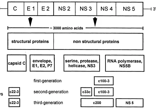
Figure 1.1: Hepatitis C virus genome and encoded proteins...17 Figure 1.2: Putative Hepatitis C virus replication...18 Figure 2.1: Oral lichen planus lesion of the buccal mucosa in a
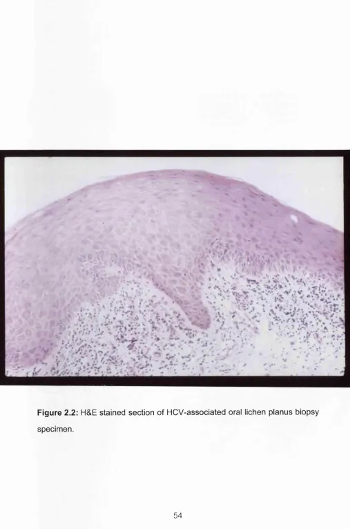
HCV-positive patient...53 Figure 2.2 H&E stained section of HCV-associated oral lichen planus
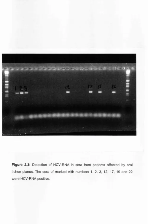
biopsy specimen... 54 Figure 2.3 Detection of HCV-RNA in sera from patients affected by oral
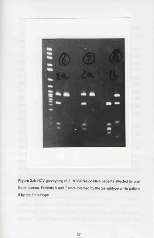
lichen planus. The sera marked with numbers 1,2,3, 12, 17, 19 and 22 were HCV-RNA positive... 66 Figure 2.4 HCV-genotyping of 3 HCV-RNA-positive patients affected by
oral lichen planus. Patients 6 and 7 were infected by the 2a subtype while patient 8 by the 1b subtype... 67 Figure 2.5 Detection of HGV-RNA in sera from HCV-positive patients
affected by oral lichen planus. Representative results of 14 cases. Sera tested in lane 4 and 12 (arrows) were HGV-RNA positive...79 Figure 2.6 Immunostaining profile of serum from one patient of the
HCV+ve/LP+ve group against monkey oesophagus, showing reactivity against epithelial nuclei (pattern I; arrowheads) and against surface-associated epithelial antigen in the basal layer (pattern II; arrows) (original magnification x 4 0 0 )... 89 Figure 2.7: Punctate cytoplasmic staining (pattern III) produced by serum
from a different patient in HCV+ve/LP+ve group (original magnification x 400). Note the apparent absence of
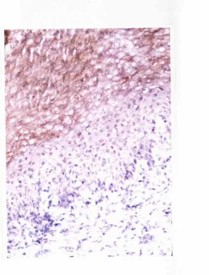
anti-nuclear reactivity in many cells (arrowheads)... 90 Figure 2.8: Expression of putative HCV antigens in oral lichen planus
lesional tissue (frozen sample). Positive cell of the
inflammatory infiltrate. Antibody used: anti-c22-0 (mAb 1) (original magnification x 400)... 102 Figure 2.9: Expression of putative HCV antigens in oral lichen planus
lesional tissue (frozen sample). Intense staining of the
keratinized layers. Antibody used: anti-c22-0 (mAb 1) (original magnification x 80)...103 Figure 2.10: Expression of putative HCV antigens in oral lichen planus
Figure 2.11 : Expression of putative HCV-associated antigens in oral lichen planus lesional tissue (paraffin-embedded sample). Positive cells of the inflammatory infiltrate. Antibody used: anti-c100-3 (mAb 2) (original magnification x 400)... 105 Figure 2.12: Expression of putative HCV-associated antigens in oral lichen
planus lesional tissue (paraffin-embedded sample), (a) staining of suprabasal kératinocytes, (original magnification x 160), (b) cytoplasmic staining of suprabasal kératinocytes, characterised by a granular appearance. Antibody used: anti-cl 00-3 (mAb 2) (original magnification x 400)... 106 Figure 2.13: Expression of putative antigens in HCV-positive liver tissue
(paraffin-embedded sample), (a) group of positive hepatocytes (original magnification x 160), (b) cytoplasmic staining of
hepatocytes, characterised by a coarse granular appaearance. Antibody used: anti-cl 00-3 (mAb 2) (original magnification x 400)...107 Figure 2.14: Graphs showing the distribution of (a) sLFA-3, (b) slCAM-1 and
(c) serum IgG levels within each patient group... 126 Figure 2.14.1: Detection of LFA-3 antigen in (a) idiopathic and (b)
HCV-associated oral lichen planus lesional tissue. In both sections LFA-3 is expressed primarly within the inflammatory infiltrate, with lower levels in adjacent regions of the epithelium and connective tissue... 126 b Figure 2.15: Severe oral lichen planus affecting the palatal mucosa...136 Figure 2.16: Squamous cell carcinoma with associated lichen planus of the
dorsum of the tongue... 137 Figure 3.1 : Risk of seroconversion after occupational exposure with
different blood-borne viruses... 162
CHAPTER 1.
1.1. Hepatitis C virus virology
Hepatitis C virus (HCV) is a positive stranded RNA virus, identified for the first time in 1989 by Choo and colleagues at the Chiron Corporation (Emeryville, California) by means of a new molecular biological technique. From a concentrate chimpanzee plasma with an high infectivity titre (10® infectious doses/ml) they extracted the total DMA and RNA which were both used as a template for the synthesis of complementary DMA (cDNA). The cDNA was inserted in a cloning vector and expressed in Escherichia coli.
Expressing proteins were immunoscreened with serum from a patient diagnosed with non-A, non-B viral hepatitis and only one clone (5-1-1) out of millions reacted (Choo et al. 1989). This clone represented the basis for the identification of a larger clone (cl 00-3) which expressed protein was used in the serological tests of first generation. By using overlapping clones, the entire sequence of the HCV genome was subsequently obtained.
Hepatitis C virus is now recognised as the main agent of the previously termed parenteral non-A, non-B viral hepatitis and one of the major causes of chronic hepatic disease world-wide. On the basis of the similarities between its nucleotide and amino acid sequence and that of the flavi- and pestiviruses, HCV has been classified within the Flaviviridae family as a separate genus (Heinz 1992). The HCV plus-stranded RNA genome comprehends approximately 9500 nucleotides and includes two untranslated regions at the 5’ and 3’ ends and an open reading frame encoding a viral polyprotein precursor of 3000 amino acids, cleaved by host and viral proteases into three structural proteins and six non-structural proteins. Structural proteins, encoded in the N-terminal region, comprehend the core protein (C) believed to be the viral capsid, and envelope proteins (El and
E2); the nonstructural proteins, encoded in the C-terminal region, include six proteins (NS2, NS3, NS4A, NS4B, NS5A and NS5B) with a number of
enzymatic activities some of which are still not fully elucidated (van Doom 1994; Brechot 1996) (Figure 1.1). The morphology of the HCV viral particle has as yet not been described in detail, however studies based on membrane filtration (Yuasa et al. 1991) and electron microscopy (Kaito et al. 1994; Li et al. 1995) suggest that the virion might be formed by a 30-50nm icosahedral nucleocapsid, surrounded by an envelope characterised by surface projections.
The precise details of HCV replication are still unclear. Replication may involve the synthesis of a negative strand intermediate which acts as a template for the synthesis of a new positive RNA genome (Yoshikura et al. 1996) (Figure 1.2). Thus the detection of the negative strand should indicate HCV replication; indeed this has been detected in the liver (Sherker et al. 1993), plasma (Sherker et al. 1993), peripheral blood mononuclear cells (Qian et al. 1992) and sperm of HCV-infected patients (Liu et al. 1994), as well as in primary and metastatic hepatocellular carcinoma tissue (Bocher et al. 1994; Kobayashi et al. 1994), and interestingly, in malignant lymphoma tissue of the parotid gland (De-Vita et al. 1995).
Due to the frequent errors in the course of replication (approximately 10^ substitutions per site per year) (Brechot 1996) and lack of repair mechanisms, HCV has an extremely variable genome and is present as quasispecies in a single infected individual (Oshima et al. 1991). This genetic variability occurs in all domains but is more frequent in a hypervariable region (HVR1 ), located at the N-terminus of E2 sequence, where a high rate of non- synonymous mutations (leading to amino acid changes and antigenically distinct variants) has been observed.
Following the complete sequencing of a number of different isolates, it was
different classification systems (Table 1.1) (Miyakawa et al. 1995). In 1994,
on the basis of a collaborative study comparing the NS5b region sequences of a world-wide panel of HCV variants, a classification was developed dividing the known viruses into main “types”, differing by 31-34% of the whole genome and numbered in order of discovery with Arabic numerals; each type is divided into “subtypes” differing up to 25%, which are identified by a lower case letter, again in order of discovery (Simmonds et al. 1994). Apart from the expensive and time-consuming sequencing of the whole genome, typing of the different viral strains can be based on the analysis of the reverse transcriptase-polymerase chain reaction (RT-PGR) products by means of different techniques such as restriction analysis (Nakao et al. 1991), hybridization with specific probes (Stuyver et al. 1993) or use of type-specific primers during the amplification step (Okamoto et al. 1992). Genotyping can also be undertaken by serological assays (Machida et al. 1992; Simmonds et al. 1993) which, although being easier, cheaper and allowing testing non- viraemic individuals, cannot discriminate between subtypes.
The distribution of the different genotypes in the HCV-positive population is not the same world-wide; genotype 1 is probably the most common variant in Europe, USA and Japan, although its prevalence may vary considerably; genotype 2 is less common in Europe than in Japan and China, genotype 3 is the most frequent in Thailand, Singapore and parts of India, genotype 4 in Egypt, Middle East and Central Africa while genotypes 5 and 6 are present in South Africa and South East Asia (McOmish et al. 1994; Anonymous 1995; Smith and Pontisso 1996). Also the distribution pattern changes according with the geographical area: in some regions particularly of Africa and Asia, only few types but many different subtypes are present, suggesting a presence of HCV for long periods and a common route of transmission; a different pattern is seen in Western Europe and North America, where there are more types in a limited number of subtypes, this pattern is consistent with
Table 1.1: Classification systems of the Hepatitis 0 Virus genotypes
System
Chayama Simmonds Okamoto Enomoto Tsukiyama New system
1 la 1 P I 1 la
11 1b 11 K1 1 1b
* * * 1c
1 1 1 2a 111 K2a 1 1 2a
111 2b IV K2b 11 2b
1 1 1 * * * * 2c
IV 3 V * * 3a
IV * * * 3b
4 * * 4a
V * * *
5a
* * * * 6a
~ 9500 nucleotides
RNA 5'h
encoded proteins
c
E 1
E2
NS2
NS3
NS4
NS5
~ 3000 amino acids
?
Structural proteins
non structural proteins
capsid C
envelope,
E 1,E 2, P7serine, protease, helicase, NS3
RNA
polymerase.
NS5B
peptides used in
current immunoassays C22-3
C22-3
first-generation
second-generation
third-generation
dOO-3
c33c dOO-3
c200 NS5
3'
+RNA
RNA Digestive endosome \
\
^
I +RNA ► Double stranded intermediate
-RNA Polyprotein
Double stranded intermediate 00
Cleavage
Assembly
Cell lysis
+RNA
RNA
+RNA
1.2. Epidemiology of hepatitis C virus infection
Hepatitis C virus infection is a world-wide health problem. The prevalence of anti-HCV antibodies among individuals at low risk for blood-borne diseases, such as volunteer blood donors without known risk factors (see below), varies considerably between geographical areas. In Europe there is a low frequency of HCV infection in Northern countries (UK 0.04% (McLindon
et al. 1995) and Norway 0.1% (Nordoy et al. 1994)), while in countries of Mediterranean area and Eastern Europe the frequency can be ten times greater. A similarly high seroprevalence has been found among 11 million Japanese blood donors (Yamaguchi et al. 1994). The HCV seropositivity rate among US blood donors varies from 0.1% and 0,7% (Sharara et al. 1996). The highest known frequencies of HCV infection has been found in developing countries, particularly in Africa (Saleh et al. 1994; el-Nanawy et al. 1995) and Middle East (Mansell and Locarnini 1995). (Table 1.2) (Abe and Inchauspe 1991 ; Allain et al. 1991 ; Amirudin et al. 1991 ; Arguillas et al. 1991 ; Chen et al. 1991; Chiaramonte et al. 1991; Dawson et al. 1991; Laskus et al. 1991; Martinez et al. 1991; Par et al. 1991; Park et al. 1991; Richards et al. 1991; Slama et al. 1991; Tsai et al. 1991; Van-der-Poel et al. 1991; Aguelles and Janot 1992; Archer et al. 1992; Darwish et al. 1992; Esteban et al. 1992; Galban et al. 1992; Garson et al. 1992; Giulivi et al. 1992; Hejjas et al. 1992; Hugo et al. 1992; Hyams et al. 1992; Hyland et al. 1992; Kashiwagi 1992; Knodler et al. 1992; Leite et al. 1992; Lin et al. 1992; Malaguti et al. 1992; Novakova et al. 1992; Zhang et al. 1992; Zufferey et al. 1992; Duraisamy et
al. 1993; Goncales et al. 1993; Goncales et al. 1993; Khan et al. 1993; Lai et al. 1993; Lytsar' et al. 1993; Ndumbe and Skalsky 1993; Sychev and Mikhailov 1993; Tang 1993; Timan et al. 1993; Tretskaia et al. 1993;
Liang et al. 1994; Lindholm 1994; Luengrojanaku! et al. 1994; MacLennan et al. 1994; Merino-Conde et al. 1994; Myl'nikov et al. 1994; Nordoy et al. 1994; Patino et al. 1994; Schwarz et al. 1994; Song et al. 1994; Suarez et al. 1994; Vasconcelos et al. 1994; Wang et al. 1994; Yamaguchi et al. 1994; Agbodjan et al. 1995; Anderson et al. 1995; Araj et al. 1995; Ayed et al. 1995; Bar et al. 1995; Bassily et al. 1995; Caspari et al. 1995; Choudhury et al. 1995; Develoux et al. 1995; Ehrmann et al. 1995; Garcia-Bengoechea et al. 1995; llako et al. 1995; Irshad et al. 1995; Jin et al. 1995; King et al. 1995; Love et al. 1995; McLindon et al. 1995; Mutimer et al. 1995; Ng et al. 1995; Quinti et al. 1995; Stern et al. 1995; Sulaiman et al. 1995; Tsega et al. 1995; Van Hoof et al. 1995; Wang 1995; Wu et al. 1995; Barna et al. 1996; Benjelloun et al. 1996; Darmadi et al. 1996; Guerrero et al. 1996; Hennig et al. 1996; JaiswaI et al. 1996; Lvov et al. 1996; Munoz-Gomez et al. 1996; Murphy et al. 1996; Maman et al. 1996; Oni and Harrison 1996; Salmeron et al. 1996; Soetjipto et al. 1996; Tang et al. 1996; Mison et al. 1997). It must be stressed that the results of most of these surveys are not comparable since a variety of tests with different predictive values have been used and confirmatory tests of the positive cases have not been always undertaken. The frequency of HCV- seropositivity in blood donors may not reflect the real proportion of HCV- infected individuals, as volunteer blood donors are a population that is negatively selected on the basis of risk factors for blood borne infections, thus the prevalence in the general population would be expected to be higher than that of blood donors; indeed few detailed studies of representative samples of the general population in France (Dubois et al. 1997), Italy (Bellentani et al.
1994) and the US (Alter 1995), reported seropositivity rates higher (1.15%, 3.2% and 1.4% respectively) than those of exclusive blood donors.
Little information is available on the incidence of HCV infection among low
risk groups. Recently a retrospective study estimated that in the US, during the last decade, 150,000 subjects have been infected per year (Alter 1995),
Table 1.2: Hepatitis C virus infection among volunteer blood donors world-wide
(a)
Country Prevalence
(%)
References
Algeria 0.18 Ayed et al. 1995.
Australia 0.14-0.78 Allain et al. 1991 ; Archer et al. 1992; Mison et al. 1997; Hyland et al. 1992.
Bangladesh 0.0 Khan et al. 1993. Belgium 0.77 Van Hoof et al. 1995. Benin 2.3 Develoux et al. 1995.
Brazil 1.14-3.1 Goncales et al. 1993; Goncales et al. 1993; Leite et al. 1992; Patino et al. 1994; Vanderborght et al. 1993; Vasconcelos et al. 1994.
Cameroon 6.4 Ndumbe et al. 1993. Canada 0.15-0.3 Giulivi et al. 1992.
China 0.0-2.5 Jin et al. 1995; Tang et al. 1996; Wang et al. 1994; Wu et al. 1995; Zhang et al. 1992; Zhang et al. 1993.
Cuba 1.5 Galban et al. 1992.
Czech Republic 0.24-0.38 Ehrmann et al. 1995; KIofera et al. 1994. ex Czechoslovakia 0.39 Novakova et al. 1992.
Egypt 14.4-26.6 Bassily et al. 1995; Danvish et al. 1992; Quinti et al. 1995. Ethiopia 1.4 Tsega et al. 1995.
France 0.63 Aguelles et al. 1992.
Germany 0.13-0.66 Caspari et al. 1995; Hennings et al. 1996; Hugo et al. 1992; Knodler et al. 1992.
Hong Kong 0.4 Lin et al. 1992.
Hungary 0.73-1.82 Barna et al. 1996; Hejjas et al. 1992; Hejjas et al. 1994; Par et al. 1991.
Iceland 0.6 Love et al. 1995.
India 0.3-1.78 Choudhury et al. 1995; Irshad et al. 1995; JaiswaI et al. 1996. Indonesia 1.6-3.1 Amirudin et al. 1991; Darmadi et al. 1996; Soetjipto et al.
1996; Sulaiman et al. 1995; Timan et al. 1993. Israel 0.4-0.66 Bar et al. 1995; Stern et al. 1995.
Italy 0.3-1.3 Chiaramonte et al. 1991; Ciuffreda et al. 1994; Lai, Malaguti et al. 1992.
Jamaica 0.3-0.4 King et al. 1995.
Japan 0.6-3.4 Hayashi et al. 1994; Honda et al. 1994; Kashiwagi et al. 1992; Yamaguchi et al. 1994.
(b)
Country Prevalence
(%)
References
Kenya 0.9 llako et al. 1995.
Korea 1.3 Park et al. 1991.
Lebanon 0.11-0.41 Araj et al. 1995; Naman et al. 1996. Malaysia 1.49-1.9 Duraisamy et al. 1993; Ng et al. 1995. Mauritius 2.1 Schwartz et al. 1994.
Mexico 0.7-1.47 Guerrero et al. 1996; Islas et al. 1994; Merino-Conde et al. 1994.
Morocco 1.1 Benjelloun et al. 1996. Netherlands 0.1 Van der Poel et al. 1991. Nigeria 2.0-3.0 Oni et al. 1996.
Northern Eurasia 0.7-10.7 Lvov et al. 1996. Norway 0.1 Nordoy et al. 1994. Philippines 2.2 Arguillas et al. 1991. Poland 2.0 Laskus et al. 1991. Rodrigues Islands 0.0 Schwartz et al. 1994.
Russia 1.37-2.5 Lystar et al. 1993; Myl’nikov et al. 1994; Sychev et al. 1993. Saudi Arabia 0.66-3.2 Abdeiaal et al. 1994; Bernvil et al. 1994.
Singapore 0.37 Wang et al. 1995.
Spain 0.5-1.2 Esteban et al. 1992; Garcia-Bengoechea et al. 1995; Martinez et al. 1991; Munoz-Gomez et al. 1996; Salmeron et al. 1996; Suarez et al. 1994.
Surinam 0.4 Van der Poel et al. 1991. Sweden 0.003 Lindholm et al. 1994. Switzerland 0.29 Zuffrey et al. 1992.
Taiwan 0.8-1.6 Chen et al. 1991 ; Luengrojanakul et al. 1994; Tsai et al. 1991.
Togo 3.3 Agbodjan et al. 1995.
Tunisia 1.09 Slama et al. 1991.
UK 0.18-0.04 Crawford et al. 1994; Garson et al. 1992; MacLennan et al. 1994; McLindon et al. 1995; Mutimer et al. 1995.
Ukraine 2.3 Tretskaia et al. 1993.
1.3. Hepatitis C virus transmission
The principal route of transmission of HCV is parenteral, through contaminated blood. Almost all subjects receiving a single unit of HCV-RNA- positive blood ultimately become HCV-infected (Aach et al. 1991), hence any recipient of blood or blood products, particularly before 1990, may be considered at risk of HCV infection. It is now evident that HCV has been the cause of up to 90% of the so called “post-transfusion non-A, non-B hepatitis” (Tremolada et al. 1991) and estimates from UK, France, and Australia indicate that 6,000 100,00 and 200,000 individuals respectively have acquired infection following blood transfusion (Zinn et al. 1995). Since the advent of screening of all blood donations for HCV, the risk of HCV acquisition following transfusion has fallen: at present the chance of becoming HCV-infected with a blood donation that passed all the screening tests is 1 in 103,000 (Schreiber et al. 1996). Besides the use of blood and blood products, other iatrogenic routes of transmission include organ (Pereira et al. 1991), tissue (Conrad et al. 1995) and bone marrow transplantation (Shuhart et al. 1994) from infected donors, haemodialysis (Alter 1995), plasmapheresis (Tao et al. 1991), endoscopy (Andrieu et al. 1995) and colonoscopy (Bronowicki et al. 1997). Transmission of HCV from health care workers (HCW) to patients is rare, although recently an HCV-positive cardiac surgeon transmitted HCV to five patients during open heart surgery (Esteban et al. 1996).
As for most blood-borne infections, injecting drug users are at high risk of HCV acquisition. Several studies, from different geographical areas, report similar prevalence rates of about 70% among injecting drug users and in the US they account for half of all newly infected individuals (Marwick 1997). Recently the use of non-injecting drugs, such as intranasal cocaine, has been significantly associated with HCV-infection (Conry et al. 1996).
Different HCV genotypes may be associated with the different routes of transmission: in Europe genotype 3a is prevalent in young people with an history of injecting drug use or tattooing (Shev et al. 1995), while genotype 1b is the most common subtype in elderly persons and those who have received blood transfusion or with no known risk factors of HCV acquisition (Pawlotsky
et al. 1995).
A contaminated hollow needle may be a source of HCV infection even in instances of accidental injury. There are several reports of HCW who contracted HCV infection following needlestick injury (Vaglia et al. 1990; Seeff 1991; Tsude et al. 1992) and recently simultaneous occupational transmission of HCV and Human Immunodeficiency Virus HIV, characterised by a very late seroconversion to both the viruses (after more than 9 months), followed by a rapid and fatal progression of the liver disease, has been documented (Ridzon et al. 1997).
infection in the human population (Mansell and Locarnini 1995). Co-infection with HIV increases the risk of vertical transmission (Zanetti et al. 1995), perhaps due to the higher serum levels of HCV-RNA, an independent risk factor for the mother-to-infant transmission (Ohto et al. 1994). The precise mode of HCV vertical transmission remains unclear (Kurauchi et al. 1993): it may occur in-utero. perinatally or even via a post-delivery route, although breastfeeding is probably not an at-risk activity (Lin et al. 1995). Household transmission in the absence of sexual contact, as in the case of siblings of
multitransfused or hemodialysis patients, is uncommon (Brackmann et al. 1993; Hou et al. 1995). Nevertheless up to 40% of HCV-infected patients have none of the known risk factors for the infection; thus there must be either unidentified or other parenteral routes of transmission -as demonstrated by the spread of HCV within a haematology ward, despite high levels of infection control (Allander et al. 1995).
1.4. Diagnosis of hepatitis C virus infection
A number of different enzyme-linked immunosorbent assays (ELISA) have been developed to detect circulating IgG antibodies to multiple viral epitopes and confirmatory recombinant immunoblot assays (RIBAs) can be utilised to exclude false positives (de Medina and Schiff 1995), which frequently occur, especially during the screening of low risk groups (McFarlane et al. 1990). The first-generation HCV ELISA was based on the c l 00-3 epitope from the nonstructural NS4 region; second-generation tests include, along with the c l 00-3 epitope, the putative core c22-3 and c33c from the NS3 region; the c33c and c l 00-3 epitopes were then substituted by a larger cloned fragment combining both of them into a c200 protein. The third generation anti-HCV ELISA includes also an antigen from the NS5 region (Figure 1.1). The latest generation serological tests can detect HCV infection 6 to 8 weeks after exposure, compared with 11-12 weeks of the first generation assays (Rubin et al. 1994), but they cannot distinguish between past or present infection, do not indicate the current phase of the disease (acute or chronic) nor predict the disease progression. Anti-HCV antibodies are present in saliva of infected patients and an assay developed to detect IgG of the oral fluids has the same sensitivity and specificity of commercially available serological tests (McIntyre et al. 1996).
At present it is not possible to detect viral antigens in blood or related fluids, thus the “gold standard” in the diagnosis of HCV infection is the detection of the viral genome by mean of RT-PCR (Okamoto et al. 1990). This test is commonly employed to distinguish HCV-seropositive patients with a resolved infection from viraemic patients, particularly among individuals with normal alanine aminotransferase (ALT) levels. In fact normal ALT values cannot exclude liver injury, as a high proportion of patients affected by HCV- associated (histologically diagnosed) chronic hepatitis may have persistently
the serum of the majority of seropositive patients with abnormal hepatic
serological tests, although false negative results are possible, due to very low viraemia or HCV-RNA limited to compartments different from serum (Schmidt et al. 1995).
Recently quantitative HCV-RNA assays have been developed and
commercialised (Jacob et al. 1997), viraemia levels being found to be a predictive factor of response to therapy.
1.5. Natural history of hepatitis C virus-related liver disease Acute hepatitis C occurs, in a minority of infected patients, 6 to 12 weeks after acquisition of the virus, while HCV RNA may become detectable in serum one week after acquisition (Farci et al. 1991). The clinical manifestations of this phase, if present, are no different from those of other viral hepatitides: mild and non-specific symptoms such as malaise, anorexia and nausea, possibly accompanied by more specific signs like jaundice and steatorrhoea and elevation of ALT serum levels. Hence the majority of infected patients have no signs of acute hepatic disease. Acute fulminant hepatitis, although possible, is rare (Wright et al. 1991; Yanagi et al. 1991).
Infection with HCV tends to cause chronic hepatic disease: in absence of treatment, up to 78% of patients show elevation of serum ALT levels for more than 6 months; but when serum HCV RNA is used as marker of persistent infection, up to 100% of the patients have been found chronically infected (Colombo 1996). With the exception of the late stages of the disease, chronic hepatitis C is usually an asymptomatic disease, often with normal or minimally elevated ALT; however histological abnormalities are present in the majority of, even apparently healthy, patients (McLindon et al. 1995; Prieto et al. 1995); for this reason liver biopsy is essential in the diagnosis and clinical management of chronically HCV-infected patients (Dusheiko et al. 1996).
Hepatic cirrhosis is a common consequence of chronic hepatitis C; in two large studies from Spain and US, cirrhosis was diagnosed in 30% and 50% of patients respectively (Tong et al. 1995; Colombo 1996); nevertheless, none of 83 women who developed chronic hepatitis following administration of HCV-contaminated Rho (D)-immunoglobulin, showed signs of progressive hepatitis or cirrhosis 15 years after the acute attack (Muller 1996). The reason for such large variations is unclear; patterns of progression may be
acquisition. However, on the basis of a large cross-sectional study, chronic hepatitis C has been recently defined as a “progressive fibrotic disease" with a progression towards cirrhosis linear according to time (Poynard et al. 1997). In the same study age at contagion, duration of infection, alcohol consumption and male gender were all factors associated with progression of fibrosis. Different HCV genotypes may also affect the severity of HCV liver disease and although data can be conflicting, an analysis of a group of liver transplant patients infected with different strains of HCV showed good evidence for a more pathogenic course of infection with subtype 1 b (Feray et al. 1995). The average time from HCV acquisition to diagnosis of cirrhosis varies between 20 and 30 years (Tong et al. 1995; Colombo 1996; Poynard et al. 1997), although a much more rapid progression of the disease is possible among immunocompromised patients, such as renal graft recipients (Chan et al. 1995) and individuals co-infected with HIV (Eyster et al. 1993).
Hepatocellular carcinoma (HCC) is strongly associated with HCV infection; it is a common finding in follow-up studies of patients with HCV infection of less than 30 years duration (Kiyosawa et al. 1990; Tong et al. 1995). Up to 76% of HCC patients may be HCV-infected (Simonetti et al. 1989) and chronic HCV infection has been suggested as the cause of the recent rise in the incidence of HCC in Japan (Okuda 1996). It has been calculated that the annual cumulative risk of developing HCC is approximately 1% in HCV- positive patients without cirrhosis and 3-10% in those with cirrhosis (BenvegnCi and Alberti 1996). Carriage of hepatitis B surface antigen (Kaklamani et al. 1991; Chiba et al. 1996), alcohol intake (Miyakawa et al. 1996; Tsutsumi et al. 1996) and possibly HCV genotype 1b (Zein et al. 1996; Bruno et al. 1997) have been indicated as possible co-factors in the
development of HCV-associated HCC, but the underlying mechanism of the association of HCV with HCC is still under investigation. Most of the patients with HCC have a previous history of cirrhosis, itself a preneoplastic condition
(Johnson 1996). Besicles HCV does not integrate in the hepatocyte genome and no transforming activity has been demonstrated for any HCV protein, thus an indirect role for HCV in the development of HCC has been proposed, although HCC in inactive or mild chronic hepatitis, not preceded by cirrhosis, has been reported (De Mitri et al. 1995).
1.6. Extrahepatic manifestations and associated conditions of hepatitis C virus infection
Hepatitis C virus infection can give rise to a broad spectrum of non-hepatic manifestations affecting different tissues and organs (Table 1.3) (Koff and
Dienstag 1995; Hadziyannis 1997). The presence of extrahepatic manifestations does not seem to relate to HCV serotype (Pawlotsky et al.
1995).
The best documented association is with essential mixed cryoglobulinaemia, a multisystemic disorder characterised by deposition of immune complexes in blood vessels resulting in a range of manifestations that goes from mild vasculitis with purpura and arthralgia to severe vasculitis with peripheral neuropathy and glomerulonephritis. The triad of purpura, weakness and arthralgia is a common presentation of cryoglobulinaemia. A direct aetiological role for HCV in the pathogenesis of essential mixed cryoglobulinaemia has been suggested on the basis of epidemiological data, detection of HCV-associated antigens in skin biopsy specimens of HCV- positive patients with essential mixed cryoglobulin, and by the frequent clinical resolution of the disease with interferon alfa (INF-a) treatment
(Cumber and Chopra 1995), in addition, in instances of HCV relapse after INF-a treatment discontinuation, cryoglobulinaemia also relapses. Hepatitis C-associated mixed cryoglobulinaemia may precede the development of non- Hodgkin's B-cell lymphoma (Ferri et al. 1994) and prevalence of HCV infection is significantly higher among patients with non-Hodgkin’s lymphoma
than control groups (Pioltelli et al. 1996); thus it appears that HCV infection may be involved in the aetiopathogenesis of a particular subset of non- Hodgkin’s lymphoma originating from mixed cryoglobulinaemia.
Membranoproliferative glomerulonephritis may be found in association with chronic hepatitis C and is usually part of the lesions associated with HCV- related mixed cryoglobulinaemia (Johnson et al. 1993).
HCV infection may give rise to porphyria cutanea tarda, a condition due to reduced hepatic uroporphyrinogen decarboxylase activity and clinically characterised by increased skin fragility and cutaneous lesions including erythema, blistering, scarring and pseudoscleroderma, occurring on exposure to sunlight. (Cribier et al. 1995). Other dermatological conditions possibly associated with HCV infection include lichen planus, polyarteritis nodosa, erythema nodosum and urticaria (Pawlotsky et al. 1995).
Thyroid disease, sometimes, but not always with an autoimmune basis, may affect more than 10% of patients with chronic HCV infection (Hadziyannis 1997). A high prevalence of diabetes mellitus has been reported in HCV liver disease (Allison et al. 1994; Grimbert et al. 1996) and a raised frequency of HCV infection has been noted among one group of diabetic patients (Simo et al. 1996); however INF-a therapy may be an aggravating factor in both these HCV-related endocrinopathies.
Autoantibodies against specific and non-specific antigens are a common finding in HCV-positive patients (Clifford et al. 1995) and chronic hepatitis showing features of both autoimmune and HCV-related hepatitis has been described (Bellary et al. 1995).
HCV infection has been reported in patients affected by Behcet’s syndrome (Munke et al. 1995) and INF may be an effective treatment of such patients (Hamuryudan et al. 1994); however only one of 224 Turkish patients with Behcet's syndrome was HCV-seropositive (Oguz et al. 1995).
Erythema multiforme triggered by HCV affecting one patient has been
described, although oral involvement was not mentioned (Antinori et al. 1991).
(B, D and G) and/or human immunodeficiency virus, injecting drug, haemophilia, etc. and their presence may influence the management of the patients and the progression of the HCV-associated liver disease.
Table 1.3: Extrahepatic manifestations of HCV infection^
Haematological and
lymphoid Aplastic anemiaMixed cryoglobulinaemia Idiopathic thrombocytopenia Non-Hodgkin’s B-cell lymphoma
Endocrine Thyroid dysfunctions(hypo- and hyperthyroidism) Diabetes mellitus
Dermatological Porphyria cutanea tarda Lichen planus
Polyarteritis nodosa Erythema nodosum Erythema multiforme Malacoplakia
Behcet’s syndrome Urticaria
Salivary gland and ocular Sialadenitis
Mooren corneal ulcer Uveitis
Kidney, neuromuscular Glomerulonephritis and joints Muscle weakness
Latent muscular abnormalities Peripheral neuropathy
Arthritis/arthralgias Rheumatoid arthritis Autoimmune and Pulmonary fibrosis miscellaneous Pulmonary vasculitis
Hypertrophic cardiomyopathy
CRST syndrome (systemic sclerosis) Antiphospholipid syndrome
Granulomas
Autoimmune hepatitis type 1 and 2 Circulating autoantibodies
1.7. Therapy of hepatitis C virus infection
Spontaneous resolution of chronic HCV infection is rare and currently the treatment of this condition is INF-a. INF-a is a cytokine produced by B
lymphocytes and monocytes following viral infection or antigenic stimulus (Wiranowska and Stewart 1981), its antiviral effects include activation of cellular ribonucleases and inhibition of viral penetration and transcription
(Davis and Hoofnagle 1986; Harper 1994).
The standard INF-a regimen in the treatment of chronic hepatitis 0 is 3
million lU administered subcutaneously three times weekly for six months. Most of the patients have a decrease in serum ALT levels and 40 to 50% have a normalisation of ALT levels (Hoofnagle and M. 1997). Suspension of
INF-a therapy is recommended in cases of lack of response after three
months, as a late response has been suggested to be unlikely (Sanchez- Tapias and Rodes 1995). An alternative to ALT levels in monitoring INF-a
therapy is HCV RNA viral load, that in most patients with a sustained response becomes undetectable within four weeks of initiating therapy (Orito et al. 1995). Unfortunately, about 50% responding patients have a relapse of ALT levels, thus only 25% of patients treated with INF-a have long-term improvement. Thus three response pattern can be described: a) non response, b) transient response, characterised by an improvement of serological parameters that is lost either during (breakthrough) or after the end of treatment (relapse), c) durable response in which the normalisation of serum ALT and clearance of HCV RNA occurs early in course of treatment and remain unchanged until the end of the follow-up period (Ryff 1997).
Since INF-a is an expensive drug that may induce side effects affecting
patient quality of life, many studies have been searching for a way to increase the long-term response-rate modifying the regimen adopted (dose and time)
or trying to identify the characteristics that make a patient a better candidate for a long-term response to the therapy. Such “predictors of response”
include viraemia, viral genotype, quasispecies heterogeneity, age and sex, liver histology, hepatic iron content, ALT level, duration and source of infection. A Consensus Panel of the National Institute of Health (NIH) has recently recommended that the length of INF-a treatment should be
increased to 12 months, as longer regimens more frequently achieve a sustained response and has indicated as factors associated with a favourable response to INF-a treatment infection with HCV genotypes 2 or 3, serum viral
levels below 1 million copies per ml, and absence of cirrhosis (Marwick 1997). A recent meta-analysis of numerous clinical trials found that INF-a is an
effective treatment for the acute phase of the infection, achieving frequent HCV RNA clearance and ALT normalisation (Camma et al. 1996). In addition a recent review suggested that INF-a treatment, especially in high dose or
long duration, may benefit HCV-related cirrhosis, reducing the risk of decompensation (Idilman et al. 1997), and of HCV-related HCC (Nishiguchi et al. 1995; Mazzella et al. 1996).
Interferon a therapy causes a transient influenza-like syndrome a few
hours after initial administration, generally well controlled with paracetamol and self-limiting. A number of other side-effects may occur in the course of treatment (Dusheiko et al. 1996; Hoofnagle and M. 1997), these include gastrointestinal upsets (nausea, vomiting diarrhoea), weight loss, neuropsychiatrie manifestations (irritability, anxiety and depression), hair loss, bone marrow suppression (thrombocytopenia, leucopenia), immune disorders (autoimmune antibodies, thyroid dysfunction) and diabetes mellitus. Some life-threatening side-effects such as myocardiopathies or cerebral vascular
haemorrhages have been reported (Bailly et al. 1997) as well as deaths due to severe bacterial infection and suicide. Most of INF-a side-effects are dose-
dependent and require reduction or cessation of therapy.
Owing to the numerous side-effects, high cost and variable efficacy of INF-a,
shown promising results is ribavirin, an antiviral agent that, combined to INF-
a, increases the percentage of sustained responses in patients with chronic
hepatitis C disease (Schalm et al. 1996; Reichard et al. 1998).
1,8. Considerations for dental treatment of an HCV-infected patient
HCV infection may seriously affect hepatic function; in particular, patients
Table 1.4: Recommendations for the dental management of an
HCV-infected patient with impaired liver function
1 With invasive procedures, careful preoperative assessment of the coagulation status of the patient, including:
• prothrombin time (PT)
• partial thromboplastin time (PTT) • platelet count
2 Great caution in the prescription of medication, in particular: • CNS depressant (barbiturates, opioids)
• hepatotoxic drugs (tetracyclines, erythromycin estolate, monoamine oxidase inhibitors, phenylbutazone)
• medications that may aggravate the haemorrhagic tendency (aspirin)
3 Consider the influence of possible HCV-associated medical conditions - mainly blood-borne infections (HIV, HBV, HGV).
4 Provide effective oral hygiene prophylaxis in cases of xerostomia, in order to reduce need for operative dentistry
CHAPTER 2.
2.1. Hepatitis C virus infection and oral lichen planus
2.1.1. Review of literature
introduction
Lichen Planus (LP) is a common mucocutaneous inflammatory disorder of uncertain aetiology (Scully and el-Kom 1985). Because of its relatively high frequency (1-2% in the general population), chronic nature and possible malignant transformation, the management of LP has a major role in day-to- day practice the oral physician has to face. Lichen planus can arise in association with a number of systemic diseases. In some instances these associations probably reflect reactions to drug therapy or represent coincidental concurrence because of the relatively high prevalence of LP, and affected patients being in middle to late life and thus liable to systemic diseases.
Associations between LP and immunologically-mediated diseases, infections and malignancies have long been suggested, but often the evidence is equivocal.
For example while sulphonylurea-like agents can cause lichenoid eruptions (Thompson and Skaehill 1994) there is no definitive evidence of a significant increased frequency of diabetes mellitus in patients with LP and vice versa.
Associations with a wide range of autoimmune disorders have been reported but most reports have included only small groups of patients and thus few helpful conclusions can be drawn of their relationship with the pathogenesis
of LP. Occasionally LP, often bullous in presentation, can arise in patients affected by malignancies.
Lichen planus is unlikely to have a strong autoimmune basis; there is no
association with HLA-DR4 haplotypes and a lack of frequent circulating autoantibodies in high titre in affected individuals (Porter et al. 1997). The lesions of LP have a predominantly CD8+ T lymphocyte infiltrate, probably mediated by local up-regulation of adhesion molecules such as intercellular
adhesion molecule-1 (ICAM-1) and lymphocyte function-associated antigen-3 (LFA-3). In addition there is an accumulation of antigen presenting cells such as Langerhans cells that may mediate local lymphocyte activation. Lichen planus thus probably represents a cell-mediated response to an antigenic trigger (Porter et al. 1997); nevertheless the identity of this trigger remains unknown.
Lichen planus and liver disease
In the last 15 years an increasingly strong association between LP and chronic hepatic disease has been suggested (Table 2.1) (Rebora et al. 1982; Korkij et al. 1984; Mobacken et al. 1984; Powell et al. 1984; Rebora and Rongioletti 1984; Katz and Pisanti 1985; Monk 1985; Scully et al. 1985; Ayala et al. 1986; Cottoni et al. 1988; del Olmo et al. 1989; GISED 1991; Gandoifo et al. 1992; el-Kabir et al. 1993; Bagan et al. 1994). Since the first report of erosive LP in a small group of patients with chronic active hepatitis (Rebora et al. 1978), as many as 64% of some groups of patients with LP, in Spain and Italy, have been reported to have chronic hepatic disease (Ayala et al. 1986; Cottoni et al. 1988; Bagan et al. 1994). In addition, a large case-control study carried out in 27 Italian hospitals between 577 patients with LP and 1031 controls, indicated that the risk of developing LP was increased among patients with a history of liver disease requiring hospital admission or specialist consultation, those who had liver biopsy and those with history of viral hepatitis; in particular the authors suggested that hepatotropic viruses different from the hepatitis B virus, namely the non-A non-B hepatitis virus, might play a significant role in the association between LP and liver disease (GISED 1991).
An association between LP and hepatitis B virus (HBV) infection has been suggested as hepatitis B surface antigen (HB^Ag) -positive patients may have double the risk of developing LP compared with HBgAg-negative patients (GISED 1991). In addition there are reports of anti-HBV antibodies in LP patients (Divano et al. 1992; Rebora 1994), of lichenoid eruption following administration of different HBV vaccines (Ciaccio and Rebora 1990; Trevisan and Stinco 1993; Aubin et al. 1994), and of an association of LP with
hepatocellular carcinoma - an HBV/HCV linked malignancy (Virgili et al. 1992). Nevertheless the majority of patients with both LP and chronic hepatic
disease are not HBV-infected.
A chronic liver disease sometimes described in association with LP is primary biliary cirrhosis (PBC) but, although some cases of oral lichenoid
lesions have occurred in PBC in the absence of penicillamine treatment (Graham-Brown et al. 1982; Powell et al. 1982; Oleaga et al. 1995), the association of LP and PBC is mostly due to the administration of this agent
Table 2.1: Studies on the possible association between lichen planus and chronic liver disease
Reference Lichen planus study
group (n° cutaneous/oral)
Abnormal liver function (%)
Chronic hepatitis (%)
Hepatic cirrhosis (%)
Liver disease overall (%)
Rebora et al. 1982 37 mucocutaneous LP 13.5 13.5
Rebora and Rongioletti, 1984 44 mucocutaneous LP 40.9 11.3 11.3
Powell et al. 1984 3897 LP" 1.3 1.3
Mobacken et al. 1984 54 oral LP (70.7% erosive) 11.1 3.7 3.7
Korkij et al. 1984 73 LP" 52.0 4.0 4.0 12b
Katz et al. 1985 15 oral LP (100% erosive) 13.3
Monk et al. 1985 55LP" 7.2
Scully et al. 1985 113 oral LP (22.1%
erosive) 7.9
Ayala et al. 1986 21 oral LP (100% erosive) 9.5 52.3 61.9
Cottoni et al. 1988 62 LP'’ (17.7% erosive) 9.6 14.5 25.8
Del Olmo et al. 1989 65 oral LP 7.6 10.7 33.8
GISED, 1991 577 LP (3.1% erosive) 18.3 21.4
Gandoifo et al. 1992 96 oral LP 5.2 7.2 24.0
El Kabir et al. 1993 180 oral LP (33.3% erosive) 25.0“ Bagan et al. 1994 187 oral LP (42.7% erosive) 21.3
Gi
' precise characteristics of LP not stated
Lichen planus and hepatitis C virus infection
Recently an association between hepatitis C virus infection and LP has been suggested that perhaps explains some aspects of the association of LP with chronic hepatic disease.
As previously mentioned, HCV infection can give rise to a wide variety of extrahepatic immunologically-mediated abnormalities including the generation of a number of tissue specific and non-specific autoantibodies (Clifford et al. 1995), cryoglobulinaemia (Cumber and Chopra 1995), autoimmune thyroiditis (Tran et al., 1993), lymphocytic sialadenitis resembling Sjogren’s syndrome (Haddad et al. 1992), diabetes (Allison et al. 1994) and possibly non-Hodgkin’s lymphoma (Ferri et al. 1994).
The first case of HCV-associated LP was reported by Mokni and colleagues in 1991 (Mokni et al. 1991), the patient was a drug addict with a HCV-related chronic active hepatitis, who develop cutaneous LP few years after HCV acquisition (the oral status was not mentioned). Following this case, many other groups reported cases of possible association between LP and HCV infection; HCV-associated hepatic disease may precede LP onset or may be diagnosed together with it; in the majority of cases mucosal involvement is present together with cutaneous lesions or even on its own (Amichai et al. 1994; Benchikhi et al. 1994; Cecchi et al. 1994; Jubert et al. 1994) and there can be coincident resolution of cutaneous and oral LP with INF-a therapy of chronic HCV-associated hepatic disease (Doutre et al.
1992). Recently a case of coincident oral LP and lymphocytic sialadenitis,
resembling Sjogren’s syndrome, has been reported (Tanei et al. 1997).
The few studies investigating the frequency of LP among HVC-positive subjects showed that five to 8% of patients with HCV-related chronic hepatic
1994; Gandoifo et al. 1994; Santander et al. 1994; Bellman et al. 1995; Favia et al. 1995; Nagao et al. 1995; Tanei et al. 1995; Carrozzo et al. 1996; Mignogna et al. 1996; Sanchez-Perez et al. 1996; Dupin et al. 1997; Imhof et al. 1997; Ingafou et al. 1997) The frequency of anti-HCV antibodies in European groups of unselected LP patients varies in different studies from about 4% (Rebora et al. 1992; Cribier et al. 1994) to 34% (Mignogna et al.
1996) but none of 55 English patients with oral LP were HCV seropositive (Ingafou et al. 1997). Twenty three percent of 40 American LP patients were HCV-seropositive by second-generation enzyme linked immunosorbent assay (ELISA) (Bellman et al. 1995), and two studies from Japan reported a
prevalence of HCV infection of 37.8% (Tanei et al. 1995) and 62% (Nagao et al. 1995). Notably, in almost all of these investigations the percentage of subjects with HCV infection in the LP study groups was much higher than that of the general population. Indeed in a recent controlled study the prevalence of HCV antibodies in a group of Italian oral LP patients was significantly higher then control subjects such that the authors recommended that patients affected by LP should be systematically screened for the presence of HCV infection (Carrozzo et al. 1996). In the same study a significant association was found between HCV infection and the erosive form of oral LP, confirming the finding of a Spanish survey (Sanchez-Perez et al. 1996) and of a previous study that found a tendency in patients with erosive oral LP to have chronic hepatic disease (Bagan-Sebastian et al. 1992).
It thus appears that there may be geographical differences in the association between HCV infection and LP. Antibodies to the GOR epitope
(GRRGQKAKSNPNRPL) (Mishiro et al. 1990) are found almost exclusively in a sub-group of autoimmune hepatitis type 2 patients who are also anti-HCV
positive and who represent the majority of autoimmune type 2 hepatitis patients in Italy (80%) but not in the UK (10%) (Michel et al. 1992). While 2 of 36 Italian patients with LP without chronic hepatic disease were HCV
seropositive, none were anti-GOR positive, whereas 11 of 20 patients with LP and chronic hepatic disease were HCV positive and 8 were also anti-GOR positive (Divano et al. 1994). This, considered with the aforementioned data, suggests that the association of LP and chronic hepatic disease may reflect infection with a particular form of HCV infection, although even the great
variability of prevalence of HCV infection within countries may have a role. Nevertheless data of French, Japanese and Italian patients, do not suggest any association between a specific HCV genotype and the development of LP (Pawlotsky et al. 1995; Nagao et al. 1996); recently, a German study found that 83% of the patients with HCV-associated LP were infected by subtype 1b; however the authors stated that such a high proportion was more likely to be related to age than that it represents an actual association of the subtype HCV-lb and LP (Imhof et al. 1997). In addition it has been shown that there are no significant differences in serum levels of HCV RNA in infected patients with or without oral LP. Similarly co-infection with hepatitis GBV-C virus, a newly described hepatitis virus sometimes also designated hepatitis G virus (HGV), seems unlikely to play a role in the development of LP in HCV infection (Nagao et al. 1996).
Interestingly HCV-associated oral LP can undergo malignant transformation (Carrozzo et al. 1997); but it seems doubtful that HCV infection alone is a risk factor for the development of oral squamous cell carcinoma as hypothesised by Nagao et colleagues presenting the case of
an HCV-positive patients with chronic hepatitis C and no oral lesions who developed oral cancer (Nagao et al. 1996), as a matter of fact the patient had been smoking 20 cigarettes and drinking 360 ml of sake a day for 40 years.
As with primary biliary cirrhosis (Seehafer et al. 1981; Graham-Brown et al. 1982; Powell et al. 1982; Oleaga et al. 1995), the development of LP in HCV
infection; indeed some cases of LP onset following INF-a therapy have been
reported (Agner et al. 1992; Dupin et al. 1994; Papini et al. 1994; Perreard et al. 1994) as well as worsening of pre-existing LP lesions (Boccia et al. 1993;
Protzer et al. 1993; Heintges et al. 1994). However this hypothesis cannot entirely explain the association of HCV infection with LP, as not all affected patients have been treated with INF-a (Carrozzo et al. 1996), there are cases
of resolution of LP following INF-a therapy as mentioned above, and recently
INF-a was found to be an effective treatment for mucocutaneous LP in a
small group of HCV-non-infected patients (Hildebrand et al. 1995). Other drugs that have been successfully adopted in the treatment of HCV-related oral LP include cyclosporine A (Papini et al. 1994), clobetasol (Mokni et al.
1996), cyclophosphamide (Aubin et al. 1994) and glycyrrhizin, a drug widely used in Japan in the treatment of chronic hepatitis C (Nagao et al. 1996).
The underlying aetiopathogenesis of HCV-related LP is unclear. Currently there is little published data of the immunological aspects of HCV-related LP; however a number of pathogenic mechanisms are possible. There may be a cell-mediated cytotoxicity to an epitope shared by HCV and damaged kératinocytes but to date there is no information of the expression of HCV in kératinocytes of the skin or mucosa in HCV-infected persons with or without LP.
It is possible that HCV-related LP has similar pathogenic mechanisms as the idiopathic form: they share the same clinical (Figure 2.1) an microscopic
(Figure 2.2) features, including a well-defined CD3+ T lymphocyte infiltrate and up-regulation of key adhesion molecules including intercellular adhesion molecule 1 (ICAM-1), very late activation antigen 4 (VLA-4) and lymphocyte function associated antigen 3 (LFA-3) (Porter et al. 1997). There is however still a need to determine if HCV RNA is expressed within the unaffected and affected mucosa of patients with HCV-related LP, to more precisely
determine if HCV-related LP is a reaction to local HCV-expression or a non
Table 2.2: Prevalence of hepatitis C virus infection in patients affected by lichen planus
CJ1 ro
Country Reference Study group Control group
Lichen Planus
HCV seropositivity
HCV
viremia Controls
HCV seropositivity
HCV viremia
France Cribieretal. 1994. 52 3.8% 112 2.6%*’
Dupin et al. 1997 102* 4.9% 306 4.5%"
Germany Imhof et al. 1997. 83 16% 14% 87 1.1%" 1.1%"
Italy Divano et al. 1992. 46 14%
Rebora et al. 1992, 29< 65% Gandoifo et al. 1994. 105* 9.5% Favia et al. 1995. 82* 20.7% Mignogna et al. 1996. 178* 34%
Carrozzo et al. 1996 70* 27.1% 21.4% 70 4.3%"
Japan Nagao et al. 1995. 45* 62% 60%
Tanei et al. 1995, 45 37.8% 45 6.7%"
Spain Bagan et al. 1994. 40 *'* 70%
Santander et al. 1994. 50 38% 30% 27 3.8%" 3.8%"
Sanchezperez et al. 1996. 78 20% 16% 82 2.4%" 2.4%"
UK Ingafoü et al. 1997. 55* 0%
USA Bellman et al. 1995, 30 23% 16% 41 4.8%"
all oral LP
^ no significant difference significant difference
%
PA
%
Figure 2.2: H&E stained section of HCV-associated oral lichen planus biopsy
specimen.
2.1.2. Epidemiological studies of the association between oral lichen
planus and infection with Hepatitis c virus
Introduction
Aims
Study 1 In view of the lack of data of the frequency of LP in patients with
HCV-associated hepatic disease, the aim of the present study was to investigate the frequency of oral LP in a large cohort of patients from central Italy with HCV-related chronic hepatic disease, compared with a similar group of patients with non HCV-related chronic hepatic disease.
Study 2 In addition, as no data on the association of HCV infection and oral LP are available from the metropolitan area of Milan, we investigated the frequency of HCV infection in the sera of a group of oral LP patients from this geographical area.
Study 3 Geographical variation in the distribution of HCV genotypes has been documented, thus in view of the knowledge that HCV genotypes may influence the clinical manifestations of HCV disease and the very limited data of HCV genotype distribution in patients with HCV-related LP, the present study analysed the distribution of HCV genotypes in a cohort of Italian patients with HCV-related oral LP.